Fakulteten för veterinärmedicin och husdjursvetenskap
Progressive Retinal Atrophy in Cats
Progressiv retinal atrofi hos katter
Aida Wolf
Uppsala 2019
Progressive Retinal Atrophy in Cats
Progressiv retinal atrofi hos katter
Aida Wolf
Handledare: Sofia Mikko, Sveriges Lantbruksuniversitet, Institutionen för
husdjursgenetik (HGEN)
Examinator: Maria Löfgren, Sveriges lantbruksuniversitet, institutionen för biomedicin och veterinär
folkhälsovetenskap
Omfattning: 15 hp
Nivå och fördjupning: Grundnivå, G2E
Kurstitel: Självständigt arbete i veterinärmedicin
Kursansvarig institution: Institutionen för biomedicin och veterinär folkhälsovetenskap Kurskod: EX0862
Program/utbildning: Veterinärprogrammet
Utgivningsort: Uppsala Utgivningsår: 2019
Elektronisk publicering: http://stud.epsilon.slu.se
Key words: Progressive retinal atrophy, retinal degeneration, cat, feline Nyckelord: Progressiv retinal atrofi, näthinneförtvining, katt
Sveriges lantbruksuniversitet
Swedish University of Agricultural Sciences
INNEHÅLLSFÖRTECKNING
Summary ... 1
Sammanfattning ... 2
Introduction ... 3
Material and Methods ... 3
Literature Review ... 4
Abyssinian ... 4
Siamese ... 5
Persian ... 6
Bengal ... 7
African black-footed cat ... 8
Discussion ... 10
1
SUMMARY
Progressive Retinal Atrophy (PRA) is an umbrella term used to describe a variety of inherited retinopathies observed in multiple species. This literature study focuses on PRA observed in five different cat breeds: Abyssinian, Siamese, Persian, Bengal and African black-footed cat. Various reports have described the mode of inheritance, age of onset, clinical and histological findings and specific genetic mutations seen in these breeds. The similarities and differences of these characteristics among these breeds are discussed.
At least four different types of PRA have been identified in these cats: rdAc (late-onset, recessively inherited rod-cone degeneration caused by a mutation in the CEP290 gene), rdy (early-onset, dominantly inherited rod-cone dystrophy, caused by a mutation in the CRX gene), an early-onset recessively inherited rod-cone degeneration caused by one of two different genetic mutations (either an AIPL1 gene mutation or an IQCB1 gene mutation) and an early-onset recessively inherited rod-cone degeneration with unknown genetic mutation.
The Abyssinian is afflicted with both rdAc and rdy. The Siamese is afflicted with one of the same mutations as the Abyssinian, rdAc. The Persian and the African black-footed cat seem to share a clinically identical early-onset recessively inherited rod-cone degeneration; however, they are caused by different genetic mutations. The Persian cat has a mutation in the AIPL1 gene and the African black-footed cat has a mutation in the IQCB1 gene. The early-onset recessively inherited rod-cone degeneration of the Bengal appears to be distinct from rdAc, rdy and the Persian (AIPL1 gene mutation). It is still unknown if the Bengal has the same mutation as the African black-footed cat.
The clinical signs and histopathology of PRA among these breeds are very similar. Hyperreflectivity and retinal vessel attenuation are seen clinically in all affected cats, whilst thinning of the outer nuclear layer and outer plexiform layer and degeneration of the photoreceptor layer are usually seen histologically in those affected.
These genetic mutations appear to be the same mutations observed in Leber’s congenital amaurosis (LCA) in humans. This opens up the possibility of using PRA affected cats as animal models of LCA in humans.
Accurately diagnosing PRA in these breeds is important to prevent the disease via selective breeding and to enhance the quality of life for those afflicted.
2
SAMMANFATTNING
Progressiv retinal atrofi (PRA), även kallad fortskridande näthinneförtvining, är ett samlingsbegrepp för att beskriva olika ärftliga ögonsjukdomar som påverkar näthinnan hos vissa arter och kan eventuellt leda till blindhet. Den här litteraturstudien fokuserar på PRA hos fem olika kattraser: abessinier, siames, perser, bengal och afrikansk svartfotad katt. Olika studier har beskrivit syndromets nedärvningsmönster, ålder vid sjukdomsdebut, kliniska och histologiska iakttagelser samt specifika genmutationer observerade hos raserna drabbade av PRA. Likheter och skillnader mellan dessa raser diskuteras i det här arbetet.
Det finns minst fyra olika typer av PRA som har identifierats hos de sex drabbade raserna. Den första är rdAc. Debuteringsåldern hos rdAc är senare jämfört med övriga typer av PRA. rdAc nedärvs via ett recessivt anlag, orsakar stav-tappdegeneration och är orsakad av en mutation i
CEP290-genen. Den andra är rdy vilket har tidig debuteringsålder, nedärvs via ett dominant
anlag, orsakar stav-tappdystrofi och är föranledd av en mutation i CRX-genen. Den tredje PRA har tidig debuteringsålder, nedärvs via ett recessivt anlag och orsakar stav-tappdegeneration. Den kan orsakas av två olika genetiska mutationer: antingen en mutation i AIPL1-genen eller en mutation i IQCB1-genen. Den fjärde PRA har tidig debuteringsålder, nedärvs via ett recessivt anlag, orsakar stav-tappdegeneration och har en okänd genetisk mutation.
Abessinier drabbas av både rdAc och rdy. Siames drabbas av samma genmutation som abessinier med rdAc. Perser och afrikansk svartfotad katt verkar ha kliniskt identiska typer av PRA där båda har tidig debuteringsålder, nedärvs via ett recessivt anlag och orsakar stav-tappdegeneration, men de orsakas av olika genmutationer. Perser har en mutation i AIPL1-genen medan afrikansk svartfotad katt har en mutation i IQCB1-AIPL1-genen. PRA hos bengal debuterar tidigt. Den nedärvs via recessivt anlag som orsakar stav-tappdegeneration, men skiljer sig från rdAc, rdy och mutationen som observerats i persers PRA. Det är fortfarande okänt vad bengal har för genmutation och huruvida det handlar om samma genmutation som drabbar afrikanska svartfotade katter.
Symtombild och histologiska undersökningar av PRA hos de här raserna är väldigt lika. Alla drabbade katter visar hyperreflektivitet och förtunning av blodkärlen i näthinnan. Histologiskt ses ett förtunnat yttre nukleärlager (outer nuclear layer) och yttre plexiformskikt (outer plexiform layer) samt degeneration av fotoreceptorlagret hos alla drabbade kattraser.
De genetiska mutationerna observerade vid PRA hos de här kattraserna verkar vara samma mutationer som finns hos människor med Lebers kongenitala amauros (LCA). Det här betyder att katter drabbade av PRA möjligtvis kan användas som djurmodeller för LCA hos människor. Det är viktigt att raserna med PRA diagnostiseras rätt för att kunna kontrollera sjukdomen genom avelsprogram och för att ge drabbade katter ett liv med hög kvalitet.
3
INTRODUCTION
There are many types of retinal atrophies in both humans and animals. The cause is often hereditary but age, nutritional deficiencies, glaucoma and toxins from drugs have also been observed to cause retinal atrophy (Barnett, 1965). Progressive Retinal Atrophy (PRA) is the term designating a group of genetic retinal diseases characterized by a progressive degeneration of the retina ultimately resulting in blindness (Millichamp, 1990). Since PRA consists of multiple hereditary retinopathies, the genetic cause, clinical signs, age of onset and histopathology of PRA differs among species and even amongst breeds (Barnett & Curtis, 1985). Changes in the retina are observed with an ophthalmoscope, an instrument used to see the interior surface of the eye also known as the fundus. There is no known treatment for PRA. Selective breeding is used to prevent this disease from affecting future individuals. PRA has been most extensively studied in dogs; more than 100 dog breeds have been found affected (Downs et al., 2011). These findings have been used to better understand analogous retinal diseases in humans, such as Leber’s congenital amaurosis (LCA) (Menotti-Raymond et al., 2010b). PRA has been described in cats also, though not as extensively as in dog breeds. The Abyssinian, the Siamese, the Persian, the Bengal and the African black-footed cat are the breeds that will be discussed in this study. The aim of this literature study is to compare and contrast PRA among these cat breeds to better understand this disease in cats. This could help provide a basis to incorporate affected cats as large animal models of human retinopathies.
MATERIAL AND METHODS
PubMed, Google Scholar, Primo and Web of Science were the databases used when searching for information and published studies. The keywords used when searching for the articles were
By Peter Hartmann at de.wikipedia, edited by Marc Gabriel Schmid Creating SVG version by Юкатан-Own work, CC BY-SA 3.0.
RPE: retinal pigment epithelium, R: rod, C: cone, OS: outer segment, IS: inner segment, ONL: outer nuclear layer, OPL: outer plexiform layer, INL: inner nuclear layer: IPL: inner plexiform layer, GC: ganglion cell.
4
(Progressive Retinal Atrophy) AND (cat or feline), IQCB1 mutation, CEP290 mutation, CRX mutation, AIPL1 mutation, polycystic kidney disease Persian and Leber’s congenital amaurosis human. A combination of the words was also used to find studies.
LITERATURE REVIEW Abyssinian
The Abyssinian is the most studied cat breed in regard to PRA. In 1982 it was suspected that there were two different types of PRA within the Abyssinian breed (Barnett, 1982). Narfström in 1983 described one of the forms of PRA as having an age of onset approximately between 1.5-2 years of age and reaching the advanced stage of the disease by 3-4 years of age (Narfström, 1983). A discoloration at the edges of the tapetal fundus was observed initially, but by 2-3 years of age the entire tapetal fundus had changed color. The advanced stage of this type of PRA is characterized by a hyperreflectivity of the tapetal fundus, attenuated retinal vessels, depigmentation of the non-tapetal fundus and a weakened pupillary light response (PLR). It was concluded that this form of PRA has an autosomal recessive mode of inheritance and affects the photoreceptor cell layer primarily (Narfström, 1983). This PRA is termed rdAc, retinal degeneration in Abyssinian cat (Narfström et al., 2009).
In an update to Narfström’s early 1983 study, Narfström and Nilsson (1983) determined the prevalence of PRA in a group of Abyssinians, aged two years or older, to be 45%. She also described a decreased electroretinogram (ERG) response detectable in affected cats before retinal changes could be observed ophthalmoscopically (Narfström & Nilsson, 1983).
In 1985, Barnett and Curtis compared the PRA in Abyssinian cats in England with that observed in Sweden by Narfström and Nilsson (1983). They concluded that the PRA in Abyssinian cats in England differs from that in Sweden. In England the disease is described as an early-onset autosomal dominant PRA, whilst in Sweden, PRA is seen in young adult Abyssinians and is caused by an autosomal recessive gene. Initial changes were detected at 4 weeks of age in the English population and consisted of mydriasis (dilated pupils) and nystagmus (involuntary eye movement). A rapid progression of the disease occurred within the next few weeks. Ophthalmoscopically, tapetal hyperreflectivity, retinal blood vessel attenuation and depigmentation in the non-tapetal region were observed (Barnett & Curtis, 1985).
In 1987, Curtis, Barnett and Leon termed the English form of PRA in Abyssinians: a rod-cone dysplasia, rdy. The initial signs were observed at 4-5 weeks of age, characterized by nystagmus and impaired PLR. By 8-12 weeks the majority of affected kittens had developed ophthalmoscopic changes such as dullness and loss of detail of the tapetal fundus, tapetal hyperreflectivity and retinal vessel attenuation. These signs progressed ultimately leading to ghost vessels and mottling on the non-tapetal fundus by 2 years of age. ERG yielded no response in those affected, light microscopy revealed thinned inner and outer photoreceptor layers and electron microscopy showed degenerative changes in both the inner and outer photoreceptor segments of the retina. Due to the absence of rod and cone responses in the ERG and the inability to distinguish between degenerating rod and cone photoreceptors using the
5
electron microscope, this form of PRA was classified as rod cone dysplasia (Curtis, Barnett & Leon, 1987).
Menotti-Raymond and colleagues conducted a study in 2007 to determine the gene defect that causes rdAc. A single nucleotide polymorphism in intron 50 of the CEP290 gene was discovered. This mutation leads to alternative splicing which leads to the insertion of four base pairs causing a frameshift in the mRNA transcript resulting in the introduction of a stop codon, and finally, the truncation of the mature protein (Narfström et al., 2009). The CEP290 gene is thought to have a vital role in ciliary transport to the primary cilia of photoreceptors (Chang et
al., 2006). This truncation can result in vital proteins not reaching the photoreceptors and
phototransduction not occurring, ultimately resulting in photoreceptor degeneration. Menotti-Raymond and colleagues suggests that the clinical signs observed in a 1985 medical dissertation by Narfström, ‘‘a marked vacuolization in the basal part of the outer segment of the rod photoreceptor, near the connecting cilium (Narfström, 1985b see Menotti-Raymond et al., 2007, p.216),’’ support the notion that photoreceptor degeneration is a result of ‘‘early abnormalities in the transport and distribution of phototransduction and/or structural proteins through the connecting cilia (Menotti-Raymond et al., 2007, p.216).’’
A 2009 study conducted by Narfström and colleagues on rdAc affected Abyssinians discovered that 82 of 87 individuals demonstrated complete concordance between rdAc genotype and phenotype (Narfström et al., 2009). This suggests that the rdAc allele is responsible for this type of PRA in the Abyssinian cat.
Menotti-Raymond conducted a study in 2010 and determined that the gene mutation causing
rdy in the Abyssinian cat is a mutation in the CRX gene. This mutation is a single base pair
deletion in exon 4 in the CRX gene on cat chromosome E2. This deletion leads to a frameshift mutation causing a premature stop codon which truncates the CRX peptide, resulting in an abnormal differentiation and development of the photoreceptors (Menotti-Raymond et al., 2010b). The CRX protein is a photoreceptor-specific transcription factor that is vital to the differentiation of photoreceptor cells (Furukawa, Morrow & Cepko, 1997), even necessary for their survival (Freund et al., 1997). This means that without a fully functioning CRX protein photoreceptor cells will not differentiate properly and eventually degenerate.
These gene mutations are also known to cause retinal dystrophy in humans. Multiple mutations, including CEP290 and CRX, have been associated with Leber’s congenital amaurosis (LCA) (den Hollander et al., 2006). LCA is an inherited human retinopathy with an early age of onset (at birth or a few months thereafter) and leading to severe visual impairment or blindness (den Hollander et al., 2006). The disease generally has an autosomal recessive mode of inheritance (den Hollander et al., 2006), however the CRX mutation appears to be inherited as an autosomal dominant (Rivolta, Berson & Dryja, 2001).
Siamese
In 1981, Carlile reported a type of PRA in Siamese cats. The average age of onset in the Siamese in this study was approximately 8.25 years of age with a progression to advanced PRA within approximately 2.5 years after initial changes. Initial changes included short-sightedness, retinal vessel attenuation and slight hyperreflectivity. These signs progressed to retinal ghost vessels,
6
general hyperreflectivity, dark and contracted optic disks and an impaired pupillary reflex leading to mydriasis. Night vision appeared to remain intact until the advanced stage of the disease, suggesting that the cones degenerated before the rods. However, complete blindness occurred in the end stage. At this stage, the outer plexiform layer and other outer adjacent layers had disappeared resulting in the inner nuclear layer lying directly on the choroid, the vascular layer of the eye directly behind the retina (Carlile, 1981).
A study conducted in 2010 by Menotti-Raymond and colleagues reported on the gene defect responsible for PRA in the Siamese and Siamese related breeds (Colorpoint Shorthair, Oriental Shorthair, Balinese and Javanese) and its prevalence in North American and European Siamese populations. Due to the high prevalence of rdAc previously reported in Abyssinian/Somali breeds (Narfström & Nilsson, 1983), these researchers wanted to determine if the rdAc allele is present in other breeds as well. Menotti-Raymond and colleagues tested 41 other breeds and 92 outbred cats leading to the detection of the rdAc allele in 34% of the breeds examined. A high frequency was detected among Siamese and Siamese related breeds. Therefore, they focused on finding the PRA mutation in these breeds. By genotyping the rdAc allele a single nucleotide polymorphism in the CEP290 gene, the same mutation causing rdAc in Abyssinian/Somali breeds, was detected. The clinical signs, mode of inheritance, age of onset and disease progression of the rdAc allele observed in Abyssinians were similar in the Siamese and Siamese related breeds. Approximately 33% of the Siamese and Siamese related breeds in North America and Europe have the CEP290 gene mutation (Menotti-Raymond et al., 2010a). The association of the CEP290 gene mutation in the Siamese, Abyssinian and humans with LCA is discussed under ‘‘Abyssinian’’ on page 5.
Persian
Several reports on various aspects of PRA in Persian cats have been published. In 1973 it was discovered by Rubin and Lipton that the PRA in Persians is inherited via an autosomal recessive trait. The age of onset was approximately 15 weeks of age. Ophthalmoscopy demonstrated: mydriasis, tapetal hyperreflectivity, retinal vascular attenuation, smaller and paler than normal optic discs and tapetal granularity. Histological changes included abnormalities in the outer segment of the retina, degeneration of the layer of rod and cone processes, thinning of the outer nuclear layer and of the outer plexiform layer (Rubin & Lipton, 1973).
In a report by Rah and colleagues (2005) it was concluded that PRA in Persians has an autosomal recessive form of inheritance. These authors reported a decrease in PLR as early as 2-3 weeks. By 16 weeks the retinal degeneration was advanced in the Persians in this study (Rah et al., 2005).
Rah, Maggs and Lyons (2006) investigated the common thought among Persian breeders that only chocolate or color-point (himalayan) Persians are at risk of PRA. They compared the prevalence of PRA among the chocolate, non-tabby solid, and himalayan coat colors as well as any correlation between PRA and polycystic kidney disease (PKD), an autosomal dominant disease with a high prevalence in Persians (Cannon et al., 2001). Using linkage analysis, they could find no association between PRA and coat color nor PRA and PKD (Rah, Maggs & Lyons, 2006).
7
In 2014, Alhaddad and others were able to describe the gene locus responsible for PRA in Persians using genome-wide association and linkage analysis. With the use of haplotype analysis, a 1.3Mb region on cat chromosome E1 was identified as being highly associated with PRA (Alhaddad et al., 2014).
In 2016, Lyons and others discovered a mutation in the AIPL1 (aryl-hydrocarbon interacting protein-like 1) gene on cat chromosome E1 resulting in a stop codon causing a truncated protein and resulting in a loss of about 40% of the normal protein. This mutation is thought to be the cause of PRA in Persians. The position of the mutation in the gene is c.577C > T of the CDS producing a p.Arg193* (Lyons et al., 2016), meaning the cytosine at nucleotide 577 on a coding DNA sequence is replaced with a thymine resulting in the amino acid Arginine changing to a stop codon (Ogino et al., 2007).
AIPL1 is vital to the function of photoreceptor phosphodiesterase-6 (PDE6), an enzyme ‘‘critical to the health and survival of rods and cones (Yadav & Artemyev, 2017, p.2)’’ AIPL1 acts as a chaperone for PDE6. If there is a mutation in the AIPL1 gene leading to a truncated AIPL1 protein, then PDE6 will no longer be able to function normally (Yadav & Artemyev, 2017). This will cause elevated intracellular cGMP levels in the retina, leading to the death of the photoreceptor cells (Wang, Tsang & Chen, 2017).
A mutation in AIPL1 is also observed in humans with LCA. As described earlier, LCA is an inherited human retinopathy most often with an autosomal recessive mode of inheritance. However, a less common autosomal dominant mode of inheritance exists as well. LCA is one of the leading causes of inherited blindness in children (Sohocki et al., 2000). Approximately 7% of LCA cases are caused by an AIPL1 gene mutation and this mutation may have an autosomal dominant mode of inheritance (Sohocki et al., 2000).
Bengal
The Bengal is the most recently reported breed of domestic cats afflicted with PRA discussed in this study. A study by Ofri and colleagues published in 2015 reported an early-onset, autosomal recessive, primary photoreceptor degeneration in Bengals. A breeding colony was established to observe clinical, electrophysiological and morphological changes of the disease over time.
At 9 weeks of age, degenerative changes were first observed. These included an increase in granularity and reflectivity of the tapetum lucidum and a mild retinal vascular attenuation. Progression to the advanced stage of disease occurred between 40-60 weeks of age and was described as tapetal hyperreflectivity and retinal ghost vessels. At this stage no ERG responses were detectable and cellular detail in the photoreceptor inner and outer segments and outer nuclear layer were indistinguishable.
Selective breeding was performed to determine if the PRA seen in Bengals and Persians is allelic. They were shown to be non-allelic; the two breeds have different forms of PRA. Mutation analysis was used to determine if the PRA in Bengals is caused by CEP290 or CRX gene mutations, the leading causes of PRA in Abyssinian and Siamese. It was concluded that
8
the Bengal is wildtype for these gene variants, meaning it has a different genetic mutation causing PRA. The mode of inheritance and age of onset of rdAc and rdy seen in these breeds do not correlate with those observed in the Bengals PRA. The cause of PRA in Bengals is currently unknown (Ofri et al., 2015).
African black-footed cat
PRA has also been found in wild cats. In 2017, Oh and others reported finding an early-onset generalized PRA in two full-sibling African black-footed cats. Mydriasis and vision deficits were first detected at 3 months of age and progressed rapidly. Both rod and cone photoreceptors were affected. Clinical and electrophysiological findings were both consistent with end-stage PRA and included tapetal hyperreflectivity, severe retinal vessel attenuation and non-recordable ERGs. Pedigree analysis demonstrated an autosomal recessive mode of inheritance. Genetic testing was conducted to determine the cause of PRA. It was confirmed that the cats were wild type for the CRX and CEP290 gene variants seen in Abyssinians and Siamese, and wild type for the AIPL1 gene variant seen in Persians. Whole genome sequencing was conducted, and multiple mutations were discovered. A 2 base pair deletion causing a frameshift mutation in the IQCB1 gene is thought to be the most probable cause due to the phenotype. However, a missense mutation in the CDH23 or DTHD1 gene may also be responsible (Oh et
al., 2017).
Mutations in the IQCB1 gene are known to cause retinal degeneration in humans. This mutation can cause non-syndromic LCA meaning that only retinal degeneration is observed in those affected. There is still a possibility, however, of patients contracting renal failure, a common disease seen in LCA patients (Estrada-Cuzcano et al., 2011).
IQCB1 encodes nephrocystin-5 (NPHP5), a protein in the connecting cilia and outer segments
of the photoreceptor cilia that helps regulate the transport of cargo molecules to the outer segment of the photoreceptor (Otto et al., 2005) and (Schäfer et al., 2008). Due to the ciliary function of this protein, a mutation would cause a disruption in this transport having negative effects on both structure and function of photoreceptors, ultimately leading to the loss of photoreceptors and retinal degeneration (Schäfer et al., 2008).
9
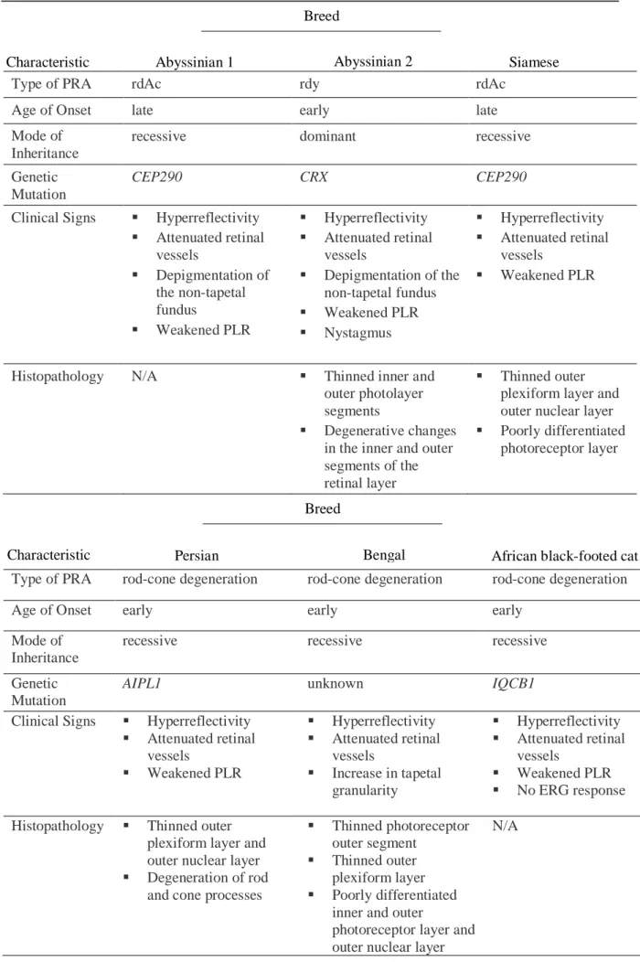
Table 1. A comparison of the characteristics of PRA in the cat breeds discussed in this study
Type of PRA rdAc rdy rdAc
Age of Onset late early late
Mode of Inheritance
recessive dominant recessive
Genetic Mutation
CEP290 CRX CEP290
Clinical Signs ▪ Hyperreflectivity ▪ Attenuated retinal vessels ▪ Depigmentation of the non-tapetal fundus ▪ Weakened PLR ▪ Hyperreflectivity ▪ Attenuated retinal vessels ▪ Depigmentation of the non-tapetal fundus ▪ Weakened PLR ▪ Nystagmus ▪ Hyperreflectivity ▪ Attenuated retinal vessels ▪ Weakened PLR
Histopathology N/A ▪ Thinned inner and
outer photolayer segments
▪ Degenerative changes in the inner and outer segments of the retinal layer
▪ Thinned outer plexiform layer and outer nuclear layer ▪ Poorly differentiated
photoreceptor layer
Type of PRA rod-cone degeneration rod-cone degeneration rod-cone degeneration
Age of Onset early early early
Mode of Inheritance
recessive recessive recessive
Genetic Mutation
AIPL1 unknown IQCB1
Clinical Signs ▪ Hyperreflectivity ▪ Attenuated retinal vessels ▪ Weakened PLR ▪ Hyperreflectivity ▪ Attenuated retinal vessels ▪ Increase in tapetal granularity ▪ Hyperreflectivity ▪ Attenuated retinal vessels ▪ Weakened PLR ▪ No ERG response Histopathology ▪ Thinned outer
plexiform layer and outer nuclear layer ▪ Degeneration of rod
and cone processes
▪ Thinned photoreceptor outer segment
▪ Thinned outer plexiform layer ▪ Poorly differentiated
inner and outer
photoreceptor layer and outer nuclear layer
N/A
Breed
Abyssinian 1 Siamese
Characteristic
Breed
Bengal African black-footed cat Characteristic Persian
10
DISCUSSION
In these five different breeds there are at least four different types of PRA, possibly five. The clinical signs in all of these breeds are very similar. Ophthalmoscopically, tapetal hyperreflectivity and retinal vessel attenuation can be observed in all affected individuals. A decreased ERG response, most often times no response, and a decreased PLR are observed in most of these breeds. The histological similarities in all breeds, where microscopic studies were conducted1, include a thinning of the outer nuclear layer and a thinning or degeneration of the
photoreceptor layer.
The differences between breeds are observed in the mode of inheritance and the age of onset. The Abyssinian has two different types of PRA, rdAc and rdy. rdAc has an autosomal recessive mode of inheritance with a later age of onset (Narfström, 1983). rdy has an autosomal dominant mode of inheritance with an early age of onset (Barnett & Curtis, 1985) and (Curtis, Barnett & Leon, 1987). The Siamese has a PRA with an autosomal recessive mode of inheritance and a later age of onset, synonymous with rdAc (Menotti-Raymond et al., 2010a). The mutation seen in rdAc, a mutation in the CEP290 gene, was discovered in both the Abyssinian and the Siamese, confirming that both breeds have the same type of PRA (Menotti-Raymond et al., 2007) and (Menotti-Raymond et al., 2010a).
All three of the remaining breeds discussed in this review, Persian, Bengal and African black-footed cat, have an autosomal recessive, early-onset, rod-cone dysplasia. Even though the clinical signs observed in these breeds are similar, the responsible genetic mutations are different. The Persian has a mutation in the AIPL1 gene (Lyons et al., 2016), the African black-footed cat has a mutation in the IQCB1 gene (Oh et al., 2017) and the mutation in the Bengal is currently unknown (Ofri et al., 2015).
The Bengal is interesting in that it was shown to have the wild-type variant for both the CEP290 and the CRX genes, meaning neither rdAc nor rdy is the cause of this type of PRA (Ofri et al., 2015). This, however, is no surprise since the mode of inheritance and age of onset of rdAc and
rdy do not correlate with those seen in the Bengal. The PRA seen in Persians appears to be
almost identical to that seen in Bengals, considering the mode of inheritance, age of onset, clinical and histological observations. However, when tested they were proven to be nonallelic (Ofri et al., 2015). This leaves the possibility that the PRA observed in African black-footed cats is the same as the PRA in Bengals. The clinical signs, histological observations, mode of inheritance and age of onset of PRA in both breeds are very similar. The Bengal has not been tested for the gene mutation observed in the African black-footed cat, IQCB1 mutation, since the Bengal study was published two years before. Due to the similarity of the PRA in these two breeds, it would be interesting to determine if the PRA in Bengals is caused by the same mutation seen in the African black-footed cat. The mutation causing PRA in Bengals could even be a previously undetermined gene mutation resulting in retinal degeneration. If this mutation is discovered the Bengal cat could also provide an animal model for retinal degeneration in humans. Further studies would have to be conducted in order to determine this.
11
The mutations in CRX, CEP290, AIPL1 and IQCB1 are all known mutations in the human retinal disease, Leber’s congenital amaurosis, an early-onset retinal degeneration causing visual deficits and blindness (den Hollander et al., 2006). These affected cats may one day prove to be valuable models for the study and treatment of LCA.
Determining these similarities between human and feline retinal degenerations not only benefits humans, but also the cats themselves. Determining gene mutations causing PRA in cats can lead to discovering PRA in additional breeds, earlier diagnosis of affected animals and prevention of blindness by selective breeding.
Visual deficits can be very stressful for both domestic and wild cats. Domestic cats that are allowed outdoors experience many risks such as traffic and wild animals. These dangers will become much more threatening if the cat is living with an undiagnosed retinopathy and has decreased vision. Indoor cats do not experience the same fatal risks as outdoor cats; however, inability to see could lead to an increased stress-level possibly resulting in the cat developing a new disease as a result of chronic stress. Wild cats with retinopathy are affected the most since they will have decreased survival and hunting skills. Their ability to see in the dark will worsen, a debilitating result of PRA for the wild cat since the majority of them hunt primarily at night. These factors significantly lessen the wild cat’s likelihood of survival.
It is also important to be aware of which breeds are affected by PRA in order to prevent breeding two breeds that have a history of PRA. This could result in the emergence of PRA in a new breed or in a specific cat, something that should be avoided to control this disease and ensure the best quality of life.
Genetic testing should be conducted on specific cats before breeding to reduce the risk of passing on these gene mutations known to cause PRA. Since there is no treatment for PRA, it is even more important to diagnose affected cats in order to help alleviate the stress that results from progressive blindness. This can be done by adapting the cat’s environment to make it easier and safer to navigate.
12
BIBLIOGRAPHY
Alhaddad, H., Gandolfi, B., Grahn, R.A., Rah, H.-C., Peterson, C.B., Maggs, D.J., Good, K.L., Pedersen, N.C. & Lyons, L.A. (2014). Genome-wide association and linkage analyses localize a progressive retinal atrophy locus in Persian cats. Mammalian Genome, vol. 25 (7–8), pp. 354–362.
Barnett, K.C. (1965). Retinal Atrophy. Veterinary Record, vol. 77 (51), pp. 1543–1560.
Barnett, K.C. (1982). Progressive retinal atrophy in the Abyssinian cat. Journal of Small Animal
Practice, vol. 23, pp. 763–766.
Barnett, K.C. & Curtis, R. (1985). Autosomal dominant progressive retinal atrophy in Abyssinian cats.
The Journal of Heredity, vol. 76, pp. 168–170.
Cannon, M.J., Barr, F.J., Rudorf, H., Bradley, K.J., Gruffydd-Jones, T.J. & MacKay, A.D. (2001). Prevalence of polycystic kidney disease in Persian cats in the United Kingdom. Veterinary
Record, vol. 149 (14), pp. 409–411.
Chang, B., Khanna, H., Hawes, N., Jimeno, D., He, S., Lillo, C., Parapuram, S.K., Cheng, H., Scott, A., Hurd, R.E., Sayer, J.A., Otto, E.A., Attanasio, M., O’Toole, J.F., Jin, G., Shou, C., Hildebrandt, F., Williams, D.S., Heckenlively, J.R. & Swaroop, A. (2006). In-frame deletion in a novel centrosomal/ciliary protein CEP290/NPHP6 perturbs its interaction with RPGR and results in early-onset retinal degeneration in the rd16 mouse. Human Molecular Genetics, vol. 15 (11), pp. 1847–1857.
Curtis, R., Barnett, K.C. & Leon, A. An Early-Onset Retinal Dystrophy With Dominant Inheritance in the Abyssinian Cat. vol. 28 (1), p. 9.
Downs, L.M., Wallin-Håkansson, B., Boursnell, M., Marklund, S., Hedhammar, Å., Truvé, K., Hübinette, L., Lindblad-Toh, K., Bergström, T. & Mellersh, C.S. (2011). A Frameshift Mutation in Golden Retriever Dogs with Progressive Retinal Atrophy Endorses SLC4A3 as a Candidate Gene for Human Retinal Degenerations. (Toland, A. E., ed) PLoS ONE, vol. 6 (6), p. e21452.
Estrada-Cuzcano, A., Koenekoop, R.K., Coppieters, F., Kohl, S., Lopez, I., Collin, R.W.J., De Baere, E.B.W., Roeleveld, D., Marek, J., Bernd, A., Rohrschneider, K., van den Born, L.I., Meire, F., Maumenee, I.H., Jacobson, S.G., Hoyng, C.B., Zrenner, E., Cremers, F.P.M. & den Hollander, A.I. (2011). IQCB1 Mutations in Patients with Leber Congenital Amaurosis. Investigative
Ophthalmology & Visual Science, vol. 52 (2), p. 834.
Freund, C.L., Gregory-Evans, C.Y., Furukawa, T., Papaioannou, M., Looser, J., Ploder, L.,
Bellingham, J., Ng, D., Herbrick, J.-A.S., Duncan, A., Scherer, S.W., Tsui, L.-C., Loutradis-Anagnostou, A., Jacobson, S.G., Cepko, C.L., Bhattacharya, S.S. & McInnes, R.R. (1997). Cone-Rod Dystrophy Due to Mutations in a Novel Photoreceptor-Specific Homeobox Gene (CRX) Essential for Maintenance of the Photoreceptor. Cell, vol. 91 (4), pp. 543–553. Furukawa, T., Morrow, E.M. & Cepko, C.L. (1997). Crx, a Novel otx-like Homeobox Gene, Shows
Photoreceptor-Specific Expression and Regulates Photoreceptor Differentiation. Cell, vol. 91 (4), pp. 531–541.
den Hollander, A.I., Koenekoop, R.K., Yzer, S., Lopez, I., Arends, M.L., Voesenek, K.E.J., Zonneveld, M.N., Strom, T.M., Meitinger, T., Brunner, H.G., Hoyng, C.B., van den Born, L.I., Rohrschneider, K. & Cremers, F.P.M. (2006). Mutations in the CEP290 (NPHP6) Gene Are a Frequent Cause of Leber Congenital Amaurosis. The American Journal of Human
Genetics, vol. 79 (3), pp. 556–561.
J.L. Carlile (1981). Feline retinal atrophy. Veterinary Record, vol. 108, p. 311.
Lyons, L.A., Creighton, E.K., Alhaddad, H., Beale, H.C., Grahn, R.A., Rah, H., Maggs, D.J., Helps, C.R. & Gandolfi, B. (2016). Whole genome sequencing in cats, identifies new models for blindness in AIPL1 and somite segmentation in HES7. BMC Genomics, vol. 17 (1). DOI: https://doi.org/10.1186/s12864-016-2595-4.
13
Menotti-Raymond, M., David, V.A., Pflueger, S., Roelke, M.E., Kehler, J., O’Brien, S.J. & Narfström, K. (2010a). Widespread retinal degenerative disease mutation (rdAc) discovered among a large number of popular cat breeds. The Veterinary Journal, vol. 186 (1), pp. 32–38. Menotti-Raymond, M., David, V.A., Schaffer, A.A., Stephens, R., Wells, D., Kumar-Singh, R.,
O’Brien, S.J. & Narfström, K. (2007). Mutation in CEP290 Discovered for Cat Model of Human Retinal Degeneration. Journal of Heredity, vol. 98 (3), pp. 211–220.
Menotti-Raymond, M., Deckman, K.H., David, V., Myrkalo, J., O’Brien, S.J. & Narfström, K. (2010b). Mutation Discovered in a Feline Model of Human Congenital Retinal Blinding Disease. Investigative Ophthalmology & Visual Science, vol. 51 (6), p. 2852.
Millichamp, N.J. (1990). Retinal Degeneration in the Dog and Cat. Veterinary Clinics of North
America: Small Animal Practice, vol. 20 (3), pp. 799–835.
Narfström, K. (1983). Hereditary progressive retinal atrophy in the Abyssinian cat. The Journal of
Heredity, vol. 74, pp. 273–276.
Narfström, K., David, V., Jarret, O., Beatty, J., Barrs, V., Wilkie, D., O’Brien, S. & Menotti-Raymond, M. (2009). Retinal degeneration in the Abyssinian and Somali cat (rdAc): correlation between genotype and phenotype and rdAc allele frequency in two continents.
Veterinary Ophthalmology, vol. 12 (5), pp. 285–291.
Narfström, L.K. & Nilsson, S.E.G. (1983). Progressive retinal atrophy in the Abyssinian cat: An update. Veterinary Record, vol. 112, pp. 525–526.
Ofri, R., Reilly, C.M., Maggs, D.J., Fitzgerald, P.G., Shilo-Benjamini, Y., Good, K.L., Grahn, R.A., Splawski, D.D. & Lyons, L.A. (2015). Characterization of an Early-Onset, Autosomal Recessive, Progressive Retinal Degeneration in Bengal Cats. Investigative Ophthalmology &
Visual Science, vol. 56 (9), p. 5299.
Ogino, S., Gulley, M.L., den Dunnen, J.T. & Wilson, R.B. (2007). Standard Mutation Nomenclature in Molecular Diagnostics. The Journal of Molecular Diagnostics, vol. 9 (1), pp. 1–6. Oh, A., Pearce, J.W., Creighton, E.K., Suedmeyer, W.K., Selig, M., Bosiack, A.P., Castaner, L.J.,
Whiting, R.E.H., Belknap, E.B. & Lyons, L.A. (2017). Early-Onset Progressive Retinal Atrophy Associated with an IQCB1 Variant in African Black-Footed Cats (Felis nigripes).
Scientific Reports, vol. 7 (1). DOI: https://doi.org/10.1038/srep43918.
Otto, E.A., Loeys, B., Khanna, H., Hellemans, J., Sudbrak, R., Fan, S., Muerb, U., O’Toole, J.F., Helou, J., Attanasio, M., Utsch, B., Sayer, J.A., Lillo, C., Jimeno, D., Coucke, P., Paepe, A.D., Reinhardt, R., Klages, S., Tsuda, M., Kawakami, I., Kusakabe, T., Omran, H., Imm, A., Tippens, M., Raymond, P.A., Hill, J., Beales, P., He, S., Kispert, A., Margolis, B., Williams, D.S., Swaroop, A. & Hildebrandt, F. (2005). Nephrocystin-5, a ciliary IQ domain protein, is mutated in Senior-Loken syndrome and interacts with RPGR and calmodulin. Nature
Genetics, vol. 37 (3), pp. 282–288.
Rah, H., Maggs, D. & Lyons, L. (2006). Lack of genetic association among coat colors, progressive retinal atrophy and polycystic kidney disease in Persian cats. Journal of Feline Medicine &
Surgery, vol. 8 (5), pp. 357–360.
Rah, H., Maggs, D.J., Blankenship, T.N., Narfstrom, K. & Lyons, L.A. (2005). Early-Onset, Autosomal Recessive, Progressive Retinal Atrophy in Persian Cats. Investigative
Ophthalmology & Visual Science, vol. 46 (5), p. 1742.
Rivolta, C., Berson, E.L. & Dryja, T.P. (2001). Dominant Leber congenital amaurosis, cone-rod degeneration, and retinitis pigmentosa caused by mutant versions of the transcription factor CRX. Human Mutation, vol. 18 (6), pp. 488–498.
Rubin, L.F. & Lipton, D.E. (1973). Retinal Degeneration in Kittens. J.A.V.M.A., vol. 162 (6), pp. 467– 469.
Schäfer, T., Pütz, M., Lienkamp, S., Ganner, A., Bergbreiter, A., Ramachandran, H., Gieloff, V., Gerner, M., Mattonet, C., Czarnecki, P.G., Sayer, J.A., Otto, E.A., Hildebrandt, F.,
Kramer-14
Zucker, A. & Walz, G. (2008). Genetic and physical interaction between the NPHP5 and NPHP6 gene products. Human Molecular Genetics, vol. 17 (23), pp. 3655–3662.
Sohocki, M.M., Perrault, I., Leroy, B.P., Payne, A.M., Dharmaraj, S., Bhattacharya, S.S., Kaplan, J., Maumenee, I.H., Koenekoop, R., Meire, F.M., Birch, D.G., Heckenlively, J.R. & Daiger, S.P. (2000). Prevalence of AIPL1 Mutations in Inherited Retinal Degenerative Disease. Molecular
Genetics and Metabolism, vol. 70 (2), pp. 142–150.
Wang, T., Tsang, S.H. & Chen, J. (2017). Two pathways of rod photoreceptor cell death induced by elevated cGMP. Human Molecular Genetics, vol. 26 (12), pp. 2299–2306.
West-Hyde, L. & Buyukmihci, N. (1982). Photoreceptor degeneration in a family of cats. JAVMA, vol. 181 (3), pp. 243–247.
Yadav, R.P. & Artemyev, N.O. (2017). AIPL1: A specialized chaperone for the phototransduction effector. Cellular Signaling, vol. 40, pp. 183–189.