1
Digital holographic microscopy – innovative and non-destructive analysis
of living cells
Z. El-Schish1, Mölder A2, Sebesta M2, Gisselsson L2, Alm K2, and Gjörloff Wingren A1,3 1
Department of Biomedical Laboratory Science, Health and Society, Malmö University, Malmö, Sweden
2
Phase Holographic Imaging AB, Lund, Sweden
3
Division of Laboratory Medicine, Malmö University Hospital, Malmö, Sweden
Abstracts of proposals for the book titled: Microscopy: Science, Technology, Applications and Education Digital holography is a novel technique that has been developed recently to study living cells. The technique is an innovative, non-destructive method with possibilities to study living cells over time. We are investigating cell number, growth, viability and death of adherent cells using digital holography, which is a novel, label-free, imaging technique for biological applications. We have recently demonstrated that digital holography is highly comparable to the conventional manual cell counting method using a hemocytometer (Mölder et al., 2008). Digital holography is a method that gives us information about the refractive index of cells, which can change under different
circumstances. The technique is cheap, fast and simple to use. The unique measurable parameters are the cell number, cell area, thickness and volume, which can be transformed to proliferation,
migration, viability and cell death. The digital holographic images produced can provide both quantitative and qualitative phase information from a single hologram. Future applications can include real-time cell monitoring of various parameters of cells of different diseases in response to clinically relevant compounds.
Keywords: digital holography, cell studies, cancer, viability, refractive index
1. Introduction
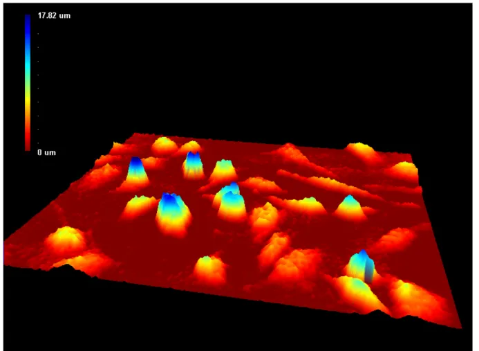
Living cells are largely transparent in ordinary microscopy, and therefore computerized analyses today require the addition of a dye to the cells. The digital holographic microscope has the unique advantage of combining the well established phase contrast microscopy with digital holography. The condition of cultured cells is analyzed using a segmentation software, that allows us to build up a data base of photographed cells (Fig. 1), measuring parameters such as optical thickness, area and shape [1]. These and other parameters can be translated into cell number, confluence, proliferation, cell death, migration and viability, using software analysis. Holography is a method of recording 3D information of an object with the help of interfering wave fronts from a laser. Holography was invented by Dennis Gabor, who proposed it a new principle of microscopy [2]. Digital holography differs from traditional holography by using a digital image sensor (CCD or CMOS) instead of the photographic plate. Interestingly, the field has recently undergone a paradigm shift with the advent of digital holography [3]. The interference pattern that the image sensor records, compresses the complete 3D information of the object in one single image. Digital holography gives information of the phase shift of the laser light that has been reflected or transmitted through the object [4]. The amount of phase shift is determined by the optical thickness, which depends on both the index of refraction inside the object and the object thickness. The phase shift can be measured with a resolution down to nanometers [5]. The principles for the digital holographic microscope have recently been reviewed by Kemper and von Bally [6].
Fig. 1 MDA-MB-231 breast cancer cells in 3D
2. Analysis of living cells
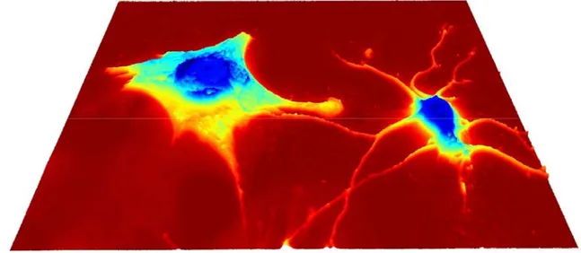
Digital holography has only recently begun to be of interest in the studies of living cells and cellular structures in the cellular and sub-cellular range for quantitative analysis combining nanometric resolution, real-time and non-invasive marker-free three dimensional observations (Fig. 2) with applications possible in cellular biology, diagnostics, genomics and proteomics, as well as in food industry [1, 7-10]. In 2006, Charrière et al used the technique for the first time to perform optical diffraction tomography of pollen grain [11]. The refractive index was determined for the different components such as the nucleus of the pollen grain, the cell wall and glycerol surrounding the pollen. These measurements could be interesting applications for material sciences and life sciences.
3
Fig. 2 3D hologram of glioma cells
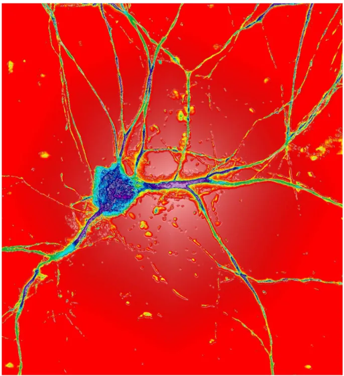
The next step was taken by analyzing the first living specimen, the testate amoeba by the same technique [12]. Indeed, the extensions of the cellular body were detected thanks to its specific refractive index contrast within the 3D reconstructions. By analyzing cortical neurons from mouse embryonical cells in primary cultures with digital holography, 3D images were obtained after numerical reconstruction [7]. Also, phase distribution changes of neuronal cell bodies were determined after hypotonic stress. When a hypotonic solution was applied on the nerve cells, the optical phase shift was decreased in the central domains of the cell body. At the same time, a phase increase was measured in the peripheral parts of the cell body, consistent with cellular surface enlargement [7]. Indeed, here the authors show that the method allows to determining independently the thickness and the integral refractive index of cells. The technique of digital holography opens up new perspectives within biomedical applications by enable both qualitative and quantitative label-free live cell imaging by the information from one single hologram [13, 14]. Recently, several investigations were performed on red blood cells (RBC). The shape, volume, refractive index and hemoglobin content are important characteristics that can be used as indicators of the physiological state of the body [15]. In this study, digital holography volume measurements of RBC were comparable with confocal microscopy and an impedance volume analyzer. Moreover, the sedimentation and membrane fluctuations of erythrocytes have been measured by digital holographic microscopy [16, 17]. The field of cell biology and cancer analysis in particular is intensively explored [8, 9, 18-21]. Many research groups are working with primary cells and established cell lines using a multitude of different techniques to measure different biological parameters, such as cell division and activation, cell death, cytotoxicity, cell cycle regulation and differentiation. Our belief is that digital holographywill be a device for applications such as on-line monitoring responses to anti-cancer drugs, analysis of subcellular features in various physiological states and in general in visualization of dynamic processes in cellular biology (Fig. 3). Development of fast and accurate evaluation tools for cancer treatments will be of great value to clinicians in deciding the most appropriate treatment for patients. A future goal is to use digital holography as a fast, automatic, and cost-efficient evaluation tool for different cancer therapies.
Fig. 3 3D hologram of neurons
3. Determination of proliferation and cell number
Automated studies for cell counting use techniques that count particles or nuclei. For those techniques to function for adherent cells, those cells must be treated to detach from the surface where they are growing. Therefore, counting of adherent cells is ideal for digital holographic microscopy and recently the first results were published [1]. Several different adherent human cell lines were tested by analyzing cells each day for a total of four days. The total phase shift was measured at 15 randomly chosen areas in the bottom of a cell culture flask. Both phase contrast and holographic pictures were automatically saved in a data base, with the ability to get information about number of cells, confluence, cell area, cell thickness and cell volume [1]. By measuring over time, changes in proliferation pattern were recorded. In parallel, the same cells were counted manually by a hemocytometer to verify the method. The results were compared and the correlation between the two methods was very high. The proliferation of the prostate cancer cell line DU145 was also analyzed with digital
5
holography, manual cell counting and MTS assay. The cell line was analyzed each day for 3 days and show comparable results with the three different methods used [Gjörloff Wingren et al, unpublished results].
4. Cell migration
Movement of cells is a central process during embryonal development, wound healing and during an immune response. The formation of tumors and metastasis arise when cells move, migrate, and attempts to find drugs that inhibits migration has been an important research field for a long time [22, 23]. Dubois et al showed that cell cultures of cancer cells could migrate in matrix gels in vitro and analyzed in three dimensions with the digital holographic microscope [19]. Several other studies show movement of cells analyzed by digital holography, sedimenting erythrocytes, HT-1080 fibrosarcoma cells and mouse fibroblasts [24, 25]. The fibroblast cells in both studies grew on and were analyzed in collagen. The migration of the cells could be followed quantitatively without the use of labeling or dyes.
5. Cell death analysis
Cell death in the form of programmed cell death or apoptosis, is another area within cell biology that is suitable for label-free quantitative analysis of cells and sub-cellular organelles using the digital holographic microscope. Cells that die of apoptosis shrink, decrease in size and get less compact [26]. Pancreatic cancer cells that were treated with toxins showed differences in volume compared to untreated control cells as analyzed by digital holography [7]. The differences were measured by the refractory index, which could be a useful tool in future analysis when investigating changes in the cytoskeleton of treated cells.
The digital holographic microscope has been used to discriminate dead cells from live cells [El-Schish and Gjörloff Wingren et al, unpublished]. Four different adherent cancer cell lines were treated with substances that induce cell death. The measurements with digital holography was performed once a day for up to three days directly in the cell culture flask to determine any changes over time. The analyzed cells were thereafter detached from the culture flask, labelled with AnnexinV-FITC and propidium iodide (PI) for a cell death-analysis by flow cytometry. The results obtained with digital holography of the treated cells showed small, shrinked cells that were fewer in number, had smaller area and a decreased optical density compared to the control cells. In parallel, the analysis by flow cytometry of the same cells revealed an increased number of cells double-positive for AnnexinV-FITC and PI. It was concluded that the obtained results by digital holography and flow cytometry were highly comparable.
6. Recombinant antibody microarrays
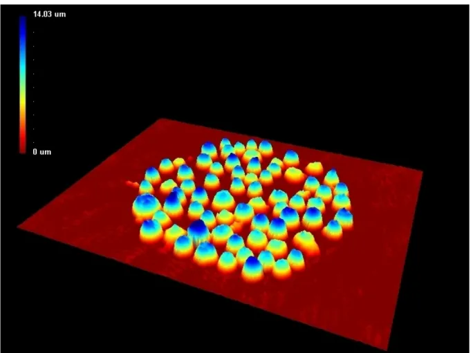
Adherent or suspension cells can also be studied by digital holography after binding to recombinant antibody microarrays (Fig. 4). The common method for this kind of phenotyping analysis is by fluorescence-labelling of the cells. Recombinant antibody microarrays can also be used for specific capture and immobilization of suspension cells, and in combination with subsequent analysis with the digital holographic microscope, holograms are numerically reconstructed and analyzed by a computer software to obtain values of mean cell area, mean cell thickness and mean cell volume (Fig. 5). Different treatments can be applied to the arrays before holographic measurements series starts and treatment outcome can be evaluated based on changes in cell volume [Nilsson and Gjörloff Wingren et al, unpublished].
Fig. 4 3D picture of antibody-targeted T leukemia cell line
Fig 5. (A) Reference diffraction pattern, (B) object diffraction pattern and (C) hologram. Numerical reconstruction of the
hologram yielded the three dimensional image of the cells (D) and the segmentation algorithm marked each individual cell (E) and analyzes cell parameters. (F) In some cases two cells were segmented as one (only part of the cell area is shown).
A
B
C
7
7. Discussion
Next step will be to treat cells with substances that inhibit cells in different stages of the cell cycle and to do analyses by digital holography. Kemper et al showed that the cellular ”dry mass” and changes in cellular density in the cell cycle could be analyzed by digital holography in yeast [27]. Other examples of interesting cellular biology applications that can be studied by digital holograhy are differentiation, wound healing and growth of cells on different surfaces. Moreover, detailed sub-cellular changes of cells transfected with DNA or siRNA could be important areas. Those treatments can affect the cellular proliferation, viability or migration. In conclusion, the digital holographic microscope gives us new information about changes in the refractive index over time, which can be transformed into measurable cellular parameters. Cancer cells are already in use, but all kinds of eukaryotic cells including stem cells, primary cells and cell lines can be included. The technique can in the future compete with conventional cell biology methods since it is cheap, fast and easy to use.
Acknowledgements The support by the Crafoord foundation, Health and Society at Malmö University, Magnus Bergwalls stiftelse, The Cancer foundation of Malmö University Hospital, Prof. Pirkko Härkönen, Dr. Christer Wingren and Emmy Nilsson are gratefully acknowledged.
References
[1] Mölder A, Sebesta M, Gustafsson M, Gisselsson L, Gjörloff Wingren A, Alm K. Non-invasive, label.free cell counting and quantitative analysis of adherent cells using digital holography. J. of Microscopy. 2008;232:240-247.
[2] Gabor D. A New Microscopic Principle. Nature. 1948;161:777-778.
[3] Schnars U, Jueptner, W, eds. Digital Holography: Digital hologram recording, numerical reconstruction and related
techniques. Berlin Heidelberg: Springer-Verlag; 2005.
[4] Sebesta M, Gustafsson M. Object characterization with refractometric digital Fourier holography. Optics Letters. 2005;30:471-473.
[5] Cuche E, Marquet P, Depeursinge C. Simultaneous amplitude-contrast and quantitative phase-contrast microscopy by numerical reconstruction of Fresnel off-axis holograms. Applied Optics. 1999;38:6994-7001.
[6] Kemper B, von Bally G. Digital holographic microscopy for live cell applications and technical inspection. Applied
Optics. 2008;47:A52-A61.
[7] Rappaz B, Marquet P, Cuche E, Emery Y, Depeursinge C, Magistretti PJ. Measurement of the integral refractive index and dynamic cell morphometry of living cells with digital holographic microscopy. Optics Express. 2005;13:9361-9373.
[8] Kemper B, Carl D, Schnekenburger J, Bredebusch I, Schäfer M, Domschke W, von Bally G. Investigations on living pancreas tumor cells by digital holographic Microscopy. J. Biomed. Opt. 2006;11:34005.
[9] Mann CJ, Yu J, Kim MK. Movies of cellular and sub-cellular motion by digital holographic microscopy. Biomedical Engineering OnLine. Available at: http://www.biomedical-engineering-online.com/content/5/1/21.
Accessed May 31, 2010.
[10] Mann CJ, Yu L, Lo CM, Kim MK. High-resolution quantitative phase-contrast microscopy by digital Holography. Optics Express. 2005;13:8693-8698.
[11] Charriere F, Marian A, Montfort F, Kuehn J, Colomb T. Cell refractive index tomography by digital holographic microscopy. Optic Letters. 2006;31:178-180.
[12] Charriere F, Pavillon N, Colomb T, Depeursinge C, Heger TH, Mitchell EAD, Marquet P, Rappaz B. Living specimen tomography by digital holographic microscopy: morphometry of testate amoeba. Optics Express. 2006;14:7005- 7013.
[13] Parshall D, Kim MK. Digital holographic microscopy with dual-wavelength phase unwrapping. Applied Optics. 2006;45:451-459.
[14] Marquet P, Rappaz B, Magistretti PJ, Cuche E, Emery Y, Colomb T, Depeursinge C. Digital holographic microscopy: a noninvasive contrast imaging technique allowing quantitative visualization of living cells with subwavelength axial accuracy. Optics Letters. 2005;30:468-470.
[15] Rappaz B, Barbul A, Emery Y, Korenstein R, Depeursinge C, Magistretti PJ, Marquet P. Comparative study of human erythrocytes by digital holographic microscopy, confocal microscopy and impedance volume analyzer. Cytometry. 2008;73:895-903.
[16] Bernhardt I, Ivanova L, Langehanenberg P, Kemper B, von Bally G. Application of digital holographic microscopy to investigate the sedimentation of intact red blood cells and their interaction with artificial surfaces. Bioelectrochemistry. 2008;73:92-96.
[17] Rappaz B, Barbul, Hoffmann A, Boss D, Korenstein R, Depeursinge C, Magistretti P, Marquet P. Spatial analysis of erythrocyte membrane fluctuations by digital holographic microscopy. Blood Cells Mol. Dis. 2009;42:228-232. [18] Carl D, Kemper B, Wernicke G, von Bally G. Parameter-optimized digital holographic microscope for high-resolution living-cell analysis. Applied Optics. 2004;43:6536-6544.
[19] Dubois F, Yourassowsky C, Monnom O., Legros JC, Debeir O, Van Ham P, Kiss R, Decaestecker C. Digital holographic microscopy for the three-dimensional dynamic analysis of in vitro cancer cell migration. J Biomed Opt. 2006;11:054032.
[20] Yu P, Mustata M, Peng L, Turek JJ, Melloch MR, French PM, Nolte DD. Holographic optical coherence imaging of rat osteogenic sarcoma tumor spheroids. Appl Opt. 2004;43:4862-4873.
[21] Hernandez-Montes M, Perez-Lopez C, Mendoza Santoyo F. Finding the position of tumor inhomogeneities in a gel-like model of a human breast using 3-D pulsed digital holography. J Biomed Opt. 2007;12:024027.
[22] Yamaguchi H, Wyckoff J, Condeelis J. Cell migration in tumors. Curr Opin Cell Biol. 2005;17:559-564. [23] Sahai E. Illuminating the metastatic process. Nat Rev Cancer. 2007;7:737-749.
[24] Langehanenberg P, Ivanova L, Bernhardt I, Ketelhut S, Vollmer A, Dirksen D, Georgiev G, von Bally G. Automated three-dimensional tracking of living cells by digital holographic microscopy. J Biomed Opt. 2009;14:014018. [25] Kemper B, Langehanenberg P, Vollmer A, Ketelhut S, von Bally G. Digital holographic microscopy - label-free 3D migration monitoring of living cells. Imaging and Microscopy. 2009;4:26-28.
[26] Kroemer G, Galluzzi L, Vandenabeele P, Abrams J, Alnemri ES, Baehrecke EH, Blagosklonny MV, El-Deiry WS, Golstein P, Green DR, Hengartner M, Knight RA, Kumar S, Lipton SA, Malorni W, Nunez G, Peter ME, Tschopp J, Yuan, J, Piacentini M, Zhivotovsky B, Melino G. Classification of cell death: recommendations of the Nomenclature Committee on Cell Death 2009. Cell Death and Differentiation. 2009;16:3-11.
[27] Rappaz B, Cano E, Colomb T, Kuhn J, Simanis V, Magistretti PJ, Marquet P. Noninvasive characterization of the fission yeast cell cycle by monitoring dry mass with digital holographic microscopy. J Biomed Opt. 2009;14:034049.


