Mandibular irregularity index stability following alveolar corticotomy
and grafting:
A 10-year preliminary study
Laith Makki
a; Donald J. Ferguson
b; M. Thomas Wilcko
c; William M. Wilcko
d; Krister Bjerklin
e;
Roelien Stapelberg
f; Anas Al-Mulla
gABSTRACT
Objective: To evaluate mandibular irregularity index stability following orthodontic treatment facilitated by alveolar corticotomy and augmentation bone grafting (Cort+).
Materials and Methods: The irregularity index of 121 orthodontically treated and 15 untreated patient study casts was analyzed at 5 years and 10 years.
Results: Cort+ resulted in significantly lower mandibular irregularity index scores at both 5 years (1.5 mm vs 4.2 mm, P , .000) and 10 years (2.1 mm vs 4.1 mm, P , .000) compared with conventionally treated patients.
Conclusions: Unmatched samples advise caution with conclusions, but orthodontic therapy combined with Cort+ enhanced the stability of the postorthodontic mandibular irregularity index for at least 10 years in this preliminary study. (Angle Orthod. 2015;85:743–749.)
KEY WORDS: Orthodontics; Corticotomy; Irregularity index; Bone grafting; Outcome stability; 10 years
INTRODUCTION
Long-term stability is a key objective in orthodontic treatment, especially satisfactory alignment of the mandibular anterior dentition as quantified by the
irregularity index. Whether long term stability of the mandibular anterior segment is affected by alveolar corticotomy and augmentation bone grafting (Cort+)1,2
has yet to be demonstrated.3
Mandibular Anterior Dental Arch Stability
A consensus of refereed orthodontic literature is that mandibular incisor crowding increases over time and there are no dependable predictors.4–7Increased crowding
shows great variation in both untreated8,9 as well as
orthodontically treated individuals. Eighteen Swedish dentists having no missing teeth or prosthodontic or orthodontic treatment were followed over a period of 40 years from ages 20 to 60; the authors found a significant increase in Little’s irregularity index (1.0 mm;P ,0.01).10The untreated mandibular arch dimensions of
15 adolescents and 18 adults were observed over a span of about 3 decades, and the rate of mandibular irregularity index increase was greater in adolescents than in adults.11
Increases in mandibular anterior crowding are also well documented in postorthodontically treated sam-ples.12,13Fifty-two females with mostly Class II
maloc-clusion were followed for nearly 2.5 decades after premolar extraction therapy; the authors reported a 1.1-mm increase in mandibular irregularity index over the 24 years.14
A mandibular irregularity index increase was reported in 96 patients following an average of
aInstructor, Advanced Orthodontics Program, European
University College, Dubai, United Arab Emirates.
bProfessor of Orthodontics and Dean, European University
College, Dubai, United Arab Emirates.
cPrivate Practice in Periodontology, Erie, PA; Departments of
Periodontology, Case Western Reserve University, Cleveland, Oh and University of Pennsylvania School of Dental Medicine, Philadelphia, PA.
dPrivate Practice in Orthodontics, Erie, PA; Department of
Orthodontics, University of Pennsylvania School of Dental Medicine, Philadelphia, PA.
eAssociate Professor, Department of Orthodontics, Faculty of
Odontology, Malmo¨ University, Sweden.
fInstructor, Advanced Orthodontics Program, European
Uni-versity College, Dubai, United Arab Emirates.
gAssistant Professor, Advanced Orthodontics Program,
Eu-ropean University College, Dubai, United Arab Emirates. Corresponding author: Dr Donald J. Ferguson, Dubai Health-care City, Bldg 27, Block D, Office 302, PO Box 53382, Dubai, UAE
(e-mail: fergusonloud@gmail.com)
Accepted: September 2014. Submitted: June 2014. Published Online: December 4, 2014
G2015 by The EH Angle Education and Research Foundation, Inc.
13 years postretention; the increase in irregularity index was significantly greater in adolescents in than adults (1.25 mm vs 0.85 mm;P 5 0.03).15
The percentage of successful orthodontic treatment outcomes, as measured by the mandibular irregularity index, diminishes as a function of time posttreatment in the absence of permanent bonded retention.6
An irregularity index score of ,3.5 mm has been designat-ed as minimal and clinically acceptable by Little et al.5,7
; a success rate of 30% at 10 years’ postretention and only 10% at 20 years’ postretention was observed in premolar extraction cases. After decades of research, the factors responsible for mandibular incisor stability remain elusive, and posttreatment stability is regarded as highly variable and unpredictable.
Booth et al.16 have demonstrated that 20 years of
fixed retention was effective in preventing the irregu-larity index from reaching scores greater than 3.5 mm. When fixed retention is not used, a consensus of the posttreatment stability literature suggests that the mandibular irregularity index will increase regardless of retainer type if retention is not permanent.5,7
In the absence of fixed retention, Rothe et al.17
compared 10-year postorthodontic samples of minimal (,3.5 mm) and high (.6 mm) irregularity index. The authors measured mandibular inferior border cortices and, by a priori application to the alveolus surrounding the mandibular anterior dentition, concluded that patients with thinner mandibular cortices were at increased risk for mandibular anterior dental relapse.
The purpose of this investigation was to evaluate mandibular irregularity index stability following ortho-dontic treatment facilitated by Cort+. The null hypoth-esis tested was that no difference in the mandibular
irregularity index 5 years and 10 years posttreatment exists in patients treated with or without Cort+. MATERIALS AND METHODS
Sample
Approval of the Institutional Review Board at the European University College was obtained to conduct this research project. Study casts of the mandibular dental arches of 136 white patients comprised the sample; 121 were orthodontically treated and 15 were not. Removable retainers (thermoplastic or Hawley-type retainers) were placed for 66 treated subjects; semipermanent, bonded canine-to-canine retainers for 31; and no retainers for 25 subjects (Table 1). Inclusion criteria for treated subjects were (1) fixed, comprehensive, straight-wire orthodontic treatment; (2) availability of mandibular study casts at pretreat-ment, immediate post-treatpretreat-ment, and a minimum of 3 years following active orthodontic treatment; and (3) availability of study casts at least 1 year after fixed retainer removal if semipermanent retainers were used. The criterion for exclusion was that study casts did not include all mandibular permanent incisors, canines, and first molars.
Patients’ records treated nonextraction with Cort+ were from a private practice; these patients had been requested to wear removable retainers posttreatment for 2 years. Cort+ samples included 43 patients at pretreat-ment and immediate posttreatpretreat-ment, 39 at 5 years, and 22 at 10 years posttreatment; age at pretreatment averaged 35.3 years. Patient records were obtained for the study as they became available during recall appointments. Every Cort+ patient who fit the inclusion criteria was
Table 1. Description of Study Subgroupsa
Post Tx Length (y)
Sample Description Short Descriptionb Sample Size(s) Age at Pre-Tx Active Tx (Pre to Post) (Post to 5 y) (Post to 10 y) Nonextraction, corticotomy+ grafting.
Removable retainers
Cort+ 43/39/22 35.3 6.8 4.3 12.1
Nonextraction. Removable retainers
Conv-5 23 23.5* 22.7* 5.3 na
Extraction (n 5 26) and nonextraction (n 5 29). Fixed—19.6 mo ( n 5 13)
Fixed—36.0 mo ( n 5 18) No retainers ( n 5 24)
Conv-10 55 12.7* 28.5* na 10.8
No treatment Untreated 15 20.5* na na 9.1*
aIncludes sample size, age at pretreatment (or first observation), active treatment time (pre-Tx to post Tx), and average length of time between posttreatment and 5-year or 10-year observation records. Note that sample size for Cort+ was 43 at pre- and posttreatment, 39 at 5 years, and 22 at 10 years posttreatment.
bCort+ indicates alveolar corticotomy and augmentation bone grafting; Conv-5, noncorticotomy 5-year sample; and Conv-10, noncorticotomy 10-year sample.
accepted, and no patient was rejected for any reason other than record nonavailability (Table 1).
The source of 5-year posttreatment records for patients treated nonextraction without corticotomy (Conv-5) were from private orthodontic practices; patients were requested to wear removable retainers for 2 years posttreatment. Conv-5 patient records were obtained as they became available during recall appointments; sample size was 23 patients and mean age at pretreatment was 23.5 years.
Study casts for treated patients, both nonextraction and extraction, with and without fixed retainers, were from a private orthodontic practice and included 10-year posttreatment study casts. Mean fixed retainer wear for the extraction (n 5 26) and nonextraction (n 5 29) cases were 19.6 and 36.0 months, respectively. The irregularity index among these four subgroups was analyzed at pre- and posttreatment as well as 10 years after active treatment using both 1-way ANOVA (parametric) and Kruskal-Wallis (nonparametric) test-ing; results showed no statistically significant differenc-es (P . .05), and the patients were pooled (n 5 55). The mean age at pretreatment of the pooled, noncorticot-omy, 10-year sample (Conv-10) was 12.7 years.
Mandibular casts of 15 adult (dentist), nonntreated subjects were part of a 40-year, follow-up sample published recently about dental arch dimensions and incisor irregularity in adults13
; only the initial and 10-year study casts were analyzed for the present investigation. Sample size was 15 and the sample averaged 20.5 years of age at the first observation time.
Procedures
Mandibular study casts representing pretreatment, immediate posttreatment, and at least 3 years post-treatment were digitally photographed. The photos were taken 90u to the occlusal plane in a standardized manner utilizing a 70-cm digital camera to study cast distance. Each photograph was secured with a millimeter ruler scale placed at the level of the mandibular occlusal plane; the scale was used to normalize the study cast images, thereby minimizing magnification errors.
ImageJ, a public domain, downloadable software program, was used as a photogrammetric measure-ment tool to record the target irregularity index on the mandibular study cast images. ImageJ has been shown to be a generally reliable and valid photogrammetric method,18especially as a technique for measuring the
irregularity index.19,20Each study cast was normalized to
the scale. The mandibular irregularity index was computed by measuring the displaced contact points of the anterior teeth as described by Little.21
Reliability of the measurement technique was evaluated by remeasuring six study cast images using ImageJ by the same investigator five times separated by at least 1 week. Intra-examiner reliability was tested using the paired t-test, and no significant statistical differences were found (P . .05)
Data were collected and stored in Excel (Microsoft, Redmond, Wash) and later transformed for use with the Statistical Package for Social Services (SPSS) software (IBM, Armonk, NY), version 15.0.1 for analysis. Homogeneity of variances was not rejected during 1-way ANOVA parametric testing and therefore Scheffe`’s post hoc test was performed to determine intergroup differences for irregularity index as well as increments of change from pretreatment to posttreat-ment, posttreatment to long term (either 5 years or 10 years), and from pretreatment to long term. The paired t-test was used to determine intragroup differences.
RESULTS
Pretreatment age for the Cort+ subjects (35.3 years) was significantly older than the other three subgroups, ie, Conv-5, Conv-10, and the untreated subgroups (23.5, 12.7, and 20.5 years, respectively; P , .000). The Cort+ sample size was 43 subjects at pretreat-ment and posttreatpretreat-ment and was 39 subjects at 5 years, compared with Conv-5 having 23 subjects. The Cort+ sample size at 10 years (n 5 22) was dissimilar to the Conv-10 (n 5 55) but not different from the nontreated subgroup (n 5 15; Table 1). Moreover, the nonextraction Cort+ active orthodontic treatment time of 6.8 months was significantly less than those of Conv-5 or Conv-10 (22.7 months and
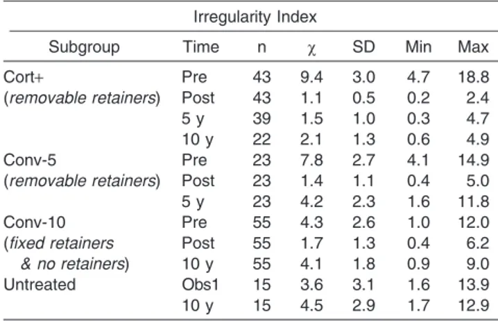
Table 2. Descriptive Statistics for Mandibular Irregularity Indexa Irregularity Index
Subgroup Time n x SD Min Max
Cort+ (removable retainers) Pre 43 9.4 3.0 4.7 18.8 Post 43 1.1 0.5 0.2 2.4 5 y 39 1.5 1.0 0.3 4.7 10 y 22 2.1 1.3 0.6 4.9 Conv-5 (removable retainers) Pre 23 7.8 2.7 4.1 14.9 Post 23 1.4 1.1 0.4 5.0 5 y 23 4.2 2.3 1.6 11.8 Conv-10 (fixed retainers & no retainers) Pre 55 4.3 2.6 1.0 12.0 Post 55 1.7 1.3 0.4 6.2 10 y 55 4.1 1.8 0.9 9.0 Untreated Obs1 15 3.6 3.1 1.6 13.9 10 y 15 4.5 2.9 1.7 12.9
* In millimeters, including sample size (n), mean (x), standard deviation (SD), and range: minimum (min) and maximum (max) for each of the four subgroups at time periods (Time) pretreatment (Pre), immediate posttreatment (Post), and either 5 or 10 years posttreatment (5 y or 10 y); the untreated subgroup included first observation (obs1) and observation at 10 years.
28.5 months, respectively; P , .000). Descriptive statistics were computed per subgroup for the man-dibular irregularity index (Table 2).
Irregularity Index
At pretreatment, the Cort+ group had a significantly higher irregularity index score (9.4 mm) than did either the Conv-10 or the untreated subgroups (4.3 mm and 3.6 mm, respectively; P , .000) but not the Conv-5 subgroup (7.8 mm;P . .05). At immediate posttreat-ment, there were no differences in mean irregularity index scores among treated subgroups. Long term, Cort+ patients demonstrated significantly lower (P , .000) irregularity index scores than did Conv-5 (1.5 mm vs 4.2 mm;P , .000) or Conv-10 (2.1 mm vs 4.1 mm; P , .000) as well as the untreated (2.1 mm vs 4.5 mm; P , .000) subgroup (Figure 1 and Table 3).
Irregularity Index Increments
The amount of irregularity index change during therapy and following removal of active orthodontic appliances was analyzed. From pre- to posttreatment,
Cort+ subjects had a significantly greater amount of reduction in irregularity index score (28.3 mm) than did Conv-5 (26.3; P 5 .01) as well as the Conv-10 subgroup (22.5 mm;P , .000). From posttreatment to 5 years, the rate of irregularity index change for Cort+ patients was significantly less than that of Conv-5 (0.4 mm vs 2.8 mm; P 5 .000). Moreover, from posttreatment to 10 years, the Cort+ (0.9 mm) irregularity index changed significantly less than did Conv-10 (2.4 mm; P , .000). From pretreatment to long term, Cort+ demonstrated significantly more reduction in irregularity index compared with Conv-5 (28.0 mm vs 23.6 mm;P 5 .000), Conv-10, as well as the untreated subgroup (26.9 mm vs 20.2 mm and 0.9 mm;P 5 .000) at 10 years (Figure 2 and Table 4). Cort+ Irregularity Index Outcomes
Paired t-tests demonstrated that the mandibular irregularity index significantly decreased during Cort+ treatment from a high at pretreatment of 9.4 mm to a low at posttreatment of 1.1 mm (P 5 .000). Significant
Figure 1. Bar chart demonstrating differences in mean irregularity index scores at the four study periods.
Table 3. Mean Irregularity Index Scores (in Millimeters) for the Four Study Subgroupsa Irregularity Index
Study Period
Cort+ (n 5 varied) Conv-5 (n 5 23) Conv-10 (n 5 55) Untreated (n 5 15)
x x P sig x P sig x P sig
Pre-Tx 9.4 (n 5 43) 7.8 NS 4.3 .000 3.6 .000
Post-Tx 1.1 (n 5 43) 1.4 NS 1.7 NS – –
5 y 1.5 (n 5 39) 4.2 .000 – – – –
10 y 2.1 (n 5 22) – – 4.1 .000 4.5 .000
aOne-way ANOVA testing revealed significant differences between Cort+ and all other subgroups except posttreatment and Conv-5s at pre-and posttreatment. X 5 mean, n 5 sample size,P sig 5 probability significance level, NS 5 not significant at P . .05.
Figure 2. Bar chart demonstrating differences in rates of irregularity index change, decreasing pretreatment to posttreatment and increasing posttreatment to long term.
increases in irregularity index were found from posttreatment to 5 years (1.1 mm vs 1.5 mm; P 5 .04), from posttreatment to 10 years (1.2 mm vs 2.1 mm; P 5 .009), and from 5 years to 10 years (1.5 mm vs 2.2 mm;P 5 .006; Table 5).
Conv-* and Untreated Irregularity Index Outcomes The Conv-5 and Conv-10 subgroups responded in similar ways, as follows:
N As expected, active orthodontic treatment signifi-cantly (P , .000) reduced the irregularity index scores in both Conv-5 and Conv-10 subgroups. N The irregularity index increased significantly (P ,
.000) from posttreatment to long term for Conv-5 (1.4 mm vs 4.2 mm) as well as for Conv-10 (1.7 mm vs 4.1 mm).
N The irregularity index for untreated subjects signifi-cantly increased (3.6 mm vs 4.5 mm, P 5 .002) during the 10-year observation period (Table 5).
DISCUSSION
A fundamental tenet in orthodontics is that mandib-ular incisor irregmandib-ularity increases as a function of time. In the present study, this principle was observed in the
mandibular anterior arch in all study subgroups. But it appeared that the process of mandibular incisor recrowding after orthodontic therapy was slowed significantly by Cort+ therapy for at least a decade.
Results of the present investigation demonstrated that the mandibular irregularity index was more stable in the Cort+ subgroup, as follows:
N The irregularity index score, while not statistically different from the conventionally treated subgroups at posttreatment, was significantly less than in all other subgroups at 5 years and 10 years.
N The increment of change in the irregularity index following orthodontic therapy was significantly less in Cort+ than in all other subgroups at 5 years and 10 years.
Results of research have also indicated an increased stability of the mandibular dental arch following Cort+.22
Clinically relevant in the present study were the findings that the irregularity index for Cort+ was greater at pretreatment and was reduced during active orthodontic treatment to a much greater extent, yet remained significantly more stable at 5 years and 10 years posttreatment. The scale of the differences is demonstrated in Figure 3.
Table 4. Rate of change for mean irregularity index scores (in millimeters) for study periodsa Irregularity Index—Increments
Study Period
Cort+ (n 5 varied) Conv-5 (n 5 23) Conv-10 (n 5 55) Untreated (n 5 15)
x x P sig x P sig x P sig
Pre to post 28.3 (n 5 43) 26.3 .01 22.5 .000 – –
Post to 5 y 0.4 (n 5 39) 2.8 .000 – – – –
Pre to 5 y 28.0 (n 5 39) 23.6 .000 – – – –
Post to 10 y 0.9 (n 5 22) – – 2.4 .000 – –
Pre to 10 y 26.9 (n 5 22) – – 20.2 .000 0.9 .000
aOne-way ANOVA revealed significant differences between Cort+ and all other subgroups at every study period. x 5 mean, n 5 sample size, P sig 5 probability significance level.
Table 5. Changes in Irregularity Index Increment Revealed by PairedT-testinga
Subgroup Irregularity Index N x 1 x 2 x-dif P sig
Cort+ (removable retainers) Pre to Post 43 9.4 1.1 28.3 .000 Post to 5 y 39 1.1 1.5 0.4 .040 Pre to 5 y 39 9.5 1.5 28.0 .000 5 to 10 y 19 1.5 2.2 0.8 .006 Post to 10 y 22 1.2 2.1 0.9 .009 Pre to 10 y 22 9.0 2.1 26.9 .000 Conv-5 (removable retainers) Pre to Post 23 7.8 1.4 26.3 .000 Post to 5 y 23 1.4 4.2 2.8 .000 Pre to 5 y 23 7.8 4.2 23.6 .000 Conv-10 (fixed or no retainers) Pre to Post 55 4.3 1.7 22.6 .000 Post to 10 y 55 1.7 4.1 2.4 .000 Pre to 10 y 55 4.3 4.1 20.2 NS
Untreated Obser1 to Obser2 15 3.6 4.5 .9 .002
aDuring and after active treatment all periods tested statistically different with one exception, that is, there was no significant difference between pretreatment and 10 years’ post treatment in the Conv-10 sample.
A number of investigators have cited an irregularity index of ,3.5 mm as ‘‘clinically acceptable’’ in describing mandibular anterior segment stability suc-cess after orthodontic treatment.5–8 The irregularity
index of ,3.5 mm at 5 years posttreatment in Cort+ was 97.4% (38 of 39 patients) compared with 47.9% (11 of 23 patients) in the Conv-5 subgroup. At 10 years posttreatment, 76.3% (17 of 22 patients) of Cort+ demonstrated a ,3.5 mm irregularity index compared with 36.4% (20 of 55 patients) in Conv-10.
Confounding factors related to the case-control study design that may have influenced our results and therefore the findings of the current investigation should be regarded as preliminary. Dissimilar sample sizes were used; the largest subgroup size difference was Cort+ (n 5 22) vs Conv-10 (n 5 55). Pretreatment ages were significantly different, with the largest spread between the Cort+ (35.3 years) and Conv-10 (12.7 years) subgroups. Even though the scale of difference in irregularity index magnitude between Cort+ and Conv-10 at 10 years posttreatment was substantial, results should be interpreted with caution given the sample age differences.
Retainer wear compliance as a confounding factor was considered. Requested of patients was 2 years of removable retainer wear (full time for 6 months followed by 1.5 years’ nighttime wear) in the Cort+ and Conv-5 subgroups. Retainer wear was rated by a patient questionnaire using a scale of 3 5 excellent, 2 5 average, and 1 5 poor. Mean retainer wear was rated 2.0 and 2.1 in the Cort+ and Conv-5 subgroups, respectively. In Conv-10, fixed retainers were removed after 19.6 months in extraction and 36 months in
nonextraction cases, leaving 7 to 8 years of unretained dentitions. The factor of retainer wear compliance as a confounding factor did not appear to influence the long-term results of the study.
Dependable predictors of postorthodontic treatment stability have not, to date, been identified. In a search for predictors of mandibular irregularity index stability, Rothe et al.17 evaluated whether the amount and
structure of mandibular bone affected the potential of postorthodontic mandibular incisor relapse. The authors compared orthodontically treated samples of 263 patients having a ,3.5-mm irregularity index with a sample of 60 with a .6.0-mm irregularity index approximately 10 years after therapy. They reported that the mean mandibular inferior border cortical thickness, as measured cephalometrically and indica-tive of the alveolus housing mandibular incisors, was significantly smaller in the relapse (.6 mm) group at all study periods. They concluded that patients with thinner mandibular cortices were at increased risk for dental relapse. This finding was confirmed 2 years later in another study by the same investigative group.23
Mandibular cortical thickness may be an important issue, and augmentation bone grafting was used in the present study in combination with orthodontic treat-ment and selective alveolar decortication.2 Greater
stability of the mandibular anterior segment in Cort+ may be due to increased cortical bone thickness following these procedures if cortical bone thinness is a risk factor for anterior segment instability and increased incisor irregularity.17,23 It has been
demon-strated that bone grafting is effective for at least 1 year following active corticotomy-facilitated treatment.24 Figure 3. Mandibular study casts with irregularity index scores of Cort+ (top row) and Conv-10 nonextraction, no retainer (bottom row) at pretreatment, posttreatment, and 10 years posttreatment, demonstrating relative differences in scale of initial malocclusion and 10 years posttreatment. The two cases are representative of the overall study results.
We also surmised that enhanced posttreatment stability is likely due to the increased turnover of tissues following corticotomy. It is reasonable to believe that there is substantial tissue memory loss due to the intensity and proximity of the insult created when full thickness flaps are elevated and the alveolar cortices are intentionally scarred with a surgical bur rotating under irrigation at high speed. However, it must be emphasized that cortical bone thickness and increased tissue turnover are ‘‘enhanced stability’’ assumptions and need further investigation.
CONCLUSIONS
N The irregularity index increased in all subgroups with time, but mandibular incisor recrowding was slowed significantly for 10 years in adult orthodontic patients following Cort+.
N Although the case-control study design of the present investigation was compromised by differences in subgroup sizes and ages, the results demonstrated enhanced treatment stability for the irregularity index compared with the stability of treatment outcomes without Cort+ as follows:
N The mandibular irregularity index following Cort+ was significantly less from immediate posttreatment to 5 years (1.5 mm vs 4.2 mm;P , .000) and 10 years (2.1 mm vs 4.1 mm;P , .000).
N The rate of irregularity index change for Cort+ was significantly less from immediate posttreatment to 5 years (0.4 mm vs 2.8 mm;P , .000) and 10 years (0.9 mm vs 2.4 mm;P , .000) compared with that of Conv-5 and Conv-10.
N Although the limitations of the imperfectly matched samples must be recognized, it may be concluded that orthodontic therapy combined with Cort+ en-hances the stability of the postorthodontic mandibu-lar irregumandibu-larity index for at least a decade.
REFERENCES
1. Wilcko WM, Wilcko MT. Accelerating tooth movement: the case for corticotomy-induced orthodontics. Am J Orthod Dentofacial Orthop. 2013;144:4–12.
2. Wilcko MT, Wilcko WM, Pulver JJ, et al. Accelerated osteogenic orthodontics technique: a 1-stage surgically facilitated rapid orthodontic technique with alveolar aug-mentation.J Oral Maxillofac Surg. 2009;67:2149–2159. 3. Mathews DP, Kokich VG. Accelerating tooth movement: the
case against corticotomy-induced orthodontics.Am J Orthod Dentofacial Orthop. 2013;144:5–13.
4. Freitas KM, de Freitas MR, Henriques JF, et al. Postreten-tion relapse of mandibular anterior crowding in patients treated without mandibular premolar extraction.Am J Orthod Dentofacial Orthop. 2004;125:480–487.
5. Little RM, Riedel RA, Artun J. An evaluation of changes in mandibular anterior alignment from 10 to 20 years postreten-tion.Am J Orthod Dentofacial Orthop. 1988;93:423–428. 6. Little RM. Stability and relapse of mandibular anterior
alignment: University of Washington studies.Semin Orthod. 1999;5:191–204.
7. Little R, Wallen T, Riedel R. Stability and relapse of mandibular anterior alignment—first premolar extraction cases treated by traditional edgewise orthodontics. Am J Orthod. 1981;80:349–365.
8. Sinclair PM, Little RM. Maturation of untreated normal occlusions.Am J Orthod. 1983;83:114–123.
9. Tibana RHW, Palagi LM, Miguel JAM. Changes in dental arch measurements of young adults with normal occlusion— a longitudinal study.Angle Orthod. 2004;74:618–623. 10. Tsiopas N, Nilner M, Bondemark L, Bjerklin K. A 40 years
follow-up of dental arch dimensions and incisor irregularity in adults.Eur J Orthod. 2013;35:230–235.
11. Eslambolchi S, Woodside DG, Rossouw PE. A descriptive study of mandibular incisor alignment in untreated subjects. Am J Orthod Dentofacial Orthop. 2008;133:343–353. 12. Boley JC, Mark JM, Sachdeva RCL, et al. Long-term
stability of Class I premolar extraction treatment. Am J Orthod Dentofacial Orthop. 2003;124:277–287.
13. Bondemark L, Holm A-K, Hansen K, et al. Long-term stability of orthodontic treatment and patient satisfaction. Angle Orthod. 2007;77:181–191.
14. Dyer KC, Vaden JL, Harris EF. Relapse revisited—again. Am J Orthod Dentofacial Orthop. 2012;142:221–227. 15. Park H, Boley JC, Alexander RA, Buschang PH. Age-related
long-term posttreatment occlusal and arch changes.Angle Orthod. 2010;80:247–253.
16. Booth FA, Edelman JM, Proffit WR. Twenty-year follow-up of patients with permanently bonded mandibular canine-to-canine retainers. Am J Orthod Dentofacial Orthop. 2008; 133:70–76.
17. Rothe LE, Bollen A-M, Little RM, Herring SW, Chaison JB, Chen CS, et al. Trabecular and cortical bone as risk factors for orthodontic relapse. Am J Orthod Dentofacial Orthop. 2006;130:476–484.
18. Kerner S, Etienne D. Root coverage assessment: validity and reproducibility of an image analysis system. Clin Periodontol. 2007;34:969–976.
19. Almasoud N, Bearn D. Little’s irregularity index: photo-graphic assessment vs study model assessment. Am J Orthod Dentofacial Orthop. 2010;138:787–794.
20. Tran AM, Rugh JD. Reliability and validity of a computer-based Little irregularity index. Am J Orthod Dentofacial Orthop. 2003;123:349–351.
21. Little RM. The irregularity index: a quantitative score of mandibular anterior alignment. Am J Orthod. 1975;68: 554–563.
22. Ferguson DJ, Makki L, Stapelberg R, Wilcko MT, Wilcko WM. Stability of the mandibular dental arch following periodontally accelerated osteogenic orthodontics therapy: preliminary studies.Semin Orthod. 2014;20(3):239–246. 23. Chaison JB, Chen CS, Herring SW, Bollen A-M. Bone
volume, tooth volume, and incisor relapse: a 3-dimensional analysis of orthodontic stability. Am J Orthod Dentofacial Orthop. 2010;138:778–786.
24. Twaddle BA, Ferguson DJ, Wilcko WM, Wilcko MT. Dento-alveolar bone density changes following accelerated ortho-dontics. AADR/IADR meeting. Available at: https://iadr. confex.com/iadr/2002SanDiego/techprogram/abstract_19697. htm. March 8, 2002.