ORIGINAL ARTICLE
Periodontitis related to cardiovascular events and mortality:
a long-time longitudinal study
Viveca Wallin Bengtsson1 &Gösta Rutger Persson1,2,3 &Johan Sanmartin Berglund4,5 &Stefan Renvert1,4,6,7 Received: 17 September 2020 / Accepted: 9 December 2020
# The Author(s) 2021 Abstract
Objective The present study assessed if individuals≥ 60 years of age with periodontitis are more likely to develop stroke or ischemic heart diseases, or at a higher risk of death for 17 years.
Material and methods At baseline individuals ≥ 60 received a dental examination including a panoramic radiograph. Periodontitis was defined as having≥ 30% sites with ≥ 5-mm distance from the cementoenamel junction to the marginal bone level. Medical records were annually reviewed from 2001 to 2018. Findings from the medical records identifying an ICD-10 code of stroke and ischemic heart diseases or death were registered.
Results Associations between periodontitis and incidence of ischemic heart disease were found in this 17-year follow-up study in all individuals 60–93 years (HR: 1.5, CI: 1.1–2.1, p = 0.017), in women (HR: 2.1, CI: 1.3–3.4, p = 0.002), and in individuals 78– 96 years (HR: 1.7, CI: 1.0–2.6, p = 0.033). Periodontitis was associated with mortality in all individuals (HR: 1.4, CI: 1.2–1.8, p = 0.002), specifically in men (HR: 1.5, CI: 1.1–1.9, p = 0.006) or in ages 60–72 years (HR: 2.2, CI: 1.5–3.2, p = 0.000). Periodontitis was more prevalent among men (OR: 1.8, CI: 1.3–2.4, p = 0.000).
Conclusions Individuals with periodontitis have an increased risk for future events of ischemic heart diseases and death. Clinical relevance Improving periodontal health in older individuals may reduce overall mortality and ischemic heart diseases. Both dental and medical professionals should be aware of the associations and ultimately cooperate.
Keywords Periodontitis . Ischemic heart disease . Mortality . Epidemiology
Introduction
Periodontitis is a chronic disease with an infectious etiol-ogy, causing an inflammatory response resulting in the breakdown of soft and hard tissues around teeth [1]. Severe periodontitis has been identified as the sixth most prevalent disease in the world [2]. In the USA, the
prevalence of periodontitis is reported to be 47% [3]. In older individuals, it is even more prevalent [4].
Cardiovascular diseases (CVDs) include all diseases asso-ciated with the heart and blood vessels, such as stroke, coro-nary heart disease, and heart failure [5]. CVDs are the most common causes of death in the USA [6]. Atherosclerosis is considered the leading cause of CVDs [7, 8]. Periodontal
* Viveca Wallin Bengtsson viveca.wallin_bengtsson@hkr.se Gösta Rutger Persson
rutger.persson@hkr.se Johan Sanmartin Berglund johan.sanmartin.berglund@bth.se Stefan Renvert
stefan.renvert@hkr.se
1 University of Kristianstad, Elmetorpsvägen 15, 29188 Kristianstad, Sweden
2
Department of Periodontics, University of Washington, Seattle, WA, USA
3 Departments of Periodontics and Oral Medicine, University of Washington, Seattle, WA, USA
4 Department of Health, Blekinge Institute of Technology, Karlskrona, Sweden
5
Department of Clinical Sciences, Lund University, Lund, Sweden 6
Dublin Dental Hospital Trinity College, Dublin, Ireland 7 Faculty of Dentistry, The University of Hong Kong, Hong Kong
SAR, China
infections may cause bacteremia triggering host systemic in-flammatory responses and chronic inflammation and related to the pathogenesis of atherosclerosis [9]. Data suggest that periodontitis is associated with subclinical atherosclerosis [7,
8,10].
Periodontitis has been associated with an increased risk for CVDs [8, 11–13]. Having periodontitis has been reported to increase the risk for stroke [14–16]. Periodontal disease has been pointed out as a risk factor for stroke, especially in men and in younger subjects [17]. In a review and meta-analysis of the literature, periodontitis was reported to be associated with the oc-currence of stroke [18]. Periodontitis has also been asso-ciated with myocardial heart infarction [19, 20]. Individuals diagnosed with the first event of acute myo-cardial infarction (AMI) were matched with subjects with no evidence of AMI. A clear association was reported between periodontitis and AMI [19]. In another control-matched study by Rydén et al. 2016[20], an increased risk for a first myocardial infarction was reported among individuals with periodontitis. In a review by Dietrich et al. [21], six case-control and cohort epidemiological studies described an increased risk for a first coronary event in individuals with diagnosed periodontitis. In gen-eral, epidemiologic data are linking periodontitis to CVDs. Periodontitis is presently believed as a CVD risk factor. Several studies assessing associations between periodontitis and CVDs are however cross-sectional co-hort or case-control studies [7,8,16]. Few studies with a prospective longitudinal study design have been reported. Accordingly, the present study aimed to assess if individ-uals≥ 60 years of age with periodontitis are more likely to develop stroke or ischemic heart diseases or are at a higher risk of death over 17 years.
Material and methods
Study individuals
Inclusion criteria selected the study individuals from the Swedish National Study of Aging and Care (SNAC). SNAC is a population-based, prospective longitudinal study in which SNAC-Blekinge is one participating research center. At the baseline in 2001–2003, an equal number of study individuals in age cohorts of 60, 66, 72, and 78 were randomly selected from the Swedish population database for Karlskrona City (The Swedish Tax Agency electronic database) and were in-vited by regular mail. All individuals in the community at age 81, 84, 87, 90, 93, and 96 years were also invited to participate at baseline, representing the older population in Karlskrona, Sweden. In total, 1402 individuals agreed to participate. All participants signed an informed consent. The principles of the
Helsinki declarations were followed. The Ethics Committee Lund, Sweden approved the study (LU 604-00, LU 744-00). Baseline inclusion criteria were as follows: (i) age between 60 and 96 years and living in the community of Karlskrona, Sweden, (ii) dentate, with one or more teeth. Exclusion crite-rion was (i) non-readable panoramic radiographs.
Medical and dental research teams at a research center in Karlskrona, Sweden, examined the study participants. The overall response rate was 62%, representing approximately 10% of the entire population≥ 60 years of age in the commu-nity. The proportion per group was 53% in individuals 60– 78 years (the randomly selected), and the response rate in 81– 96 years was 47%.
Radiographic measurement
An analogue panoramic radiograph using a standard exposure of 75 kV/10 mA was obtained using an Orthopantomograph (OP 100, Instrumentarium, Tuusula, Finland; film Kodak T-Mat G/RA, intensifying screen Kodak Lanex Regular, film processor Durr XR 24). The radiographic measurements were made from the panoramic radiographs exposed at baseline between 2001 and 2003. Among the initial 1402 radiographs, 858 readable radiographs meeting the inclusion criteria of pre-senting with at least one tooth were included in the present study.
An independent, experienced examiner (REP) performed the radiographic measurements. The examiner was masked to medical conditions, gender, age, and survival status of the study individuals. Bone loss was measured, based on the num-ber of interproximal sites, as percent loss of bone from the enamel cement junction (CEJ) to the highest marginal bone level on the mesial and distal surfaces of each tooth. Alveolar bone loss≥ 5-mm distance from CEJ to marginal bone level on≥ 30% of sites was used as the definition of periodontitis. Intra-class coefficient (ICC) analysis between randomly se-lected cases for double assessments regarding the reproduc-ibility of the distance between CEJ to the apex was 0.93 (95% CI: 0.91–0.96, p < 0.01) between the first and second reading.
Medical examination
Cerebrovascular diseases (stroke) and ischemic heart diseases were registered from an electronic medical database at the research center of the general hospital in Karlskrona and fol-lowing the International Statistical Classification of Diseases and Related Health Problems 10th revision ICD-10 codes (ICD-10): ICDI60-69 for stroke and ICD I20-25 for ischemic heart diseases. A physician (JB) annually reviewed the medi-cal database, including all medimedi-cal records between 2001 and 2018, assessing medical records of the participating individ-uals in the 17 years following the baseline examination. Any findings from the medical records identifying an ICD-10 code
were identified as a positive finding. Death or the first event of a stroke or ischemic heart disease was recorded as an event. Information on diabetes type 2, hypertension, or smoking was identified from a self-reported questionnaire on medical his-tory at baseline, with a focus on a hishis-tory of acute myocardial infarction (AMI) or a history of stroke. Smoking included both current and former smoking versus non-smoking includ-ed only those individuals with no current nor former history of smoking.
Statistics
The Statistical Package for the Social Sciences (SPSS) Predictive Analytics Software (PASW) 25.0 statistical soft-ware package (SPSS Inc., Armonk, NY, USA) for personal computer (PC) was used in the analyses. The data were ana-lyzed using descriptive and inferential statistics. Dichotomous data were analyzed using the Pearsonχ2test, and by Mantel– Haenszel common odds ratio. Survival statistics with Cox regression analysis, method enter, was used to study adjusted associations. A multivariable adjustment was made for age, body mass index (BMI)≥ 30, diagnosis of diabetes mellitus type 2, gender, hypertension, history of acute myocardial in-farction (AMI), history of stroke, periodontitis, and smoking. Proportional hazards assumption was evaluated graphically with“log-log” plots. Time was defined as months from inclu-sion (dental examination) to either stroke, respectively ische-mic heart diseases or death outcome or censoring due to em-igration, death, or end of follow-up. Statistical significance was declared atp < 0.05.
Results
Demographic data
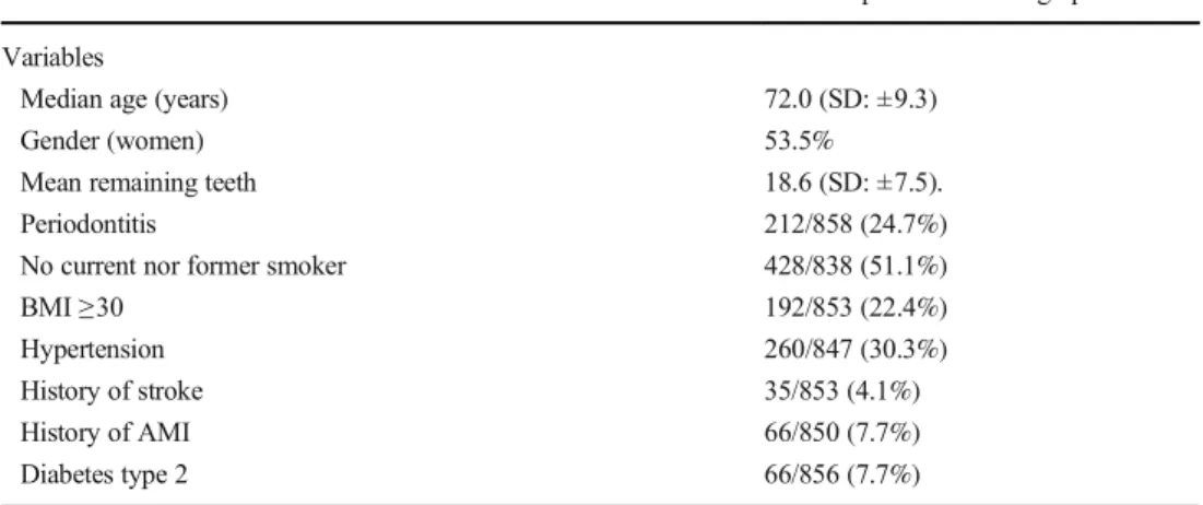
Data were derived from 858 individuals (women 53.5%). During the 17-year follow-up period, 492/858 (57.3%) died, and 51/858 (5.9%) moved away. The ages at base-line varied between 60 and 93 years with median age 72.0 years. On average, the individuals had 18.6 remain-ing teeth (SD: ± 7.5). Approximately half of the individ-uals 428/838 (51.1%) reported that they did not or had never smoked. At the baseline examination, periodontitis was declared in 212/858 (24.7%) (Table 1). Men had a higher prevalence of periodontitis than women, 121/212 (57.1%) (OR: 1.8, CI: 1.3–2.4, p = 0.000). Data were derived from 858 individuals (women 53.5%) with 471/ 858 (54.9%) in ages 60-72 years, young old (YO), and 378/858 (45.1%) in ages 78-96 years, old-old (OO) 78– 96 years age group, including 471/858 (54.9%) respec-tively 387/858 (45.1%) individuals.
Periodontitis and incidence of ischemic heart diseases
or stroke during the follow-up period
The cumulative incidence of ischemic heart diseases between 2001 and 2018 was 203/858 (23.7%), with men 102/203 (50.2%) and women 101/203 (49.8%) (OR: 1.2, CI: 0.9–1.7, p = 0.221). The incidence of ischemic heart diseases was 57.2 incidences per year and 6668 per 100,000 and year. The cu-mulative incidence of stroke was 118/858 (13.8%), with 60/ 118 (50.8%) men respectively 58/118 (49.2%) women (OR:1.2, CI: 0.8-1.8, p=0.308). Stroke incidence was 24.86 per year, which corresponds to 2898 strokes per 100,000 per-sons and year.
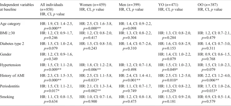
Cox regression analysis based on baseline data with peri-odontitis as an independent variable and incidence of the first event of a stroke or ischemic heart diseases as the dependent variable and with adjustment for the variables age group, BMI ≥ 30, diabetes type 2, gender, hypertension, history of acute myocardial infarction, history of stroke, and smoking, was used. Periodontitis increased the risk for ischemic heart dis-eases in all individuals (HR: 1.5, CI: 1.1–2.1, p = 0.017) (Fig.1), in women (HR: 2.1, CI: 1.3–3.4, p = 0.002), and in the OO group (HR: 1.7, CI: 1.0–2.6, p = 0.033) (Table2). No significant association was identified between periodontitis and stroke, in neither of all individuals, women, men, YO, and OO (Table3).
Associations to mortality during the follow-up period
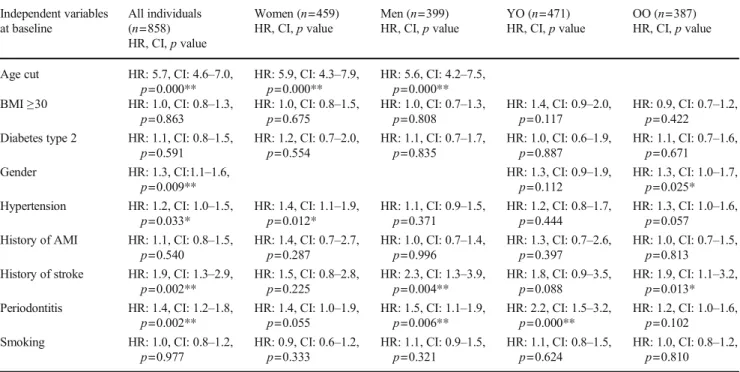
Data on the cause of death was not available, but during the 17-year follow-up period, 492/858 (57.3%) had died. Among those who had died, 160/492 (32.5%) had periodontitis whereas among those who were alive, 52/366 (14.2%) had periodontitis (OR: 2.9, CI: 2.1–41.1, p = 0.000). In individuals with periodontitis, 160/212 (75.5%) (62 women and 98 men) had died and 52/212 (24.5%) (29 women and 23 men) were still alive at the end of the study in 2018 (OR: 1.7, CI: 1.1–2.7, p = 0.028). Cox regression analysis with periodontitis as the independent variable and mortality as the dependent variable and with adjustment for the variables age group, BMI≥ 30, diabetes type 2, gender, hypertension, history of acute myo-cardial infarction (AMI), history of stroke, and smoking, was used. Periodontitis increased the risk for all-cause mortality in all individuals (HR: 1.4, CI:1.2–1.8, p = 0.002) (Fig.2), in men (HR: 1.5, CI: 1.1–1.9, p = 0.006), and in the YO group (HR: 2.2, CI: 1.5–3.2, p = 0.000). No associations in women or the OO individuals between periodontitis and mortality were identified (HR: 1.4, CI: 1.0–1.9, p = 0.055), respectively (HR: 1.2, CI: 1.0–1.6, p = 0.102) (Table4).
A significant association was found between periodon-titis and the number of lost teeth (OR: 1.1, CI: 1.1–1.1, p = 0.000) using binary logistic regression analysis. In a second Cox regression analysis, the number of lost teeth
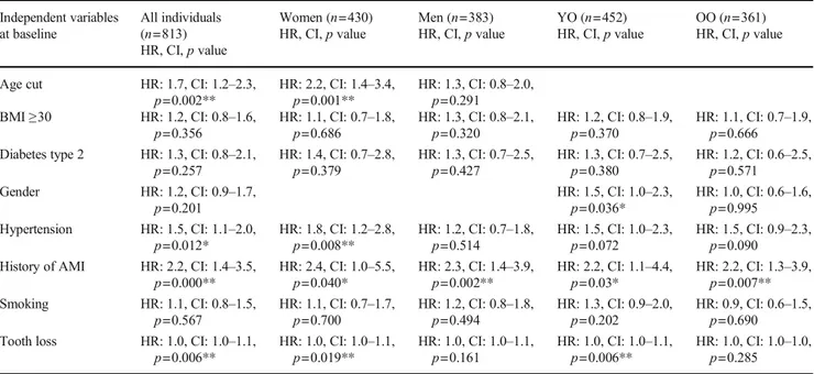
at baseline was used as a proxy for periodontitis with the number of lost teeth as an independent variable and in-cidence of the first event of a stroke or ischemic heart diseases as the dependent variable and with adjustment for the following variables: age group, BMI ≥ 30, diabe-tes type 2, gender, hypertension, history of acute myo-cardial infarction, history of stroke, and smoking. The number of lost teeth increased the risk for ischemic heart diseases in all individuals (HR: 1.0, CI: 1.0–1.1, p = 0.006), in women (HR: 1.0, CI: 1.0–1.1, p = 0.019), and in the YO group (HR: 1.0, CI: 1.0–1.1, p = 0.006) (Table 5). No significant association was identified be-tween the number of lost teeth and stroke, in neither of all individuals, women, men, YO, and OO (Table 6). Using the number of lost teeth as an independent vari-able and incidence of mortality as the dependent varivari-able
adjusting for the same variables as above, the number of lost teeth increased the risk for mortality in all individ-uals (HR: 1.0, CI: 1.0–1.0, p = 0.000) and in women (HR: 1.0, CI: 1.0–1.0, p = 0.000) (Table 7).
Discussion
The present study identified that over the 17-year follow-up period, periodontitis increased the risk of future incidences of ischemic heart diseases in all individuals, in women, and in the OO age group. In woman with periodontitis, the HR for the incidence of ischemic heart disease was 2.1. Another study has confirmed that after the menopause, women have a higher incidence of AMI compared to age-matched men [22] which is in agreement with the results of the present study in older Table 1 Baseline 2001–2003,
characteristics of the study individuals
≥1 remaining tooth, dental examination, readable panoramic radiographn=858 Variables
Median age (years) 72.0 (SD: ±9.3)
Gender (women) 53.5%
Mean remaining teeth 18.6 (SD: ±7.5).
Periodontitis 212/858 (24.7%)
No current nor former smoker 428/838 (51.1%)
BMI≥30 192/853 (22.4%)
Hypertension 260/847 (30.3%)
History of stroke 35/853 (4.1%)
History of AMI 66/850 (7.7%)
Diabetes type 2 66/856 (7.7%)
AMI acute myocardial infarction, BMI body mass index, Periodontitis = alveolar bone loss ≥ 5-mm distance from the CEJ to marginal bone level on≥ 30% of sites. Self-reported at baseline: no current nor former smoker, hypertension, history of stroke, history of AMI, and diabetes type 2
Fig. 1 Cox regression curves: 17-year cumulative ischemic heart disease survival of the total pop-ulation, comparing individuals with and without periodontitis
people. There are few other longitudinal studies concerning periodontitis and incidences of CVDs. In a Danish national register–based cohort study with a follow-up period of 15 years, patients with periodontitis were reported to have an increased risk of CVDs [23]. In the study by Hansen et al. (2013), ICD codes were used to define periodontitis.
This classification of periodontitis is different from the one used in the present study. Additionally, the age of the study population was > 18 years, and gender differences were not reported making comparisons between results observed in the present study impossible. A Korean nationwide cohort follow-up study of 7.6 years showed a dose-dependent association
Table 3 Associations between stroke and different independent variables including periodontitis by Cox regression analysis Independent variables at baseline All individuals (n=858) HR, CI,p value Women (n=459) HR, CI,p value Men (n=399) HR, CI,p value YO (n=471) HR, CI,p value OO (n=387) HR, CI,p value
Age cut HR: 3.4, CI: 2.3–5.1,
p=0.000** HR: 3.6, CI: 2.0p=0.000** –6.4, HR: 3.2, CI: 1.8p=0.000** –5.7, BMI≥30 HR: 0.7, CI: 0.4–1.1,
p=0.157 HR: 0.7, CI: 0.3p=0.282 –1.4, HR: 0.8, CI: 0.4p=0.479 –1.6, HR: 1.0, CI: 0.5p=0.953 –2.0, HR: 0.5, CI: 0.3p=0.098 –1.1, Diabetes type 2 HR: 0.8, CI: 0.4–1.7,
p=0.613 HR: 0.6, CI: 0.2p=0.474 –2.2, HR: 1.0, CI: 0.4p=0.924 –2.7, HR: 0.4, CI: 0.1p=0.245 –1.8, HR: 1.2, CI: 0.5p=0.704 –2.8, Gender HR: 1.3, CI: 0.9–1.9,
p=0.188 HR: 1.2, CI: 0.6p=0.587 –2.2, HR: 1.3, CI: 0.8p=0.294 –2.2, Hypertension HR: 1.8, CI: 1.2–2.6,
p=0.006** HR: 2.1, CI: 1.2p=0.013* –3.6, HR: 1.6, CI: 0.9p=0.118 –2.8, HR: 1.7, CI: 0.9p=0.128 –3.1, HR: 1.8, CI: 1.1p=0.019* –3.1, History of stroke HR: 3.7, CI: 2.0–6.9,
p=0.000** HR: 2.7, CI: 1.0p=0.052 –7.5, HR: 4.6, CI:2.1–10.2, p= 0.000** HR: 6.2, CI: 2.7–14.4, p= 0.000** HR: 2.3, CI: 0.9–6.1, p=0.088 Periodontitis HR: 1.2, CI: 0.8–1.8,
p=0.442 HR: 1.3, CI: 0.7–2.4,p=0.485 HR: 1.2, CI: 0.6–2.1,p=0.627 HR: 1.1, CI: 0.5–2.3,p=0.865 HR: 1.2, CI: 0.7–2.0,p=0.535 Smoking HR: 1.0, CI: 0.7–1.5,
p=0.915 HR: 0.8, CI: 0.4–1.4,p=0.381 HR: 1.2, CI: 0.7–2.1,p=0.544 HR: 1.2, CI: 0.7–2.3,p=0.496 HR: 0.9, CI: 0.6–1.6,p=0.834 BMI body mass index, Periodontitis = alveolar bone loss ≥ 5-mm distance from the CEJ to marginal bone level on ≥ 30% of sites, YO young-old 60– 72 years,OO old-old 78–93 years, *p < 0.05, **p < 0.01
Table 2 Associations between ischemic heart diseases and different independent variables including periodontitis by Cox regression analysis Independent variables at baseline All individuals (n=858) HR, CI,p value Women (n=459) HR, CI,p value Men (n=399) HR, CI,p value YO (n=471) HR, CI,p value OO (n=387) HR, CI,p value
Age category HR: 1.9, CI: 1.4–2.5,
p=0.000** HR: 2.5, CI: 1.6p=0.000** –3.8, HR: 1.4, CI: 0.9p=0.098 –2.2, BMI≥30 HR: 1.2, CI: 0.9–1.7,
p=0.246 HR: 1.2, CI: 0.8–2.0,p=0.417 HR: 1.3, CI: 0.8–2.2,p=0.304 HR: 1.3, CI: 0.8–2.0,p=0.284 HR: 1.2, CI: 0.7–2.1,p=0.479 Diabetes type 2 HR: 1.5, CI: 1.0–2.4,
p=0.079 HR: 1.5, CI: 0.8–3.0,p=0.243 HR: 1.4, CI: 0.7–2.6,p=0.310 HR: 1.6, CI: 0.8–2.9,p=0.153 HR: 1.4, CI: 0.7–3.0,p=0.311 Gender HR: 1.2, CI: 0.9–1.6,
p=0.349 HR: 1.4, CI: 1.0–2.2,p=0.079 HR: 0.9, CI: 0.6–1.5,p=0.768 Hypertension HR: 1.5, CI: 1.1–2.0,
p=0.009** HR: 1.8, CI: 1.2–2.8,p=0.006** HR: 1.2, CI: 0.7–1.8,p=0.498 HR: 1.5, CI: 1.0–2.3,p=0.049* HR: 1.5, CI: 1.0–2.3,p=0.079 History of AMI HR: 2.3, CI: 1.5–3.5,
p=0.000** HR: 2.5, CI: 1.1–5.8,p=0.033* HR: 2.4, CI: 1.4–4.1,p=0.001** HR: 2.5, CI: 1.2–5.0,p=0.010* HR: 2.2, CI: 1.2–4.0,p=0.006** Periodontitis HR: 1.5, CI: 1.1–2.1,
p=0.017* HR: 2.1, CI: 1.3–3.4,p=0.002** HR: 1.1, CI: 0.7–1.7,p=0.749 HR: 1.3, CI: 0.8–2.2,p=0.229 HR: 1.7, CI: 1.0–2.6,p=0.033* Smoking HR: 1.1, CI: 0.8–1.5,
p=0.634 HR: 1.0, CI: 0.7–1.6,p=0.900 HR: 1.2, CI: 0.8–1.8,p=0.475 HR: 1.3, CI: 0.9–2.0,p=0.181 HR: 0.9, CI: 0.5–1.4,p=0.579 AMI acute myocardial infarction, BMI body mass index, Periodontitis = alveolar bone loss ≥ 5-mm distance from the CEJ to marginal bone level on ≥ 30% of sites,YO young-old 60–72 years, OO old-old 78–93 years, *p < 0.0.5, **p < 0.01
with tooth loss and incident myocardial infarction, heart fail-ure, and ischemic stroke, especially in individuals with peri-odontitis [24]. The circumstances between the study above and our study are not the same. The classification of periodon-titis was in the study by Lee et al. [24] not defined, and the study individuals included were from 20 years, and the mean age not mentioned. Also, the included individuals had a his-tory of a CVD event, meaning that they were at an increased risk for a subsequent CVD [25].
Another main finding in this study was that individuals with periodontitis were at a higher risk to die during the
17-year follow-up compared to individuals without a diagnosis of periodontitis. Recently published data have shown that peri-odontitis increased the risk for all-cause mortality within 15 years [23]. In a 3-year follow-up study, individuals with severe periodontitis developed the combined endpoint (myo-cardial infarction, stroke/transient ischemic attack, cardiovas-cular death, and death caused by stroke) more often compared to individuals without periodontitis (18.9% versus 14.2%) [26]. As we were unaware of the exact reasons for death in the present study, such a combined endpoint was not possible to include. In the present study, it was demonstrated that in
Table 4 Associations between mortality and different independent variables including periodontitis by Cox regression analysis Independent variables at baseline All individuals (n=858) HR, CI,p value Women (n=459) HR, CI,p value Men (n=399) HR, CI,p value YO (n=471) HR, CI,p value OO (n=387) HR, CI,p value
Age cut HR: 5.7, CI: 4.6–7.0,
p=0.000** HR: 5.9, CI: 4.3p=0.000** –7.9, HR: 5.6, CI: 4.2p=0.000** –7.5, BMI≥30 HR: 1.0, CI: 0.8–1.3,
p=0.863 HR: 1.0, CI: 0.8–1.5,p=0.675 HR: 1.0, CI: 0.7–1.3,p=0.808 HR: 1.4, CI: 0.9–2.0,p=0.117 HR: 0.9, CI: 0.7–1.2,p=0.422 Diabetes type 2 HR: 1.1, CI: 0.8–1.5,
p=0.591 HR: 1.2, CI: 0.7–2.0,p=0.554 HR: 1.1, CI: 0.7–1.7,p=0.835 HR: 1.0, CI: 0.6–1.9,p=0.887 HR: 1.1, CI: 0.7–1.6,p=0.671 Gender HR: 1.3, CI:1.1–1.6,
p=0.009** HR: 1.3, CI: 0.9–1.9,p=0.112 HR: 1.3, CI: 1.0–1.7,p=0.025* Hypertension HR: 1.2, CI: 1.0–1.5,
p=0.033* HR: 1.4, CI: 1.1–1.9,p=0.012* HR: 1.1, CI: 0.9–1.5,p=0.371 HR: 1.2, CI: 0.8–1.7,p=0.444 HR: 1.3, CI: 1.0–1.6,p=0.057 History of AMI HR: 1.1, CI: 0.8–1.5,
p=0.540 HR: 1.4, CI: 0.7–2.7,p=0.287 HR: 1.0, CI: 0.7–1.4,p=0.996 HR: 1.3, CI: 0.7–2.6,p=0.397 HR: 1.0, CI: 0.7–1.5,p=0.813 History of stroke HR: 1.9, CI: 1.3–2.9,
p=0.002** HR: 1.5, CI: 0.8–2.8,p=0.225 HR: 2.3, CI: 1.3–3.9,p=0.004** HR: 1.8, CI: 0.9–3.5,p=0.088 HR: 1.9, CI: 1.1–3.2,p=0.013* Periodontitis HR: 1.4, CI: 1.2–1.8,
p=0.002** HR: 1.4, CI: 1.0–1.9,p=0.055 HR: 1.5, CI: 1.1–1.9,p=0.006** HR: 2.2, CI: 1.5–3.2,p=0.000** HR: 1.2, CI: 1.0–1.6,p=0.102 Smoking HR: 1.0, CI: 0.8–1.2,
p=0.977 HR: 0.9, CI: 0.6–1.2,p=0.333 HR: 1.1, CI: 0.9–1.5,p=0.321 HR: 1.1, CI: 0.8–1.5,p=0.624 HR: 1.0, CI: 0.8–1.2,p=0.810 AMI acute myocardial infarction, BMI body mass index, Periodontitis = alveolar bone loss ≥ 5-mm distance from the CEJ to marginal bone level on ≥ 30% of sites,YO young-old 60–72 years, OO old-old 78–93 years, *p < 0.05, **p < 0.01
Fig. 2 Cox regression curves: 17-year cumulative death survival of the total study population, com-paring individuals with and with-out periodontitis
men and the YO (60–72 years), there was an association be-tween periodontitis and mortality. Young individuals (30– 40 years) with periodontitis and missing molars have been reported to have an increased risk for early death over 16 years [27]. Missing teeth, due to periodontal disease, could be a proxy for a previous inflammation and partly explain the
identified association with an increased risk for mortality among young-old in the present study that is in line with the results from the study by Lee et al. [24]. In another recent study,≥ nine missing teeth were also associated with mortality [28]. In the present study, an association between periodontitis and the number of lost teeth at baseline was found. When lost Table 5 Associations between ischemic heart diseases and different independent variables including the number of lost teeth by Cox regression analysis Independent variables at baseline All individuals (n=813) HR, CI,p value Women (n=430) HR, CI,p value Men (n=383) HR, CI,p value YO (n=452) HR, CI,p value OO (n=361) HR, CI,p value
Age cut HR: 1.7, CI: 1.2–2.3,
p=0.002** HR: 2.2, CI: 1.4p=0.001** –3.4, HR: 1.3, CI: 0.8p=0.291 –2.0, BMI≥30 HR: 1.2, CI: 0.8–1.6,
p=0.356 HR: 1.1, CI: 0.7–1.8,p=0.686 HR: 1.3, CI: 0.8–2.1,p=0.320 HR: 1.2, CI: 0.8–1.9,p=0.370 HR: 1.1, CI: 0.7–1.9,p=0.666 Diabetes type 2 HR: 1.3, CI: 0.8–2.1,
p=0.257 HR: 1.4, CI: 0.7–2.8,p=0.379 HR: 1.3, CI: 0.7–2.5,p=0.427 HR: 1.3, CI: 0.7–2.5,p=0.380 HR: 1.2, CI: 0.6–2.5,p=0.571 Gender HR: 1.2, CI: 0.9–1.7,
p=0.201 HR: 1.5, CI: 1.0–2.3,p=0.036* HR: 1.0, CI: 0.6–1.6,p=0.995 Hypertension HR: 1.5, CI: 1.1–2.0,
p=0.012* HR: 1.8, CI: 1.2–2.8,p=0.008** HR: 1.2, CI: 0.7–1.8,p=0.514 HR: 1.5, CI: 1.0–2.3,p=0.072 HR: 1.5, CI: 0.9–2.3,p=0.090 History of AMI HR: 2.2, CI: 1.4–3.5,
p=0.000** HR: 2.4, CI: 1.0–5.5,p=0.040* HR: 2.3, CI: 1.4–3.9,p=0.002** HR: 2.2, CI: 1.1–4.4,p=0.03* HR: 2.2, CI: 1.3–3.9,p=0.007** Smoking HR: 1.1, CI: 0.8–1.5,
p=0.567 HR: 1.1, CI: 0.7–1.7,p=0.700 HR: 1.2, CI: 0.8–1.8,p=0.494 HR: 1.3, CI: 0.9–2.0,p=0.202 HR: 0.9, CI: 0.6–1.5,p=0.690 Tooth loss HR: 1.0, CI: 1.0–1.1,
p=0.006** HR: 1.0, CI: 1.0–1.1,p=0.019** HR: 1.0, CI: 1.0–1.1,p=0.161 HR: 1.0, CI: 1.0–1.1,p=0.006** HR: 1.0, CI: 1.0–1.0,p=0.285 AMI acute myocardial infarction, BMI body mass index, Tooth loss = number of lost teeth, YO young-old 60–72 years, OO old-old 78–93 years, *p < 0.05, **p < 0.01
Table 6 Associations between stroke and different independent variables including the number of lost teeth by Cox regression analysis Independent variables at baseline All individuals (n=817) HR, CI,p value Women (n=431) HR, CI, p value Men (n=385) HR, CI,p value YO (n=447) HR, CI,p value OO (n=366) HR, CI,p value
Age cut HR: 3.3, CI: 2.1–5.1,
p=0.000** HR: 3.2, CI: 1.7p=0.000** –6.0, HR: 3.4, CI: 1.9p=0.000** –6.2, BMI≥30 HR: 0.7, CI: 0.4–1.1,
p=0.140 HR: 0.7, CI: 0.3p=0.256 –1.3, HR: 0.8, CI: 0.4p=0.454 –1.5, HR: 1.0, CI: 0.5p=0.961 –2.0, HR: 0.5, CI: 0.3p=0.088 –1.1, Diabetes type 2 HR: 0.8, CI: 0.4–1.7,
p=0.542 HR: 0.6, CI: 0.2p=0.422 –2.1, HR: 1.0, CI: 0.4p=0.958 –2.6, HR: 0.4, CI: 0.1p=0.232 –1.8, HR: 1.1, CI: 0.5p=0.754 –2.7, Gender HR: 1.3, CI: 0.9–2.0,
p=0.151 HR: 1.2, CI: 0.7p=0.554 –2.2, HR: 1.3, CI: 0.8p=0.272 –2.3, Hypertension HR: 1.7, CI: 1.2–2.6,
p=0.007** HR: 2.0, CI: 1.1–3.5,p=0.020* HR: 1.6, CI: 0.9–2.7,p=0.129 HR: 1.6, CI: 0.8–3.1,p=0.147 HR: 1.8, CI: 1.1–3.1,p=0.021* History of stroke HR: 3.6, CI: 1.9–6.6,
p=0.000** HR: 2.5, CI: 0.9p=0.070 –6.9, HR: 4.6, CI:2.1–10.1, p=0.000**
HR: 6.1, CI: 2.6–14.1,
p=0.000** HR: 2.2, CI: 0.9p=0.103 –5.8, Smoking HR: 1.0, CI: 0.7–1.5,
p=0.909 HR: 0.8, CI: 0.4–1.4,p=0.353 HR: 1.2, CI: 0.7–2.2,p=0.483 HR: 1.2, CI: 0.7–2.2,p=0.535 HR: 1.0, CI: 0.6–1.6,p=0.881 Tooth loss HR: 1.0, CI: 1.0–1.0,
p=0.593 HR: 1.0, CI: 1.0–1.1,p=0.249 HR: 1.0, CI: 1.0–1.0,p=0.717 HR: 1.0, CI: 1.0–1.1,p=0.552 HR: 1.0, CI: 1.0–1.0,p=0.921 BMI body mass index, Tooth loss = number of lost teeth, YO young-old 60–72 years, OO old-old 78–93 years, *p < 0.05, **p < 0.01
teeth at baseline were used as a proxy for periodontitis, an association between all-cause mortality in all individuals was found. Such an association is in line with the results obtained using periodontitis as the independent variable in the Cox regression analysis. However, lost teeth at baseline were also associated with mortality among women and to an increased risk for ischemic heart diseases in all individuals, in women, and in the YO group. Accordingly, using lost teeth as a proxy for periodontitis does not seem to be an adequate approximation for periodontal disease.
Deaths by CVDs have decreased during the latest 60 years, as a result of preventive care and advances in medicine [29]. Many older individuals are using preventive medications that may delay or even prevent a CVD event from occurring [30]. Recent data reported that the numbers of missing teeth were related to heart failure and myocardial infarction. In con-trast, the number of teeth missing was not significantly related to stroke in a longitudinal study with a median follow-up time of 15.8 years [31]. In another study, the same tendency was reported; missing teeth (≥ five missing teeth) were statistically associated with an event of coronary heart disease and acute myocardial infarction. In contrast, tooth loss was not associ-ated with stroke in a 13-year prospective longitudinal study [28].
In the literature, different classifications and parameters for periodontitis have been used. Missing teeth have also been proposed as a proxy for current or past periodontitis as it is considered to reflect an accumulation of oral inflammation
[32]. Correctly, if untreated, periodontitis may result in tooth loss and is one of the primary reasons for tooth loss in adult-hood [33]. In a recent study by Lee et al. [24], edentulous individuals demonstrated the highest cardiovascular risk. It is, however, difficult to be sure of the reason for missing teeth unless it is reported in the dental records. In the present study, it was not possible to within certainty decide the reason for tooth loss. Alveolar bone loss, as used in the present study as a proxy for periodontal disease, is an indicator of a patient who has had periodontal inflammation during a period.
The prevalence of periodontitis defined by loss of al-veolar bone was relatively low (24.7%). The reported prevalence of periodontitis in individuals 65 years and older has been reported to be 70% in the USA and with increasing prevalence with increasing age [3]. Differences in prevalence may be related to the different classifica-tions of periodontitis used. Eke et al. [3], when defining “total periodontitis,” included mild, moderate, and severe stages of periodontitis and used clinical attachment–level loss as an indicator for periodontitis. The definition of periodontitis used in the present study is based on bone loss of≥ 5 mm from the CEJ to the alveolar bone level on ≥ 30% of sites. The use of the 5-mm level was chosen to account for possible measurement errors. Individuals de-fined as mild periodontitis cases in the study by Eke et al. [3] were not defined as periodontitis patients in the pres-ent study, which to some degree may explain the differ-ences in prevalence figures reported in the two studies. Table 7 Associations between mortality and different independent variables including the number of lost teeth by Cox regression analysis Independent variables at baseline All individuals (n= 810) HR, CI,p value Women (n=427) HR, CI,p value Men (n=383) HR, CI,p value YO (n=448) HR, CI,p value OO (n=362) HR, CI,p value
Age cut HR: 5.0, CI: 4.0–6.2,
p=0.000** HR: 4.8, CI: 3.5p=0.000** –6.6, HR: 5.2, CI: 3.8p=0.000** –7.1, BMI≥30 HR: 1.0, CI: 0.8–1.2,
p=0.913 HR: 1.0, CI: 0.7–1.4,p=0.839 HR: 0.9, CI: 0.7–1.3,p=0.658 HR: 1.3, CI: 0.9–1.9,p=0.219 HR: 0.9, CI: 0.6–1.2,p=0.347 Diabetes type 2 HR: 1.0, CI: 0.7–1.3,
p=0.806 HR: 1.0, CI: 0.6–1.8,p=0.815 HR: 0.9, CI: 0.6–1.4,p=0.641 HR: 0.8, CI: 0.4–1.5,p=0.528 HR: 1.0, CI: 0.7–1.5,p=0.943 Gender HR: 1.4, CI: 1.1–1.7,
p=0.002** HR: 1.5, CI: 1.0–2.1,p=0.022 HR: 1.4, CI: 1.1–1.7,p=0.014 Hypertension HR: 1.2, CI: 1.0–1.5,
p=0.040* HR: 1.4, CI: 1.0–1.9,p=0.025* HR: 1.1, CI: 0.8–1.5,p=0.393 HR: 1.1, CI: 0.8–1.6,p=0.597 HR: 1.3, CI: 1.0–1.6,p=0.060 History of AMI HR: 1.1, CI: 0.8–1.6,
p=0.403 HR: 1.5, CI: 0.8–2.9,p=0.212 HR: 1.0, CI: 0.7–1.5,p=0.813 HR: 1.2, CI: 0.6–2.3,p=0.580 HR: 1.1, CI: 0.7–1.5,p=0.710 History of stroke HR: 1.7, CI: 1.1–2.5,
p=0.012* HR: 1.3, CI: 0.7–2.4,p=0.426 HR: 2.0, CI: 1.2–3.5,p=0.013* HR: 1.7, CI: 0.8–3.3,p=0.149 HR: 1.8, CI: 1.1–2.9,p=0.032 Smoking HR: 1.0, CI: 0.8–1.2,
p=0.956 HR: 0.9, CI: 0.6–1.2,p=0.337 HR: 1.2, CI: 0.9–1.5,p=0.311 HR: 1.1, CI: 0.8–1.6,p=0.555 HR: 1.0, CI: 0.8–1.2,p=0.828 Tooth loss HR: 1.0, CI: 1.0–1.0,
p=0.000** HR: 1.0, CI: 1.0–1.1,p=0.000** HR: 1.0, CI: 1.0–1.0,p=0.050 HR: 1.1, CI: 1.0–1.1,p=0.000** HR: 1.0, CI: 1.0–1.0,p=0.090 AMI acute myocardial infarction, BMI body mass index, Tooth loss = number of lost teeth, YO young-old 60–72 years, OO old-old 78–93 years, *p < 0.05, **p < 0.01
Individuals affected with bone loss in older ages may reflect a long history of periodontitis and accordingly a long time of an inflammatory response.
The new accepted classification of periodontitis in-cludes clinical variables (CAL and probing pocket depth) as well as bone loss in radiographs [34]. The bone loss reflects the accumulated progressive effect of periodontitis over a long time [35, 36]. Clinical parameters partly reflecting the inflammatory activity could be more tran-sient, giving the information of the periodontal status at that specific moment. Studies have reported that the mean proportion of bone loss increased with age, but the pro-portion of teeth with periodontal pockets remained un-modified. In another study, the alveolar bone loss progressed with age but was limited after the age of 50 [37]. In older individuals, gingival recession is the main reason for attachment loss [38]. Crestal bone height and CAL have shown a good correlation [39]. It has been proven that attachment loss precedes radiographic evi-dence of crestal alveolar bone loss during periods of peri-odontal disease activity [40] whereas, over time, these differences seem to level off [39]. In our study, the limit of alveolar bone loss≥ 5-mm distance from CEJ to mar-ginal bone level reflects a definitive bone loss and such a bone loss is present on≥ 30% of sites corresponding to a general disease distribution.
One limitation with the present study was that the most fragile and medically compromised individuals were not able to participate. The fact that the most fragile individuals did not participate may have affected the results, possibly lowering the associations between periodontitis and CVDs. The indi-viduals in the present study were 60 years or older. While the follow-up period was long, obviously many died during the study. The causes of death were not known, but the main reasons for death are still CVDs [41]. The consequence of not knowing the reasons for death makes it impossible to include mortality by CVDs in a combined endpoint, among others, which should have to strengthen the associations be-tween periodontitis and CVDs. The risk factors in the Cox regression analysis were adjusted only from the baseline data, which is another limitation in the study.
The long-time follow-up and that CVD events were easy to follow and control is a strength with the present study. It would be interesting to study if well-designed preventive den-tal programs can influence the incidence of CVDs in long-term studies. Intervention studies are needed to verify a valid link between periodontitis and CVDs.
In conclusion, this study demonstrated that in older adults, periodontitis was a statistical risk indicator for ischemic heart diseases. Over the time studied, periodontitis was significantly associated with mortality. This is the first long-time follow-up report on periodontitis and the incidence of cardiovascular diseases and death.
Acknowledgments We acknowledge the contribution by Associate Professor Rigmor Persson (REP), University of Washington, who ana-lyzed the radiographs. We also want to acknowledge Ms. Ingrid Jonasson, Blekinge Institute of Technology, Karlskrona, Sweden, who collected the clinical dental data and Ms. Johanna Renvert, University of Kristianstad, for data management. We are specifically grateful to the study participants. The study was accomplished within the context of the Swedish National Graduate School for Competitive Science on Aging and Health (SWEAH). The Ministry of Health and Social Affairs, Sweden; the participating county councils, municipalities, and university departments; and the Swedish National Study on Aging and Care (SNAC) (www.snac.org) supported the study. The study was also supported by the Research Foundation at Kristianstad University, Sweden.
Funding Open Access funding provided by Kristianstad University.
Compliance with ethical standards
Conflict of interest The authors declare that they have no conflict of interest.
Ethical approval All procedures performed in the study, involving hu-man participants, were following the ethical standards of the regional research ethics committee and with the 1964 Helsinki declaration and its later amendments.
Informed consent All individuals included in the study signed an in-formed consent.
Disclaimer The sponsors had no role in any aspect of the study design or reporting of findings.
Open Access This article is licensed under a Creative Commons Attribution 4.0 International License, which permits use, sharing, adap-tation, distribution and reproduction in any medium or format, as long as you give appropriate credit to the original author(s) and the source, pro-vide a link to the Creative Commons licence, and indicate if changes were made. The images or other third party material in this article are included in the article's Creative Commons licence, unless indicated otherwise in a credit line to the material. If material is not included in the article's Creative Commons licence and your intended use is not permitted by statutory regulation or exceeds the permitted use, you will need to obtain permission directly from the copyright holder. To view a copy of this licence, visithttp://creativecommons.org/licenses/by/4.0/.
References
1. Bartold PM, van Dyke TE (2013) Periodontitis: a host-mediated disrupture of microbial homeostasis unlearning learned concepts. Periodontology 2000(62):203–217. https://doi.org/10.1111/j.1600-0757.2012.00450.x
2. Kassebaum NJ, Bernabé E, Dahiya M, Bhandari B, Murray CJ, Marcenes W (2014) Global burden of severe periodontitis in 1990–2010: a systematic review and meta-regression. J Dent Res 93:1045–1053.https://doi.org/10.1177/0022034514552491
3. Eke PI, Dye BA, Wei L, Thornton-Evans GO, Genco RJ (2012) CDC Periodontal Disease Surveillance workgroup: James Beck, Gordon Douglass Roy Page. Prevalence of periodontitis in adults in the United States: 2009 and 2010. J Dent Res 91:914–920
4. Eke PI, Wei L, Borgnakke WS, Thornton-Evans G, Zhang X, Lu H, McGuire LC, Genco RJ (2016) Periodontitis prevalence in adults,≥ 65 years of age, in the USA. Periodontology 2000(72):76–95.
https://doi.org/10.1111/prd.12145
5. Waters AM, Trinh L, Chau T, Bourchier M, Moon L (2013) Latest statistics on cardiovascular disease in Australia. Clin Exp Pharmacol Physiol 40(347–356):10.111/1440–11681.12079 6. Roth GA, Johnson CO, Abate KH et al (2018) Global burden of
cardiovascular diseases collaboration, the burden of cardiovascular diseases among US states, 1990-2016. JAMA Cardiol 3:375–389.
https://doi.org/10.1001/jamacardio.2018.0385
7. Desvarieux M, Demmer RT, Rundek T, Boden-Albala B, Jacobs DR Jr, Papapanou PN, Sacco RL (2003) Oral Infections and Vascular Disease Epidemiology Study (INVEST). Relationship be-tween periodontal disease, tooth loss, and carotid artery plaque: the Oral Infections and Vascular Disease Epidemiology Study (INVEST). Stroke 34:2120–2125
8. Lockhart PB, Bolger AF, Papapanou PN et al (2012) American Heart Association Rheumatic Fever, Endocarditis, and Kawasaki Disease Committee of the Council on Cardiovascular Disease in the Young, Council on Epidemiology and Prevention, Council on Peripheral Vascular Disease, and Council on Clinical Cardiology. Periodontal disease and atherosclerotic vascular disease: does the evidence support an independent association? A scientific state-ment from the American Heart Association. Circulation 125: 2520–2544.https://doi.org/10.1161/CIR.0b013e31825719f3
9. Aoyama N, Suzuki JI, Kobayashi N, Hanatani T, Ashigaki N, Yoshida A, Shiheido Y, Sato H, Kumagai H, Ikeda Y, Akazawa H, Komuro I, Izumi Y, Isobe M (2017) Periodontitis deteriorates peripheral arterial disease in Japanese population via enhanced sys-temic inflammation. Heart Vessel 32:1314–1319.https://doi.org/ 10.1007/s00380-017-1003-6
10. Söder PO, Söder B, Nowak J, Jogestrand T (2005) Early carotid atherosclerosis in subjects with periodontal diseases. Stroke 36: 1195–2000
11. Scannapieco FA, Bush RB, Paju S (2003) Associations between periodontal disease and risk for atherosclerosis, cardiovascular dis-ease, and stroke. A systematic review. Annuals of Periodontology 8:38–53.https://doi.org/10.1902/annals.2003.8.1.38
12. Kinane D, Bouchard P, Group E of European Workshop on Periodontology (2008) J Clin Periodontol 35:333–337.https://doi. org/10.1111/j.1600-051X.2008.01278.x
13. Cullinan MP, Seymour GJ (2013) Periodontal diseases and health: consensus report of the Sixth European Workshop on Periodontology. Periodontology 2000 62:271–286.https://doi.org/ 10.1111/prd.12007
14. Reyes L, Herrera D, Kozarov E, Roldán S, Progulske-Fox AJ (2013) Periodontal bacterial invasion and infection: contribution to atherosclerotic pathology. Clinical Periodontology 40:S30– S50.https://doi.org/10.1111/jcpe.12079
15. Schenkein HA, Loos BG (2013) Inflammatory mechanisms linking periodontal diseases to cardiovascular diseases. Jorunal of Clinical Periodontology 40:S51–S69.https://doi.org/10.1111/jcpe.12060
16. Tonetti MS, Van Dyke TE (2013) Working group 1 of the joint EFP/AAP workshop. Periodontitis and atherosclerotic cardiovascu-lar disease: consensus report of the Joint EFP/AAP Workshop on Periodontitis and Systemic Diseases. J Periodontol 84(4 Suppl):24– 29.https://doi.org/10.1902/jop.2013.1340019
17. Lafon A, Pereira B, Dufour T et al (2014) Periodontal disease and stroke: a meta-analysis of cohort studies. Eur J Neurol 21(1155– 61):e66–e67.https://doi.org/10.1111/ene
18. Grau AJ, Becher H, Ziegler CM, Lichy C, Buggle F, Kaiser C, Lutz R, Bültmann S, Preusch M, Dörfer CE (2004) Periodontal disease as a risk factor for ischemic stroke. Stroke 35:496–501.https://doi. org/10.1161/01.STR.0000110789.20526.9D
19. Persson GR, Ohlsson O, Pettersson T (2003) Chronic periodontitis, a significant relationship with acute myocardial infarction. Eur Heart J 24(23):2108–2115.https://doi.org/10.1016/j.ehj.2003.10. 007
20. Rydén L, Buhlin K, Ekstrand E et al (2016) Periodontitis increases the risk of a first myocardial infarction: a report from the PAROKRANK study. Circulation 133:576–583.https://doi.org/ 10.1161/CIRCULATIONAHA.115.020324
21. Dietrich T, Sharma P, Walter C (2013) The epidemiological evi-dence behind the association between periodontitis and incident atherosclerotic cardiovascular disease. J Clin Periodontol 14:S70– S84.https://doi.org/10.1111/jcpe.12062
22. Stramba-Badiale M, Fox KM, Priori SG, Collins P, Daly C, Graham I, Jonsson B, Schenck-Gustafsson K, Tendera M (2006) Cardiovascular diseases in women: a statement from the policy conference of the European Society of Cardiology. Eur Heart J 27:994–1005
23. Hansen GM, Egeberg A, Holmstrup P, Hansen PR (2016) Relation of periodontitis to risk of cardiovascular and all-cause mortality (from a Danish Nationwide Cohort Study). Am J Cardiol 118: 489–493.https://doi.org/10.1016/j.amjcard.2016.05.036
24. Lee HJ, Choi EK, Park JB, Han KD, Oh S (2019) Tooth loss predicts myocardial infarction, heart failure, stroke, and death. J Dent Res 98:164–170.https://doi.org/10.1177/0022034518814829
25. Park HW, Yoon CH, Kang SH, Choi DJ, Kim HS, Cho MC, Kim YJ, Chae SC, Yoon JH, Gwon HC, Ahn YK, Jeong MH, KAMIR/ KorMI Registry (2013) Early- and late-term clinical outcome and their predictors in patients with ST-segment elevation myocardial infarction and non-ST segment elevation myocardial infarction. Int J Cardiol 169:254–261
26. Reichert S, Schulz S, Benten AC, Lutze A, Seifert T, Schlitt M, Werdan K, Hofmann B, Wienke A, Schaller HG, Schlitt A (2016) Periodontal conditions and incidence of new cardiovascular events among patients with coronary vascular disease. J Clin Periodontol 43:918–925.https://doi.org/10.1111/jcpe.12611
27. Söder B, Jin LJ, Klinge B, Söder PO (2007) Periodontitis and pre-mature death: a 16-year longitudinal study in a Swedish urban pop-ulation. J Periodontal Res 42:361–366
28. Liljestrand JM, Havulinna AS, Paju S, Männistö S, Salomaa V, Pussinen PJ (2015) Missing teeth predict incident cardiovascular events, diabetes, and death. J Dent Res 94:1055–1062.https://doi. org/10.1177/0022034515586352
29. GBD 2017 Risk Factor Collaborators (2018) Global, regional, and national comparative risk assessment of 84 behavioural, environ-mental and occupational, and metabolic risks or clusters of risks for 195 countries and territories, 1990-2017: a systematic analysis for the Global Burden of Disease Study 2017. Lancet 392:1923–1994.
https://doi.org/10.1016/S0140-6736(18)32225-6 Erratum in: Lancet 2019; 393:132. Lancet 2019;393:e44
30. van der Vorst EPC, Peters LJF, Müller M, Gencer S, Yan Y, Weber C, Döring Y (2019) G-protein coupled receptor targeting on mye-loid cells in atherosclerosis. Front Pharmacology 10:531.https:// doi.org/10.3389/fphar.2019.00531
31. Holmlund A, Lampa E, Lind L (2017) Oral health and cardiovas-cular disease risk in a cohort of periodontitis patients. Atherosclerosis 262:101–106. https://doi.org/10.1016/j. atherosclerosis.2017.05.009
32. Holmlund A, Lind L (2012) Number of teeth is related to athero-sclerotic plaque in the carotid arteries in an elderly population. J Periodontol 83:287–291.https://doi.org/10.1902/jop.2011.110100
33. Natto ZS, Aladmawy M, Alasqah M, Papas A (2014) Factors con-tributing to tooth loss among the elderly: a cross-sectional study. Singap Dent J 35:17–22.https://doi.org/10.1016/j.sdj.2014.11.002
34. Papapanou PN, Sanz M, Buduneli NS, Dietrich T, Feres M, Fine DH, Flemmig TF, Garcia R, Giannobile WV, Graziani F, Greenwell H, Herrera D, Kao RT, Kebschull M, Kinane DF,
Kirkwood KL, Kocher T, Kornman KS, Kumar PS, Loos BG, Machtei E, Meng H, Mombelli A, Needleman I, Offenbacher S, Seymour GJ, Teles R, Tonetti MS (2018) Periodontitis: consensus report of workgroup 2 of the 2017 World Workshop on the Classification of Periodontal and Peri-Implant Diseases and Conditions. J Periodontol 89(Suppl 1):173–182.https://doi.org/ 10.1002/JPER.17-0721
35. Needleman I, Garcia R, Gkranias N (2018) Mean annual attach-ment, bone level, and tooth loss: a systematic review. J Periodontol 89(Suppl 1):120–139.https://doi.org/10.1002/JPER.17-0062
36. Reddy MS, Geurs NC, Jeffcoat RL, Proskin H, Jeffcoat MK (2000) Periodontal disease progression. J Periodontol 71:1583–1590 37. Persson RE, Hollender LG, Persson GR (1998) Assessment of
al-veolar bone levels from intraoral radiographs in subjects between ages 15 and 94 years seeking dental care. J Clin Periodontol 25: 647–654
38. Billings M, Holtfreter B, Papapanou PN, Mitnik GL, Kocher T, Dye BA (2018) Age-dependent distribution of periodontitis in
two countries: findings from NHANES 2009 to 2014 and SHIP-TREND 2008 to 2012. J Periodontol 89(Suppl. 1):140–158.https:// doi.org/10.1002/JPER.17-0670
39. Machtei EE, Hausmann E, Grossi SG, Dunford R, Genco RJ (1997) The relationship between radiographic and clinical changes in the periodontium. J Periodontal Res 32(8):661–666
40. Goodson JM, Haffajee AD, Socransky SS (1984) The relationship between attachment level loss and alveolar bone loss. Journal of Clinal Periodontology 11:348–359
41. GBD (2017) Causes of Death Collaborators (2018) Global, region-al, and national age-sex-specific mortality for 282 causes of death in 195 countries and territories, 1980-2017: a systematic analysis for the Global Burden of Disease Study 2017. Lancet 10:1736–1788.
https://doi.org/10.1016/S0140-6736(18)32203-7
Publisher’s note Springer Nature remains neutral with regard to jurisdic-tional claims in published maps and institujurisdic-tional affiliations.