Faculty of Veterinary Medicine and Animal Science
Genetic variation in genes associated with
canine brachycephaly
Genetisk variation i gener associerade med brakycefali hos
hund
Genetic variation in genes associated with brachycephaly
Genetisk variation i gener associerade med brakycefali
Elin Johansson
Supervisor: Katja Nilsson, Swedish University of Agricultural Sciences, Department of Animal Breeding and Genetics
Examiner: Erling Strandberg, Swedish University of Agricultural Sciences, Department of Animal Breeding and Genetics
Credits: 30 credits
Level: Second cycle, A2E
Course title: Independent project in Animal Science
Course code: EX0872
Course coordinating department: Department of Animal Breeding and Genetics
Place of publication: Uppsala
Year of publication: 2019
Cover picture: Elin Johansson
Online publication: https://stud.epsilon.slu.se
Keywords: Brachycephaly, dog, genetic variation, SMOC2, BMP3, DVL2
Domestication followed by controlled breeding has generated many dog breeds, displaying high morphological variation. One type of dogs with a dis-tinct morphology is brachycephalic dogs, characterized by a shortened muz-zle, a wide head and widely spaced eyes. The phenotype of brachycephalic dogs has been associated with several health issues, where one of the most obvious is the Brachycephalic Obstructive Airway syndrome (BOAS) which affects the breathing capacity and thermoregulation of the dog. Several stud-ies have aimed to identify the molecular background of the shorter snouts in brachycephalic dogs. So far, mutations in genes such as Bone Morphogenic Protein 3 (BMP3), Fibroblast growth factor 4 (FGF4), SPARC-related mod-ular calcium binding gene 2 (SMOC2) and DISHEVELLED 2 (DVL2) are thought to be associated with the altered skull shape causing canine brachy-cephaly. The aim of this master thesis was to investigate the genetic variation in the genes SMOC2, BMP3, and DVL2 in the Swedish population of four brachycephalic breeds; Boston Terrier, English Bulldog, French Bulldog and Pug. 102 privately owned brachycephalic dogs were genotyped for the muta-tion in the SMOC2 gene, addimuta-tionally, 45 of these dogs were also genotyped for the mutations in the BMP3 and DVL2 gene.
The mutant variant of the SMOC2 gene was fixated in all four breeds. The mutant variant of the BMP3 gene had high allele frequency in all breeds. In Bull type breeds (Boston Terrier, English Bulldog and French Bulldog) the mutant version of the DVL2 gene was fixated, whilst all Pugs were tested wild type for the DVL2 mutation. Low genetic variation will make it more chal-lenging to improve health through genetic selection within the breeds.
Keywords: Brachycephaly, dog, genetic variation, SMOC2, BMP3, DVL
Abstract
Domesticering följt av kontrollerad avel har genererat många hundraser med stora variationer i utseende. En typ av hundar som uppvisar ett distinkt utse-ende är brakycefala (trubbnosiga) hundar, karaktäriserat av en kort nos, brett huvud och med stort avstånd mellan ögonen. Fenotypen hos brakycefala hun-dar har kopplats till hälsoproblem där ett av de mest påtagliga problemen är Brachycephalic obstructive airway syndrome (BOAS), ett syndrom som på-verkar hundens andning och förmåga att reglera sin kroppstemperatur. Fler-talet studier har utförts för att identifiera de molekylära bakomliggande orsa-kerna till den kortare nosen hos brakycefala hundar. Hittills har mutationer i gener så som Bone Morphogenic Protein 3 (BMP3), Fibroblast growth factor 4 (FGF4), SPARC-related modular calcium binding gene 2 (SMOC2) och DISHEVELLED 2 (DVL2) kunnat kopplas till den avvikande huvudformen hos brakycefala hundar. Syftet för detta examensarbete var att undersöka den genetiska variationen i generna SMOC2, BMP3 och DVL2 för den svenska populationen av bostonterrier, engelsk bulldogg, fransk bulldogg och mops. Totalt genotypades 102 privatägda brakycefala hundar för mutationen i
SMOC2 genen, dessutom genotypades 45 av dessa hundar för mutationerna i BMP3 och DVL2. Denna studie pekar på att mutationen i SMOC2 var fixerad
i de fyra raserna. I de fyra raserna var allelfrekvensen för den muterade BMP3 allelen hög. I de tre raserna av bull-typ (bostonterrier, engelsk bulldogg och fransk bulldogg) var mutationen i DVL2 fixerad, medan alla mopsar var ge-notypade som vildtyp. Låg genetisk variation kommer göra det mer utma-nande att förbättra hälsan genom genomisk selektion inom raserna.
Nyckelord: Brakycefali, trubbnos, hund, genetisk variation, SMOC2, BMP3, DVL2
Sammanfattning
1 Introduction 9
1.1 The history of the dog 9
1.1.1 Genetic Diversity in dogs 10
1.2 Brachycephaly 13
1.2.1 The Genetics behind Brachycephaly 15
1.3 Aim of study 23
2 Material and Method 24
2.1 Recruitment of dogs 24
2.1.1 Swedish inventory of Brachycephalic dogs 24
2.2 DNA extraction and quantification 26
2.3 PCR amplification 27 2.3.1 Primer design 27 2.3.2 PCR conditions 28 2.4 Sanger Sequencing 28 2.5 TaqMan assays 29 3 Results 31 3.1 GAPDH 31 3.2 Dilution series 31 3.3 SMOC2 31 3.4 Sanger Sequencing 33 3.4.1 BMP3 33 3.4.2 DVL2 34 3.5 BMP3 35 3.6 DVL2 36
4 Discussion and conclusions 38
4.1.1 SMOC2 38
4.1.2 BMP3 39
4.1.3 DVL2 40
4.2 Strengths and weaknesses with this thesis 41
4.3 Relationship in the studied population 42
4.4 Phenotypic variation 43
4.5 Conclusions 44
5 Possible future scenarios for the brachycephalic breeds 45
5.1.1 Enough genetic variation 45
5.1.2 Not enough genetic variation in the breeds 49
5.1.3 Breed standards and breed-specific breeding instructions 51
5.2 Weaknesses in study and future perspectives 52
References 55
Acknowledgements 63
Appendix A Laboratory Protocols 64
Appendix B, Supplementary information and Popular scientific summary in
1.1 The history of the dog
Man’s best friend, the dog (Canis lupus familiaris), is one of the most popular pets today and is, in many cases, considered as a highly beloved family member. The origin of the dog, being the first animal to be domesticated, is thought to have begun around 11 000– 30 000 years ago (Thalmann et al. 2013; Freedman et al. 2014). Both the exact temporal and geographical origin of the dog is however controversial, and some studies suggests a single domestication event in East Asia while others suggest several domestication events in different geographic locations (Vila 1997; Frantz et al. 2016; Wang et al. 2016). While the answer to where and when the domestication occurred remains indefinite, genetic studies suggest that the dog de-scends from the gray wolf (Canis lupus) (Wayne 1993; Freedman & Wayne 2017).
The dog has evolved through a mutually beneficial relationship with humans, sharing food sources and living space. Modern dog breeds are the result of at least two genetic bottlenecks, where the population size has drastically increased. One bottleneck being the domestication event and the other through the man ruled inten-sive selection to create breeds (Lindblad-Toh et al. 2005). Dogs have been selec-tively bred to support human needs, such as hunting, herding, obedience, guarding, rescuing and for companionship. This selection has generated a large number of dog breeds, displaying large variety in behaviour, size, head shape, coat colour and coat texture (Lindblad-Toh et al. 2005; Schoenebeck & Ostrander 2013). Today, 346 dog breeds are recognised by the Federation Cynologique Internationale (FCI, 2019), spanning from the small Chihuahua to the big Great Dane, from the hairless Xo-loitzcuintle to the coated Afghan hound, from the short-nosed Pug to the long-nosed Collie. No other domesticated species show such variations in phenotype and behaviour as the ones displayed among dog breeds (Stockard 1941).
The many dog breeds surely display a high morphological variation. For exam-ple, the skull shape can differ remarkably and might be a breed-defining feature. Skull shape can be divided into three categories, a short head which is technically called “brachycephalic” (e.g. Pug and French Bulldog), a long head which is tech-nically called “dolichocephalic” (e.g. sighthounds and collies) and in between as an intermediate is the shape called “mesocephalic” or “mesaticephalic” (e.g. Beagle) (Evans & Miller 2013).
The morphological variation shown in the dog is a result of controlled breeding and the formation of breed standards. Typically, in the creation of a breed, a small number of dogs have been used. The small number of founders followed by a small number of males used for breeding, have reduced the effective population size in the dogs and created genetic drift, resulting in a reduced genetic diversity within breeds and a higher variation between breeds (Ostrander & Wayne 2005).
1.1.1 Genetic Diversity in dogs
The genetic diversity can be evaluated in several ways, for example by using gene-alogical data (i.e. pedigree data), where the average inbreeding coefficient (F) being one of the most widely accepted tools. At a population level, the inbreeding level must be evaluated with caution since “inbreeding” can have different meaning, de-pending on how its computed. When analysing pedigrees, the coefficient of inbreed-ing is often defined as the probability that two alleles at a given locus are identical by descent (IBD). Other useful tools for evaluating diversity when using pedigree data is the effective population size and ancestral constitutions (Leroy et al. 2009; Leroy 2011).
More recent ways to assess the genetic diversity within-breeds is the use of mo-lecular data by using genetic markers (Leroy et al. 2009). Genetic markers like au-tosomal microsatellites, mitochondrial DNA, Y chromosome markers and Single nucleotide polymorphisms (SNPs) have been used to study genetic polymorphisms, degree of heterozygosity and phylogeny (Irion et al. 2003).
Irion et al. (2003) assessed genetic diversity by studying 100 microsatellites for 28 breeds recognised by the American kennel club (AKC). The breeds were selected from each of the seven recognised groups in AKC. An average of 41 dogs per breed was sampled and screened. By determining the allele frequencies and number of alleles at specific marker loci, the genetic polymorphism was determined. Total het-erozygosity (i.e., the probability that two gametes randomly picked from a
population carries different alleles), the average heterozygosity among subpopula-tions and the average breed heterozygosity was calculated. The study showed that the total heterozygosity was high for all breeds, with an average of 0.618 and with only three breeds with an average breed heterozygosity below 0.5. It was also found that a decrease in population size and longer time since the breed recognition, de-creased both average breed heterozygosity and number of alleles in Hardy-Wein-berg equilibrium. After breed recognition, forces such as founder effects, bottle-necks like changes in population size and usage of popular sires will continue to decrease the genetic diversity in breeds (Irion et al. 2003).
Leroy et al. (2009) performed a study with the aim to assess the genetic diversity with-in dog breeds using both genealogical and molecular data. In total, 1514 dogs from 61 different breeds, representing all 10 groups (see supplementary information for details) recognised by Fédération Cynologique Internationale (FCI) was sampled and genotyped using 21 autosomal markers. The mean value for non-biased hetero-zygosity was calculated to 0.62 (Leroy et al. 2009), consistent with the result from the Irion study (Irion et al. 2003).
Pedersen et al. (2016) performed a study to assess the genetic diversity within the English Bulldog breed. The control group consisted of 102 bulldogs used for breeding and additional 37 bulldogs submitted for diagnostic tests was used as a case group. The study used several techniques to estimate the genetic diversity within the breed; sequencing the mitochondrial D-loop to decide maternal haplo-types, using 33 Short tandem repeat (STR) markers, in total seven Dog Leukocyte Antigen (DLA) class I and class II markers and lastly using six Y-specific markers for deciding paternal haplotypes. Additionally, SNP assay information from ten bulldogs and ten Standard poodles from other GWAS studies was used to obtain information of runs of homozygosity (ROH) in the breed. The observed heterozy-gosity for the English bulldog was calculated to 0.575 when including both cases and controls. By analysing the GWAS data, the alleles shared among all individuals, the runs of homozygosity were larger and involved more chromosomes when com-paring the English bulldog to the Standard poodle. When analysing the maternal haplotypes, five haplotypes were identified. Three of the five haplotypes were iden-tified in 90.9 % of the dogs in the control group, these three haplotypes were also found in other brachycephalic breeds, including mastiffs. For the paternal haplo-types, four haplotypes were identified. One of these, named Haplotype 1 was dom-inant and present in 93.1 % of the 44 males studied. Remaining haplotypes were observed at each 2.3 %. The dominant haplotype 1 were also found in breeds such as French Bulldog, Staffordshire Bull terrier, Miniature Bull terrier, Bull terrier, Beagle, Coton de Tulear and Mastiffs. The shared maternal and paternal haplotypes
between mentioned breeds might give an insight to the bulldogs’ ancestry. For the STR markers, most loci had one or two alleles dominating in frequency, highest allele number was 11, whereas the lowest allele number was three. Out of 33 loci, 19 loci had a single allele with an allele frequency over 50 %, six loci with one allele with allele frequency of 70 % or higher and one locus fixed among almost all studied dogs. However, when analysing the internal relatedness (IR) implying how related the parents are and the effect on population fitness, the average IR for English bull-dog was 0.007. As a comparison, a litter born from two sibling parents would have an IR of 0.25. When adjusting the IR using allele frequencies from village dogs, the IR increases to 0.34 implying a high degree of inbreeding. Village dogs have been shown to have genetic links to most modern breeds and since random breeding oc-curs in these types of populations, the population serves as a reservoir of ancestral genetic diversity inherited by descent. An increased adjusted IR was also observed in Standard poodles, but to a lower degree. The study indicates that the English bulldog has lost genetic diversity through small founder population and intensive phenotypic selection in breeding, which may make it hard to select for changes in the breed when it comes to healthier phenotypes (Pedersen et al. 2016).
Domestication followed by the formation of dog breeds with closed stud books, hard phenotypic selection, inbreeding and usage of popular sires have resulted in loss of genetic diversity. In some breeds, later bottlenecks where the population has decreased because of war, isolation or economic depression have also affected the genetic diversity (Ostrander & Wayne 2005). The high usage of popular males and a closed gene pool reduces the genetic diversity and increasing the incidence of in-herited diseases within the breed. In many dog breeds, line breeding (i.e. mating between related individuals) have been carried out to maintain desired traits in the breed (Mellersh 2008). Loss of genetic diversity is not always correlated to in-creased disease incidence and poor health, but has been associated with unhealthy physiological and morphological traits in many dog breeds (Farrell et al. 2015). With low genetic variation within in a breed, it can be hard to eliminate deleterious traits in the population once recognized. A low genetic variation will also limit the ability to select for new traits or to, for example, improve unhealthy phenotypes (Pedersen et al. 2016).
1.2 Brachycephaly
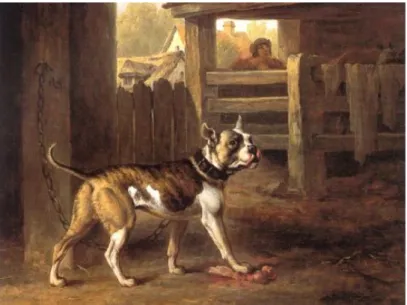
The morphology of dogs with a short muzzle is highly deviant, both from other dog breeds but also from their ancestor, the wolf. The brachycephaly phenotype is char-acterised by a shortened muzzle, a wide head with widely spaced eyes and is often considered as “flat faced”, due to altered growth of basisphenoid and basioccipital bones in the skull (Packer et al. 2015). Brachycephalic breeds are for example Pug, Boxer, English bulldog, French Bulldog, Shih tzu and Japanese Chin. Some brach-ycephalic breeds, like the Bulldogs and the Boston terrier is thought to share a com-mon ancestor. The early bulldog, often referred to as Bandogs, was used in bull-baiting and organized dog fights. These dogs were described as brave and brute dogs with huge jaws. Baiting with Bandogs were forbidden 1835 in England, and the number of Bandogs decreased drastically but the breed was rescued and refined, resulting in the modern bulldog (American Kennel Club).
The pug is, despite its resemblance with bulldogs, thought to be a very old breed descending from Asia (American kennel club).
Figur 1. A painting by Philip Reinagle from 1790, showing a Bulldog.
Many of the brachycephalic breeds are very popular world-wide as companion dogs. In Sweden, both the French bulldog and the Pug are popular breeds and were ranked number 10th and 16th respectively, in 2016 in number of registrations in the
Swedish kennel club(SKK, 2016). Despite the popularity of these breeds, the brach-ycephalic phenotype is associated with several health issues.
A recent analysis of disease prevalence included more than 1.27 million dogs, thereof 184 748 brachycephalic dogs, the analysis was performed under a nine-year
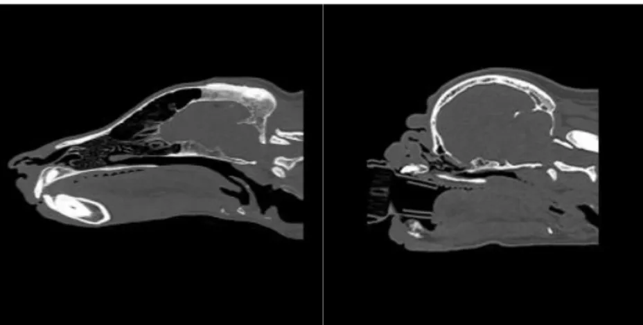
period. The report identified some conditions more frequently occurring in brachy-cephalic dogs, compared to non-brachybrachy-cephalic dogs. For example, the short nose combined with shallow orbits makes the eyes vulnerable, increasing the risk of eye injuries such as corneal ulcer, ocular traumas and conjunctivitis. The study showed that there is a three to four-time higher risk of cornea injuries in brachycephalic dogs, compared to non-brachycephalic dogs. A shortened skull and compact body may result in excessive skin and skin folds, increasing the risk for skin problems such as fungal skin diseases and pyoderma (Feng et al. 2017). However, one of the most obvious issues related to the brachycephaly conformation is the Brachyce-phalic Obstructive Airway Syndrome (BOAS), which predominately affects brach-ycephalic dogs. In a risk factor analysis performed by Njikam et al. (2009) it was found that brachycephalic dogs were 38 times more likely to have BOAS, compared to non-brachycephalic dogs (Njikam et al. 2009). The skull of a brachycephalic dog is significantly shorter, but the soft tissues of the head are not proportionally reduced as the skull. This is problematic, since the size reduced skull will contain too much soft tissue, which leaves little space for the passage of air (Harvey 1989). Conse-quently, BOAS is a respiratory syndrome where soft tissues blocks the dogs airways during respiration (Packer et al. 2015). BOAS is characterized by anatomical abnor-malities such as elongated soft palate, hypoplastic trachea, narrowed nostrils and distortion of the pharyngeal soft tissues which causes resistance to airflow and re-stricted breathing (Fawcett et al. 2019). Breathing for brachycephalic dogs requires more laboured breathing to produce a higher negative pressure to cope with the re-sistance of the airflow (Koch et al. 2003).
Figure 2. The figure shows Computed tomography scans of a German Shepherd (left) and a Pug (right). Picture: Cambridge University.
Clinical signs of BOAS includes noisy respiratory sounds such as stridor (wheez-ing) and inspiratory stertor (snor(wheez-ing), gagging, vomiting, regurgitation, syncope (fainting), dyspnoea (shortness of breath) and exercise intolerance (Riecks et al.
2007). The affected breathing might lead to increased sensibility to heat and im-paired capability to thermoregulate (Davis et al. 2017), which might lead to a col-lapse when the dog is excited or hot (Koch et al. 2003).
A study performed by Packer et al. (2015) showed that the BOAS risk increases as the length of the muzzle shortens. In the 154 studied brachycephalic dogs, BOAS was only present in dogs with a muzzle length less than half of the cranial length (length from stop to the occipital protuberance). A thicker neck and obesity was also factors found to increase the BOAS risk (Packer et al. 2015).
Dogs affected of severe BOAS might, in some cases, need treatment such as weight loss, housing in cool environment and anti-inflammatory medicine. Some severe cases of BOAS might also require surgery (Riecks et al. 2007) in order to widen the nostrils or/and short the soft tissue blocking the airways. A recent study by Liu et al. (2017) investigated the outcomes and prognostic factors of surgical treatments in pugs, French and English Bulldogs. The dogs in the study had an im-proved respiratory function after surgery, although the respiratory function in 68 % of the dogs remained compromised. Age, the level of laryngeal collapse and body condition was found to be the main prognostic factors for BOAS surgery (Liu et al. 2017).
1.2.1 The Genetics behind Brachycephaly
Studies have revealed quantitative trait loci (QTL) on canine familiaris chromo-somes (CFA) 1, 5, 18, 26, 32 and on the X chromosome, associated with the skull length in dogs (Boyko et al. 2010; Marchant et al. 2017). So far, mutations in genes such as Bone Morphogenic Protein 3 (BMP3), Fibroblast growth factor 4 (FGF4) and SPARC-related modular calcium binding gene (SMOC2) are thought to be as-sociated with the altered skull shape causing canine brachycephaly (Boyko et al. 2010; Schoenebeck et al. 2012; Marchant et al. 2017).
SMOC2
Marchant et al., 2017 found that the QTL on CFA1 was highly significant and as-sociated with brachycephaly. A 187.7 kb critical interval in CFA1 was common among 30 out of 37 brachycephalic dogs participating in the study. Five variants were retained in this interval, one being a Long interspersed nuclear element (LINE-1) detected within the SMOC2 gene. This LINE-1 insertion was predicted to cause a premature stop after exon 8 of the canonical 13-exon transcript, causing a fivefold reduction of the SMOC2 mRNA expression. However, Marchant et al. (2017) claims that it remains unclear if the isoforms are translated but predicts that the protein
products would shear within the thyroglobulin-like domain, resulting in the protein missing the extracellular calcium-binding-domain. The study suggests that the in-sertion if the LINE-1 solely explains 36 % of the shortened face displayed in brach-ycephalic dogs in the study. By studying chick embryos, SMOC2 expression was observed in the pharyngeal arches and shown to have temporal expression during the development of the later mandible and viscerocranium (Marchant et al. 2017).
Mansour et al. (2018) genotyped 109 brachycephalic dogs from eleven breeds; Boston Terrier, Boxer, Brussels Griffon, Bull Mastiff, Bulldog, Cavalier King Charles Spaniel, French Bulldog, Lhasa Apso, Pekingese, Pug and Shih Tzu for the SMOC2 insertion. 80 of the 109 genotyped brachycephalic dogs was found to be homozygous, eight heterozygous (three Boston Terriers, three Bull Mastiffs, one Lhasa Apso and one Shih Tzu) and 21 dogs was found to be homozygous for the wild-type variant (one Boxer, six Bull Mastiffs, ten Cavalier King Charles Spaniel, two Lhasa Apso and two Pekingese).
The SMOC2 gene is one of two homologues, where SMOC1 and SMOC2 are both members of Secreted protein acidic and rich in cysteines (SPARC) protein family, also known as BM-40. The members of the SPARC protein family, all con-tains an extracellular calcium-binding (EC) domain and a follistatin-like (FS) do-main, (Termine et al. 1981; Maier et al. 2008). SPARC is a matricellular protein that is secreted into the extracellular matrix, but these proteins are not involved in primary structures in this location. Matricellular proteins like SPARC modulates cell function by interacting with hormones, proteases, cytokines and binding to growth factors and cell-surface receptors. SPARC proteins are expressed in high levels during embryonic development, in adult tissue both SPARC proteins are pre-sent in tissues undergoing repair or remodelling due to processes like wound healing or disease. (Bornstein 2009). SMOC2 mRNA has been found to be expressed in many different tissues, such as spleen, skeletal muscle, ovary, testis, brain, thymus, lung, liver and in the heart (Vannahme et al. 2003). SMOC2 has also been demon-strated to stimulate endothelial cell proliferation and migration, but also to be in-volved in formation of new capillaries (Rocnik et al. 2006).
Liu et al. (2008) investigated SMOC2 expression in mouse embryos and found that SMOC2 was distributed in the pharyngeal arches (i.e. the developing face), de-veloping limbs and somites (Liu et al. 2008). Somites are epithelial cell clusters positioned bilaterally of the neural tube, which gives rise to cartilage, tendons, skel-etal muscle, the vertebrae and ribs and the dermis of the back (Gilbert & Barresi 2016). Feng et al. (2009) examined gene expression during the development of the mouse facial prominences. Gene expression in the developing facial prominences
in mouse embryos were investigated between embryonic day 10.5 and 12.5 and it was found that several genes are expressed during the development, one gene being
SMOC2, expressed in varying levels in samples from the maxillary, mandibular and
frontonasal area(Feng et al. 2009).
In a case with dental defects in humans, Bloch-Zupan et al. (2011) found two cousins each homozygous for a mutation in the canonical-splice donor site in the first intron of the SMOC2 gene. A zebra fish model was used to further investigate effects of mutations in SMOC2 and the effect on dental development. Two types of morpholino knockdowns were created, one to target the initiation codon and one to target the boundary between exon 2 and intron 2 (Bloch-Zupan et al. 2011). Mor-pholino oligonucleotides (MO) is a tool to inhibit the translation of RNA transcripts. MOs is typically a oligomer of 25 morpholino bases that by complementary base pairing binds to the target RNA (Bill et al. 2009). When studying embryos carrying MO, the teeth were smaller with different shape and some dermal bones in the jaws and the fifth ceratobrancial bone were missing calcification, indicating that the skull is affected in morphants. Zebrafishes with the MOs also displayed a smaller head. Though, it should be noted that the development of teeth in zebra fish resembles the development in vertebrates, but the anatomy differs compared with for example hu-mans. The knockdown of smoc2 showed to effect expression of genes such as dlx2b,
bmp2a and pitx2, all involved in tooth development (Bloch-Zupan et al. 2011).
Melvin et al. (2013) identified several genes that alter craniofacial conformation in zebrafish. Eight genes were fully analysed and grouped depending on what struc-ture the alterations would affect; genes essential for formation of the ventral viscer-ocranium, genes essential for development of anterior neurocranium and genes crit-ical for morphogenesis of all neural crest derived structures (including viscerocra-nium and neurocraviscerocra-nium). Smoc2 was analysed as one of the genes essential for the formation of viscerocranium. Two MOs were used to knockdown smoc2, one trans-lation blocking MO to the 5´untranslated region (UTR) and one splice-blocking MO targeting a splice donor site for exon 11. Both MOs reduced the size of the head and eye abnormalities, 10 % in UTR MOs and 50-60 % of the splice site morphants were affected. The ceratohyals (hyoid horn) were flattened and inverted in morphants, compared to controls. The splice site donor MO altered the processing of smoc2 RNA significantly, suggesting the observed affected craniofacial phenotype in mor-phants are due to loss of wild-type smoc2 transcripts (Melvin et al. 2013).
The craniofacial defects investigated in these studies suggests that Smoc genes encode proteins that have a functional role in the development of the facial skeleton.
BMP3
By investigating the QTL at chromosome 32, a critical interval of 85 kb spanning the bone morphogenic protein 3 (BMP3) gene was found (Schoenebeck et al. 2012). After filtering, a Single nucleotide polymorphism (SNP) encoding for a missense mutation was found, nearly fixated among small brachycephalic dogs in the study. The mutation, called BMP3F452L, is caused by a C to A transversion at position CFA
32:5231894, where C being the ancestral allele and A being the derived allele. The missense mutation causes a nonsynonymous substitution, changing the amino acid phenylalanine to leucine (Schoenebeck et al. 2012).
By using a zebra-fish model, bmp3 mRNA expression was found as highly dy-namic, first appearing during mid-somitogenesis (i.e. formation of somites) and later expressed throughout the head, brain, ventricles and posterior somites. 48 hours af-ter fertilization, the bmp3 expression is found in pectoral fins, the pharyngeal arch region, in the jaw structures and in the heart. The early expression of bmp3 in the cranial structure suggested a role for Bmp3 in craniofacial development. By knock-ing down endogenous Bmp3 activity usknock-ing translation-blockknock-ing antisense MO, it was found that MO treated zebrafish embryos demonstrated severe deficiency in the jaw development. Multiple cartilage elements that forms the viscerocranium and neurocranium where lost or underdeveloped in knocked downed embryos. The re-sults of the study indicates that Bmp3 is required for craniofacial development in zebra fishes and that the identified BMP3F452L gene variant influences skull shape
in brachycephalic dogs. (Schoenebeck et al. 2012).
Bone morphogenic proteins (BMPs), being multi-functional growth factors, be-longs to the transforming growth factor β superfamily (TGFβ). The TGFβ pathway plays a central role in signalling networks controlling bone development, tissue re-pair, cell growth, cell differentiation and cell proliferation in metazoans. The TGFβ family can be divided into several subfamilies, including protein families such as TGF-β, BMPs, activin and growth differentiation factors (GDF). TGFβ initiate sig-nalling through specific interactions with receptors on the surface of target cells (Massagué et al. 2000).
BMPs are involved in embryonic development and in multiple different cellular function in adult animals. BMP signalling is involved in heart, neural and cartilage development. It also plays an important role in the postnatal bone formation (re-viewed in Chen et al., 2004). BMP3, also known as osteogenin, works as an antag-onist for other BMPs. BMP3 antagonises the ability of BMP2 to induce differentia-tion and cell commitment of osteogenic cells. Bmp3 is suggested to negatively affect bone density, where Bmp3 null mice (Bmp3 -/-) showed to have twice as much
trabecular bone (spongy bone), compared to non-mutated littermates. Though, no differences in the size of the femur were observed and there were no difference in bone density between wild-type and heterozygous littermates (Daluiski et al. 2001).
By studying Xenopus embryos, Gamer et al. (2005) found that BMP3 inhibits activin and BMP-4 signalling. The activin type II receptor (ActRII) is a common receptor that binds to multiple ligands, such as activin and several BMPs. Binding between ActRII, activin or BMPs is required for patterning during the development of germ layers in the embryo. The binding between activins and BMPs and the re-ceptor phosphorylates rere-ceptor regulated Smad proteins (R-Smad), R-Smad to-gether with other transcriptional regulatory proteins regulates transcription of target genes. It was showed that if BMP-3 binds to its receptor (ActRII), BMP-3 works as an antagonist against activin and BMP-4. BMP-3 reduces the phosphorylation of R-Smad, and thereby inhibits the mesoderm-inducing activities of activin and BMP-4. By injecting BMP-3 in Xenopus embryos, overexpression of BMP-3 presents a phe-notype with deviant tail formation, a shortened and curved body, reduced eyes and head structures and enlarged cement glands. However, the overexpression of BMP-3 could be rescued by co-injections of a truncated type II activin receptor (xActRIIB), which rescued the body axis and head structure in a dose dependent manner (Gamer et al. 2005).
FGF4
In the study by Marchant et al.(2017), a retrogene for the Fibroblast growth factor 4 (FGF4) located on chromosome CFA 18 was associated with the size of the neu-rocranium centroid size in brachycephalic dogs (Marchant et al. 2017). Earlier stud-ies on FGF4 by Parker et al. (2009) identified the FGF4 retrogene on chromosome 18 that causes short legs (canine chondrodysplasia), present in dog breeds such as Dachshunds and Corgis (Parker et al. 2009).
The fibroblast growth factor family, composed of several secreted proteins, func-tions in the earliest stages of the embryonic development, organogenesis and in adult tissue where they act as homeostatic factors important for maintenance of the tissue, tissue repair and regeneration and metabolism. FGFs signals to receptor tyrosine kinases and intracellular non-signalling proteins (i.e. intracellular FGFs) that serves as cofactors. The FGF family is conserved and can be found in both invertebrates and vertebrates and is expressed in almost all tissues (reviewed in Ornitz & Itoh, 2015).
By investigating RNA expression in mice, Niswander and Martin (1992) found that Fgf-4 expression is important during early embryonic development. Fgf-4
mRNA could be detected at late blastocyst stage in cells giving rise to embryonic
lineages. Later, in early gastrulation, Fgf-4 was expressed in the primitive streak where definitive endoderm and mesoderm are formed. After the establishment of the three germ layers, Fgf-4 mRNA was detected in branchial arch units, somite myotome and in the apical ectodermal ridge where the limb buds develop. Expres-sion was also found in tooth bud, suggesting that Fgf-4 has multiple roles during the development of the embryo.
Boulet et al., (2004) studied Fgf8 and Fgf4 in mice embryos and found that mice lacking Fgf4 in apical ectodermal ridge had normal limbs. In Fgf8 mutants, the limb development was severely affected, with some skeletal elements of the limb miss-ing. When creating mutants lacking both Fgf4 and Fgf8 in the forelimb apical ecto-derm ridge, the forelimb does not develop, due to that the limb mesenchyme fails to survive in absent of both FGFs. When inactivating FGf4 and Fgf8 in both forelimb and hindlimb, all limbs fails to develop. It was suggested that Fgf8 is necessary for maintenance or initiation of Sonic hedgehog (Shh) and that Fgf4 partially can com-pensate for the loss of Fgf8 in development of the distal limb (Boulet et al. 2004).
FGFs is also thought to be involved in the development of the hair follicle. Salmon Hillbertz et al. (2007) found that ridged dog breeds, such as Rhodesian Ridgeback and Thai Ridgeback, are either heterozygous or homozygous for a large duplication. The duplication that is thought to be the causative mutation for the ridge, included genes such as FGF3, FGF4, FGF19, ORAOV1 and the 3’ end of CCND1, encoding for cyclin D1. The ridge also predisposes dermoid sinus, a con-genital developmental disorder (Salmon Hillbertz et al. 2007), caused by an incom-plete separation of the neural tube and the skin .
DVL2
A recent study by Mansour et al. (2018) identified a frameshift mutation in the Wnt pathway gene Dishevelled Segment Polarity Protein 2 (DVL2) located on canine chromosome CFA 5, associated with the screw tail that is characteristic for English bulldogs, French bulldogs and Boston terriers. The frameshift mutation, caused by a deletion of a C (g.32195043_32195044del), preserves the majority of the DVL2 protein, though the last 48 amino acids at the C terminus of the protein is replaced by novel sequence of 26 amino acid residues. The results indicated that the Wnt-dependent phosphorylation of the mutant variant DVL2 protein was reduced, com-pared to the wild type variant. Out of 177 dogs from screw tailed breeds, 171 dogs were found homozygous for the mutation in DVL. The exception was six Boston Terriers, where four individuals were heterozygous, and two individuals were ho-mozygous wild type. The pugs tested was found to be of wild types, as expected,
since the tail of a pug is of full length without malformations of the caudal vertebras (Mansour et al. 2018). Mutations in Dishevelled genes in humans is characterised by craniofacial, limb and vertebral malformations, causing a syndrome named Robi-now syndrome. Affected individuals often displays distinctive facial features such as widely spaced eyes, a wide forehead, short nose and a broad mouth (Genetics Home Reference 2019). Due to the resemblance between the phenotype of humans affected by the Robinow syndrome and the phenotype displayed in brachycephalic dogs, it is possible that the DVL2 mutation in screw tail dogs also have an effect on the brachycephalic phenotype (Mansour et al. 2018).
The first dishevelled mutation was identified in Drosophila, where a viable allele was associated with a phenotype with disoriented hairs on the body and wings. It was later shown that Dishevelled being a signalling molecule, works as a key com-ponent in the Wnt signalling pathway (Boutros & Mlodzik 1999).
Wnt proteins are one of the major families of developmentally important signal-ling molecules, involved in processes such as embryonic induction, generation of cell polarity and cell fate specification during embryonic development and tissue homeostasis. It is also suggested that Wnt signalling is involved in cancer genesis in humans (Cadigan & Nusse 1997). One of the best understood Wnt gene is the wingless (wg) gene, essential for segmentation and patterning events in the fruit fly
Drosophila melanogaster. Wg is required in several events during embryo
develop-ment, such as in patterning of the segmented ectoderm in the trunk, the development of the head, the central nervous system and the legs. Wg is also required for growth of the wing blade in the development of the wing disc, hence the name wingless. In the adult fly, Wg is required for patterning of bristles and body hair, in antennae, eyes and genitalia (reviewed in Klingensmith & Nusse, 1994).
Wnt genes have been identified in invertebrates and vertebrates, with ortholog
relationship between species such as Wnts in Drosophila and in humans. Between vertebrates, the Wnt genes are highly conserved, sharing sequence identity and gene structure. It has been showed that signaling by members of the Wnt family have diverse functions during embryogenesis, and has been suggested that Wnt genes play roles in the development of different structures of the brain, development of dorsal-neural tube derivatives, development of kidneys, hair growth, limb polarity and the development of the placenta. Mutations in Wnt genes has also been associ-ated with shortened anterior-posterior axis and female infertility due to defects dur-ing development of female reproductive organs (rewieved in Miller, 2001). In mouse, mutations in Wnt genes have been associated with defects in the tail, the tailbud and in caudal somites (Cadigan & Nusse 1997).
The Wnt pathway is activated when the Wnt ligand binds to the two membrane receptors, Frizzled (Fz) and low-density lipoprotein receptor related protein 6 (LRP6) or LRP5. The complex of ligand and receptors, together with the recruitment of Dishevelled results in phosphorylation of LRP6/LRP5 and the recruitment of the axin complex to the receptors. Next, the axin complex is inhibited, which allows β-catenin located in the cytoplasm to enter the nucleus and form complexes with DNA-bound T cell factor/lymphoid enhancer factor (TCF/LEF), where β-catenin serves as a co-activator which activates Wnt target gene expression. In absence of Wnt, β-catenin in the cytoplasm is constantly degraded by the axin complex and the Wnt target genes are repressed (MacDonald et al. 2009). Proteins encoded by the dishevelled genes works as important transducers in Wnt pathways, that conducts the Wnt signal to downstream cellular machinery (Na et al. 2007).
The Dishevelled proteins have three known structural domains, DIX (Dishev-elled and Axin) domain at the N-terminus, PDZ (Post-synaptic density protein-95) and DEP (Dishevelled, Egl-10 and Pleckstrin). By investigating DVL2, Gammons et al. (2016) found that the DEP domain, binding to Frizzle receptors is essential for the Wnt-dependent signaling to β-catenin. The PDZ domain was dispensable for the β-catenin response (Gammons et al. 2016).
Tadjuidje et al. (2011) showed that both Dvl2 and Dvl3 plays a part in early embryo development in the Xenopus. It was found that both Dvl2 and Dvl3 are ma-ternally encoded for. Before the zygotic gene activation (ZGA), where the embryo starts to produce its own transcripts and proteins, development is dependent of stored maternal factors in the oocyte. Genes encoding for such factors are referred to as maternal effect genes, are important for the oogenesis, enables ZGA and the progression of the early embryo development (Kim & Lee 2014). By studying
Xenopus oocytes, it was found that Dvl2 and Dvl3 mRNA are expressed early in the
development and decreases during the developmental process. Dvl2 was later found to increase after the ZGA when the zygotic transcription starts, but Dvl3 remained low. Dvl1 was not detected until later in the development. By using different anti-sense oligos to block translation of Dvl2 and Dvl3, it was found that maternal de-pletion of Dvl2 or Dvl3 results in similar phenotypes. Dede-pletion of Dvl resulted in shortened body axis, blastopores not closing and open neural folds. The heads were also found to be smaller in Dvl depleted embryos. Since maternal depletion of either Dvl2 or Dvl3 affects early development, it was suggested that Dvl2 and Dvl3 are not redundant to each other and that the proteins might interact with each other. Dvl2 and Dvl3 was found to be required for activation in the c-jun N-terminal kinase
(JNK) pathway which is important for the convergence extension (Tadjuidje et al. 2011), where cell movements results in elongation of the body axis.
Xing et al. (2018) studied the effects of mutations in zygotic Dvl, maternal Dvl and the effects when combining mutations in both zygotic and maternal Dvl in zebrafishes. By generating mutated lines for the five known dvl genes in zebrafish, it was found that only dvl2 and dvl3 were maternally expressed. Maternal and zy-gotic mutants for four out of five of the dvl genes developed normally and survived to adult zebrafish. Dvl2 mutants were however found to have defects. Zygotic dvl2 (Zdvl2) mutants displayed a slightly reduced anterior-posterior axis, compared to wild type embryos. Half of the Zdvl2 mutants survived to adult, one third of the surviving females could not spawn and all male mutants were not able to produce offspring due to absent courtship behaviour. Embryos with mutations in both ma-ternal and zygotic dvl2 (MZdvl2) developed craniofacial defects, fused eyes and the pharyngeal cartilages protruded outwards. MZdvl3 embryos displayed just a weak axis extension defect. When generating double heterozygote, (dvl2+/-; dvl3+/-), these
embryos showed a slight reduction of the axis extension. However, when mating two double heterozygous fishes, embryos displayed a shortened anterior-posterior axis, a compressed head and reduced swim bladder. Absence of zygotic Dvl2 and Dvl3 resulted in moderate convergence extension defects, while absence of both zygotic and maternal Dvl3 and Dvl3 generated severe convergence extension de-fects. Progressive reduction of Dvl dosage was found to gradually induce anterior-posterior patterning defects, ranging from anterior-posterior deficiency to complete lack of the trunk and tail. In conclusion, Xing et al. (2018) found that Dvl3, and in particular Dvl2, is important in the convergence extension movement and in anterior-posterior patterning during embryonic development (Xing et al. 2018).
1.3 Aim of study
The aim of this thesis was to investigate the genetic variation in three of the genes associated with canine brachycephaly by looking into the Swedish population of four brachycephalic dog breeds; English bulldog, French bulldog, Pug and Boston terrier.
2.1 Recruitment of dogs
In total, data from 102 privately owned brachycephalic dogs (44 males and 58 fe-males) were used in this thesis. The dogs were phenotypically described at six gath-erings, held between 17th of September 2018 and 20th of January 2019. Three of the
gatherings were organised by the Swedish kennel club (SKK), one gathering was organised by SKK and the English Bulldog club and two were organised by SKK and the French Bulldog club. The gatherings held by SKK and SKK/English dog club were held at SKKs facility, while gatherings organised by the French Bull-dog club were held in Bull-dog training facilities at two different locations.
Information from 18 English Bulldogs (9 males and 9 females), 48 French Bull-dogs (23 males, 24 females and one with unknown sex), 21 Pugs (7 males and 14 females) and 15 Boston terriers (5 males and 10 females) were used for this thesis. The dogs in the inventory was between 9 months and 13 years old. In addition, DNA from one Labrador Retriever, one Golden Retriever, two Pugs and two Chinese Crested Dogs were used.
2.1.1 Swedish inventory of Brachycephalic dogs
The data used in this project was collected as part of a bigger Nordic research study with the aims to investigate the phenotypic and genotypic variation in the four breeds; English Bulldog, French Bulldog, Pug and Boston terrier, and if it’s possible to by selection achieve changes in anatomy to reduce the predisposition of BOAS. The ambitions in the Swedish project was to recruit 50 – 100 dogs of each breed, approximately 25 dogs of each breed should later be selected and genotyped. The inventory is a part of the SKK’s ambition to reduce extreme conformations in
2
Material and Method
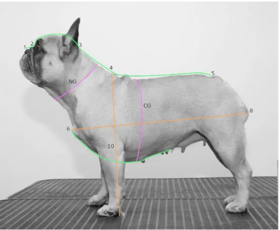
Swedish dogs. At the gatherings, the procedure consisted of four stations. Station one, where the dog owner filled in information of the dog, such as birth date, sex, registration number, medical information and gave consent to participation in the study. At the second station, a veterinary inspection was performed to record meas-urements of the dog and parameters such as respiration and circulation. Some of the measurements taken is illustrated in figure 3 below.
Figure 3. Illustration of some of the measurements taken during the inventories (Photo and illustration, Elin Johansson.
The skull length was measured from the stop (2) to the external occipital protuber-ance at the back of the skull (3). The snout length was measured from the stop (2) to the nose tip (1). Other measurements taken were the neck girth (NG), neck length (3 to 4), chest girth (CG), back length (4 to 5), body length (6 to 8), height at withers (4 to 9), elbow height (10 to 9) and chest length measured between the top of the breast bone (6) and the end of the sternum (7). By dividing the snout length with the skull length, the craniofacial ration was obtained. The degree of nostril stenosis was also graded according to a four graded scale from open nostrils to severe stenosis, described by (Liu et al. 2017). The dogs were also weighed, and the body condition
score was assessed, were a scale from one (undernourished) to nine (obese) was used. The dog’s skin and presence of excessive skin folds were also examined.
At a third station, DNA was collected, and the dog was photographed from dif-ferent angles (profile, front, back and from above). At the last station, additional conformation traits were recorded, such as height at shoulders, height at elbow, back length, body length, neck girth, chest girth and chest length. The placement and appearance of eyes, nose and ears and front and rear angles, appearance of paws, top line, wrinkles on body and nose folds and the length of tail and its appearance were also assessed. The dog’s movement was scored when letting the dog trot back and forth on the floor.
Each dog was given a specific identification number at the gathering, partly to easy have access to an overview of the number of dogs in the inventory and partly to handle the information more objectively by just using the inventory number and/or breed information.
A second master thesis was done within the same Swedish project, with the aim to investigate the phenotypic variance correlated to BOAS in the Swedish popula-tion of four brachycephalic breeds; English Bulldog, French Bulldog, Pug and Bos-ton Terrier and discuss their welfare implications (Bertilsson, 2019).
2.2 DNA extraction and quantification
Buccal cells were collected from the participating dog’s cheeks by two or three bris-tle cytology brushes. The cytology brushes were left to air dry for a couple of minutes and later placed in a marked paper envelope. The envelopes with the cytol-ogy brushes was then stored in room temperature until DNA extraction. The enve-lopes were stored between one day and four months, depending on when the sample was collected and when the extraction was performed.
Genomic DNA was isolated using the QIAsymphony® SP automated system with the QIAsymphony DSP DNA Midi Kit (QiaGen, ID 937236, Hilden, Ger-many) (protocol in appendix I).
To determine the nucleic acid concentration in the samples, spectrophotometry was used by loading 2 µl of the samples on a NanoDrop™ 8000 Spectrophotometer (Thermo Fisher Scientific), this was performed on eight of the samples. Due to the large variation in nucleic acid concentration and the uncertainty of how much of the
nucleic acid is derived from dog’s own buccal cells and how much is from bacteria or feed residues in the mouth, it was decided not to use the spectrophotometer on the other samples.
To ensure that the samples contained sufficient amount of canine genomic DNA, a polymerase chain reaction (PCR) of the canine housekeeping gene Glyceralde-hyde 3-phosphate dehydrogenase (GAPDH) was set up, using the HotStarTaq® kit (QiaGen ID: 203601, Hilden, Germany) (protocol 1, Appendix A). In total, the PCR was set up for nine samples, including eight brachycephalic dogs and one Labrador Retriever. A gel electrophoresis was performed to segregate the PCR products for validation.
A dilution series was performed to investigate which concentrations that could work for a PCR. Three samples were picked out, based on the nucleic acid concen-tration measured with NanoDrop™, one sample with high, intermediate and low concentration was chosen. The samples were diluted to concentrations 1:10 and 1:100 and a second PCR for the GAPDH gene was set up (Appendix A, protocol 1). This procedure was mainly performed to assess the least possible amount of ge-nomic DNA to use from each sample due to the expected limited amount of gege-nomic DNA template obtained from the cheek swab samples available in this study.
In total, DNA from five additional dogs were used in the study as positive con-trols. DNA from one Labrador Retriever, one Golden Retriever, two Pugs and two Chinese Crested Dogs. For the Retrievers and the additional Pug, the DNA extrac-tion was made from whole blood. For the two Chinese Crested Dogs, cytology brushes were used as described for the brachycephalic dogs above.
2.3 PCR amplification
2.3.1 Primer design
In order to detect the LINE-1 insertion in the SMOC2 gene, PCR primers for geno-typing described in Marchant et al. (2017) was used. The primers were checked, using the Primer3 software. Primers were obtained from TAG Copenhagen A/S (Denmark).
In total, three primers were used (table 1). All three primers were used in the same reaction. The melting temperatures of the primers were between 55.3°C and 60°C, with a GC content between 47.8% and 70 % (Appendix A, protocol 2).
Table 1. Primers used for amplification of SMOC2.
Primer Primer sequence Size
SMOC2 F GGC AGG GGA TGG GGA AGC CT 531 bp
SMOC2 R (wild type) ACT GTG TGC TTT GCC CAA ACT CA
SMOC2 F GGC AGG GGA TGG GGA AGC CT 698 bp
SMOC2 R (mutated) TGC CCA TAA AGT TCA GGG TCC ACT
Heterozygous will show as two visible bands on an agarose gel.
2.3.2 PCR conditions
For 102 dogs, the DNA sequences containing the SMOC2 gene was amplified using the kit HotStarTaq®. The total reaction volume of the PCR was 10 µL, composed of; 1x PCR Buffer (15 mM MgCl2, ph 8.7) + dNTP [0.25 mM] + primers [0.3
µM/primer] + 0.125 µL Taq Polymerase [0.025 U/µL] + 1 µL DNA and addition of RNase-free water until the completion of the 10 µL reaction volume. One negative control per master mix was included, were the DNA was substituted with 1 µL of water (Appendix A, protocol 3)
PCR reactions were performed in a thermal cycler (ProFlex PCR system, Ap-plied Biosystems by Life Technologies), using a touchdown PCR program, accord-ing to followaccord-ing protocol. Initiation step: 95 °C for 2 minutes. 3-step cycle (20 cy-cles) Denaturation step: 98 °C for 10 seconds. Annealing 72 - 52 °C (reduced with 1 °C/cycle) for 30 seconds. Extension: 72 °C for 45 seconds. Followed by another 3-step cycle; 98 °C for 10 seconds, 55 °C for 30 seconds, 72 °C for 45 seconds. Finishing synthesis step: 72 °C for 7 minutes. The lid heat was put on 98 °C. There-after, infinite hold in 4 °C until the products were quantified on an 2 % agarose gel, using a 100 bp precision size marker. The gel electrophoresis was run in 110V for approximately two to three hours, depending on the size of the gel.
The PCR protocol was kindly provided through personal communication with Marchant, T (March 2019).
2.4 Sanger Sequencing
Since no dog in this study had a known genotype for the BMP3 and DVL2, Sanger Sequencing with capillary electrophoresis was performed to obtain the genotype for
three dogs, one Golden Retriever, one English Bulldog and one Pug, and use these dogs as positive controls in later TaqMan assays. Two sets of M13 tailed primers per gene, flanking the SNP were used (table 2).
Table 2. Primers used for sequencing BMP3 and DVL2 genes.
Primer Primer sequence Source
BMP3 1 F GGTGATGATACAGGAGATTGTGCCAAA Schoenenbeck et al., 2012 BMP3 1 R CTGCCAGGTTATCTGCAAGCACAAG Schoenenbeck et al., 2012
BMP3 2 F TCATGCCACCATCCAGAGTA Primer 3
BMP3 2 R GCCAGGTTATCTGCAAGCAC Primer 3
DVL2 1 F CGGCTAGCTGTCAGTTCTGG Mansour et al., 2018 DVL2 1 R CAGTGAGTCTGAGCCCTCCA Mansour et al., 2018
DVL2 2 F CCTGGGCTCCATCCCTAT Primer 3
DVL2 2 R GCGCCCTACATAACATCCAC Primer 3
PCR reactions were performed according to the BigDye® Direct Sequencing Kit Protocol (Applied Biosystems®, Inc) (see Appendix A, protocol 3). PCR reactions were performed in a ProFlex™ PCR system (Applied Biosystems®), later the PCR-amplicons were purified using the BigDye Directs Cycle Sequencing Kit, following manufacturer’s instructions. Templates were prepared to be sequenced both in for-ward and reverse orientation. PCR sequence products were analysed on the ABI 3500XL Genetic Analyzer (Applied Biosystems®).
2.5 TaqMan assays
For genotyping of the SNPs in the BMP3 and DVL2 gene, TaqMan® SNP genotyp-ing assays were used. In total, 45 brachycephalic dogs and one non-brachycephalic dog (25 females, 20 males, 1 unknown) were genotyped for polymorphism in each of the two genes, BMP3 and DVL2. The genotyping assays were designed according to custom manufacturer specifications.
The reaction was carried out in a 96 well plate. The reaction volume was 15 µL per well and was consisting of 7.5 µL TaqMan® Universal PCR Master Mix [2X], 0.38 µL TaqMan® Genotyping assay mix (primers + probes) [40X], 5.62 µL DNase-free water and 1.5 µL DNA template. The software StepOne™ Software v2.3 and the instrument StepOnePlus ™ (Applied Biosystems®) Real-time PCR system generated the results. Cycle conditions according to following protocol: Pre PCR stage in 50 °C for 2 minutes, polymerase activation in 95 °C for 10 minutes,
followed by a touch-down series starting with denaturation in 95 °C for 15 seconds followed by cycling stage of 50 cycles with annealing/extension in 60 °C/55°C for
BMP3 and DVL2 respectively, followed by 95°C for 15 seconds and an
anneal-ing/extension step in 55°C for 1 minute. Finally, a hold stage of 60 °C for 30 sec-onds.
The TaqMan assay relies on two sequence specific primers (forward and reverse) for PCR amplification. The assay also contains two allele specific TaqMan probes, each probe can detect one of the two alleles. The probes carry a dye label at the 5’end, a so called “reporter”. Commonly, VIC® reporter is used for allele one and FAM™ reporter for allele two. At the 3’ end, the probe carries a non-fluorescent quencher bound to a minor groove binder (MGB). Each probe anneals specifically to the complementary sequence, depending on which allele is present. At denatura-tion, the primer pair and the corresponding probe anneals to the sequence. At polymerisation, probes hybridized to the target gets cleaved. The cleavage separates the reporter dye from the quencher and increases the fluorescence of the reporter, which can be detected by the device. The fluorescence generated during PCR am-plification indicates which of the alleles that are present in the sample (Ther-moFisher Scientific).
For genotyping of BMP3, VIC® reporter was used to detect the wild-type allele C (allele 1) and FAM™ reporter was used to detect the derived A allele (allele 2). For genotyping of DVL2, VIC® reporter was used to detect C allele (allele 1) for wild-type and FAM™ reporter was used for the derived A allele (allele 2).
The dogs with known genotyped was used as positive controls, other marked as unknown. After sequencing, the results were analysed using StepOne™ software and the ThermoFisher Cloud.
Figure 4. Schematic figure of the TaqMan assay for BMP3. Primers are marked out as arrows.
3.1 GAPDH
For eight samples, a PCR was setup against the housekeeping gene GAPDH to en-sure that the samples contained enough canine DNA for later PCR of the SMOC2 gene. All eight samples generated a band of correct size of 108 bp on the agarose gel. It was concluded that the canine DNA concentration in the samples should be sufficient for further testing.
3.2 Dilution series
Since there was limited amount of DNA from each dog in the study, a dilution series was performed to check if the samples could be diluted so that less DNA could be used for each testing. Three of the eight samples described above was diluted to 1:10 and 1:100 and tested for the GAPDH gene. However, for one sample, the DNA concentration of DNA after diluting was probably too low to get amplified, resulting in no visible bands after gel electrophoresis. Consequently, it was decided not to dilute DNA samples from the buccal swabs for further testing.
3.3 SMOC2
By setting up a PCR, the genotypes for the SMOC2 gene was obtained. Amplifica-tion products were visualised on an agarose gel (figure 6), where the wild type allele is expected to generate a fragment of 531bp, and the mutated allele is expected to generate a fragment of 698 bp.
Figure 6. Agarose gel electrophoresis. Agarose gel electrophoresis showing SMOC2 PCR amplicons from different individuals. One band at about 700 bp indicates that the individual is homozygous for the mutation. Sample 1002, displaying two bands, is heterozygous for the mutation, carrying both the mutated LINE-1 insertion and the wild-type haplotype.
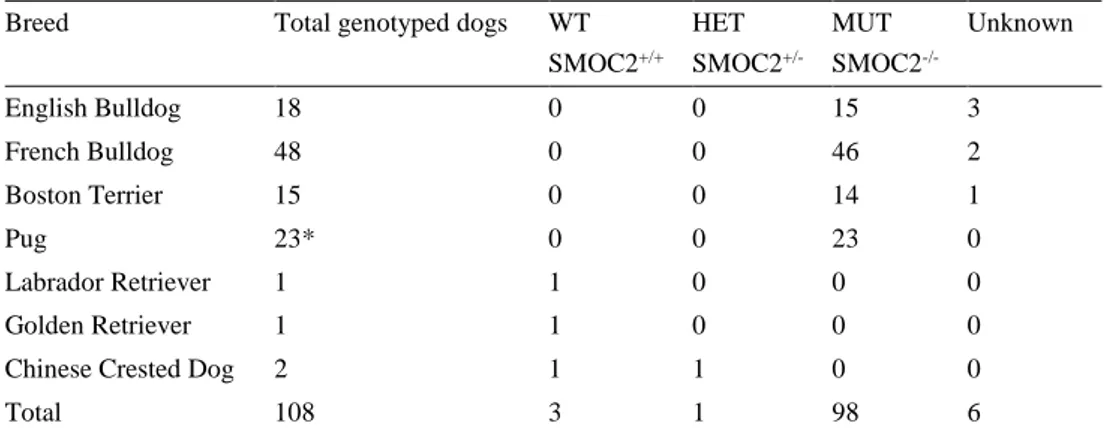
Table 3. SMOC2 LINE-1 insertion.
Breed Total genotyped dogs WT
SMOC2+/+ HET SMOC2 +/-MUT SMOC2 -/-Unknown English Bulldog 18 0 0 15 3 French Bulldog 48 0 0 46 2 Boston Terrier 15 0 0 14 1 Pug 23* 0 0 23 0 Labrador Retriever 1 1 0 0 0 Golden Retriever 1 1 0 0 0
Chinese Crested Dog 2 1 1 0 0
Total 108 3 1 98 6
*Including DNA extracted from blood from two additional pugs, not participating in the inventory.
Out of 108 dogs genotyped for the SMOC2 mutation, 102 dogs (98 brachycephalic dogs and four non brachycephalic dogs) were successfully genotyped. All 98 suc-cessfully genotyped brachycephalic dogs were found to be homozygous for the mu-tation. As a control group, four non brachycephalic dogs were genotyped, consisting of one Labrador and one Golden Retriever and two Chinese crested dogs. One dog, a Chinese crested dog was found to be heterozygous for the SMOC2 mutation, the
other three dogs of the control group was genotyped as homozygous for the wild-type variant.
The allele frequency for the mutated allele when calculated for all brachyce-phalic dogs is 1.
3.4 Sanger Sequencing
By using Sanger sequencing, the genotypes for BMP3 and DVL2 for three dogs were obtained.
3.4.1 BMP3
The Golden Retriever was found homozygous for the ancestral C allele. Both the Pug and the English Bulldog was found homozygous for the derived A allele (Figure 7).
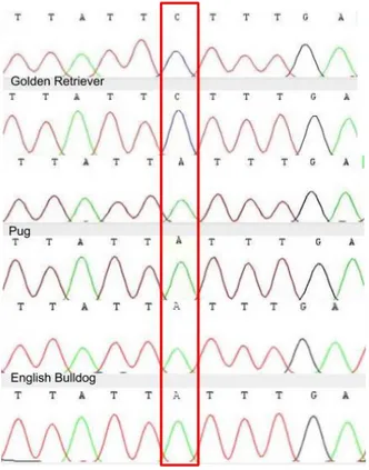
Figure 7. The figure shows the output for all three sampled dogs, Golden Retriever forward and reverse strand on top, Pug forward and reverse in the middle and English Bulldog forward and reverse bottom. The red box indicates the SNP position.
3.4.2 DVL2
The Golden Retriever and the Pug was found homozygous for the ancestral C allele. The English Bulldog was found homozygous for the deletion, causing the mutation.
Figure 8. Visualisation of the SNP using Sanger Sequencing. The figure shows the output for all three sampled dogs. Golden Retriever front and reverse strand on top, Pug forward and reverse strand in the middle and English Bulldog forward and reverse strand bottom. The red box indi-cates the SNP position.
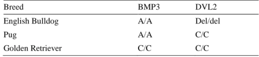
In summary, the English Bulldog was found homozygous for the mutations in both BMP3 and DVL2. The Pug was found homozygous for mutated variant of BMP3 and homozygous for the ancestral variant of DVL2. The non-brachycephalic Golden Retriever was homozygous for the ancestral variant in both studied genes (Table 4).
Table 4. Genotypes for BMP3 and DVL2 of three dogs.
Breed BMP3 DVL2
English Bulldog A/A Del/del
Pug A/A C/C
3.5 BMP3
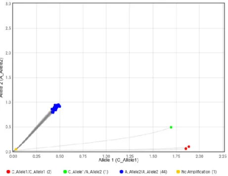
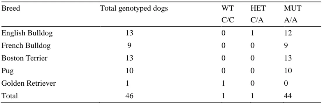
For BMP3, a TaqMan® SNP genotyping assays was performed to obtain the geno-types of 46 dogs (Figure 9). The C allele is presumed as the ancestral allele, called allele 1. The A allele is presumed as the derived (mutated) allele, called allele 2. Additionally, an artificial heterozygote sample was created by mixing DNA from the sequenced Golden Retriever, homozygous for allele A and the sequenced Eng-lish Bulldog, homozygous for allele C. In total, 44 brachycephalic dogs were found homozygous for the A allele (Table 5). One English Bulldog was labelled as heter-ozygous, carrying one A allele and one C allele. The non-brachycephalic dog, a Golden Retriever was labelled as homozygous for the C allele. Also, the created artificial heterozygous sample was labelled as homozygous for the C allele.
Figure 9. The two red dots in the right corner represents the samples found homozygous for allele A, the sequenced Golden Retriever and the artificial heterozygote. The green dot close to the middle represents the English Bulldog labelled as heterozygous. The blue upper cluster represents all other brachycephalic dogs, homozygous for the C al-lele.
Table 5. BMP3 status variant across breeds
Breed Total genotyped dogs WT
C/C HET C/A MUT A/A English Bulldog 13 0 1 12 French Bulldog 9 0 0 9 Boston Terrier 13 0 0 13 Pug 10 0 0 10 Golden Retriever 1 1 0 0 Total 46 1 1 44
The allele frequency for the mutated allele when calculated for all four breeds to-gether is 0.988.
3.6 DVL2
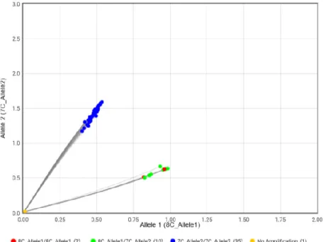
For DVL2, a TaqMan® SNP genotyping assays was performed to obtain the geno-types of 46 dogs (Figure 10). The C allele is presumed as the ancestral allele, called allele 1. The deletion causing the mutation is called allele 2. As described for the BMP3, an artificial heterozygous was created also for this genotyping assay. In total, 35 of the brachycephalic dogs was labelled as homozygous for the mutation. All ten genotyped pugs and the sequenced Golden Retriever was labelled as heterozygous. The artificial heterozygote sample is found just slightly above the lower cluster.
Figure 10. The two red dots in the lower green cluster in the plot represents the
se-quenced homozygous Golden Retriever and the sese-quenced homozygous Pug. All other green dots in the lower cluster is Pugs. The green dot slightly above the lower cluster represents athe artificial heterozygous. The upper blue cluster repre-sents all English Bulldogs, French Bulldogs and Boston Terriers.
Table 6. DVL2 status (DVL2c.2044delC) variant across breeds
Breed Total genotyped dogs WT
C/C HET C/del MUT Del/del English Bulldog 13 0 0 13 French Bulldog 9 0 0 9 Boston Terrier 13 0 0 13 Pug 10 10 0 0 Golden Retriever 1 1 0 0 Total 46 11 0 35
Allele frequency for the mutated allele when calculated for all four breeds together is 0.75. When calculating the allele frequency for just the bull type breeds (Boston Terrier, English Bulldog and French Bulldog), the allele frequency for the mutated allele is 1.
The aim of this thesis was to investigate the genetic variation in known genes asso-ciated with brachycephaly in dogs. Three genes were studied, SMOC2, BMP3 and
DVL2. In total, 102 brachycephalic dogs were genotyped for the mutation in the SMOC2 gene and 46 of these dogs were also genotyped for the mutations in the BMP3 gene and DVL2 gene respectively.
4.1.1 SMOC2
The SMOC2 LINE-insertion is thought to highly influence the brachycephalic phe-notype. Marchant et al. (2017) suggest that the size effects of the associated SMOC2 haplotype accounts for 36 % of the facial length variation in the dogs in their study. In this thesis, all 102 successfully genotyped brachycephalic dogs were found to be homozygous for the SMOC2 LINE-1 insertion. Consequently, the allele frequency for the derived allele was calculated to 1 in all studied breeds, suggesting that the mutation is or is close to fixation.
In a communication from Bannasch (2019), the allele frequency for the LINE-1 insertion was calculated to 0.97 for dogs of Bull type (Boston Terrier, Bulldog and French Bulldog) and 0.75 for Asian type breeds (Pug, Pekingese and Shih Tzu). The high allele frequency in the bull type breeds is similar when compared to the result in this thesis. For the Pugs, representing the Asia type breeds, the allele frequency in this study was higher since all individuals were found homozygous for the muta-tion.
In contrast to the study by Mansour et al. (2018), were three of the studied Boston Terriers was found heterozygous, all brachycephalic dogs were found homozygous in this thesis.