END-TO-END AUDIT: COMPARISON OF TLD AND
LITHIUM FORMATE EPR DOSIMETRY
Emelie Adolfsson, Paulina Wesolowska, Joanna Izewska, Eva Lund and Åsa Carlsson
Tedgren
The self-archived postprint version of this journal article is available at Linköping
University Institutional Repository (DiVA):
http://urn.kb.se/resolve?urn=urn:nbn:se:liu:diva-164697
N.B.: When citing this work, cite the original publication.
Adolfsson, E., Wesolowska, P., Izewska, J., Lund, E., Carlsson Tedgren, Å., (2019), END-TO-END AUDIT: COMPARISON OF TLD AND LITHIUM FORMATE EPR DOSIMETRY, Radiation Protection Dosimetry, 186(1), 119-122. https://doi.org/10.1093/rpd/ncy289
Original publication available at:
https://doi.org/10.1093/rpd/ncy289
Copyright: Oxford University Press (OUP) (Policy B - Oxford Open Option A)
END-TO-END AUDIT – COMPARISON OF TLD AND LITHIUM
FORMATE EPR DOSIMETRY
Emelie Adolfsson1*, Paulina Wesolowska2, Joanna Izewska2, Eva Lund3, Åsa Carlsson Tedgren4
1 Department of Radiation Physics and Department of Medical and Health Sciences, Linköping University,
Linköping, Sweden
2International Atomic Energy Agency, Vienna, Austria
3Department of Medicine and Health Sciences, Linköping University, Linköping, Sweden
4Department of Medical and Health Sciences, Linköping University, Linköping, Sweden and Department of
Medical Radiation Physics and Nuclear Medicine, Section for Radiotherapy Physics and Engineering, Karolinska University Hospital, Stockholm Sweden
The aim of this study was to test two different solid state dosimetry systems for the purpose of end-to-end audits of radiotherapy VMAT technique; a lithium formate EPR system and a lithium fluoride TLD system. As a complement to the solid state systems, ion chamber measurements were performed. A polystyrene phantom with a PTV and an OAR structure was scanned using CT. A VMAT dose plan was optimized to deliver 2 Gy to the target volume and to minimize the dose to the OAR. The different detectors were inserted into the phantom and the planned dose distribution was delivered. The measured doses were compared to the TPS calculated doses. Good agreement was found between the TPS calculated and the measured doses, well accepted for the dose determinations in remote dosimetry audits of VMAT treatment technique.
INTRODUCTION
Verification of the quality of radiotherapy through independent dosimetric audits is nowadays generally accepted. The International Atomic Energy Agency and the World Health Organization (IAEA/WHO) have since 1969 performed mailed audits using thermoluminescent dosimetry (TLD) for high-energy photon beams [1]. Today, the two largest audit networks are currently operated by the IAEA [2, 3] and the Imaging and Radiation Oncology Core Houston (IROC, Houston, USA) [4] and the interest in developing systems on the national level is high. Dosimetry audits are classified into different levels, ranging from output measurements in reference conditions to verification of the complete treatment chain in anthropomorphic phantoms (end-to-end tests) [5, 6]. The choice of the audit level should be consistent with the national audit programme and should address the needs of participating clinics [7]. Advanced treatment techniques such as intensity modulated radiotherapy (IMRT) and volumetric modulated arc therapy (VMAT) have now been introduced at several radiotherapy centers. The complexity of inverse dose planning and pre-treatment verification further increases the importance of auditing.
Solid state detectors are in general advantageous to use
for phantom based remote dosimetry audits. The phantom can be prepared and loaded with the detectors before sending to the audit clinic and hence reduce the risk for handling mistakes. TLDs are commonly used in audits due to their long accessibility on the market and well-known behavior in MV photon beams. However, TLD has disadvantages such as destructive reading process and cumbersome handling procedures requiring experienced operators. Hence several newly-developed audit systems have chosen other dosimetry systems. Electron paramagnetic resonance (EPR) dosimetry using alanine is an accepted standard dosimetric method and is used as a secondary standard at both The National Physical Laboratory (NPL, UK) [8] and at the Physikalisch-Technische Bundesanstalt (PTB, Germany) [9]. For EPR dosimetry, the readout process is non-destructive to the signal which allows several readouts of the same dosimeter.
An alternative to alanine for EPR dosimetry is lithium formate monohydrate (referred to as lithium formate in the following text). Lithium formate has dosimetric properties at least as good as those of alanine. Both lithium formate and alanine have a linear dose response which makes them suitable for measurements of high doses (target) and low doses (organs at risk (OARs)) simultaneously. Compared to alanine, lithium formate has up to seven times higher sensitivity [10, 11]. The lithium formate system has shown to be robust; there is no detectable signal fading occurring one month after irradiation (a sufficient time period for the complete audit procedure) and it is insensitive to variations in the
storage temperature (important issue when detectors are being sent by post) [12]. The response of lithium formate detectors is independent of the dose rate and beam quality in the ranges that are relevant for radiotherapy [13]. Lithium formate has previously been used for verification of IMRT treatments [13] and for end-to-end audits [14].
The aim of this study was to compare a lithium formate EPR system, handled by Linköping University, Sweden, to the IAEA lithium fluoride TL system for the purpose of end-to-end audits in a solid phantom. Inter-institution comparison of dosimetry systems is of benefit for better understanding the advantages and limitations of the systems. The results of such comparison could show possible systematic errors but also be used for testing the consistency of the systems. As a complement and an independent verification of the dose, ionization chamber measurements were performed. This project was a part of a coordinated research group (IAEA Coordinated Research Project E2.40.18), where a method for end-to-end audit of advanced treatment techniques was developed [15]. MATERIAL AND METHODS
Phantom
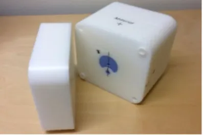
A solid head phantom designed by the IAEA for use in end-to-end remote IMRT audits was used in this study [16]. The phantom includes one target region (PTV) and one organ at risk (OAR). The phantom (figure 1) was made of white polystyrene (density of 1.04 g/cm3)
and the PTV and OAR was made of solid water (density of 1.032 g/cm3) to be able to be visualized in
the CT images. Two measurement points were situated in each structure, i.e. in total there were four measurement points; PTV superior and inferior, OAR superior and inferior.
Fig 1. The polystyrene phantom including PTV and OAR inserts in blue (solid water).
Detectors
The three detector systems used in this work were: Semiflex ionization chamber 31010 (PTW, Freiburg, Germany), lithium formate EPR detectors and TLD powder capsule. Their properties and calibration procedures are described below.
EPR lithium formate
The EPR detectors were cylinders with the length of 4.8 mm and the diameter of 4.5 mm. The detectors consisted of 90% lithium formate monohydrate (HCO2
Li H2O) powder bound in 10% paraffin and pressed to
tablets. Signal evaluation was performed with a Bruker EleXsys E580 spectrometer equipped with the standard resonator (4102 ST). The spectrometer settings were: microwave power 20 mW, modulation frequency 100 kHz, modulation amplitude 12 G, receiver gain 60 dB, sweep width 30 G and sweep time 168 s. Each dosimeter was read out three times in a rotating schedule to eliminate any possible sensitivity variation of the spectrometer during the day (no external reference was used in the resonator). All preparations and settings used were made according to a standardized workflow explained in [17].
Three detectors were used in the same measurement position in the audit phantom and the mean signal from them was used to determine the absorbed dose.
The absorbed dose to water calibration was performed in a 6 MV linear accelerator photon beam using a Farmer type ionization chamber with the traceability to BIPM primary standard. For calibration details see [18].
TLD lithium fluoride
The TL detectors were plastic capsules of 2.5 cm length with 0.5 cm diameter, with the inner dimensions of 1.9 cm length and 0.3 cm diameter filled in with a TLD-100 powder (Harshaw, USA). The powder from one capsule was sufficient to prepare four samples which were read using the Fimel PCL-3 reader. The reference TLDs used for the system calibration were irradiated in the reference conditions [19] on Nordion X200 Co-60 unit. The standard IAEA protocol [7] was used for the detectors preparation and evaluation.
Ionization chamber
The Semiflex ionization chamber was chosen for the measurements as it has the outer dimensions that are comparable to the two other systems. The sensitive volume is 0.125 cm3. The Semiflex chamber was cross
calibrated with a Farmer type ionization chamber with the traceability to BIPM primary standard.
Experimental method
The phantom preloaded with either TLD or EPR lithium formate was scanned in a Siemens Somotom Sensation open CT scanner (Siemens Healthcare GmbH, Erlangen, Germany) using a head CT protocol. The CT DICOM file was transferred to the Eclipse treatment planning system (TPS) where the PTV, OAR and detector volumes were contoured (different volumes for the three detector types). A VMAT plan was created using the analytical anisotropic algorithm (AAA) version 13.6. Figure 2 shows the configuration of detectors for irradiation. Two fractions of the dose plan were delivered for each dosimeter type, i.e. a total dose of 4 Gy to the PTV, the corresponding dose to the OARs was 1.1 Gy x 2. The measured doses were compared to the calculated TPS doses in the appropriate volumes corresponding to each detector. Corrections were made for the accelerator daily output. The irradiations were repeated for TLD and the ionization chamber, however it was not possible for the EPR due to technical problems at the time of measurements.
Fig 2. The positions of the detectors in the phantom seen from the side. The effective measurement volumes are marked in dark grey and the solid water parts in light blue. The ionization chamber measurements were performed at one position at the time while solid plugs were inserted in the others.
Uncertainty estimation budget
Absorbed dose to water determined by the EPR dosimetry system has a combined standard uncertainty of 2% with the main contributing factor being the type B uncertainty of the ionization chamber used for the system calibration (1.7%) [19]. Other factors of
relevance are related to the homogeneity within the
dosimeter batch and the readout process. The overall
Fig. 3. The percentage difference between the TPS calculated doses and the measured doses for EPR (circles), TLD (diamonds) and the ionization chamber (crosses).
combined uncertainty for the TLD system is 1.7%. The uncertainty in the calibration of the TLD system arise largely from the uncertainty in the ionization chamber calibration of 0.5%, reproducibility in the phantom positioning of 0.2%, solid water to water dose correction of 0.5%, dosimeter irradiation positions corrections of 0.03% and the readout uncertainty of 0.48%.
RESULTS AND DISCUSSION
The percentage difference between the TPS calculated and the measured doses in each detector system respective volume are shown in figure 3.
In the PTV structure, the measured absorbed dose agreed with the TPS calculated doses within each system’s respective uncertainty. Also, in a comparison between the three detector systems, the agreement is within 2.2%.
In the OAR the difference between the calculated and the measured doses was expected to be larger due to inhomogeneous dose distribution in this area. This can also be seen in figure 3. Some uncertainty for the TPS calculated doses is introduced by contouring the sensitive volumes of each detector. The slice thickness is 2 mm, hence the resolution is not better than 2 mm in the scan direction, which is a limitation when contouring small objects. Also, a position error of 1 mm in one of the three translational directions would cause a difference of about 1% in mean dose to the volume.
CONCLUSIONS
This study showed that both the TL and the EPR PTV_S PTV_I OAR_S OAR_I PTV_S PTV_I OAR_S OAR_I EPR TLD PTV_S OAR_S Ion chamber PTV_I OAR_I
remote dosimetry audits of VMAT dose plans. The two systems show similar results within their respective uncertainties which also agrees well with measurements made with the ionization chamber. ACKNOWLEDGEMENTS
This work was supported by the Swedish Cancer foundation (CF), contract number 110322. The IAEA IAEA Coordinated Research Project E2.40.18 is also acknowledged.
REFERENCES
1. Izewska J and Andreo P. The IAEA/WHO TLD postal programme for radiotherapy hospitals. Radiotherapy and Oncology 2000;54:65-72. 2. Izewska J, Andreo P, Vatnitsky S and Shrott KR.
The IAEA/WHO TLD postal dose quality audits for radiotherapy: a perspective of dosimetry practices at hospitals in developing countries. Radiotherapy and Oncology 2003;69:91-97. 3. Izewska J, Bera P and Vatnitsky S. IAEA/WHO
TLD Postal dose audit service and high precision measurements for radiotherapy level dosimetry. Radiation Protection Dosimetry 2002;101:387-392. 4. Followill DS, Evans DR, Cherry C, et al. Design,
development, and implementation of the Radiological Physics Center’s pelvis and thorax anthropomorphic quality assurance phantoms. Medical Physics 2007;34:2070-2076.
5. Kron T, Hamilton C, Roff M and Denham J. Dosimetric intercomparison for two Australasian clinical trials using an anthropomorphic phantom. International Journal of Radiation Oncology*Biology*Physics 2002;52:566-579.
6. Izewska J, Georg D, Bera P, et al. A methodology for TLD postal dosimetry audit of high-energy radiotherapy photon beams in non-reference conditions. Radiother. Oncol. 2007;82:67-74. 7. SSDL Newsletter 66: Setting up a Dosimetry Audit
Centre: Infrastructure and Resources https://www-pub.iaea.org/MTCD/Publications/PDF/Newsletters /SSDL-66.pdf 2017.
8. Sharpe PHG, Rajendran K and Sephton JP. Progress towards an alanine/ESR therapy level reference dosimetry service at NPL. Appl. Radiat. Isot. 1996;47:1171-1175.
9. Anton M. Development of a secondary standard for the absorbed dose to water based on the alanine
EPR dosimetry system. Appl. Radiat. Isot. 2005;62:779-795.
10. Lund E, Gustafsson H, Danilzuk M, et al. Formates and dithionates: sensitive EPR-dosimeter materials for radiation therapy. Appl. Radiat. Isot. 2005;62:317-324.
11. Vestad TA, Malinen E, Lund A, Hole EO and Sagstuen E. EPR dosimetric properties of formates. Appl. Radiat. Isot. 2003;59:181-188.
12. Adolfsson E, Karlsson M, Alm Carlsson G, et al. Investigation of signal fading in lithium formate EPR dosimeters using a new sensitive method. Phys. Med. Biol. 2012;57:2209-2217.
13. Gustafsson H, Lund E and Olsson S. Lithium Formate EPR Dosimetry for verification of calculated dose distributions prior to intensity modulated radiation therapy. Phys. Med. Biol. 2008;53:4667-4682.
14. Adolfsson E, Gustafsson H, Lund E, Olsson S and Carlsson Tedgren A. A method for dosimetry audit of IMRT and VMAT. Radiotherapy and Oncology 2014;111: S285.
15. SSDL Newsletter 64: Development of Quality Audits for Advanced Technology in Radiotherapy Dose Delivery https://www-pub.iaea.org/MTCD/Publications/PDF/Newsletters /SSDL-64.pdf. 2016.
16. Wesolowska P, Almady B, Adolfsson E, et al., Pilot study of a remote end-to-end dosimetry audit for IMRT and VMAT treatments, in ESTRO 35. 2016.
17. Adolfsson E, Carlsson Tedgren Å, Alm Carlsson G, Gustafsson H and Lund E. Optimisation of an EPR dosimetry system for robust and high precision dosimetry. Radiation Measurements 2014;70:21-28.
18. Antonovic L, Gustafsson H, Alm Carlsson G and Carlsson Tedgren Å. Evaluation of a lithium formate EPR dosimetry system for dose measurements around 192Ir brachytherapy sources.
Med. Phys. 2009;36:2236-2247.
19. Andreo P, Burns DT, Hohlfeld K, et al., Absorbed Dose Determination in External Beam Radiotherapy, IAEA TRS 398: An International Code of Practice for Dosimetry Based on Standards of Absorbed Dose to Water, in IAEA Technical Report Series no 398 (Vienna:
International Atomic Energy Agency). 2000, IAEA: Vienna, Austria.