Department of Medical Biochemistry and Microbiology
Biomedical laboratory science programme
Degree project 15hp, 2011
Phylogenetic characterization of equine influenza
viruses from Swedish outbreaks from 1979 to 2001
Binnaz Acar
Supervisor: Siamak Zohari
Department of Virology, Immunobiology and Parasitology
National Veterinary Institute
2
Abstract
Introduction: Equine influenza virus, an influenza type A virus, belongs to the family of
Orthomyxoviridae. Equine influenza is a major cause of respiratory disease in horses and
outbreaks have severe economical repercussions for the horse industry. It is considered to be endemic in Sweden and between 1997 and 2006 there have been around 10 to 40 outbreaks every year. The objective of this study was to do a phylogenetic characterization of equine influenza outbreaks that occurred in Sweden during a twenty year period.
Methods: The haemagglutinin and neuraminidase gene of 14 samples and the complete
genome of three samples collected over the span of 20 years were sequenced. The viral RNA were extracted, amplified with OneStep RT-PCR and sequenced.
Results & Discussion: The phylogenetic tree and deduced amino acid sequence of HA1 illustrated that different lineages of equine influenza virus has circulated simultaneously in the Swedish horse population. The isolates mainly belonged to pre-divergence-, Eurasian- and American lineages. To characterize equine influenza viruses is important for vaccine strain selection, to fully understand the disease and how the virus evolves.
Keywords: H3N8; Phylogeny; Genetic; Vaccine; Antigenic drift
Introduction
Equine influenza virus, influenza type A virus, belongs to the family of
Orthomyxoviridae. Based on antigenic
differences between nucleocapsid protein and matrix proteins, viruses of this family are divided into five genera: influenza type A, B, C, thogotovirus and Isa virus.
Influenza C can infect humans and swine and causes mild upper respiratory tract infections. Influenza B is known to infect humans and seals. This subtype causes a severe infection to the lower respiratory tract and can consequently cause
pneumonia and or encephalitis. Influenza A virus, which infects naturally a wide
variety of different species including humans, pigs, equine and a wide range of avian species, is the most problematic.
Influenza A viruses can further be divided into different subtypes depending on the antigenic variants of the surface glycoproteins; the haemagglutinin (HA) and the neuraminidase (NA). There are
currently 16 HA (H1-H16) and 9 NA (N1-N9) subtypes and a virus consist of one of each glycoprotein subtype (Capua & Alexander, 2008).
Wild aquatic birds are the major
reservoirs of influenza A viruses in nature, predominantly in orders Anseriformes (particularly ducks, geese and swans) and Charadriformes (particularly gulls, terns and waders) (Olsen et al., 2006).
Transmission from the natural reservoirs to a wide range of other species including humans, equine, feline, canine, swine and domestic birds occur frequently but they seldom establish lasting lineages in these new hosts (Gibbs & Anderson, 2010).
The influenza A virus is an enveloped virus, pleomorphic in shape that can appear as spherical and tubular (illustrated with figure on cover page) with a size range between 80-120 nm in diameter. The envelope consists of a lipid bilayer and surface glycoproteins such as HA, NA and
3 an ion channel; membrane protein (M2).
Beneath the lipid membrane is a lining of viral matrix protein (M1). The inner part of the virion contains eight single-stranded RNA segments, the genome encodes for 11 proteins. The RNA segments are coated with nucleoprotein (NP) and each segment is coupled with an RNA polymerase component; polymerase basic 1 (PB1), polymerase basic 2 (PB2) and polymerase acidic (PA). Other component in the virion is nuclear export protein (NEP).
The RNA genome of influenza A virus is ca. 13 600 nt, and the length of each segment is except for HA and NA greatly conserved. The segments are ordered from 1-8 in decreasing length. The genome for HA and NA (segment 4 and 6) is 1779 nt and 1413 nt respectively and the
polypeptide HA is 566 and NA 454 amino acids in length. Segment 1 and 2 (PB2, PB1) have 2341 nt and the polypeptide consists of 759 and 757 amino acids. Segment 2 also codes for a second smaller polypeptide (PB1-2F). Segment 3 is 2233 nt and codes for PA which has 716 amino acids. The NP polypeptide has 498 amino acids and the gene is made up of 1565 nt. Segment 7 codes for matrix proteins M1 and M2 which are 252 and 97 amino acids respectively, the genome is 1027 nt in length. NS1 and NEP protein is encoded from segment 8 which has 890 nt. The polypeptide NS 1 is 230 and NEP 121 amino acids in length.
The 3’ and 5’ ends of the segments have non-coding region which vary in length, these regions contains e.g. promoter
activity and mRNA polyadenylation signal. Proteins involved in viral replication are PB1, PB2, PA and NP (Schnitzler & Schnitzler, 2009). The M1 protein is the
most abundant virion protein and is a type-specific antigen of influenza viruses. It has lipid binding regions and is believed to add to the virions rigidity. M2 is a pH activated ion channel that allows protons to enter into the virion during uncoating. The NS1 protein is a non-structural protein that is expressed abundantly in early infection. One of NS1:s function is to inhibit the cellular anti-viral interferon response. NEP is believed to mediate the export of viral ribonuclear proteins from the nucleus of host cell to the cytoplasm.
The surface glycoproteins HA and NA has a very important role in the
pathogenicity of the virus. Host specificity is determined by the HA (Schnitzler & Schnitzler, 2009) and NA surface
glycoproteins; they determine how efficient the virus is at attaching and fusing with host cell and after production of new virions, release from cell. HA have several functions, some of which are to serve as a viral attachment protein and start fusion of the envelope with the cell membrane. NA has an enzymatic activity that cleaves sialic acid on glycoproteins; this facilitates release from host cell surface and prevents virion proteins from aggregating (Myers & Wilson, 2006).
HA binds to sialic acid attached to galactose either by α2,3-Gal or (SA)-α2,6-Gal linkages. The specificity of the virus depends on which conformation the virus binds to. Human influenza viruses binds predominantly to (SA)-α2,6-Gal that are well represented in human epithelial cells. Avian strains bind to (SA)-α2,3-Gal which are predominant in avian cells (Capua & Alexander, 2008). Swine tissue expresses both types of conformations and are therefore considered to be “mixing
4 vessels” for human, avian and swine
influenza strains. A genetic reassortment of influenza strains in a “mixing vessel” can produce a virus that is able to cross the species barrier; this was seen in the 2009 influenza (A) H1N1 pandemic. This mechanism where HA and NA RNA segments reassort and produce a new antigenic subtype of virus is called antigenic shift. Another important mechanism, antigenic drift, is when the mutations in the HA and NA genes cause minor changes to the surface glycoproteins. These changes are important because they allow the virus to evade the host immune response and infect populations that are supposedly protected by either acquired immunity or vaccination (Schnitzler & Schnitzler, 2009).
Equine influenza was first isolated in the Czech Republic 1956
(A/equine/1/Prague/56). The subtype isolated was a H7N7 strain and later identified as the main reason for equine influenza outbreaks around the world. In Miami, Florida, 1963 a second strain was isolated and subtyped as H3N8
(A/equine/2/Miami/63) and until late 1970s both strains circulated simultaneously. But since then the H7N7 strain has not been isolated from horses showing clinical signs. This virus is now believed to have gone extinct (Gibbs & Anderson, 2010).
The strain circulating in Europe and America at present is H3N8 and has since late 1980s diverged into two distinct lineages; Eurasian and American. The American lineage has further separated into the Kentucky, South American and Florida lineages Clade 1 and Clade 2 (Qi et al., 2010). These lineages are now
co-circulating worldwide without any
inclination to geographical locations (Ito et
al., 2008).
Equine influenza is a major cause of respiratory disease in horses and is the source of numerous large outbreaks in vaccinated and naïve horse populations. These outbreaks have severe economical repercussions for the horse industry (Bryant
et al., 2011).
Aerosol, wind and nose-to-nose contact are some of the ways equine influenza virus can be transmitted to horses. The
incubation period is 1-3 days and shedding of virus begins 24 hours after infection and can continue for 7-10 days. The virus infects any breed, sex and age and throughout the year. The morbidity of infected horses in vulnerable populations can be up to 100 % while the mortality is relatively low; however this depends on the health of the horse and the virus strain the horse is infected with. The clinical signs of an influenza infection in horses are fever, lethargy and anorexia. The most prominent symptom is a repetitive harsh dry cough that can last up to 3 weeks (Myers & Wilson, 2006).
The spread of equine influenza is mostly due to the fact that horses are transported internationally for e.g. competitions and breeding. There are regulations in place that should prevent the spread such as health examinations and quarantines but unfortunately these are often proven insufficient to stop spread to local horses. Due to continuous transport of race horses new variants quickly reach far away continents. In most countries equine
influenza is considered to be endemic in the native populations, with the exception of New Zeeland and Iceland where no
5 outbreaks of equine influenza has occurred
to date (Gibbs & Anderson, 2010). Equine influenza is considered to be endemic in Sweden and between 1997 and 2006 there have been around 10-40
outbreaks every year. A widespread and severe outbreak occurred in Sweden in 2007 where roughly 200-300
non-vaccinated horses were infected in 50-60 areas. Vaccination was during that time obligatory for horses participating in all races except for trotting. In 2008 The Swedish Trotting Association decided that vaccination would be mandatory for all horses participating in races1, 2.
Vaccines are to date the only medical way to prevent outbreaks of equine influenza. They have been used as prophylaxis and the goal is to completely prevent infection. When an infection of influenza occurs the immune system responds by activating the cell-mediated response and production of antigen specific serum IgG and secretory IgA antibodies. The immunity derived from the natural response to infection can last at least one year. In contrast, inactivated adjuvant vaccines do not activate this kind of immune response. A vaccination results in low levels of neutralizing antibodies in nasal secretions. It fails to stimulate mucosal IgA production and the cell-mediated immune response is insufficient. The fallout is infected horses with no clinical signs but nevertheless shedding
1 International Collating Center (ICC) available
from http://www.aht.org.uk/icc/linksicc.html Accessed on 14 April 2011.
2 National Veterinary Institute (SVA) available
from
http://www.sva.se/sv/undersida/Nyhetsarkiv/Epidem i-av-hastinfluensa-bland-svenska-hastar-/ Accessed
on 14 April 2011.
virus (Myers & Wilson, 2006). Vaccines target the viral glycoproteins such as HA. Protection, as mentioned earlier, is not 100 %. This is also due to the mutation rate of the influenza virus and the resulting antigenic drift. The produced antibodies protecting against one specific strain might not work on a slightly different one (Bryant
et al., 2011).
There are many different diagnostic tests available to confirm a diagnosis of equine influenza, some of which are through virus isolation, ELISA, RT-PCR and
haemagglutinin inhibition (HI) test. The usual material used in diagnostic testing is nasopharyngeal swabs from horses.
By virus isolation it is possible to detect the HA activity within the amnion or allantoic of an inoculated embyonated hens’ egg. This is performed by a
haemagglutinin inhibition test with specific antisera to H3N8 and H7N7 viruses. If antibodies to influenza virus are present in the sample they will prevent attachment of the virus to the erythrocytes and
haemagglutination will be inhibited. Virus isolation is the only test that yields virus isolate that can be used in further research; but it can take weeks until results are visible. The ELISA detects viral nucleoproteins through the use of monoclonal antibodies. This test is less sensitive when there are samples with low titers of virus but can yield results within 15 minutes of setting up the test. The qRT-PCR is a sensitive, specific and relatively fast test that can detect influenza virus with primers for the nucleoprotein or matrix gene, although it does not discriminate between ongoing and passing infections (Myers & Wilson, 2006).
6 The purpose of this study was to follow
the development of the equine influenza outbreaks that occurred in Sweden during a twenty year period. The collected samples were sequenced and a phylogenetic tree constructed.
Materials and methods
Samples
In this study a total of 14 equine
influenza A viruses collected over the span of 20 years were sequenced. The samples were chosen with regards to area and year of isolation. The samples were clinical material that had previously been analyzed for diagnostic purposes. The samples were de-identified when stored for research purposes.
Full length genome for 3 strains was determined along with sequence data for two surface glycoproteins HA and NA. Viruses characterized in this study were obtained from the repository at the
Swedish National Veterinary Institute. The seed stock of the influenza A viruses were grown in the allantoic cavity of 9-day-old embryonated hens’ eggs and were stored at –80C until further use.
RNA extraction
The RNA from the samples was extracted by a method optimized at the National Veterinary Institute (SVA). 250 µL of sample was added to 750 µL of Trizol reagent (Invitrogen Corp., Carlsbad, CA) this causes cell lysis and virus
inactivation. To get a phase separation and easily collect the RNA, 250 µL of
chloroform was used. The samples were then centrifuged at 4C, 12879 × g for 10 min. The aqueous phase was transferred to new tubes and iso-propanol added to precipitate the RNA. Following this the samples incubated in room temperature for 10 min and then centrifuged at 4C, 12879 × g for 10 min. The supernatant was discarded and the RNA pellet washed with 500 µL of 70 % ethanol and then
centrifuged at 4C, 8944 × g for 10 min. The wash was repeated once but only centrifuged for 5 min. The supernatant was discarded and the tubes were left to air dry for 20 min and then the pellets were resuspended in 20 µL of RNase-free water (water treated with 0.1% v/v
diethylpyrocarbonate (DEPC) for at least 1 hour at 37°C and then autoclaved (at least 15 min)). The samples were immediately put on ice and stored at -20C. Negative controls were used in each extraction to make sure there was no contamination of the samples during extraction. The controls were either allantoic fluid from
non-inoculated embryonated hens’ eggs or DEPC-water.
Real-Time PCR
A real-time PCR (qPCR) using
QIAGEN OneStep RT-PCR kit (QIAGEN, Hilden, Germany) of the samples was carried out to ensure the quality of the extracted RNA and to make sure there was no contamination of the negative controls. The qPCR was conducted on the M-gene; the sequences for forward primer was 5´AGATGAGTCTTCTAACCGAGGTCG 3´, reverse primer was
5´TGCAAAAACATCTTCAAGTCTCTG 3´ and probe
TCAGGCCCCCTCAAAGCCGA-7 TAMRA 3´. The concentrations of primers
and probe were 0.2 µM, dNTP 0.2 µM and MgCl2 1.25 mM. A volume of 0.6 µL of enzyme mix with 0.06 µL of Invitrogen RNaseOUT Recombinant Ribonuclease Inhibitor was added with 2 µL of RNA-template. DEPC-water was used to get the reaction volume up to 15 µL. The qPCR was run for 50C for 30 min for reverse-transcription, 95C for 15 min for activation of HotStarTaq DNA Polymerase,
inactivation of reverse-transcription and denaturing of cDNA template. Cycling conditions were denaturing at 95C for 10 s, annealing and extension at 58C for 20 s, this is repeated 40 times. Final extension was at 50C for 1 min.
Reverse-Transcriptase PCR
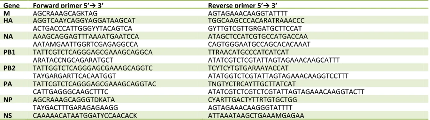
The samples were amplified using the QIAGEN OneStep RT-PCR (QIAGEN, Hilden, Germany) kit according to the manufacturer’s instructions to perform a multiplex RT-PCR. The larger genes were amplified using two primer pairs for each gene that enabled the maximum coverage of the amplified genes when sequencing. The primers used in this study are given in Table 1.To determine the quality of all the RT-PCR products gel electrophoresis was conducted. The concentration of the agarose gel was 1.5 % and DNA was stained with BIOTIUM GelRed Nucleic Acid Stain (BIOTIUM, Hayward, USA).
Prior to sequencing, PCR products were purified with Shrimp Alkaline Phosphatase (SAP) and Exonuclease I (ExoI) (Thermo Fisher, Waltham, USA). The enzyme SAP degrades excess dNTP:s and ExoI degrades single stranded DNA in a 3'→5' direction. This step reduces interference when sequencing.
BigDye sequencing reaction and
percipitation
BigDye Terminator v3.1 Cycle Sequencing Kit (Applied Biosystems, Foster City, CA) was also used according to kit protocol.
BigDye sequencing PCR-products were precipitated by adding 2 µL of sodium acetate solution pH 5.2 and 50 µL of 99 % ethanol to each tube. The samples were incubated in room temperature for 20 min and then centrifuged for 20 min at 4C; 15115 × g.The supernatant was removed and 250 µL of 75 % ethanol was added and the samples centrifuged as previously mentioned. All the ethanol was removed and the tubes with pellet were left to air dry. The samples were stored at 4C until sequencing was performed.
The pellets were resuspended in 13 µL of formamide and vortexed right before Sanger sequencing.
Sequence analysis and generation
of phylogenetic tree
After sequencing, assembly of sequences, removal of low-quality sequence data, nucleotide sequence translation into protein sequence, additional multiple sequence alignments and processing were performed with the Bioedit software version 7.0.4.1, with an engine based on the Custal W algorithm. Blast homology searches (www.ncbi.nlm.nih.gov/blast) were used to retrieve the top fifty
homologous sequences for sequenced gene from the GenBank database. The
phylogenetic analysis, based on complete genome
8
Table 1. RT-PCR primers for influenza A, subtype H3N8. For longer genes multiple primers were used.
nucleotide and amino acids sequences were constructed with Molecular
Evolutionary Genetics Analysis (MEGA, version 5) software using neighbor-joining tree inference analysis with the Tamura-Nei model, with 2000 bootstrap
replications to assign confidence levels to branches.
Results
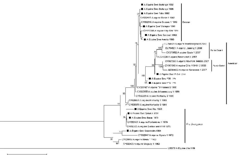
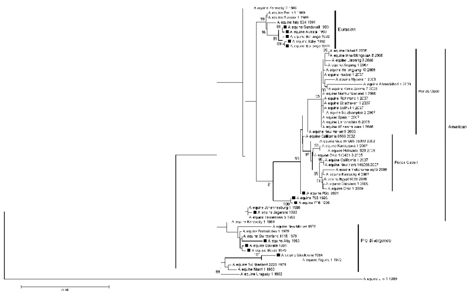
The complete NA (Fig. 1) and HA (Fig. 2) genome sequence was used to construct the phylogenetic tree of equine influenza virus isolated from 14 different Swedish outbreaks from 1979-2001. To highlight the five different lineages; Pre-divergence, Eurasian, American and the sub-lineages Florida Clade 1 and 2 reference sequences from GenBank was used. The reference sequences were chosen with regards to difference in country and the period viruses were isolated.
Phylogenetic analysis
The isolates grouped primarily in the pre-divergent-, American- and Eurasian lineages. The older isolates dated from 1979-1984 were grouped with the pre- divergent lineage while older isolates from 1992-1996 (excluding Jägarsro/1993) grouped with the Eurasian lineage. The
isolates from the American lineage dated from 1993-2001. As described by Bryant et
al., 2011, virus isolated and characterized
from 2008-2009 group with the sub-lineages Florida Clade 1 and 2.
The same clusters could be seen with NA as for HA, except for the phylogenetic analysis for NA showed that the isolates Borlänge/1992, Borlänge/1996 and Täby/1992 were identical to each other while HA Borlänge/1992 was slightly different from the others. The NA gene of Visingsö/1990 grouped with the Eurasian lineage.
The phylogenetic tree for HA give evidence that there have been different equine influenza viruses co-circulating in the Swedish equine population; e.g. Borlänge/1996, P35/1996 and P16/1996; where the first one belongs to the Eurasian lineage and the two latter to the American lineage. Solvalla/1991 clustered with Borås/1979 in the pre-divergent lineage. Stockholm/1984 was more like the first equine influenza isolated, Miami/1/1963, and Jägarsro/1993 grouped with the early American lineage.
A/equine/Jilin/1/1989 was an avian influenza virus that infected equines in China and Mongolia and therefore did not group with the equine influenza viruses.
Gene Forward primer 5’→ 3’ Reverse primer 5’→ 3’ M AGCRAAAGCAGKTAG AGTAGAAACAAGGTATTTT HA AGGTCAAYCAGGYAGGATAAGCAT TGGCAAGCCCACARATRAAACCC ACTGACCCATTGGGYYTACAGTCA GYTTGTCGTTGRGATGCTTCCAT NA AAAGCAGGAGTTTAAAATGAATCCA ATAGCTCCATCGTGCCATGACCAA AATAMGAATTGGRTCGAGAGGCCA CAGTGGGAATGCCAGCACACAAAT PB1 TATTCGTCTCAGGGAGCGAAAGCAGGCA TTRAACATGCCCATCATCAT ARATACCNGCAGARATGCT ATATCGTCTCGTATTAGTAGAAACAAGCATTT PB2 TATTGGTCTCAGGGAGCGAAAGCAGGTC TCYTCYTGTGARAAYACCAT TAYGARGARTTCACAATGGT ATATGGTCTCGTATTAGTAGAAACAAGGTCCTTT PA TATTCGTCTCAGGGAGCGAAAGCAGGTAC TNGTYCTRCAYTTGCTTATCAT CATTGAGGGCAAGCTTTC ATATCGTCTCGTCTCGTATTAGTAGAAACAAGGTACTT NP AGCRAAAGCAGGGTDKATA CYARTTGACTYTTRTGTGCTGG TAYGACTTTGARAGAGAAGG AGTAGAAACAAGGGTATTTT NS CAAAAACATAATGGATYCCAACACK ATTAAATAAGCTGAAAMGAGAA
9
Fig. 1. Phylogenetic analysis of the NA genome. The isolates sequenced in this study are marked with a black square. The analysis was done by Neighbour-joining method and bootstrap values were obtained after 2000 replicates. Values higher than 80 % are shown. The tree is drawn to scale and the horizontal distances represent number of base substitutions/site (0.05). The vertical distance is for spacing only.
10
Fig. 2. Phylogenetic analysis of the HA genome. The isolates sequenced in this study are marked with a black square. The analysis was done by Neighbour-joining method and bootstrap values were obtained after 2000 replicates. Values higher than 80 % are shown. The tree is drawn to scale and the horizontal distances represent number of base substitutions/site (0.05). The vertical distance is for spacing only.
Comparison of deduced amino
acid sequences of HA gene
The amino acid alignment (Fig. 3) shows differences in several important antigenic sites between strains that have been co-circulating e.g. Borlänge/1996 and P35/1996 co-circulated in Sweden but belong to different lineages. P35/1996 has in site B2 a total of six amino acid changes compared to the reference strain,
Borås/1979, which is three more than Borlänge/1996; the changes are 204-Lys→Gln, 205-Glu→Gln and
208-Lys→Glu. In antigenic site D2, P35/1996 is identical to the reference strain while Borlänge/1996 has two substitutions. P16/1996 and P35/1996 are isolates that co-circulated on the Swedish island Gotland during 1996 and are identical to each other except for amino acid 40 where the former has a serine residue and the latter leucine. PGol/2001 is also from Gotland but was isolated much later and has evolved from the previous strains. All strains circulating in Gotland belonged to the American lineage.
The strains circulating in 1992
(Borlänge and Täby) have three differences in amino acid composition. Täby has on antigenic site A, position 150; arginine instead of glycine and outside the antigenic site at 159-Thr→Met and at residue 540-Phe→Leu.
Borås/1979 and Solvalla/1991 are very similar apart from four amino acid
Fig. 3. Multiple amino acid sequence alignment of the HA1 gene of Swedish isolates. Residues identical to A/Equine/Borås/1979 are indicated with dots and differences are shown with single alphabet substitutions. Significant antigenic sites are shaded and major substitutions highlighted in yellow for clarity. The antigenic sites were determined with accordance to Barbic et al., 2009; Qi et al., 2010 and Ito et al., 2008. * indicates the pre-divergence lineage; # indicates the Eurasian lineage and ¤ indicates the American lineage.
Discussion
The objective of this report was to phylogenetically characterize equine influenza viruses causing outbreaks in Swedish horse populations. The results showed that through the years there have been several different strains of equine influenza virus belonging to different lineages co-circulating in the Swedish equine population.
The cause of influenza epidemics is thought to be the result of the virus ability to through mutations change the antigenic sites that antibodies recognize. This allows the influenza virus to escape the host immune system and therefore cause recurring infections (Barbic et al., 2009; Ito et al., 2008). There are five antigenic sites determined in human H3 influenza viruses (A-E) which are further divided into regions (A1, A2, A3, B1, B2, C1, C2, D1, D2, D3, E1, and E2) (Ito et al., 2008). For significant antigenic drift in human H3 there has to be at least two to four amino acid changes in two different antigenic sites (Ito et al., 2008; Martella, et al., 2007) Since vaccination is at present the only way to prevent outbreaks,
characterization of antigenic drift from old and circulating strains is very important to determine if the vaccine is up to date or not. There have been outbreaks in Europe due to vaccine failiure e.g. in Croatia 2004 described by Barbic et al., 2009. Another risk with vaccines that are mismatched with the cirulating strain is that the horses get a short-lived and mild disease but still shed large quantities of virus. This can fuel the spread and cause infection in the equine population instead of protecting against already circulating strains. This can be the case with the three Swedish strains (Borlänge/1996; P16/1996 and P35/1996)
that co-circulated in 1996 and where the first one belongs to the Eurasian and the two latter to the American lineage. Daly et
al., 1996 observerd that it is more likely
that antigenic sites B and D undergo changes in amino acid composition and also suggested that antigenic site B is under greater selective pressure. This site is consideration to be a “hot spot” for changes in amino acid sequence (Ito et al., 2008). This theory can be applied to this study where the greatest changes in amino acid sequence could also be seen in antigenic sites B and D.
Avesta/1993 and Sundsvall/1993 were co-circulating and have identical amino acid composition but the phylogenetic analysis demonstrate that they are not identical. This could indicate that Avesta/1993 might have a mute single point mutation in the genome that differs from Sundsvall/1993.
Even though there are 12 years between the isolation of Solvalla/1991 and
Borås/1979 they cluster together and are very similar in amino acid sequence apart from four residues. This can indicate “frozen evolution” (Borchers et al., 2005) or that the strain isolated in 1991 has been circulating for a time but “evaded”
detection. “Frozen evolution” is a term Endo et al., 1992 coined when describing a virus that ciculates over a long period of time without significant antigenic and genetic variation. Their study illustrated that there are multiple evolutionary lineages of H3N8 circulating simultaniously.
Screening of equine influenza viruses in racehorses and the monitoring of vaccine efficiency is important to prevent
14 economical disaster for the horse industy.
Competition horses are often vaccinated as it is mandatory for participants but the remaining horse population is usually not. These un-vaccinated populations can act as virus reservoirs (Elton et al., 2011) and subsequently be the sourse of reemergence of old strains and caus new infections and outbreaks in the vaccinated population. To detect the possible “frozen evolution” strains and prevent vaccine failiure these horses should be included in screening programs.To really understand the evolution of equine influenza and the effectiveness of vaccines a broader
perspective needs to be applied to research surrounding this area.
Equine influenza vaccines need to be regularly updated to include protection agains new emerging strains. Vaccines usually include two or three strains to cover the circulating lineages (Elton et al., 2011). In 2010 new recommandations to update the equine influenza vaccines were made by the Expert Survaillance Panel (ESP). According to ESP there is no need to include a Eurasian lineage to current vaccine but they should contain a representative for Florida Clade 1 e.g. A /South Africa/2003-like or A/Ohio/2003-like and for Clade 2 viruses
A/Richmond/1/20073.
The diagnostic methods used in veterinary medicine for influenza A
infections have mostly been virus isolation and/or haemagglutinin inhibition. Direct
3
World Organisation for Animal Health (OIE) available from http://www.oie.int/publications-and-documentation/bulletins-online/ Accessed on 27 May 2011.
Flu antigen ELISA has been used in clinics for rapid detection of influenza A virus infections. The test detects the influenza A virus genetically conserved nucleoproteins with monoclonal antibodies. In a
comparative study of diagnostic methods for influenza A virus Quinlivian et al., 2004 showed that Direct antigen Flu ELISA was the least sensitive compared to virus isolation and RT-PCR. Studies has shown that RT-PCR is the most sensitive and specific method to detect Influenza A virus infections in horses. It can detect samples with few copies of genetic
material and also “dead” viruses which can be perceived as a pro and a con (Diaz-Mendez et al., 2010; Quinlivian et al., 2004). The RT-PCR used in this study was originally developed for general screening of avian influenza viruses but could be implemented to detect the genome for equine influenza viruses, old as well as more recent strains. This study confirms that OneStep RT-PCR is a very sensitive and specific method to easily amplify influenza genome (RNA) to cDNA in one tube.
This study provided important
molecular and genetic information of the surface glycoproteins of equine influenza viruses circulating in the Swedish equine population and will hopefully provide scientific information to allow a better understanding of the disease.
Acknowledgment
The Swedish Farmers' Foundation for Agricultural Research (SLF, H0947319) financially supported this work.
15
References
Barbic, L., Madic, J., Turk, N., & Daly, J. (2009). Vaccine failure caused an outbreak of equine influenza in Croatia.
Veterinary Microbiology, 133, 164-171.
Borchers, K., Daly, J., Stiens, G., Kreling, K., Kreling, I., & Ludwig, H. (2005). Characterization of three equine
influenza A H3N8 viruses from
Germany (200 and 2002): Evidence of
frozen evolution. Veterinary
Microbiology, 107, 13-21.
Bryant, N. A., Rash, A. S., Woodward, A. L., Medcalf, E., Helwegen, M., Wohlfender, F., Cruz, F., Herrmann, C., Borchers, K., Tiwari, A., Chambers, T. M., Newton, J. R., Mumford, J. A., & Elton, D. M. (2011). Isolation and characterization of equine influenza viruses (H3N8) from Europe and North America from 2008 to 2009. Veterinary Microbiology, 147, 19-27.
Capua, I., & Alexander, D. J. (2008). Ecology,
epidemiology and human health
implications of avian influenza viruses: why do we need to share genetic data?.
Zoonoses and Public Health, 55, 2-15.
Daly, J. M., Lai, A. C., Binns, M. M., Chambers, T. M., Barrandeguy, M., Mumford, J. A. (1996). Antigenic and genetic evolution of equine H3N8 influenza A viruses. Journal of General
Virology, 77, 661-671.
Diaz-Mendez, A., Viel, L., Hewson, J., Doig, P., Carman, S., Chambers, T., Tiwari, A., & Dewey, C. (2010). Survaillance of equine respiratory viruses in Ontario.
The Canadian Jornal of Veterinary Research, 74, 271-278.
Elton, D., & Bryant, N. (2011). Review article: HBLS's advances in equine veterinary science and practice. Equine Veterinary
Journal, 43, 250-258.
Endo, A., Pecoraro, R., Sugita, S., Nerome, K. (1992) Evolutionary pattern of the H3 haemagglutinin of equine influenza viruses: multiple evolutionary lineages and frozen replication. Archives of
Virology, 123, 73-87.
Gibbs, E. P., & Anderson, T. C. (2010). Equine and canine influenza: a review of current
events. Animal Health Research
Reviews, 11, 43-51.
Ito, M., Nagai, M., Hayakawa, Y., Komae, H., Murakami, N., Yotsuya, S., Asakura, S., Sakoda, Y., & Kida, H. (2008). Genetic analyses of an H3N8 influenza virus isolate, causative strain of the outbreak of equine influenza at the Kanazawa Racecourse in Japan in 2007. Journal of
Veterinary Medical Science, 70,
899-906.
Martella, V., Elia, G., Decaro, N., Di Trani, L., Lorusso, E., Campolo, M., Desario, C., Parisi, A., Cavaliere, N., & Buonavoglia, C. (2007). An outbreak of equine influenza virus in vaccinated horses in Italy is due to H3N8 strain closely related to recent North American representatives of the Florida sub-lineage. Veterinary Microbiology, 121, 56-63.
Myers, C., & Wilson, W. D. (2006). Equine influenza virus. Clinical Techniques in
Equine Practice, 5, 187-196.
Olsen, B., Munster, V. J., Wallensten, A., Waldenstrom, J., Osterhaus, A. D., & Fouchier, R. A. (2006). Global patterns of influenza A virus in wild birds.
Science, 312, 384-388.
Schnitzler, S. U., & Schnitzler, P. (2009). An update on swine-origin influenza virus A/H1N1: a review. Virus Genes, 39, 279-292.
Qi, T., Guo, W., Huang, W. Q., Li, H. M., Zhao, L. P., Dai, L. L., He, N., Hao, X. F., & Xiang, W. H. (2010). Genetic evolution of equine influenza viruses isolated in China. Archives of Virology,
155, 1425-1432.
Quinlivian, M., Cullinane, A., Nelly, M., van Maanen, K., Heldens, J., & Arkins, S. (2004). Comparison of sensitivities of virus isolation, antigen detection, and nucleic acid amplification for detection of equine influenza virus. Journal of