CAN WE REDUCE THE ONSET
AND RECIDIVISM OF CRIME WITH
NON-INVASIVE BRAIN
STIMULATION?
A SYSTEMATIC REVIEW OF THE EFFECTS OF
TRANSCRANIAL DIRECT CURRENT
STIMULATION ON RESPONSE INHIBITION
MARIA TERESA VAOS SOLANO
Degree project in Criminology Malmö University 15 credits Health and SocietyOne-year Masters’ degree 205 06 Malmö Criminology Program
CAN WE REDUCE THE ONSET
AND RECIDIVISM OF CRIME WITH
NON-INVASIVE BRAIN
STIMULATION?
A SYSTEMATIC REVIEW OF THE EFFECTS OF
TRANSCRANIAL DIRECT CURRENT
STIMULATION ON RESPONSE INHIBITION
MARIA TERESA VAOS SOLANO
Vaos Solano, M.T. Can we reduce the onset and recidivism of crime with non-invasive brain stimulation? A systematic review of the effects of transcranial direct current stimulation on response inhibition. Degree project in criminology 15 Credits. Malmö University: Faculty of Health and Society, Department of Criminology, 2018.
Deficits in executive functions, specifically in response inhibition (RI), have been reported in antisocial behavior, conduct disorder, attention-deficit/hyperactivity disorder (ADHD), etc. Individuals with deficits in RI have a high probability to show non-adapted social behavior that can lead to crime. Many studies have shown that transcranial direct current stimulation (tDCS), a noninvasive brain stimulation (NIBS) technique, modulate the activity of the prefrontal cortex and the functions involved in executive control and RI. This article aims to review the literature on the effect of tDCS on RI and executive control and to highlight research avenues to develop therapeutic alternatives to prevent onset and recidivism of crime. A systematic review of the literature was performed in the Libsearch database
following PRISMA method. Ten studies were selected showing tDCS modulation of RI measured with the Stop Signal and the Go-NoGo task. Eight of the studies
showed gains on RI with tDCS versus sham. The data led to consideration of tDCS as a new therapeutic alternative to improve RI and hence prevention of onset and recidivism on crime. Individual differences, targeted brain areas, the polarity of electrodes and long-term learning effects are further discussed as crucial
considerations for future studies.
Keywords: anti-social behavior, crime, deviant behavior,executive functions, noninvasive brain stimulation, response inhibition, transcranial direct current stimulation
CONTENTS
LIST OF ABBREVIATIONS ... 1
INTRODUCTION ... 2
BACKGROUND ... 3
Transcranial Electrical Stimulation (tES) a Noninvasive Brain Stimulation Technique ... 3
Transcranial Direct Current Stimulation (tDCS) ... 5
Executive Functions ... 6
Response inhibition or inhibitory control ... 6
Assessment Tasks ... 7
Aim and Objectives ... 7
METHODOLOGY ... 8
Study Selection ... 8
Ethical considerations ... 9
RESULTS ... 10
tDCS Gains versus Sham ...11
Effects of tDCS Activation / Deactivation of the Right / Left Hemisphere on RI ...11
Effects of Trials per Session ...15
Duration of the Effects ...15
DISCUSSION ... 18
Impact of tDCS Montage and Protocol on RI ...18
Duration of the Effects ...20
CONCLUSION ... 21
REFERENCES ... 23
APPENDIX A ... 33
Appendix A. PRISMA checklist ...33
APPENDIX B ... 35
Appendix B. Figure 2 (Appendix B1), Figure 3 (Appendix B2) and Figure 4 (Appendix B3)showing main results of the effects of tDCS activation/deactivation of the right/left brain’s hemispheres. ...35
1
LIST OF ABBREVIATIONS
ADHD Attention-Deficit-Hyperactivity-Disorder
DLPC Dorso-Lateral Prefrontal Cortex
EF’s Executive Functions
HD High Definition
IFC Inferior Frontal Cortex
IFG Inferior Frontal Gyrus
LTD Long-Term Depression
LTP Long-Term Potentiation
M1 Primary Motor Area
NIBS Non-Invasive Brain Stimulation
NMDA N-methyl-D-aspartate
OFC Orbito Frontal Cortex OFG Orbito Frontal Gyrus PFC Pre-Frontal Cortex
PRISMA Preferred Reporting Items for Systematic Reviews PSMA Pre-Supplementary Motor Area
RI Reactive Inhibition RT Reaction Time SOA Superior Orbital Area SST Stop Signal Task
tDCS Transcranial Direct Current Stimulation tES Transcranial Electric Stimulation
2
INTRODUCTION
In the field of criminology, a potential tool to modify deviant or maladapted behavior is transcranial direct current stimulation (tDCS). The purpose of the present review is to analyze the neuromodulation effects that tDCS have on executive control and response inhibition (RI), cognitive processes closely related to antisocial and deviant behavior,and its potential efficacy to apply in the prevention of crime.
The cost of crime can be quantified in economic figures, calculated in institutional, legal costs, loss of property and productivity, though the intangible cost to the health and well-being of the victims, offenders, and relatives as well as the fear of being a victim is even more detrimental to society (Cohen, 2008; Tan & Haining, 2016). According to McCollister, French & Fang (2010), the tangible plus intangible cost per-offense in 2008 in USA was $8,982,907 for murder, $240,776 for rape/sexual assault, $107,020 for aggravated assault, and $42,310 for robbery. However, they recognize as an important limitation of the study the no inclusion of psychological and other indirect costs.
An increasing literature in criminology stresses the need for a biopsychosocial approach (Andrade, Barbosa & Lozada, 2012; Engel, 1978; Hart, Ray & Ksir, 2009). Biopsychosocial views are based on a holistic understanding of crime as an
interaction of biological and psychological factors with social environment (Scarpa, Tanaka & Chiara Haden, 2008). Individual biological and psychological
characteristics of offenders are therefore most relevant to comprehend criminal behavior and devise individual treatments with the aim to avoid the onset and recidivism of crime. Individual risk factors for criminal behavior include antisocial personality behavior (Fridell, Hesse, Jaeger & Kühlhorn, 2008;), low self-control (Wolfe, Reisig, Michael & Holtfreter, 2016) impulsivity (Vazsonyi, Cleveland & Wiebe, 2006), substance abuse (Pérez & Ruiz, 2017), and conduct disorder (Liabø & Richardson, 2007). All these factors have in common a deficit in executive functions (EFs), specifically in inhibitory control or RI. Deficits in EFs have been related to primary and recidivist crime (Seruca & Silva, 2015).
A growing body of evidence is showing that noninvasive brain stimulation (NIBS), like tDCS, have the potential to enhance EFs deficits (Clark et al., 2012; Crivelli et al., 2016; Ditye, Jacobson, Walsh & Lavidor, 2012; Jacobson, Javitt, & Lavidor, 2011;Javadi & Cheng, 2013), and therefore to be a useful treatment to avoid offender’s onset and recidivism. However, study results are inconsistent, and not much is known about the mechanisms underlying these effects as well as the interaction of interventions with individual differences and diverse cognitive processes. The results also vary according to the type of task, the time of the
assessment, and stimulation and montage parameters used in the study. For instance, a meta-analysis by Dedoncker, Brunoni, Baeken, and Vanderhasselt (2016), revealed that tDCS had differential effects on healthy participants vs. patients. Healthy
3
densities with the effect more pronounced in women. In contrast, neuropsychiatric samples scored greater in accuracy when tDCS was applied online vs. offline protocol tasks. Only one systematic review was found dealing with the effects of NIBS on EFs involved in impulsivity (Brevet-Aeby, Brunelin, Iceta, Padovan & Poulet, 2016). However, in this review, the authors considered all types of NIBS jointly with cognitive processes that have been related to impulsivity such as attention, inhibitory control, risk-taking, delay discounting planning, and working memory. Also, they considered the effects on tasks as varied as the retrieval-induced forgetting task, the face-word interference task, the Stroop color-word matching task and the 2 N-back. These tasks measure different cognitive processes where
distinctive areas of the brain are involved. Therefore, the conclusion of the review was a general view of how NIBS works on different cognitive processes that have been related to impulsivity. In contrast, the present review focus on studies assessing RI with the Stop Signal Task (SST) and the Go-NoGo task, thus avoiding the
measurement of other cognitive processes that are not directly related to RI. Also, the present review concentrates on tDCS, allowing a more accurate conclusion, since the different NIBS techniques act in a distinctive way and need specific stimulation and assembly protocols.
Before describing the study in detail, a theoretical background will be introduced. First, a brief description of the non-invasive brain stimulation technique, tDCS, to provide the reader with information about its principles, evidence of impact on
cognitive abilities and mechanisms. Second, a brief introduction to EFs and RI to
substantiate the rationale for its choice as important cognitive processes involved in criminality. Third, a description of the cognitive tasks that assess RI, the SST, and the Go-NoGo task, as well as the Trail Making Tests A & B (TMTA & B), assessing EF´s. Finally, the aim and specific objectives of the study will be stated.
BACKGROUND
Transcranial Electrical Stimulation (tES) a Noninvasive Brain Stimulation Technique
Employing current as a therapeutic technique dates back to antiquity. The use of electric sea creatures is reported as early as A.D. 43 to treat headache, gout,
hemorrhoids, and neuralgia. The invention in 1744 of an electric portable container, made possible the storage and control of the electrical discharges, making
Electrotherapy a conventional treatment to heal neuralgia, contractions, and
paralysis. In the mid-twentieth century, electrical stimulation of the brain began to be used in research and treatment of psychological disorders (for a review see Cohen - Kadosh, 2015). However, electric stimulation rapidly fell into oblivion, likely due to an increase in popularity of pharmacological treatments. It will not be until the publication of the study by Nitsche and Paulus (2000) that interest in electrical stimulation of the brain will resume (Coffman, Clark & Parasuraman, 2014). A further reason why this technique, although readily available for decades, did not
4
gain more attention in human research, is that different parameters
like the position of the electrodes, current intensities, and stimulation duration are critical to change cerebral excitability. Therefore, much research has had to be conducted before discovering patterns that work consistently (Nitsche & Paulus, 2000). Nowadays, tES is delivered through two or more electrodes placed on the scalp, above the area of interest, with a low intensity between 1 to 2mA. After
approximately 5 minutes of this weak current passing through the scalp, it alters neural activity under electrodes and connected subcortical structures (Nitsche et al., 2003; Weber, Messing, Rao, Detre, & Thompson-Schill, 2014). Several factors, like electrodes size and positioning, in addition to the intensity and duration of the stimulation, will influence the impact of tES. By varying these parameters, different effects are induced. For instance, electrode’s positioning is critical, because although electrical fields are comparatively non-focal, the placement of the electrodes is associated with the amount of current is delivered and the brain region to be
stimulated (Guleyupoglu, Schestatsky, Edwards, Fregni & Bikson, 2013). The size of electrodes is also relevant, bigger ones, reducing current density, therefore diffusing the current in a larger area. Eventually, selecting a montage will depend on the specific results the researcher wants to achieve. Studies show considerable variability regarding specific characteristics of the stimulation protocol (e.g., number of
sessions, electrode placement, current intensity, and features) as well as the type of cognitive assessment performed. Any variation on the montage and the protocol will impact the results, for this reason, the interpretation of the studies becomes
complicated (Saturnino, Antunes & Thielscher, 2015). A systematic review by Elmasry, Loo, and Martin (2015), reported that tES + cognitive training enhanced performance on most of the tasks showing improvements in working memory, cognitive control, approximate number sense and mathematical skills.
Sham
A sham condition is like a placebo or fake treatment. In a sham condition, the researcher goes through the same motions as in an active condition without
performing the treatment. In a sham session, the same level of current than in a real condition is delivered, but for a shorter time, usually 20 or 30 seconds. Such a brief induction is not enough to produce neuronal excitability, but it causes the same tingling in the scalp as an experimental condition (Kessler, Turkeltaub, Benson & Hamilton, 2012; Prichard, Weiller, Fritsch & Reis, 2014).The sensations caused by both real and sham tES rapidly disappear thanks to habituation (Gandiga, Hummel, & Cohen, 2006). In research with tES, the sham condition is compared with the active condition to control for placebo effects.
Safety
A growing body of literature indicates that tES is a safe, noninvasive method of brain stimulation (Fregni, Boggio, Nitsche & Pascual-Leone, 2005). For instance, in a study with 131 healthy subjects undertaking 183 actives and 94 sham tDCS sessions, no serious adverse effects were found (Kessler et al., 2012). For more information about tES see review by Cohen -Kadosh (2015).
5
Transcranial Direct Current Stimulation (tDCS)
Principles
TDCS consists of the application of a weak electric current over the scalp using conductive electrodes. In tDCS, one electrode is used as the anode (positive) and the other one as a cathode (negative). Anodal stimulation enhances excitability, while cathodal stimulation diminishes it. The reason for this is most probably that anodal stimulation leads to neuronal depolarization and increasing neuronal excitability while cathodal stimulation has the opposite effect, decreasing cortical excitability through hyperpolarization (Nitsche et al., 2003).
Evidence of impact on cognitive abilities
Although tDCS has been shown to improve cognitive performance in both a healthy (Sparing, Dafotakis & Meiste, 2008), and clinical population (Nitsche, Boggio & Pascual-Leone, 2009), results are inconsistent. Contradictory findings might be explained by the different parameters, like timing and placement of the electrodes used in the studies (Martin, Liu, Alonzo, Green & Loo, 2014), together with the diverse tasks employed for measurement. Much research focusing on the
enhancement of cognitive performance has targeted attention processes. Many frontal areas have been shown to work in attentional processes, and the right inferior frontal gyrus (IFG) seems to be especially critical for RI. For example, Ditye et al. (2012) reported that 1,5mA of anodal tDCS during 15 minutes on the right IFG combined with cognitive training improved the ability to inhibit responses on the SST task, a measure of RI. Also, Jacobson et al. (2011) found 10 minutes of anodal 1mA tDCS applied on the right IFG before subjects performed the SST, enhanced RI. The study also showed that stimulation of right IFG, but not stimulation of the right anterior gyrus, improved RI. An interesting study by Clark et al. (2012) used brain imaging together with tDCS to improve learning of tasks. In this case, the task was the identification of concealed objects in naturalistic surroundings. Subjects received 30 min of 2.0 mA tDCS on-line over the right IFG and right parietal cortex. TDCS significantly improved learning and performance in comparison with a sham condition, suggesting better selective attention and RI. In nonclinical populations, RI performance also improved with anodal stimulation of the left dorsolateral prefrontal cortex (DLPFC). The studies by Fecteau et al. (2007) and Boggio et al. (2010), reported a decrease in risk-taking, a behavior associated with an increased inhibitory control. Similarly, Boggio et al. (2007) reported an enhanced inhibitory response in a Go-NoGo task compared to sham. Both studies utilized an intensity of 2mA. Another study measuring the time the gaze remained fixated on a threat in highly
trait-anxious individuals, reported that participants receiving anodal tDCS on the left DLPFC showed a significant decrease, suggesting an improvement of attentional control (Heeren, Baeken, Vanderhasselt, Philippot & de Raedt, 2015). Working memory performance has also been shown to improve with tDCS. For example, Gladwin, den Uyl, Fregni and Wiers (2012) found the application of 10 min of 1mA anodal tDCS on the left DLPFC, improved performance in a Sternberg task
measuring working memory and selective attention. Another study by Mulquiney, Hoy, Daskalakis, and Fitzgerald (2011) also found improvement on a 2-back working memory task. However, the improvement was just shown in response time
6
(RT) and not accuracy, and no effect was found on the Sternberg task. Moreover, Javadi and Cheng (2013) and Javadi and Walsh (2012), found significant results with the application of 1.5mA and 1mA, respectively, anodal tDCS delivered for 20 min on the left DLPFC in a long-term verbal memory task.
For evidence of the implication of other frontal areas in attentional processes see reviews by Juan and Muggleton (2012); Clark, Squire, Merrikhi, and Noudoost (2015). For evidence of the clinical efficacy of tDCS in psychiatric disorders, see review by Kekic, Boysen, Campbell, and Schmidt (2016).
Mechanism
The precise mechanisms by which tDCS influences behavior are not known, however, previous research reports that the electrical disturbance of the cortical neurons environment caused by the delivery of tDCS, can cause the separation of electric charge within dendrites and cell body, producing a change in membrane potential (Bikson, Radman & Datta, 2006). Additionally, the after-effects of tDCS seem to depend on the N-methyl-D-aspartate receptor (also known as the NMDA receptor) suggesting an interaction of tDCS with glutamatergic systems, essential for learning processes (Liebetanz, Nitsche, Tergau & Paulus, 2002; Bliss &
Collingridge, 1993).
Executive Functions
The EFs are responsible for modulating and regulating behavior in a flexible and adaptive way in a conscious or non-conscious way. A deficit in EFs has been related to deviant and antisocial behavior (Gil-Fenoy, García-García, Carmona-Samper & Ortega-Campos, 2018; Wenner, Bianchi, Figueredo, Rushton & Jacobs, 2013; Wikström & Sampson, 2006). The study of patients with brain lesions and neuroimaging studies have shown that the EFs arise from the interconnection of different areas such as the parietal cortex, the basal ganglia and other brain areas with the prefrontal cortex (PFC). This control system allows a complex and adapted behavior beyond the simple stimulus-response associations (Bari & Robbins, 2013). An extensive meta-analysis performed by Ogilvie, Shum, Stewart, and Chan (2011), showed a strong association between a deficit in EFs and criminality (i.e., individuals who committed at least one crime during their lifetime).
Response inhibition or inhibitory control
Inhibition is an important aspect of executive control. It consists of the suppression of information or behaviors that are not important or appropriate at a certain time. According to Miller and Cohen (2001), there are 4 main forms of inhibition: (1) interruption of well-trained or previously valid behaviors, also called reactive
inhibition (2) prevention of irrelevant information that interferes with other processes
(3) restriction of inappropriate behavior in a certain social context, also called
proactive inhibition (4) remove irrelevant information from the working memory. RI deficits are frequently reported in adult male sexual offenders (pedophiles/child molesters and uncategorized sexual offenders (see review by Adjorlolo & Egbenya, 2016)as well as in the susceptibility for the initiation of drug taking (Groman, James
7
& Jentsch, 2009). Also, RI deficits were found in a study by Prateeksha, Roopesh and Vijayasagar (2014) in children with conduct disorder in comparison with typically developing children. Moreover, a genetically determined failure of RI function has been reported by Biederman and Faraone (2005) in subjects exhibiting a collection of attention-deficit-hyperactivity disorder (ADHD) -like traits (impulsivity and hyperactivity). In fact, ADHD symptoms have been related to self-report
offending (Gudjonsson, Sigurdsson, Adalsteinsson & Young, 2013), higher rates of recidivism in juvenile offenders (Van der Put, Asscher & Stams, 2016), and long-term risk for later antisocial involvement (Mohr-Jensen & Steinhausen, 2016). For a review see Bari and Robbins (2013) as well as Blair, Veroude, and Buitelaar (2016). Studies with functional magneto-resonance imaging (fMRI) have shown that the lateral PFC is implicated in processes of inhibition (Willcutt, Doyle, Nigg, Faraone & Pennington, 2005). Also, there is evidence of the implication of the
pre-supplementary motor area (PSMA) and inferior frontal cortex (IFC) during RI (Allen et al., 2018).
Assessment Tasks
The Go-NoGo and SST are employed to asses RI. While there are other tasks that measure inhibitory processes, each may capture several aspects of control involving distinctive constructs and unique neuronal mechanisms (Dimoska-Di Marco,
McDonald, Kelly, Tate, & Johnstone, 2011). In the laboratory, the Go-NoGo and SST are usually used to measure the proactive and reactive inhibitory response of the subjects.In a Go-NoGo task, the participants respond to a target in the Go (proactive inhibition) condition and must inhibit the response in the NoGo (reactive inhibition) condition. Measures of accuracy and reaction time (RT) are taken. In the SST, the participants respond to a stimulus when it is presented but they must inhibit the response if they are shown a signal to stop (reactive inhibition). Measures of RT are taken. Participants with impulse control problems, such as ADHD, have difficulty stopping the response (reactive inhibition) (Purves et al., 2013). Functional
neuroimaging studies have shown that both NoGo and Stop trials activate mainly the right IFC, the PSMA, and basal ganglia (Aron, Poldrack & Robbins 2004). The right IFC and the PSMA seem to work in combination and to be the critical cortical areas for controlling and stopping the behavior (Aron & Poldrack, 2006).
The TMTA & B are utilized to measure executive control and “have been hypothesized to reflect a wide variety of cognitive processes including attention, visual search and scanning, sequencing and shifting, psychomotor speed, abstraction, flexibility, ability to execute and modify a plan of action, and ability to maintain two trains of thought simultaneously” (Salthouse, 2011, p. 222).
Aim and Objectives
The aim of the present review is to analyze the neuromodulation effects of tDCS on executive control and RI, investigating the influence of the montage, individual differences as well as the duration of the effects. Because a deficit in RI is related to deviant behavior, the potential efficacy of tDCS to modulate RI can be applied in the prevention and treatment of antisocial and deviant behavior.
8
Objectives:
• To analyze the impact of tDCS montage and protocol on the efficacy to modulate RI
• To analyze the impact of individual differences on the efficacy of tDCS to modulate RI
• To analyze the duration of the effects of tDCS on RI
METHODOLOGY
The search for papers was conducted in April 2018 in the databases, MEDLINE, ScienceDirect, Social Sciences Citation Index, Academic Search Elite, and Science Citation Index. The following keywords were used:” response inhibition and
transcranial current stimulation”, “response inhibition and tDCS”, “inhibitory control and transcranial current stimulation”, “inhibitory control and tDCS”, “executive functions and transcranial current stimulation, “executive functions and tDCS”, “executive control and transcranial current stimulation”, and “executive control and tDCS”. Each of these combinations was made on an individual search query. Keywords had to appear in the title or abstract and the search was restricted to scholarly and peer review publications. No restrictions were placed on the language or year of publication. Results from all search terms were combined and duplicate records were removed. The abstracts of the remaining articles were screened. The full text of each potentially relevant study was retrieved and reviewed in detail. The inclusion criteria of studies in this systematic review were: (1) original and empirical articles dealing with use of tDCS to investigate effects on EFs, specifically in RI (2) the studies must have a sham group (3) the tasks to measure RI must be the Go-NoGo task and/or the SST. The exclusion criteria were: review articles, theoretical articles, meta-analyses, case reports, animal studies, studies using other NIBS techniques, like TMS, and studies measuring RI with other tasks other than the Go-NoGo and SST tasks. Full-text articles from the same research groups were
compared regarding the risk of duplicate populations
Study Selection
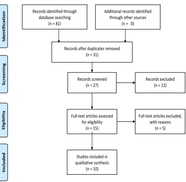
Based on this search strategy, 31 unique records could be identified. A total of 12 records were excluded after a screening of titles, abstracts, and article format
reviews. A further 4 articles were not accessible for the full text. The resulting 15 full texts could then be retrieved. These 15 full-text articles were analyzed regarding the assessment of RI. In total, 10 primary studies published between 2014 and 2017 (Nejati, Najian, Salehinejad, Nitsche & Javadi, 2017; Doruk, Gray, Bravoa, Pascual-Leone & Fregni, 2014; Kwon & Kwon, 2013; Kwon, Kwon & Tremblay, 2016; Cunillera et al., 2016; Castro-Meneses, Johnson & Sowman, 2016;Stramaccia et al., 2015; Dambacher et al., 2015; Nieratschker at al., 2015;Hogeveen et al., 2016) were included for further analyses in this systematic review. All other full texts (N = 5) were excluded due to: (1) a lacking description of the protocol used for stimulation,
9
not describing if the task was performed On/Off line or if the other outcome
measures, eye tracking and EEG, interacted and modified the RI measures (Lapenta, Sierve, de Macedo, Fregni & Boggio, 2014); (2) the assessment of RI in the study by Loftus, Yalcin, Baughman, Vanman & Hagger (2015), and Bender Filmer and Dux (2017) was made with the Stroop color-word matching task and the forced choice reaction time respectively. These tasks might be measuring other cognitive
components not related to RI, or assessing different processes than the SST and the Go/No-Go task, as studies have shown that the correlation between scores on these tasks is weak, indicating that “the Stroop and Go-NoGo tasks tap different aspects of selective attention and response inhibition” (Oka et al., 2012, p. 377); (3) the
outcome measure was working memory and not RI (Gill, Shah-Basak, Priyanka & Hamilton, 2015), and (4) the study by Strobach, Soutschek, Antonenko, Flöel and Schubert (2015 ) was excluded because the outcome measures were based on a dual task (visual- auditory), and the aim of the study was the improvement of these combined dual tasks and not of RI.
The two articles by Kwon and Kwon (2013; 2016), were explored to assess the risk of duplicate populations. It was decided to include both studies as relevant
differences in the protocol of stimulation as well as the outcome measures were found between them. An exception from the exclusion criteria was made for one of the studies (Doruk, et al., 2014) measuring EFs with the TMTA & B instead of the Go-NoGo or SST tasks. It was decided to include this study because it was the only one found measuring long-term effects (one month after stimulation). Even if direct conclusionscannot be drawn, at least it will give a glimpse of how tDCS affects long-term executive control.
For a detailed PRISMA (Moher, Liberati, Tetzlaff & Altman, 2009) flow diagram and checklist, see Figure 1 and Appendix A respectively.
Ethical considerations
The 10 empirical studies reviewed were approved by a local research ethics
committee and reported no conflicts of interest. Informed consent was obtained in all studies from the participants before the intervention. Regarding participants of reduced competence, children in the study by Nejati et al. (2017) younger than 16 years old, informed consent was obtained from the parents, with authors stating that participants were free to withdraw at any time. Confidentiality of the data was not discussed in any of the studies, however, 5 of the studies stated to follow the ethical principles of the declaration of Helsinki. The participants in the studies by Hogeveen et al. (2016), and Cunillera et al. (2016) were paid with cash, and in the study by Castro-Meneses et al. (2016), participants were awarded cash or student credits. It is worth noticing that participants did not specifically consent to the use of the data in the present review. However, it was considered that the present review’s potential benefits were relevant enough for the use of existing data. Also, this review was limited to studies already in the public domain. The data from the 10 studies were presented in an accurate and fair way, although some of the data was not fully transcribed because was not relevant for the review. For instance, results of tDCS on
10
other tasks than the SST or the Go-NoGo task or measures of other cognitive processes.
Figure 1. PRISMA flow diagram of selected studies investigating the effects of tDCS on RI
RESULTS
Study Characteristics
Records identified through database searching (n = 81) Scr e e n in g In cl u d e d El ig ib il ity Id e n ti fi cat ion
Additional records identified through other sources
(n = 0)
Records after duplicates removed (n = 31)
Records screened (n = 27)
Records excluded (n = 12)
Full-text articles assessed for eligibility
(n = 15)
Full-text articles excluded, with reasons
(n = 5)
Studies included in qualitative synthesis
11
Two from the ten studies reviewed had participants diagnosed with neurological deficits, one by Nejati et al. (2017) had children with ADHD, and Doruk et al., (2014) had participants diagnosed with Parkinson disease. All the other studies had healthy samples. The size of the sample of the studies ranged from 13 to 115 with the fewer subjects in the study by Cunillera et al. (2016) and more subjects in the study by Stramaccia et al., (2015). No direct or confounding effects of gender were found in any of the studies reviewed. Most of the studies, probably due to the low number of participants, used repetitive measures, with the entire sample on all conditions. All the studies randomly assigned subjects to the different conditions although many used the same participants for all conditions. Even in this case, all the studies report a random counterbalance of conditions. All the studies included a sham condition and subjects were blind to conditions. Five of the studies (Nejati et al., 2017; Cunillera et al., 2016; Castro‑Meneses et al., 2016; Stramaccia et al., 2015; and Nieratschker at al., 2015), used a pre-test or control, obtaining a baseline measure of RI, with the other five articles lacking a pre-test measure.
tDCS Gains versus Sham
Eight of the ten studies reviewed report gains of tDCS versus sham. From these 8 studies, five report a gain in shorter reactive inhibition, one report a gain in accuracy, one report gains in proactive inhibition and one report a gain in executive control (For a summary see Table 1).
The study by Dambacher et al. (2015) did not find any effect of anodal tDCS applied to the right IFG and cathodal tDCS applied to the left IFG on RI measured with the Go-NoGo task. Finally, Nieratschker at al., (2015), found an impairment effect of cathodal tDCS applied to the left DLPFC only in individuals with the Val/Val genetic profile. Theoretically, this profile is related to a lower activity in the dopaminergic system, also found in individuals with ADHD.
Effects of tDCS Activation / Deactivation of the Right / Left Hemisphere on RI
RI or inhibitory control impairment causes automatic responses and inappropriate actions related to emotional and behavioral control. Previous research found that the right DLPFC and the right IFG areas of the brain are involved in the control of responses, and most studies stimulate these areas. However, after the review of the 10 selected articles, it was observed that it was important to make a distinction between the studies that activate and/or deactivate the right and /or left hemisphere of the brain. The nature and particularities of tDCS, makes this current to act
differently to other NIBS techniques. The area of the brain under the anode electrode it is said to be activated, and the area under the cathode electrode to be deactivated. Most of the studies reviewed followed this principle and used tDCS in this way.
Activationof the right hemisphere: IFG, DLPFC, PFC, and OFC
When positive stimulation (+ anodal tDCS) is delivered, the current causes a depolarization of the resting membrane potential, which increases neuronal excitability by decreasing the threshold of the cell firing.
12
The right IFG. This area was chosen to be activated and therefore the anode
electrode or positive pole was placed on the right IFG by Cunillera et al., (2016); Stramaccia et al., (2015); Dambacher et al. (2015), and Hogeveen et al., (2016). Two studies (Stramaccia et al., 2015; Hogeveen et al., 2016) reported an improvement in RI with shorter signal stop reaction time (SSRT) or reactive inhibition, on the stimulation group compared to sham. The study by Stramaccia et al. (2015) report improvements in the SSRT with conventional tDCS and with high definition (HD) tDCS. In contrast to conventional tDCS, which uses large sponge electrodes, HD-tDCS uses small electrodes targeting more specific areas. One study (Cunillera et al., 2016) found improvement on proactive inhibition but no effect on reactive
inhibition, meaning that subjects showed improvement in acquiring the strategy of longer RT to the Go signal but they didn’t show a reduced RT to the NoGo signal. Proactive and reactive inhibition might imply different mechanisms, and RI related to impulsivity and antisocial behavior has been shown to be related to both.
However, different areas of the brain could be in play. Finally, one study didn’t find any effects in comparison with sham (Dambacher et al., 2015).
All the studies were performed with healthy young subjects (22-25 years old), the duration of stimulation ranged between 10 and 20 minutes, and the intensity of the current ranged between 1-1.5 mA.
The size of the electrodes did not influence the effects of tDCS on RI, with both, the study using the largest ones, 35 cm², and the study using the smallest electrodes, 9cm², not finding any effect in reactive inhibition. Also, the study using HD- tDCS versus conventional tDCS didn’t find any difference in the effects, with both conditions improving RI. This suggests that it is not necessary to focalize the activation on a smaller area of the right IFG.
There were no differences between Online and Off-line stimulation as with both effects are reported.
All the studies finding improvement on reactive inhibition measured RI with the SST. In contrast, the study that found no effects measured RI with the Go-NoGo task. Another important difference is that in the study finding no effects, the montage of tDCS was bilateral, with the cathode/anode placed on the contralateral IFG. It suggests that the placement of the negative pole in the contralateral IFG canceled the effects of the activation in the IFG.
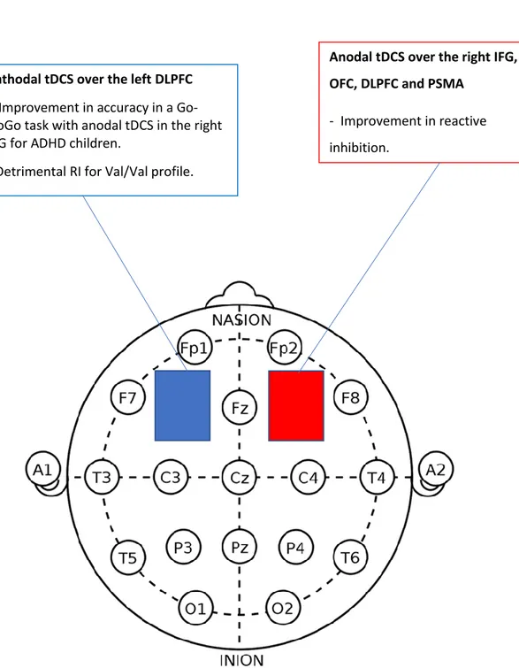
In sum, the activation of the right IFG improved RI when measured with the SST, and when the placement of electrodes was the anode on the right IFG and the cathode in a neutral area.
The right orbitofrontal cortex (OFC). This area was activated in one of the
studies (Nejati et al., 2017) finding improvement in accuracy in the Go-NoGo task in children diagnosed with ADHD. The right OFC is close to the right IFG, especially in children. The tDCS doesn’t provide a focused activation of a small specific area, and in children, because of a closer proximity between brain regions, it is possible that the area targeted extended beyond the OFC. However, it is also possible that the OFC is a better candidate to improve the kind of RI measured with a Go-NoGo task
13
which was employed in the study by Nejati et al. (2017) Also, the cathode or negative pole was placed on the left DLPC, and the effects could be due to the combination of this placement and not just to the activation of the right OFC. The OFC has been associated with RI after the response to a stimulus is reversed, which is best suited to the Go-NoGo task than the SST. Moreover, the OFC might be a specific area to activate for children with ADHD but not for a different population.
The right PFC. This area was activated in the study by Castro‑Meneses et al.
(2016), reporting a reduction of the RT in a SST in healthy adults, and improving reactive inhibition in manual and vocal responses. However, the PFC is a big area that includes IFG, DLPFC, and orbito-frontal gyrus (OFG) among others. The authors report that the placement of the anodal electrode, based on the 10–20 EEG system (Jasper 1958), was over the intersection point of a line between T4-Fz and a line between F8-Cz, which correspond to the right IFG as identified by Jacobson et al. (2011). Therefore, this study could be considered as effects of tDCS on the right IFG. Also, this study adds to the findings of the previous ones that the effects are seen in vocal inhibition in addition to manual reactions.
The right DLPFC. This area was activated in two studies, the one by
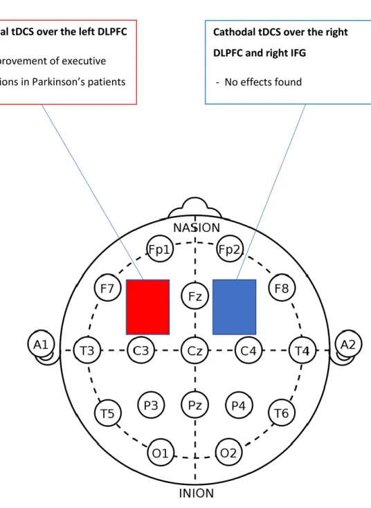
Stramaccia et al., (2015), found no effects of stimulation on RI measured with the SST. However, in the study by Doruk et al., (2014), the authors report an
improvement in executive control assessed with the TMT-B task in Parkinson’s patients.
In sum, anodal tDCS of the right PFC improves RI measured with the Go-NoGo and SST. For results of the effects of anodal tDCS on the right PFC see Figure 2 on Appendix B1.
Activation of the left hemisphere: DLPFC
The left DLPFC. This area was activated in two studies (Doruk et al., 2014;
Nejati et al., 2017). The study by Nejati et al. (2017) found no effects, but Doruk et al. (2014) reported an improvement on executive control. However, clear differences can be seen in both studies. First, subjects in the first study were children with ADHD, and in the second, Parkinson patients with a mean age of 61. Also, Nejati et al. (2017) applied a bilateral montage with the cathode electrode in the right DLPFC, meanwhile Doruk et al, placed the cathode on the contralateral supraorbital area (SOA), an area over the eye used often as a neutral area. Moreover, Doruk et al. assessed executive control with the TMTA & B tasks and Nejati et al. (2017) used the Go-NoGo task to assess RI. Finally, in the study by Doruk et al. (2014) subjects were stimulated during 10 sessions along 2 weeks in comparison with the Nejati et al. (2017) study were subject’s stimulation condition was done during one session. Worth noticing is that in the study with Parkinson patients, the effects persisted after one month in comparison with subjects in the sham condition. However, the
activation of the right DLPFC also generated the same improvements. This suggests that the tDCS irrespective of activation of the left or right DLPFC improve EFs specifically in subjects with Parkinson.
14
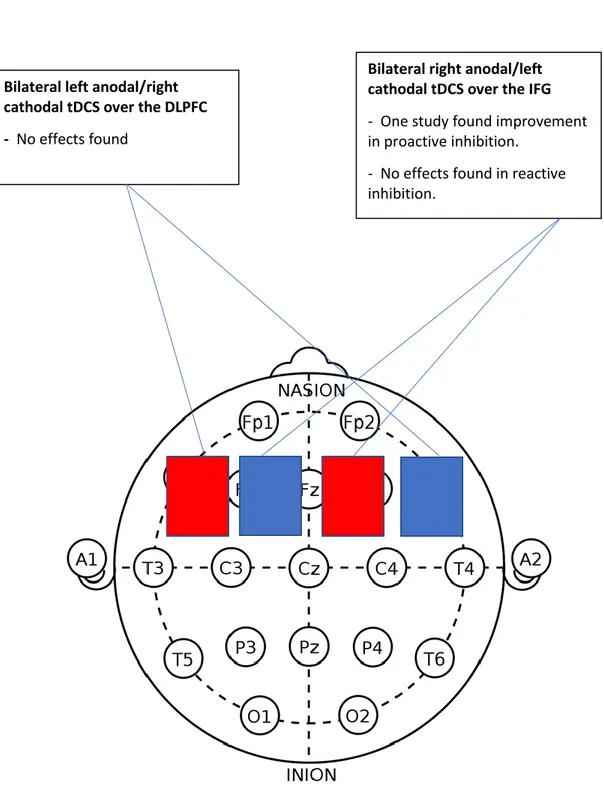
In sum, the results suggest that a bilateral montage, the anode in the left DLPC and the cathode in the right DLPFC does not improve RI (see Figure 3 on Appendix B2). Also, that anodal tDCS of the left and right DLPFC can enhance RI specifically in individuals with Parkinson's disease, and that the effects can be long-term (see Figure 4 on Appendix B3).
Activation of the primary motor area (M1) and the premotor supplementary area (PSMA)
Two studies applied the anode to the M1 and PSMA (Kwon & Kwon, 2013), and to the PSMA (Kwon et al., 2016). They found that the simulation of the PSMA
improved RI in an SST with a shorter reactive inhibition time, both on and offline (see Figure 2). However, no effects were found when activating the M1. The PSMA is involved in cognitive processes of preparation and inhibition of motor movements. In contrast, the M1 area is directly involved in the immediate movement. This
suggests that RI is a cognitive process that precedes the movement and it is not a physical reflex.
Deactivation of the right hemisphere: DLPF and IFG
When negative stimulation (cathodal tDCS) is delivered, the current causes a hyperpolarization of the resting membrane potential, increasing the threshold of the cell firing. Therefore, the application of the cathode to a brain area will “deactivate that area”. What it does is to elevate the threshold making it more difficult for the neuron to fire.
The right DLPFC. The study by Nejati et al., (2017) did not find any effect on
RI when the cathode was placed on the right DLPFC and the anode was applied to the left DLPFC. Likewise, the study by Stramaccia et al., (2015) did not find any effects with the cathode on the right DLPFC and the anode on the left superior orbital area (SOA).
The right IFG. The study by Dambacher et al. (2015) did not find any effect
when the cathode was on the right IFG and the anode on the left IFG.
In sum, the deactivation of the right hemisphere in contrast to the activation shows no effects on RI (see Figure 4 on Appendix B3).
Deactivation of the left hemisphere: IFG and DLPFC
The left IFG. The study by Dambacher et al. (2015) did not find any effect when the anode was on the right IFG and the cathode on the left IFG.
The left DLPFC. This area was deactivated in two studies. The study by Nejati
et al., (2017), shows that deactivation of this area together with the activation of the right OFG improves accuracy in ADHD children. In contrast, deactivation of the left DLPFC with the anode applied to the orbit area produced a detrimental RI effect in individuals with a genetic profile causing a lower level of dopamine in the PFC, like the low dopamine levels reported in children with ADHD (Nieratschker at al., 2015). It is interesting to note that the two studies assessed RI with the Go-NoGo task and both samples had subjects with lower levels of dopamine in the PFC. However,
15
important differences can be observed between the studies. First, in one of the studies the sample are children and in the other adults. Also, one report improvement in accuracy and the other reports detrimental effects on RT on the NoGo signal. Finally, and most important, in the study finding improvement in RI (Nejati et al., 2017)the anode electrode was placed on the right OFC, while in the study by Nieratschker at al. (2015), the anode was placed on the contralateral orbit area, an area often used as a reference neutral area. Moreover, the impairment effects found in the study by Nieratschker at al. (2015), were selective for adults with the Val/Val genetic profile and the deactivation of this area can lower the threshold of the neurons even more for these individuals. The fact that the study by Nejati et al. (2017) found improvement effects on RI with the activation at the same time of the right OFC, suggest that the right hemisphere activation is more important than the deactivation of the left hemisphere for enhancement effects (see Figure 2 on Appendix B1).
Effects of Trials per Session
Nine of the ten studies reviewed involved only one session of tDCS, however, the number of trials varies across studies with a range of 50 to 1142 trials. Enhancement effects were reported irrespective of the number of trials
.
Duration of the Effects
The study by Doruk et al. (2014) was the only one assessing long time effects with 10 sessions over two weeks and reporting offline effects of tDCS on executive control measured 2 weeks and one month after stimulation.
With one session and a stimulation time range of 10min – 21.75 min, the effects of tDCS did not last longer than 72 hours on accuracy, 24 hours in reactive inhibition, one week in proactive inhibition, and 48 hours to obtain an impairment effect on reactive inhibition.
It is interesting that effects are found even after only 10 minutes of stimulation. However, what it seems to be crucial is to start measuring after at least five minutes of activating the area. For instance, the study by Dambacher et al. (2015) with the longest stimulation duration, 21.75 minutes, did not find any effect. Maybe because the assessment was started immediately after applying the current, without waiting for the five recommended minutes to stimulate the area.
16
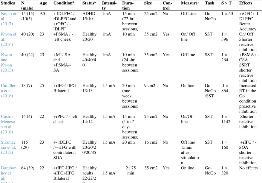
Table 1. Studies Included in the Systematic Review with Criteria Included for Review
Studies N (male)
Age Conditionª Statusᵇ Intensi-ty
Dura-tion
Size Con-trol
Measureᶜ Task S × T Effects Nejati et al. (2017) 15 (15) /10(5) 9.5 + lDLPFC / -rDLPFC and +rOFC / - lDLPF ADHD 15/10 1mA 15 min (72-hr between sessions)
25 cm2 No Off Line Go-NoGo 1 × 50 +rOFC / -l DLPFC Better Accuracy Kwon et al. (2016) 40 (20) 23 +PSMA / - left cheek Healthy 20/20
1mA 10 min 35 cm2 Yes On/ Off line SST 1 × 396 On/ Off Shorter reactive inhibition Kwon and Kwon (2013) 40 (22) 23 +M1/-SA and +PSMA/- SA Healthy 40/40/4 0 1mA 10 min (24 -hr between sessions)
35 cm2 Yes Off line SST 1 × 264 +PSMA / -CSA SSRT shorter reactive inhibition Cuniller a et al. (2016) 13 (7) 25 +rIFG/-lIFG Bilateral Healthy 13/13 1.5 mA 20 min (one week between sessions) 9 cm2 No On line Go-NoGo /SST 1 × 864 Increased RT in the Go condition proactive inhibition Castro‑ Menese s et al. (2016) 14 (4) 22 +rPFC / - left cheek Healthy 14/14 1.5 mA 15 min (1 to 7 days between sessions) 25 cm2 No On/Off line SST 1 × 1142 Shorter reactive inhibition Stramac cia et al. (2015) 115 (29) 23 +- rDLPC /+-rIFG with contralateral SOA Healthy 20/20/2 0/20/35
1.5 mA 20 min 16 cm2 No Off line 15min after stimulatio n SST 1 × 160 +rIFG / -SOA shorter reactive inhibition Dambac her et al. (2015) 64 (39) 22 +rIFG-lIFG / -rIFG+lIFG Bilateral Healthy adults 22/22/2 0 1.5 mA 21.75 min
35 cm2 Yes On line Go-NoGo
1 × 320
17
Table 1 (continued)
Studies N (male)
Age Conditionª Statusᵇ Intensi-ty
Dura-tion
Size Con-trol
Measureᶜ Task S × T Effects Nieratsc hker at al. (2015) 41 (9) 24 +rOrbit -lDLPFC Healthy 41/41 1 mA 20 min/48 hr between sessions 35 cm2 No On line Go-NoGo 1 × ns. Detrimental effects for the Val/Val genetic profile Doruk et al. (2014) 18 (12) 61 +lDLPF /+ rDLPC with contralate-ral SOA Parkin-son 6/5/7 2mA 20 min 35 cm2
Yes Off line -2 weeks and 1 month after stimulatio n TMTA & B 10 over 2 weeks / no trials Lasting one-month effects on TMT-B on the 2 conditions Hogeve en et al. (2016) 46 (ns) 25 +(HD) rIFG / + rIFG with cathode on Cz Healthy 15/15/1 6 1 mA 20 min 25 cm2 HD= 4× Ɵ 1cm
Yes On line SST 1 × 192 shorter reactive inhibition with convention-nal and HD
Note. PSMA (Pre-Supplementary Motor Area); SOA (Supraorbital Area); + lDLPFC (anodal left Dorsolateral Prefrontal Cortex); - rDLPC
(cathodal right Dorsolateral Prefrontal Cortex); + rOFC (anodal right Orbito Frontal Cortex); + M1 (anodal primary Motor area); SST (Stop- Signal task); SSRT (Stop Signal Response Time); Trail Making Tests A & B (TMTA & B); HD (High Definition); Cz (midline sagittal plane); ADHD ( Attention Deficit Hyperactivity Disorder); S ×T (Sessions × trials); ns (not stated); +rIFG (anodal right Inferior Frontal Gyrus); - rIFG (anodal right Inferior Frontal Gyrus); + lIFG (anodal left Inferior Frontal Gyrus): - lIFG (cathodal left Inferior Frontal Gyrus); ns (not stated). ªOne of the conditions is always a sham condition
ᵇSample characteristics and subjects per condition
18
DISCUSSION
The purpose of the present review was to analyze the neuromodulation effects of tDCS on executive control and RI, and its potential efficacy to apply in the
prevention and treatment of antisocial and deviant behavior. Specifically, to clarify the most effective montage and protocol to improve deficits in RI as well as
individual differences and duration of the effects. Individual risk factors for criminal behavior include antisocial personality behavior (Fridell et al., 2008), low self-control (Wolfe et al., 2016) impulsivity (Vazsonyi et al., 2006), substance abuse (Pérez & Ruiz, 2017), and conduct disorder (Liabø & Richardson, 2007). All these factors have in common a deficit in RI (Blair et al, 2016; Monette, Bigras & Guay, 2015). Therefore, the improvement of the deficits in RI could help to decrease the likelihood that a person commits a crime. Also,the influence of individual
differences as well as the duration of the effects that tDCS has on RI are crucial factors to consider if in the future we want to apply this technique to prevent onset and recidivism of crime
The keywords selected for the search restricted it to tDCS effects on RI assessed with the SST and Go-NoGo task. In the studies employing many assessment tasks, only the results in the SST and Go-NoGo task were reviewed. An exception was done with the study by Doruk et al. (2014), evaluating executive control with the TMT-A & B, because was the only article found investigating long-term effects Despite systematic literature search, there is a possibility that certain studies have been missed or excluded and the present review may not be exhaustive. The impact that tDCS montage, protocols, and individual differences have on RI, together with the duration of the effects, were analyzed.
The studies reviewed emphasized the application of the simulation to activate, applying the anode, or to deactivate an area, applying the cathode. Also, the studies ‘results were dependent on the hemisphere where the stimulation was applied, right or left. Most studies stimulated the IFG and the DLPFC, but some applied tDCS on the OFC, PFC and the M1 and PSMA.
Differences between the right and left hemisphere, activation or deactivation tDCS were observed. Results also varied depending on individual differences. Duration of the effects was dependent on the number of sessions but not in the number of trials.
Impact of tDCS Montage and Protocol on RI
Collectively, the review of the studies reveals a pattern where activation or anodal tDCS of the right PFC causes an improvement in RI, and the deactivation or cathodal tDCS of the left hemisphere shows contradictory results, possibly determined by individual differences. In accordance with previous research, gains of stimulation versus sham were found with the electrodes applied to the right IFG, OFG and DLPF. This might be since tDCS apply a non-focused current on the scalp that affects a large area of the brain. Also, the fact that the application of HD- tDCS
19
produced the same effects than the conventional tDCS suggest that stimulation of an extensive area of the right PFC is involved in inhibitory control. The activation or anodal tDCS of the PSMA, also improved RI in the SST, which reinforces the hypothesis that RI is a cognitive process that precedes physical action and that a dysfunction of this process can lead to the inability to restrict a movement. Of
interest would be to investigate if a deficit in the performance of the PSMA is related to impulsivity and/or low self-control and if a treatment with tDCS might improve this behavior.
Interestingly, the results of the present review suggest that activation of an area is more important than deactivation when applying tDCS to improve RI. Also, a bilateral assembly, such as the anode in the left DLPFC and the cathode in the right DLPFC, does not produce gain compared to the sham condition. This suggests that the effects cancel each other out, or that the use of tDCS to deactivate an area is not effective due to the nature of the direct current. In contrast to other tES techniques, in a direct current the cathode acts as a reference of the direction of the current, since the direct current cannot travel very far until it begins to lose energy, and only in one direction, from the anode to the cathode, with electrons that travel from the anode and exit through the cathode (ElectronicsCoach, 2018). If the objective is to
deactivate an area of the brain, other NIBS techniques, such as transcranial magnetic stimulation, may be a better option since tDCS seems to work better by activating and not deactivating areas of the brain.
Moreover, specific areas of the PFC seem to respond to different assessment tasks employed to measure RI. For instance, anodal tDCS of the right IFG improved specifically RI as measured by the SST, and anodal tDCS of the right OFC improved RI as measured with the Go-NoGo task. Although both tasks rely on an inhibitory mechanism, each might capture different aspects of inhibitory control. In the future, it is crucial to investigate what are the deficits in the inhibitory mechanisms that are specific for the different disorders involved in deviant behavior and consider that different tasks assess various aspects of the same cognitive process when designing studies and interpreting the results.
A possible limitation of the study reporting no gains (Dambacher et al., 2015) is that tDCS was applied at the same time the subjects started the Go-NoGo task, not leaving the established 5 minutes necessary for the area to be activated. This might have biased the results. Also, in contrast to the other studies that performed
ANOVAS or ANCOVAS, the statistics the authors choose, a MANOVA, with the gender as a factor and the pre-test measures discounted from the posttest measures, could have further reduced the chance to find an effect. Most importantly, this was the only study that applied bilateral tDCS, meaning that the activation of the right/left IFG was done at the same time as the deactivation of the right/left IFG. Moreover, the results of the study by Nieratschker at al., (2015), reporting an impairment effect of cathodal tDCS on the left DLPF in RI measured with a Go-NoGo task, should be taken with caution. First, the authors’ aim was to prove that the Val/Val gene is related to a lower dopamine level in the left DLPFC and that for subjects with this profile a deactivation of this area would generate a detriment in
20
response control. However, they did not apply the anode to activate this area and see if an improvement of RI would occur. Also, it is important to consider that they did not quantify the dopamine levels and that their conclusions relied just on a
theoretical model. However, this study suggests that the effects of tDCS might be moderated by the levels of dopamine in the PFC.
In sum, from the present review can be concluded that most effective to improve reactive inhibition on a non-clinical sample assessed with the SST seems to be a montage where the anode is placed on the right IFG or the PSMA, and the cathode in a neutral area, like the contralateral orbital area. The size of electrodes, the intensity of the current, duration of the stimulation, the use of HD or conventional tDCS, number of trials, and gender, did not affect the results in the articles reviewed. The assembly for clinical samples seems to be different, with the DLPFC appearing as an important region to stimulate to enhance RI, especially in individuals with a deficit in the dopaminergic system.
Impact of Individual Differences
The review shows individual differences to be essential to consider in the design of protocols and montages. For instance, the deactivation or cathodal tDCS of the left DLPC had detrimental effects in a Go-NoGo task for subjects with a low dopamine level in the PFC and no effects on subjects with a normal level of dopamine. Moreover, anodal tDCS of the right OFC together with cathodal tDCS of the left DLPFC improved accuracy in a Go-NoGo task in ADHD children. The symptoms of ADHD have been suggested to arise from a deficit in cortical inhibitory
neurotransmission (Demirtas-Tatlidede, Vahabzadeh-Hagh & Pascual-Leone, 2013) and deficits in the dopaminergic system (Biederman & Faraone, 2005). It is possible that the left DLPFC is an area specifically sensible for subjects with a low level of dopamine in the PFC.
The activation or anodal tDCS of the DLPFC (left and right) seems toimprove executive control specifically in individuals diagnosed with Parkinson. It is possible that in these patients an enhancement effect can be seen because the activation of both, left and right DLPFC, activates the basal ganglia through the dopaminergic system (Tomoko et al., 2013), which is directly connected with the DLPFC, and it is affected in this pathology. Therefore, the activation of the left and right DLPFC might work specifically in individuals with a pathologically low activity of the dopaminergic system.
In sum, individual differences such as neurological diagnoses and dopamine levels should be considered as the same stimulation protocol will show different effects. When using tDCS as a treatment for deviant behavior, individual differences should be considered, and montage and protocols adjusted accordingly.
Duration of the Effects
It is well known that the underlying mechanisms for online and offline effects of tDCS are different with online effects related to changes in polarization in neural membranes, whereas offline effects involve more complex processes such as long-term potentiation (LTP) and long-long-term depression (LTD) inducing long-long-term
21
synaptic plasticity (Nitsche et al., 2003). Online and offline (no more than 15
minutes after stimulation) effects were measured at 9 of the 10 studies. As five of the reviewed studies used repeated measures, it was possible to calculate the maximum duration of the effects of a session with tDCS. Thus, it was concluded that the duration of the effects of a session with tDCS lasted no more than 24 hours. Offline, long time effects (more than 24 hours) were assessed only in one of the articles. The study by Doruk et al. (2014) was included in this review because being the only one to measure long-term effects. However, in contrast to the other studies, the
assessment task was a TMT-A & B task and not the SST or Go-NoGo task and it had a clinical sample with Parkinson’s patients. Therefore, it is inviable to draw
conclusions. However, the study suggests that it is possible for the effects of tDCS to last for at least one month after 10 sessions with tDCS.
Interestingly, the number of trials, or cognitive training, in one session does not seem to affect the results. For instance, Doruk et al. (2016) report improvement in RI one month after tDCS without cognitive training. This suggests that tDCS without training was effective in Parkinson patients. This result contradicts previous studies and theory that relies on a simultaneous cognitive training and stimulation for the treatment to be effective (Oldrati, Colombo & Antonietti, 2018).
In sum, the duration of the effects of tDCS on RI seems to be related to the number of sessions and not to the number of trials in each session. The long-term
potentiation involving a learning process is crucial if tDCS is to be used as a tool to prevent and treat deviant behavior. Therefore, the design of prevention and treatment programs to improve deficits in RI, and hence, deviant behavior, should include several sessions with tDCS.
An important limitation in the application of tDCS on the prevention of onset and recidivism in crime is the lack of longitudinal designs with all studies except one listed in this review measuring short-term effects. The only study using repeated sessions found interesting lasting effects that may lead to plasticity and learning processes.To be able to apply tDCS to prevent and treat deviant behavior, in the future, research should focus on longitudinal designs together with neuroimaging to assess long time learning effects and synaptic plasticity. Another limitation is the age of the participants, with 8 of the 10 studies reviewed having a range between 22-25 years old, one study had a mean age of 9.5 years (Nejati et al., 2017), and another 61 years old (Doruk et al., 2014). Thus, conclusions about tDCS effects on children and the elderly are limited. In the future, more studies with children are necessary to understand better if tDCS can be used as a tool to prevent the onset of crime. Moreover, to decrease recidivism, studies with young and older offenders are needed.
22
The present review shows that tDCS can modulate RI. Deficits in RI are frequently reported in adult male sexual offenders (pedophiles/child molesters and
uncategorized sexual offenders (Adjorlolo & Egbenya, 2016) as well as in the
susceptibility for the initiation of drug taking (Groman et al., 2009). Also, RI deficits are found in children with conduct disorder (Prateeksha, et al.,2014), and ADHD (Biederman & Faraone, 2005). At the same time, ADHD has been related to higher rates of recidivism in juvenile offenders (Van der Put et al., 2016), and long-term risk for later antisocial involvement (Mohr-Jensen & Steinhausen, 2016). Therefore, the treatment of the deficits in RI could prevent the onset and recidivism of crime. Given the high detrimental impact that crime has on society, and the lack of effective measures and treatments, tDCS offers a promising new approach to treat deviant behavior, and hence, decrease onset and recidivism of crime. In contrast to drugs, tDCS is non- invasive, easy, cheap, and safe to apply. Important considerations for the use of tDCS are the different effects found depending on individual differences, and the lack of studies measuring long-term effects.
In conclusion, although further research needs to be undertaken, it seems possible to modulate RI trough tDCS and hence, to improve conducts with a high likelihood of leading to criminal behavior. Therefore, in the field of criminology, a potential tool to modify deviant or maladapted behavior is tDCS.
23
REFERENCES
Adjorlolo, S., & Egbenya, D. L. (2016). Executive functioning profiles of adult and juvenile male sexual offenders: A systematic review. Journal of Forensic
Psychiatry & Psychology, 27(3), 349-375. doi:10.1080/14789949.2016.1141431 Allen, C., Singh, K. D., Chambers, C. D., Verbruggen, F., Verbruggen, F., Allen, C.,
Verbruggen, F. (2018). Evidence for parallel activation of the
pre-supplementary motor area and inferior frontal cortex during response inhibition: A combined MEG and TMS study Royal Society Publishing.
doi:10.1098/rsos.171369
Andrade, S., Jenny Marcela Barbosa, Ñ., & Claudia Ximena Lozada, R. (2012). Biopsychosocial risk factors influence in the development of dissocial disorder in Colombian adolescents. Revista Internacional De Psicología, Vol 12, Iss 01 (2012), (01),
Aron, A.R., Poldrack, R. A., & Robbins, T. W. (2004). Inhibition and the right inferior frontal cortex. Trends in Cognitive Sciences, 8(4), 170-177. doi: 10.1016/j.tics.2004.02.010
Aron, AR., & Poldrack, RA. (2006). Cortical and subcortical contributions to stop signal response inhibition: role of the subthalamic nucleus. Journal of
Neuroscience, 2006
Bari, A., & Robbins, T. W. (2013). Inhibition and impulsivity: Behavioral and neural basis of response control. Progress in Neurobiology, 108, 44-79. doi:
10.1016/j.pneurobio.2013.06.005
Bender, A. D., Filmer, H. L., & Dux, P. E. (2017). Transcranial direct current
stimulation of superior medial frontal cortex disrupts response selection during proactive response inhibition Academic Press Inc. doi:
10.1016/j.neuroimage.2016.10.035
Biederman, J., & Faraone, S. V. (2005). Attention-deficit hyperactivity disorder.
Lancet,
366(9481), 237-248. doi:10.1016/S0140-6736(05)66915-2
Bikson, M., Radman, T., & Datta, A. (2006). Rational modulation of neuronal processing with applied electric fields. Conference Proceedings: Annual
International Conference of the IEEE Engineering in Medicine and Biology Society. IEEE Engineering in Medicine and Biology Society. Annual
24
Blair, R. J. R., Veroude, K., & Buitelaar, J. K. (2016). Review article: Neuro-cognitive system dysfunction and symptom sets: A review of fMRI studies in youth with conduct problems. Neuroscience and Biobehavioral Reviews, doi: 10.1016/j.neubiorev.2016.10.022
Bliss, T. V., & Collingridge, G. L. (1993). A synaptic model of memory: Long-term potentiation in the hippocampus. Nature, 361(6407), 31-39.
Boggio, P. S., Villani, A. B., Zaghi, S., Fecteau, S., Pascual-Leone, A., Fregni, F. (2010). Modulation of risk-taking in marijuana users by transcranial direct
current stimulation (tDCS) of the dorsolateral prefrontal cortex (DLPFC) doi:
10.1016/j.drugalcdep.2010.06.019
Boggio, P. S., Bermpohl, F., Vergara, A. O., Muniz, A. L. C. R., Nahas, F. H., Leme, P. B., Fregni, F. (2007). Research report: Go-no-go task performance
improvement after anodal transcranial DC stimulation of the left dorsolateral prefrontal cortex in major depression. Journal of Affective Disorders, 101, 91-98. doi: 10.1016/j.jad.2006.10.026
Brevet-Aeby, C., Brunelin, J., Iceta, S., Padovan, C., & Poulet, E. (2016). Review article: Prefrontal cortex and impulsivity: Interest of noninvasive brain stimulation. Neuroscience and Biobehavioral Reviews, 71, 112-134. doi: 10.1016/j.neubiorev.2016.08.028
Castro-Meneses, L., Johnson, B. W., & Sowman, P.F. (2016). Vocal response inhibition is enhanced by anodal tDCS over the right prefrontal cortex.
Experimental Brain Research, 234(1), 185-195.
doi:10.1007/s00221-015-4452-0
Clark, K., Squire, R. F., Merrikhi, Y., & Noudoost, B. (2015). Visual attention: Linking prefrontal sources to neuronal and behavioral correlates. Progress in
Neurobiology, 132, 59-80.
doi:http://dx.doi.org.ludwig.lub.lu.se/10.1016/j.pneurobio.2015.06.006 Clark, V. P., Coffman, B. A., Mayer, A. R., Weisend, M. P., Lane, T. D. R., Calhoun, V. D., Wassermann, E. M. (2012). TDCS guided using fMRI significantly accelerates learning to identify concealed objects. Neuroimage,
59(1), 117-128.
doi:http://dx.doi.org.ludwig.lub.lu.se/10.1016/j.neuroimage.2010.11.036 Coffman, B. A., Clark, V. P., & Parasuraman, R. (2014). Review: Battery powered
thought: Enhancement of attention, learning, and memory in healthy adults using transcranial direct current stimulation. Neuroimage, 85(-), 895-908. doi: 10.1016/j.neuroimage.2013.07.083
Cohen - Kadosh, R. (2015). Modulating and enhancing cognition using brain stimulation: Science and fiction. Journal of Cognitive Psychology, 27(2), 141.
25
Cohen, M. A. (2008). The effect of crime on life satisfaction. The Journal of Legal
Studies, S325. doi:10.1086/588220
Crivelli, D., Pala, F., Finocchiaro, R., Grippa, E., Lecci, G., & Balconi, M. (2016). 68. The effect of a neuromodulation protocol on executive functions in healthy elderly: Psychometric and EEG evidences. Clinical Neurophysiology, 127e148. doi: 10.1016/j.clinph.2015.09.076
Cunillera, T., Fuentemilla, L., Brignani, D., Miniussi, C., Cucurell, D.,
Castro-Meneses, L., Sowman, P. F. (2016). The right inferior frontal cortex in response
inhibition: A tDCS–ERP co-registration study Academic Press Inc. doi:
10.1016/j.neuroimage.2015.11.044
Dambacher, F., Schuhmann, T., Sack, A.T., Lobbestael, J., Arntz, A., Brugman, S., Mondini, S. (2015). No effects of bilateral tDCS over inferior frontal gyrus on
response inhibition and aggression Public Library of Science. doi:
10.1371/journal.pone.0132170
Dedoncker, J., Brunoni, A. R., Baeken, C., & Vanderhasselt, M. (2016). A Systematic Review and Meta-Analysis of the Effects of Transcranial Direct Current Stimulation (tDCS) Over the Dorsolateral Prefrontal Cortex in Healthy and Neuropsychiatric Samples: Influence of Stimulation Parameters. Brain Stimulation, 9501-517. doi: 10.1016/j.brs.2016.04.006
Demirtas-Tatlidede, A., Vahabzadeh-Hagh, A. M., & Pascual-Leone, A. (2013). Can noninvasive brain stimulation enhance cognition in neuropsychiatric disorders? Neuropharmacology, 64, 566-578. doi:
http://dx.doi.org.ludwig.lub.lu.se/10.1016/j.neuropharm.2012.06.020
Dimoska-Di Marco, A., McDonald, S., Kelly, M., Tate, R. & Johnstone, S. (2011). A meta-analysis of response inhibition and Stroop interference control deficits in adults with traumatic brain injury (TBI). Journal of Clinical and Experimental Neuropsychology, 33(4), 471-485. doi:10.1080/13803395.2010.533158 Ditye, T., Jacobson, L., Walsh, V., & Lavidor, M. (2012). Modulating behavioral
inhibition by tDCS combined with cognitive training. Experimental Brain
Research, 219(3), 363-368. doi:10.1007/s00221-012-3098-4
Doruk, D., Gray, Z., Bravo, G. L., Pascual-Leone, A., & Fregni, F. (2014). Effects of tDCS on executive function in Parkinson's disease. Neuroscience Letters,
58227-31. doi: 10.1016/j.neulet.2014.08.043
ElectronicsCoach. (2018, April 30). Difference between AC and DC. Retrieved from https://electronicscoach.com/difference-between-ac-and-dc.html