RESEARCH
Using GafChromic film to estimate the effective dose from
dental cone beam CT and panoramic radiography
A Al-Okshi
1, M Nilsson
1, A Petersson
1,2, M Wiese
2and C Lindh*
,11
Department of Oral and Maxillofacial Radiology, Faculty of Odontology, Malm¨o University, Malm ¨o, Sweden;2Department of Radiology, University of Copenhagen, Copenhagen, Denmark
Objectives: To demonstrate the feasibility of GafChromic® XR-QA2 (ISP Corp., Wayne, NJ) as a dosemeter when performing measurements of the effective dose from three cone beam CT (CBCT) units and to compare the doses from examinations of three common dental clinical situations. A second aim was to compare the radiation doses for three digital panoramic units with the doses for the CBCT units.
Methods: The CBCT units used were Veraviewepocs 3De®(J Morita MFG Corp., Kyoto, Japan), ProMax®3D (Planmeca, Helsinki, Finland) and NewTom VGi®(Quantitative Radiology, Verona, Italy). GafChromic XR-QA2 films were placed between the selected layers of the head and neck of a tissue-equivalent human skull (RANDO® phantom; The Phantom Laboratory, Salem, NY). The exposure parameters were set using the automatic exposure control function of the units. Depending on the availability, medium and smaller field of view (FOV) scanning modes were used. The effective dose was estimated using the 2007 International Commission on Radiological Protection formalism.
Results: The lowest effective dose of a CBCT unit was observed for ProMax 3D, FOV 43 5 cm (10mSv), the highest for NewTom VGi, FOV 8 3 8 cm—high resolution (129 mSv). The range of effective doses for digital panoramic machines measured was 8–14 mSv.
Conclusions: This study demonstrates the feasibility of using radiochromic films for dental CBCT and panoramic dosimetry.
Dentomaxillofacial Radiology (2013) 42, 20120343. doi: 10.1259/dmfr.20120343
Cite this article as: Al-Okshi A, Nilsson M, Petersson A, Wiese M, Lindh C. Using GafChromic film to estimate the effective dose from dental cone beam CT and panoramic radiography. Dentomaxillofac Radiol 2013; 42: 20120343.
Keywords: radiation dosage; cone beam computed tomography; film dosimetry
Introduction
Panoramic radiography has long been a common di-agnostic imaging technique in dentistry owing to its low dose and large area for evaluation, which includes bone and teeth in the same image.1Cone beam CT (CBCT) is a more recent technology that has significant potential for a number of clinical situations requiring CBCT im-aging.2Although CBCT provides additional information, it may result in higher radiation doses than conventional imaging procedures, such as intraoral and panoramic.2,3
Although the absorbed doses from oral and maxillofa-cial radiology procedures are usually low, no exposure to radiographs can be regarded as completely free of risk. The measurement of absorbed organ doses is needed to estimate the effective dose associated with diagnostic radiographic imaging. The traditional way of determining the effective dose in oral and maxillofacial radiology is by measuring the organ doses using thermoluminescent dosemeters (TLDs) and head phantoms.3–6
In rotating irradiation geometry with collimated ra-diation fields, the dose distribution will show more or less steep dose gradients. This is a major problem if you want to map or sample the dose distribution with a
*Correspondence to: Dr Christina Lindh, Department of Oral and Maxillofacial Radiology, Faculty of Odontology, Malm ¨o University, SE 205 06 Malm ¨o, Sweden. E-mail:christina.lindh@mah.se
Received 28 September 2012; revised 10 April 2013; accepted 11 April 2013
reasonable degree of accuracy using TLDs. The geom-etry drawback encouraged us to choose a method with higher spatial resolution for mapping the dose distri-butions in a phantom. The GafChromic®XR-QA film (ISP Corp., Wayne, NJ) is designed specifically as a quality assurance (QA) tool for radiology and dosime-try applications. In combination with flatbed document scanners,7it has been used for measuring medical8and dental9CBCT doses and CT doses.10,11Some of the char-acteristics of this film have been studied: energy depen-dence8and angular dependence.10One of the advantages of this film is that it provides a high-resolution image with no chemical processing.12The film is not sensitive to visual light and can therefore be handled in ambient light. Ionizing radiation induces polymerization within the active layer of the film, changing its reflectance and making it appear darker. The darkening of the film is dependent on the radiation exposure received. The dark-ening of the film occurs instantaneously as the reaction immediately creates a polymer dye complex within the active layer of the film.13
The aim of this study was to demonstrate the feasi-bility of the GafChromic film as a dosemeter for use in rotating dental radiography and apply this technique for estimation of the effective doses from three CBCT units and three panoramic units.
Materials and methods Cone beam CT units
The CBCT units selected for this study were Veraviewepocs 3De®(J Morita MFG Corp., Kyoto, Japan), ProMax® 3D (Planmeca, Helsinki, Finland) and NewTom VGi® (Quantitative Radiology, Verona, Italy). All CBCT units used flat panel detectors (FPDs) based on similar prin-ciples, i.e. a scintillating CsI layer and a light-sensitive silicon photodiode matrix. Exposure parameters and protocols used are given inTable 1.
Panoramic units
The panoramic machines selected for this study were Veraviewepocs 3De (J Morita MFG Corp., Kyoto, Japan), ProMax 3D and ProMax (Planmeca) ProMax was used with a photostimulable phosphor plate system (PSP) (DX-S digitizer; Agfa HealthCare, Mortsel, Belgium), ProMax 3D was used with a charge coupled device detector (CCD) and Veraviewepocs 3De was used with the same FPD as for the CBCT mode. The pa-rameters of voltage (kV), tube current (mA) and expo-sure time (s) for each scan of the CBCT and panoramic units were fixed at the units’ manufacturer-recommended settings for an average adult patient (Table 1).
Phantom and the GafChromic film XR-QA2
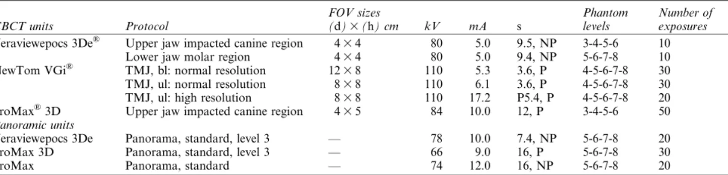
The phantom (RANDO®; The Phantom Laboratory, Salem, NY) was a small adult skull surrounded by soft tissue-equivalent material (Figure 1). Using the posi-tioning aid provided for the scanner, the phantom was positioned mimicking a typical patient examination.
GafChromic films were placed between four selected levels in the head and neck of the phantom for each radiographic technique to record the distribution of the absorbed radiation dose. For detailed information re-garding placement of the films, seeTable 1.
Measurements were performed using GafChromic XR-QA2 films that were scanned with an Epson® Per-fection 4990 Photo flatbed scanner (Seiko Epson Corp., Nagano, Japan). To be able to translate the blackening of the film to absorbed dose, the film has to be calibrated before dosimetric application. The dose–response curve of the GafChromic film was determined using an ion-ization chamber (Radcal 10X6-6®, a Radcal model 9660 ion chamber digitizer and a Radcal model 2186 elec-trometer; Radcal Corp., Monrovia, CA). A standard X-ray tube for medical radiology (A-196®; Varian Medi-cal Systems, Inc., Salt Lake City, UT) with a standard collimating device (Svendx SX100-MF; Santax Medico A/S, Aarhus, Denmark) was used for irradiation. The ionization chamber was placed on top of a 255-cm-thick
Table 1 Technical parameters of selected CBCT and panoramic exposure protocols and sites in which GafChromic®XR-QA2 films were placed in the phantom
CBCT units Protocol FOV sizes (d)3 (h) cm kV mA s Phantom levels Number of exposures Veraviewepocs 3De® Upper jaw impacted canine region 4
3 4 80 5.0 9.5, NP 3-4-5-6 10
Lower jaw molar region 43 4 80 5.0 9.4, NP 5-6-7-8 10
NewTom VGi® TMJ, bl: normal resolution 123 8 110 5.3 3.6, P 4-5-6-7-8 30
TMJ, ul: normal resolution 83 8 110 6.1 3.6, P 4-5-6-7-8 30 TMJ, ul: high resolution 83 8 110 17.2 P5.4, P 4-5-6-7-8 20 ProMax®3D Upper jaw impacted canine region 43 5 84 10.0 12, P 3-4-5-6 50 Panoramic units
Veraviewepocs 3De Panorama, standard, level 3 — 78 10.0 7.4, NP 5-6-7-8 20
ProMax 3D Panorama, standard, level 3 — 66 9.0 16, P 5-6-7-8 30
ProMax Panorama, standard — 74 12.0 16, NP 5-6-7-8 20
bl, bilateral; CBCT, cone beam CT; FOV, field of view; Level 3, level 3 of autoexposure used for adults; NP, not pulsed radiation; P, pulsed radiation; TMJ, temporomandibular joint; ul, unilateral.
GafChromic XR-QA2 film is manufactured by ISP Corp., Wayne, NJ; Veraviewepocs 3De units by J Morita MFG Corp., Kyoto, Japan; NewTom VGi by Quantitative Radiology, Verona, Italy; and Promax 3D and Promax by Planmeca, Helsinki, Finland.
styrofoam slab to minimize the backscatter. A Raysafe® (Unfors Raysafe AB, Billdal, Sweden) semi-conducting detector was placed adjacent to the ionization chamber for use as an exposure monitor when the film was ir-radiated. The detector was connected to a Raysafe Xi electrometer.
The output (mGy/mAs) of the X-ray tube was de-termined using 60 kV, 80 kV and 120 kV X-ray tube
potentials. The ionization chamber was thereafter re-moved and replaced with pieces of the GafChromic film that were irradiated with absorbed doses (in air) up to 200 mGy. It was found that the dose–response curve for 60 kV, 80 kV and 120 kV coincided. Owing to the very small spectral dependence of the ion chamber used for the output measurements, the calibration was not sen-sitive to tube potential, and the same calibration curve could thus be used for all three CBCT units. This is in agreement with what was found for the GafChromic XR-CT film.14This film has identical physical properties and an identical calibration curve to the GafChromic XR-QA film. The film used in this study is a new ver-sion, XR-QA2, which has a slightly higher sensitivity. According to our results, there is no reason to believe that the spectral dependence should differ from that of the XR-CT film, indicating that their atomic compositions are very similar. A piece of film that did not undergo any irradiations was scanned together with the other films and used for background subtraction. The pieces of films were scanned to an ordinary 24-bit red–green–blue (RGB) im-age; a 24-bit RGB image was used for simplicity reasons to train inexperienced operators. No colour channel se-lection or suppression was used, and images were stored as 24-bit RGB images. These images were read with Image J® (see http://rsbweb.nih.gov/ij/), following background subtraction, and converted to black-and-white 8‐bit images. The mean pixel values were measured in each film square using rectangular regions of interest (ROIs). The mean pixel values were used to construct a dose– response diagram. The equation of the dose–response curve (Figure 2) was used for converting the net pixel value distributions found in the phantom measurements to the absorbed dose distribution.
Before the GafChromic films were positioned for the CBCT scans, a scout image was taken to ensure correct positioning of the phantom and that the field of view
Figure 1 RANDO® phantom head (The Phantom Laboratory, Salem, NY)
Figure 2 Dose–response calibration curve. The x-axis is the film response represented by the net pixel value change and the y-axis is the corresponding absorbed dose
(FOV) included the whole anatomical ROI. The dose measurements did not include the scout images. After loading with GafChromic XR-QA2 films, the phantom was exposed several times (Table 1) to provide a reliable measurement. Later, these values were divided by the number of exposures to provide one individual value for each region.
For the skin (entrance) dose measurements, TLDs were used. The TLDs were calibrated using a secondary standard 60Co beam and a pre-defined, reproducible geometric set-up with a plexiglass phantom containing the dosemeters. Prior to the dose measurements in the clinical situation, the dosemeters were annealed at 400 °C for 10 min. For every group of 20 dosemeters, 4 were used for reference irradiation in the 60Co beam and 2 were used for background correction, leaving 14 dose-meters available for clinical dosimetry. The dosedose-meters were read in a Thermo Scientific Harshaw 5500 TLD® reader (Thermo Fisher Scientific Inc., Reading, UK). The signal was corrected for the dosemeter’s higher sensitivity to diagnostic X-ray energies, and was there-after converted to the absorbed dose.
Scanning system
An Epson Perfection 4990 PHOTO scanner was used. Warming up the scanner provided a more stable light source and more consistent optical density readings.15 The films were scanned in the same orientation, and within the region of the scanner, which had been pre-viously determined to be the most uniform in sensitivity. The Image J programme was used for converting the net pixel value distributions found in the phantom meas-urements to the absorbed dose distributions.
Dose estimations
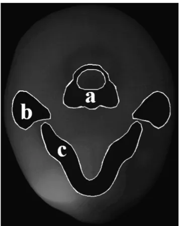
The mean absorbed dose to organs (parotid gland, oral mucosa, extrathoracic airways, bone surfaces, red bone marrow, skin, brain and thyroid gland) and tissue types that were irradiated were estimated by superimposing ROIs on the dose distribution matrices (Figure 3) and calculating the mean value inside each ROI. This was repeated for all film sheets in the phantom.
The equivalent dose for an organ/tissue was calcu-lated as the product of the mean absorbed dose to that organ/tissue and the fraction of that organ/tissue that was irradiated. For the skin surface of the exposed head and neck, we used a simple model. The irradiated area in CBCT imaging, which represented the irradiated skin surface of the head and neck area, was Y (cm²) and the total skin surface area was 1.9 m² (19 000 cm²).16The fraction of the skin area was assumed to be Y/19 000. The estimation of fraction of the irradiated bone surface was based on the total bone area (100 000 cm2)17 and bone area irradiated for each protocol. Owing to higher attenuation in the bone, a conversion factor for Dbone/
Dsoft-tissue of 4 was used. The fraction of the red bone
marrow irradiated (cervical vertebrae and the mandib-ular ramus) was assumed to be 2% for the temporoman-dibular joint (TMJ) lower jaw and panoramic protocol
and,1% for the upper jaw scan.16 The brain fraction estimation is just a crude estimation depending on the radiation geometry of each protocol. The fractions of organs irradiated in each protocol are shown inTable 2. The effective dose was then estimated as the sum of the organ/tissues’ equivalent dose multiplied by their tissue-weighting factor according to the International Commission on Radiological Protection (ICRP) 2007 recommendations.18
Result
For CBCT units, the results were split up by dividing the units into two categories: medium FOV (used for TMJ) and small FOV (used for maxillary impacted ca-nine and mandibular molar area). This allows for a better comparison between protocols, as different FOV sizes are used for different subsets of patients.
Table 3gives the absorbed organ doses and effective doses for medium FOV (TMJ) protocols. The effective dose ranged between 45mSv and 129 mSv. The highest absorbed dose was in the parotid salivary gland. The effective dose of the examination with high resolution was nearly three times higher than that for normal re-solution with the same FOV (83 8 cm). Table 3 also shows the results for the small FOV protocols. The ef-fective dose ranged between 10mSv and 22 mSv.
Figure 3 Region of interests on the dose distribution matrices; (a) vertebra, (b) parotid gland and (c) mandible
The dosimetric results of panoramic imaging are shown in Table 3. The highest absorbed doses were found in the parotid salivary glands (0.65–1.0 mGy). Effective doses range between 8mSv and 14 mSv. The unit with the highest effective dose is the ProMax using a PSP as a receptor; the unit with the lowest effective dose is the ProMax 3D.The effective dose was 1.8 times higher in ProMax than in ProMax 3D.
For comparison purposes, the effective doses were also calculated as multiples of the average dose of panoramic radiograph of the same study and as a percentage of the average annual dose of 760mSv in Sweden from natural background radiation.19 Moreover, the excess cases of fatal cancer in 1 million people irradiated was calculated using a risk coefficient of 5.53 1022Sv211(Table 4).
The highest excess cases were associated with the scan protocol used for the TMJ—high resolution, whose ef-fective dose is about 62 days of exposure to background radiation. The lowest fatal cancer excess cases of the CBCT scans were associated with the ProMax 3D upper impacted canine protocol. Its effective dose corresponds to approximately 5 days of exposure to natural back-ground radiation. On the other hand, the effective dose
of panoramic radiography was between 4 and 7 days of natural exposure.
Discussion
The results of this study should be interpreted with care owing to the complex relationship between image quality, size of the scanned volume and absorbed dose to different tissues. The main purpose of this study was to develop and test GafChromic film dosimetry rather than to compare the clinical performance of imaging devices.
There are a number of ways to estimate the effective dose. All of them include assumptions, which result in limitations and uncertainties.20
The effective dose corresponds to the risk that also a uniformly distributed dose with the same value in the whole body would represent. It gives a general indi-cation of the level of risk for the X-ray examination in question. It takes into account different organs’ sensitivities to induction of severe late effects and is the preferred quantity for comparing the detrimental
Table 2 Estimated fraction of tissue irradiated by primary and scattered radiation for CBCT and panoramic scan protocols Fraction irradiated Upper jaw impacted canine region 43 4 Lower jaw molar region 43 4
TMJ: normal resolution 123 8 Upper jaw impacted canine region 43 5 Standard panoramic TMJ: normal resolution 83 8 TMJ: high resolution 83 8
Tissue Primary/scattered Primary/scattered Primary/scattered Primary/scattered Primary/scattered
Parotid glands 0.70/0.30 1.00/0.00 1.00/0.00 0.70/0.30 1.00/0.00
Oral mucosa1 extrathoracic airways 1.00/0.00 1.00/0.00 1.00/0.00 1.00/0.00 1.00/0.00 Brain 0.10/0.90 0.00/1.00 0.20/0.80 0.10/0.90 0.10/0.90 Bone surfaces 0.01/0.99 0.02/0.98 0.02/0.98 0.01/0.99 0.02/0.98 RBM ,0.01/.0.99 0.02/0.98 0.02/0.98 ,0.01/.0.99 0.02/0.98 Skin 0.01/0.99 0.01/0.99 0.03/0.97 0.02/0.98 0.02/0.98 Thyroid 0.00/1.00 0.00/1.00 0.00/1.00 0.00/1.00 0.00/1.00
CBCT, cone beam CT; RBM, red bone marrow; TMJ, temporomandibular joint.
Table 3 Absorbed organ dose (mGy) and effective dose (mSv) for CBCT and panoramic scan protocols
CBCT-small FOV CBCT-medium FOV Panorama
Veraviewepocs® 3De Veraviewepocs 3De ProMax® 3D NewTom VGi® NewTom VGi NewTom VGi Veraviewepocs 3De ProMax 3D ProMax Tissue Upper jaw (43 4) Lower jaw (43 4) Upper jaw (43 5) TMJ NR (123 8) TMJ NR (83 8) TMJ HR
(83 8) Standard Standard Standard
Parotid glands 1.890 0.900 0.840 2.400 2.130 5.700 0.700 0.650 1.000 Oral mucosa1 extrathoracic airways 0.120 0.600 0.070 2.300 1.800 5.200 0.240 0.156 0.240 Brain 0.001 NS 0.001 0.230 0.150 0.420 0.002 0.001 0.002 Bone surfaces 0.065 0.108 0.040 0.122 0.102 0.306 0.017 0.009 0.014 RBM NS 0.036 NS 0.048 0.040 0.120 0.005 0.003 0.004 Skin 0.017 0.015 0.012 0.074 0.062 0.186 0.001 0.001 0.001 Thyroid 0.001 0.050 0.001 0.020 0.020 0.040 0.020 0.013 0.020 Effective dose 21 22 10 56 45 129 11 8 14
CBCT, cone beam CT; FOV, field of view; HR, high resolution; NR, normal resolution; NS, not significant; RBM, red bone marrow; TMJ, temporomandibular joint.
Veraviewepocs 3De units are manufactured by J Morita MFG Corp., Kyoto, Japan; NewTom VGi by Quantitative Radiology, Verona, Italy; and Promax 3D and Promax by Planmeca, Helsinki, Finland.
effects from different exposure situations to large populations.18
Most studies of dose distribution measurements in oral and maxillofacial radiography are based on TLDs.3–6 The dosemeters are placed inside a phantom in small cavities, which have been drilled in a regular pattern in every slice of the phantom. TLDs have the advantage of being rather sensitive and can measure the absorbed dose down to at least 0.5 mGy with sufficient accuracy. They also have some major drawbacks, namely: (1) They must be handled with extreme care, and the
whole dose measuring procedure, including calibra-tion, is very time-consuming.
(2) Their energy dependence in the diagnostic energy range will result in their response being dependent on the amount of scatter at the measurement point. As the amount of scatter varies within the phantom, the uncertainty of the dose values will increase. (3) The dosemeters are 33 3 3 1 mm3. In an irradiation
geometry, where the dose gradients are as steep as 25% per mm, it is obvious that the positioning of the radiation field in relation to the dosemeters can heavily affect the dose values measured.
Radiochromic films, initially intended for dose measurement in radiotherapy, are now also available with higher sensitivity for X-ray diagnostic purposes as GafChromic XR-QA, XR-QA2 and XR-CT. There are some advantages of GafChromic films compared with TLDs, such as easy preparation and adjustable size of the film. The reading process and the digitization procedure for a set of three film sheets take a few sec-onds, whereas around 1 min or more is necessary for reading one TLD. Furthermore, the GafChromic film will present a continuous “analog”-like dose distribu-tion, where the limit for spatial resolution is set by the pixel size when digitizing the image in the flatbed scanner. Recent studies have shown that it is possible to use ordinary office flatbed document scanners for radio-chromic film scanning.7,21The film response depends on
the film type, batch number and scanning parameters. As for any radiation dosimetry system, uncertainties exist.12 The attenuation of the film was determined experi-mentally and was found to be two times higher than that of the soft tissue. Thus, the film thickness of 25mm corresponds to 50mm of soft tissue, which is negligible for the dose measurement geometry. However, one sit-uation that can occur and can affect the dose meas-urements is when the central beam of the X-ray field coincides with the film plane. Here, there exist primary photon paths directed along the film plane, which will lead to underestimation of the absorbed dose because of the higher attenuation in the film. We have exper-imentally determined the underestimation to be in the order of 10%. This will, however, only occur if the central axis of the radiation field coincides with the film plane. We have deliberately avoided this in all measurement situations.
In all settings of different units, the salivary gland tissue received the highest amount of radiation expo-sure. The salivary gland tissue tends to be in the centre of the imaging field and receives nearly constant expo-sure during the rotation of the gantry. This is a major reason for the increased effective dose seen when using the newest ICRP 2007 guidelines,18as the salivary gland tissue had not been previously included in the calculations. The effective doses of the ProMax 3D-CBCT esti-mated in the present study were lower than those pre-viously reported.2–4However, the main explanation for the lower measured doses is likely to be the increase in copper filtration of the X-ray beam and the difference in FOV. A study by Ludlow and Ivanovic22was based on an early version of the ProMax 3D-CBCT unit. Begin-ning in 2008, those units were equipped with an addi-tional 0.5 mm of copper filtration to reduce the dose. We also found that the effective dose for Veraviewepocs 3De-CBCT (21mSv) is higher than that for the ProMax 3D-CBCT (10mSv) for upper jaw at small FOV. Again, the greatest contribution to the lower measured doses is probably an increase in the copper filtration of the X-ray beam for the ProMax 3D unit and a short exposure
Table 4 Effective dose and risk as multiple of average panoramic images, days of natural background dose in Sweden and risk of cancer
Unit/protocol Effective dose (mSv) Dose as multiple of average panoramic radiographa
Days of per capita natural background (2.08mSv per day)
Excess cases of fatal cancer in 1 million peopleb
Veraviewepocs 3De®/upper jaw 21 1.9 10 1.2
Veraviewepocs 3De/lower jaw 22 2.0 11 1.2
ProMax®3D/upper jaw 10 0.9 5 0.6
NewTom VGi®/TMJ, NR (123 8) 56 5.1 27 3.9 NewTom VGi/TMJ, NR (83 8) 45 4.1 22 2.5 NewTom VGi/TMJ, HR (83 8) 129 11.7 62 7.1 Veraviewepocs 3De/panorama 11 1.0 5 0.6 ProMax 3D/panorama 8 0.7 4 0.4 ProMax/panorama 14 1.3 7 0.8
Veraviewepocs 3De units are manufactured by J Morita MFG Corp., Kyoto, Japan; NewTom VGi by Quantitative Radiology, Verona, Italy; and Promax 3D and Promax by Planmeca, Helsinki, Finland.
HR, high resolution; NR, normal resolution; TMJ, temporomandibular joint.
a
Average of three units: ProMax, ProMax 3D, Veraviewepocs 3De (11mSv).
time of 2.8–8.3 s, combined with a pulsed output. The ProMax 3D unit used was an upgraded unit of the version that was manufactured in 2011. In the newest model, manufactured in 2012, some parameters are changed for three-dimensional exposure; however, the mAs is constant and the dose should not be affected.
From the results, the effect of FOV positioning can be observed. Comparing an upper jaw canine region with a lower jaw molar region scan from the Veraviewepocs 3De-CBCT, it is clear that there were large differences regarding the absorbed dose for the parotid salivary glands and oral mucosa. On the other hand, the dose to the thyroid was very low because it was outside of the primary beam for all protocols.
The NewTom VGi provides two levels of resolution of the same FOV (83 8): high and normal. When the high resolution was selected, the calculated effective dose was 129mSv. If the normal resolution is chosen, the present study shows that the effective dose can be reduced to about 35% of that with high resolution. When comparing the effective dose from a study by Ludlow,23it can be seen that a higher effective dose for high resolution (172mSv) and a normal resolution (51mSv) of the same FOV were found than those in this study (129–45 mSv). Also, the effective dose of FOV (123 8) normal resolution (56 mSv) is lower when com-pared with the study by Ludlow (69mSv).23Difference in dosimetry and variation in phantom position can again account for these differences.
The absorbed dose produced by a CBCT unit is de-pendent on the imaging parameters used (tube poten-tial, mAs); pulsed beam vs continuous beam; amount, type, and shape of beam filter; full 360° rotation vs partial rotation; limited vs full FOV; and resolution setting. Some of these factors, such as type of beam and filtration, are unique to a specific unit, whereas other factors, such as FOV, are under the control of the operator. In general, smaller FOV, lower radiation dose5and a shorter scan-ning time all result in a lower total dose of radiation.
Also, the dose levels are lower in a CBCT scan when compared with multislice (CT) scanners.22
We measured the absorbed dose during panoramic exposure with three digital panoramic units equipped with different detectors. Effective doses ranged between 8mSv and 14 mSv. When PSP, CCD and FPD units were compared, the effective dose of the panoramic unit using the PSP receptor (14mSv) was higher than those of the CCD and FPD units (8–11 mSv).
When the exposure settings are considered, the pan-oramic machine (ProMax) with the highest dose uses 74 kV, the highest tube current (12 mA) and the longest exposure time (16 s). The ProMax 3D-Panoramic, yielding the lowest dose, operates at the lowest tube current (9 mA). Differences in the doses measured depend not only on the tube potential, mA and filter but also on the actual expo-sure time, i.e. if the X-rays are continuous or pulse. The sizes of the radiation field also play a significant role.
Ludlow et al3 evaluated a ProMax (CCD based) panoramic machine operated at 68 kV and 13 mA with a 16 s exposure time and found an effective dose of 24.3mSv using the ICRP 2007 tissue weights. In our study, an effective dose of 14mSv was found. Difference in the type of dosimetry, variation in exposure settings and phantom composition and position can account for these differences.
The use of CBCT for diagnosis, dental implant plan-ning and orthodontic treatment is a subject of intense discussion among dental practitioners. The risk associ-ated with exposing a patient to higher levels of radiation must be weighed against the improvements in patient care and the information that is gained through the use of CBCT. This issue must be carefully considered.
In conclusion, GafChromic film can be utilised to map the dose distribution and measure the absorbed organ/tissue dose of CBCT and panoramic radiography. The use of small FOV and standard resolution reduces the dose when compared with larger FOVs of the same ROI or higher resolution.
References
1. White SC, Pharoah MJ. The evolution and application of dental maxillofacial imaging modalities. Dent Clin North Am 2008; 52: 689–705, v.doi: 10.1016/j.cden.2008.05.006.
2. European Commission. Cone beam CT for dental and maxillo-facial Radiology: evidence based guidelines. Radiation Protection Publication 172. Luxembourg, Germany: European Commission; 2012 (accessed 27 June 2012). Available from:http:/ec.europa.eu/ energy/nuclear/radiation_protection/doc/publication/172.pdf 3. Ludlow JB, Davies-Ludlow LE, White SC. Patient risk related to
common dental radiographic examinations: the impact of 2007 International Commission on Radiological Protection recom-mendations regarding dose calculation. J Am Dent Assoc 2008; 139: 1237–1243.
4. Pauwels R, Beinsberger J, Collaert B, Theodorakou C, Rogers J, Walker A, et al. Effective dose range for dental cone beam computed tomography scanners. Eur J Radiol 2012; 81: 267–271. doi: 10.1016/j.ejrad.2010.11.028.
5. Qu XM, Li G, Ludlow JB, Zhang ZY, Ma XC. Effective radia-tion dose of ProMax 3D cone beam computerized tomography scanner with different dental protocols. Oral Surg Oral Med Oral
Pathol Oral Radiol Endod 2010; 110: 770–776. doi: 10.1016/ j.tripleo.2010.06.013.
6. Ludlow J, Davies-Ludlow L, Brooks S, Howerton WB. Dosime-try of 3 CBCT devices for oral and maxillofacial radiology: CB Mercuray, NewTom 3G and i-CAT. Dentomaxillofac Radiol 2006; 35: 219–226.doi: 10.1259/dmfr/14340323.
7. Boivin J, Tomic N, Fadlallah B, Deblois F, Devic S. Reference dosimetry during diagnostic CT examination using XR-QA ra-diochromic film model. Med Phys 2011; 38: 5119–5129. doi: 10.1118/1.3622607.
8. Tomic N, Devic S, DeBlois F, Seuntjens J. Reference radio-chromic film dosimetry in kilovoltage photon beams during CBCT image acquisition. Med Phys 2010; 37: 1083–1092. 9. Rampado O, Bianchi SD, Peruzzo Cornetto A, Rossetti V, Ropolo
R. Radiochromic films for dental CT dosimetry: a feasibility study. Phys Med 2012.doi: 10.1016/j.ejmp.2012.06.002.
10. Brady S, Yoshizumi T, Toncheva G, Frush D. Implementation of radiochromic film dosimetry protocol for volumetric dose assessments to various organs during diagnostic CT procedures. Med Phys 2010; 37: 4782–4792.
11. Rampado O, Garelli E, Ropolo R. Computed tomography dose measurements with radiochromic films and a flatbed scanner. Med Phys 2010; 37: 189–196.
12. Devic S. Radiochromic film dosimetry: past, present, and future. Phys Med 2011; 27: 122–134.doi: 10.1016/j.ejmp.2010.10.001. 13. Rink A, Vitkin IA, Jaffray DA. Suitability of radiochromic
me-dium for real-time optical measurements of ionizing radiation dose. Med Phys 2005; 32: 1140.
14. Butson MJ, Cheung T, Yu PK. Measurement of energy depen-dence for XRCT radiochromic film. Med Phys 2006; 33: 2923–2925.
15. Paelinck L, De Neve W, De Wagter C. Precautions and strategies in using a commercial flatbed scanner for radiochromic film dosimetry. Phys Med Biol 2007; 52: 231.doi: 10.1088/0031-9155/52/1/015. 16. International Commission on Radiation Protection. Basic
ana-tomical and physiological data for use in radiological protection: reference values. ICRP publication 89. Ann ICRP 2002; 32: 1–277. 17. Jee WSS. The skeletal tissues. In: Weiss L (ed). Histology: cell and tissue biology. 5th edn. Kidlington, UK: Elsevier Science Ltd.; 1983. pp. 206–254.
18. International Commission on Radiation Protection. Recommen-dations of the International Commission on Radiation Protection.
ICRP publication 103. Ann ICRP 2007; 37: 1–332.doi: 10.1016/ j.icrp.2007.10.003.
19. Vattenfall. Radiation. Reference to Vattenfall AB Environmental Product Declarations S-P-00021 and S-P-00026. 2010 [accessed 15 March 2012]. Available from:http://www.vattenfall.com/en/file/ Radiation_12808068.pdf
20. Thilander-Klang A, Helmrot E. Methods of determining the ef-fective dose in dental radiology. Radiat Prot Dosimetry 2010; 139: 306–309.doi: 10.1093/rpd/ncq081.
21. Thomas G, Chu R, Rabe F. A study of GafChromic XR Type R film response with reflective-type densitometers and economical flatbed scanners. J Appl Clin Med Phys 2003; 4: 307–314.doi: 10.1120/1.1621373.
22. Ludlow JB, Ivanovic M. Comparative dosimetry of dental CBCT devices and 64-slice CT for oral and maxillofacial radiology. Oral Surg Oral Med Oral Pathol Oral Radiol Endod 2008; 106: 106–114.doi: 10.1016/j.tripleo.2008.03.018.
23. Ludlow JB. Effective doses of NewTom VGi variable volume dental CBCT unit. Annual meeting of the American Association of Dental Research (AADR). Tampa, FL (cited 21–24 March 2012). Available from:http://iadr.confex.com/iadr/2012tampa/webprogram/ Paper1570