SING YEE YEUNG
STIMULI-RESPONSIVE LIPID
BILAYER MIMICS FOR PROTEIN,
VIRUS AND CELL RECOGNITION
MALMÖ UNIVERSIT Y HEAL TH AND SOCIET Y DOCT OR AL DISSERT A TION 20 1 8:7 SIN G YEE YEUN G MALMÖ UNIVERSIT S TIMULI-RESPONSIVE LIPID BIL A YER MIMICS FOR PR O TEIN, VIRUS AND CELL REC OGNITION
S T I M U L I - R E S P O N S I V E L I P I D B I L A Y E R M I M I C S F O R P R O T E I N , V I R U S A N D C E L L R E C O G N I T I O N
Malmö University
Health and Society, Doctoral Dissertation 2018:7
© Copyright Sing Yee Yeung 2018
Front illustration: Assembly of reversible self-assembled monolayers By Jia Yee Yeung (yeungjiayee@gmail.com)
ISBN 978-91-7104-926-1 (print) ISBN 978-91-7104-927-8 (pdf) ISSN 1653-5383
SING YEE YEUNG
STIMULI-RESPONSIVE LIPID
BILAYER MIMICS FOR PROTEIN,
VIRUS AND CELL RECOGNITION
Malmö University, 2018
Faculty of Health and Society
Department of Biomedical Science
CONTENTS
ABBREVIATIONS ... 7
ABSTRACT ... 9
POPULÄRVETENSKAPLIG SAMMANFATTNING ... 10
LIST OF PUBLICATIONS ... 12
BIOMIMETIC INTERFACES INSPIRED BY NATURE ... 14
MULTIVALENT INTERACTIONS AT THE CELL SURFACE ... 18
Sialic Acid and Influenza Viruses ... 18
Tripeptide Arginine-glycine-aspartic acid (RGD) and Cell Adhesion ... 20
MOLECULAR DESIGN FOR 2D SELF-ASSEMBLY ... 22
Self-Assembled Monolayers (SAMs) ... 23
REVERSIBLE SELF-ASSEMBLED MONOLAYERS (RSAMS) ... 25
STATE OF THE ART ... 30
Supported Lipid Bilayers (SLBs) ... 30
Stimuli-Responsive Surfaces ... 35
Light-Responsive ... 35
Electrical-Responsive ... 37
Chemical-Responsive ... 38
Surfaces based on Host-Guest Chemistry ... 40
AIM OF THE THESIS ... 42
SURFACE CHARACTERIZATION TECHNIQUES ... 44
Ellipsometry ... 44
Infrared Reflection Absorption Spectroscopy (IRAS) ... 45
RESULTS AND DISCUSSIONS ... ... ... 48
Amphiphiles Design and Synthesis ... 48
Surface Fabrication and Characterization ... 50
Mechanistic Insights into Layer Formation ... 51
Engineering Functional rSAMs ... 53
CONCLUSION AND OUTLOOK ... 57
ACKNOWLEDGEMENTS ... 59
ABBREVIATIONS
2D two-dimensional
AFM atomic force microscopy
CB[n] cucurbit[n]urils CD cyclodextrins
CTA cetyltrimethylammonium
CTB cholera toxin B
cyclo(RGDfK) pentapeptide cyclo(-Arg-Gly-Asp-d-Phe-Val-)
DMNPB 3-(4,5-Dimethoxy-2-nitrophenyl)-2-butyl ester
DOPC 1,2-dioleoyl-sn-glycero-3-phosphocholine DOTAP 1,2-dioleoyl-3-trimethyl ammonium propane DPPC 1,2-dipalmitoyl-sn-glycero-3-phosphocholine
ECM extracellular matrix
EggPC egg phosphatidylcholine
FRAP fluoresence recovery after photobleaching
IP inositol phosphates
IP4 inositol tetraphosphate
IRAS infrared reflection-absorption spectroscopy
MBA 4-mercaptobenzoic acid
MDSA 10-mercaptodecanesulfonic acid
MHA 16-mercaptohexadecanoic acid
MUA 11-mercaptoundecanoic acid
PAM pentamidine
PBA phenylboronic acid
PEG polyethyleneglycol
PGAPMA poly(3-gluconamidopropyl methacrylamide)
PHEMA poly(hydroxyethyl methacrylate)
RGD arginine-glycine-aspartic acid
RGD-Cp GRGDS-terminated cyclopentadiene
rSAMs reversible self-assembled monolayers
SAMs self-assembled monolayers
SLBs supported lipid bilayers
SUVs small unilamellar vesicles
TR-DHPE texas-red
ABSTRACT
The most well-studied two-dimensional biomimetic cellular membrane models are self-assembled monolayers (SAMs) and supported lipid bilayers (SLBs). The former has the advantage of control over ligand density, homogeneity and orientation, allowing unambiguous interaction studies. It however lacks long-range lateral mobility, which is one of the most important aspects of cellular membranes. SLBs are laterally mobile but they are fragile and instable upon exposure to air. Literature examples that contain all the above desirable characteristics with stimuli-responsiveness to fabricate biomaterials for biosensing or modulating cell adhesion are rare. We here report on an adaptable platform, reversible self-assembled monolayers (rSAMs), featuring strongly enhanced affinity towards influenza viruses as compared to SAMs, lateral mobility to investigate glycan-lectin interactions and tunable surface dynamics to modulate cell adhesion. This new system utilizes noncovalent amidinium-carboxylate ion pairs for building up stable and ordered two-dimensional assemblies, akin to lipid bilayers but with a simple preparation process, stimuli-responsiveness and fast on/off rates.
POPULÄRVETENSKAPLIG
SAMMANFATTNING
Levande celler begränsas utåt av ett cellmembran som skyddar cellen från omvärlden. Membranet består av ett dubbelskikt av ett slags fett som kallas lipider och proteiner. Lipider är amfifila långsmala molekyler med en vattenälskande huvud-grupp bunden till en vattenavstötande kolkedjesvans. Membranet bär även på sockermolekyler som likt en skog, täcker cellens yta utåt. Dessa fungerar som vakter för cellerna och "pratar" med externa celler eller molekyler vilket påverkar vad som händer inne i cellen. Molekylerna i lipidskiktet är rörliga, och anpassar sig geometriskt till de yttre molekylerna, cellerna eller organismerna som dom "pratar" med. Forskare har studerat dessa beteenden i flera år med hjälp av modellytor där molekylerna innehar fasta positioner. Dessa modeller saknar således dynamiken som finns i cellmembranen. I det här arbetet presenterar vi en serie amidin-funktionella amfifila molekyler som skräddarsytts i vårt laboratorium för att binda och organisera sig på en fast yta och uppträda likt cellens membran.
Vid ett förhöjt pH-värde är ytan - i det här fallet en karboxylsyra belagd guldyta - negativt laddad och amfifilerna positivt laddade. Dessa attraheras därför till varandra under uppbyggnad av ett tätt packat skikt vilket påminner om lipiddubbelskiktet med avseende på dess rörlighet men skiljer sig från dessa i ett viktigt hänseende. Skiktbildningen är i vårt fall snabb och spontan och kan med en pH förskjutning till den sura sidan kastas om så att skiktet försvinner. Detta gör så att ytan kan återanvändas.
I det här arbetet har vi visat två biomimetiska tillämpningar av dessa membranliknande ytor. I den första spelar ytorna rollen som värdcell vid en influenzavirus infektion. Här har vi visat att virus binder med extremt hög
affinitet till ytorna vilket vi klart kunnat härleda till ytans dynamik. Detta tillåter ultrakänslig virusdetektion. I den andra biomimetiska tillämpningen har vi designat ytorna för att efterlikna den levande cellens naturliga omgivning den s.k. extracellular matrix (ECM). Vi har här visat en unik kombination av ytans dynamik och reversibilitet för kontrollerad cell adhesion, modulering och differentiering samt återvinning av celler. Vi tror därför att dessa framsteg kommer att få stor betydelse inom flera olika områden. Här ska särskilt nämnas som verktyg för känslig detektion av virus infektioner och som redskap för utveckling av artificiella organ.
LIST OF PUBLICATIONS
I. Yeung, S. Y.; Mucha, A.; Deshmukh, R.; Boutrus, M.; Arnebrant, T.;
Sellergren, B. Reversible self-assembled monolayers (rSAMs): adaptable surfaces for enhanced multivalent interactions and ultrasensitive virus detection. ACS Central Science, 3 (2017) 1198-1207
II. Yeung, S. Y.; Ederth, T.; Pan, G.; Malcaité, E.; Chaturvedi, V.;
Sellergren, B. pH-switchable lipid bilayer-like monolayers with ultrahigh lectin affinity, Submitted.
III. Yeung, S. Y.; Ederth, T.; Pan, G.; Cicénaité, J.; Cárdenas, M.;
Arnebrant, T.; Sellergren, B. Reversible self-assembled monolayers (rSAMs) as robust and fluidic lipid bilayer mimics. Langmuir, 34 (2018) 4107-4115.
IV. Yeung, S. Y.; Pan, G.; Ederth, T.; El-Schich, Z.; Tran, T.; Holmqvist,
B.; Sellergren, B.; Reversible self-assembled monolayers (rSAMs) with tunable surface dynamics modulate cell adhesion behaviour, Manuscript.
Contributions
Paper I. Design of molecules and experiments with input from coauthors. Synthesis and characterization of all molecules. Performance of all experiments apart from fluorescence recovery after photobleaching (FRAP). Co-analysis of results and co-writing of manuscript with BS with input from coauthors.
Paper II. Design of molecules and experiments with input from coauthors. Synthesis and characterization of all molecules. Performance of all experiments, apart from FRAP. Analysis of results and writing of manuscript with input from coauthors.
Paper III. Design of molecules and experiments with input from coauthors. Characterization of all molecules. Performance of all experiments with assistance from JC, apart from FRAP. Analysis of results and writing of manuscript with input from coauthors.
Paper IV. Design of molecules and experiments with input from coauthors. Partake in experiments with GP and ZE (cell biology), BH (cell staining and fluorescence microscopy) and TT (synthesis and characterization of molecules). TE prepared the self-assembled monolayer (SAM) coated gold cell culture plates. Analysis of results and writing of manuscript with input from coauthors.
Related publications, not included in the thesis.
I. Pan, G.; Shinde, S.; Yeung, S. Y.; Jakštaité, M.; Li, Q.; Wingren,
A. G.; Sellergren, B. An epitope-imprinted biointerface with dynamic bioactivity for modulating cell-biomaterial interactions, Angewandte Chemie International Edition, 56 (2017) 15959-5963.
II. Mucha, A.; Deshmukh, R.; Yeung, S. Y.; Sellergren, B. A dynamic
platform for pH-triggered assembly of close packed protein multilayers and ultrasensitive biosensors, Submitted.
BIOMIMETIC INTERFACES INSPIRED
BY NATURE
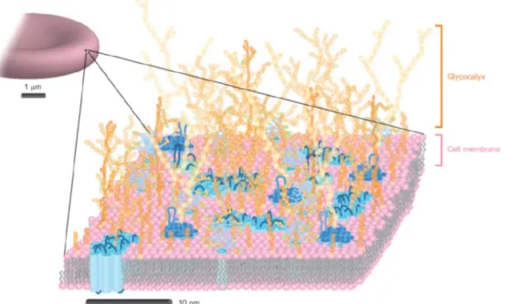
The cellular membrane delimits the cellular components from the external environment. Thousands of lipids, carbohydrates and proteins constitute this complex surface and pre-organize themselves into molecular fingerprints to mediate cell adhesion and communication (Figure 1). Most of these activities rely on multiple associations and spatial organization of the receptors and ligands, mainly proteins and carbohydrates to achieve high avidity and selectivity. As these bioactive ligands are associated with the underlying
Figure 1. Cartoon illustration of a red blood cell surface. The lipids, proteins and carbohydrates are coloured pink, blue and orange respectively. Image adapted with permission from ref 1. Copyright 2011 Springer Nature.
dynamic lipid bilayer or proteins, the resulting spatiotemporal organization further complicates the understanding and modulation of these biological processes ex situ. To circumvent issues with the complexity of cellular membranes, chemists and material scientists design simplified two-dimensional (2D) or 3D biomimetic models to interrogate the basic cellular functions and to construct materials for biosensing, tissue engineering and regenerative medicine.1-5
In order to mimic the functions of cellular membranes, an understanding of the structural organization of its main constituent, the lipid bilayer is required. The lipid bilayer is an organized assembly formed by phospholipids to minimize unfavourable exposure to the aqueous environment. Through a combination of hydrogen bonding and hydrophobic forces, the phospholipids align parallel to each other with the phosphate head groups orientated outwards to the aqueous environment and the hydrocarbon tail groups facing inwards (Figure 2). Multiple noncovalent interactions, specifically, van der Waals and hydrophobic forces that hold the structure together give lipid bilayer its unique liquid crystalline characteristic. The liquid crystalline order, long-range lateral mobility and parallel orientation of the lipid tails define the orientation of the associated surface proteins and carbohydrates and render
these ligands mobile in 2D space.6-8
The area of chemistry crucial in understanding the forces that drive the formation of these assemblies is termed “Supramolecular Chemistry”. The term coined by Jean-Marie Lehn is defined as “chemistry beyond the molecule bearing on the organized entities of higher complexity results from the association of two or more chemical species held together by intermolecular
forces”.9 Based on these principles, molecules designed with complementary
molecular recognition elements can self-assembly in a permissive environment Figure 2. Cartoon illustration of a cross-sectional view of a phospholipid bilayer. The red circles and black tails represent the phosphate and hydrocarbon groups respectively.
to create well-defined and organized supramolecular architectures. The outstanding feature of the aforementioned scaffold is its dynamic character due to its ability to reversibly associate, dissociate and rearrange their
molecular components in response to external stimuli.10-13
Decorating supramolecular scaffolds (supported lipid bilayers (SLBs), host-guest, peptide or nucleotide-based assemblies) with bioactive ligands for engineering functional materials is particularly attractive due to their biomimetic characteristics and stimuli-responsiveness. Attempts to introduce carbohydrate or protein moieties further the understanding of lateral and conformational dynamics in multivalent glycan-protein/glycan-glycan interactions and cell adhesion processes. In combination with their intrinsic chemical- or physical-responsiveness, supramolecular-based materials drive advancements in drug delivery, biosensing, chemical biology and cell-based studies and engineering.14-17
2D platforms due to their compatibility with surface characterization and microscopy techniques and high throughput evaluation tools are of key importance for biomaterial development in biosensing, high throughput screening and cell studies. In glycan-based investigations, immobilization of the glycans onto solid substrates facilitate the detection of pathogens and quantitative analysis of glycan−protein interactions.2-3, 18-21
In cell biology, the ability to immobilize cell adhesion molecules in a controlled fashion further the understanding of cell response towards physical and chemical features of the underlying substrate. This manipulation of cell behavior can influence cell growth and differentiation, which is of great interest in technological development of tissue engineering and regenerative medicine.5, 15, 17, 22-23
One of the most well-established 2D platforms is self-assembled monolayers (SAMs), where the ligands are typically immobilized based on thiol-gold or
silyl ethers-SiO2 reactions/interactions on the surface. These layers are easy to
prepare with excellent control over surface composition, density and
orientation of the ligands.24-25
The nature of these interactions for immobilization, however, precludes long-range mobility, which is an
important feature of cellular membranes.26
Investigations of laterally mobile glycans or proteins, based on supported lipid bilayers (SLBs), uncover the importance of translational dynamics in multivalent-based interactions and cell adhesion and differentiation. The downsides of SLBs are their instability
upon air exposure, complex preparation process, poor long-term stability in cell culture and lack of stimuli-responsiveness. 16-17, 27-31
Here we aim to resolve some of the stated issues relating to glycan-based platforms for biosensing and microarrays and development of biomaterials for fundamental cell studies, tissue engineering and regenerative medicine with a novel 2D platform, reversible self-assembled monolayers (rSAMs).
MULTIVALENT INTERACTIONS
AT THE CELL SURFACE
Cell surface glycans and proteins are receptors and ligands for modulating cellular recognition and adhesion. As singular receptor-ligand interaction is often weak, multiple associations are needed to achieve high avidity required to hold the cells into tissues or immobilize a free-floating cell or pathogen onto
a surface. During monovalent binding, the free energy released, ∆ ∆
∆ is the sum of enthalpic, ∆ and entropic, ∆ contributions. In other
words, the binding process is dependent on the strength of interaction and the changes in degrees of freedom between and in the ligands and receptors,
∆ ∆ ∆ ∆ ∆ . In the
presence of multivalent interactions, after the binding of the first receptor and ligand, the remaining ligands and receptors lose translation, rotational and conformation entropy and subsequent binding become less favourable. To minimize the entropic penalties in multivalent binding, careful design of the scaffold and spacer is required to optimize binding affinity.32-34
In this chapters, the key parameters in designing sialic acid and peptide arginine-glycine-aspartic acid (RGD) based scaffolds towards influenza virus binding and cell adhesion respectively will be discussed in detail.
Sialic Acid and Influenza Viruses
As a first step prior to the infection process, influenza viruses adhere onto host cells through multiple interactions between sialic acids on the cellular membrane and the sialic acid binding lectin, hemagglutinin on the virus surface (Figure 3A). Designing multivalent sialic acid scaffolds for inhibiting or binding influenza viruses require careful attention to the virus surface morphology. A model influenza virus is spherical and measures 80-120 nm across. The surface is covered predominantly by approximately 500
hemagglutinin proteins spaced 10-12 nm apart with each lectin containing 3
sialic acid binding pockets 4-5 nm away from each other (Figure 3B).32, 35
Multivalent sialic acid scaffolds designed for binding influenza viruses have been investigated as drug targets or biosensors since the 1990s and several
review sections summarizing these works have been published.32-33, 36 Earlier
studies were based on linear polymers and lipid-based scaffolds such as liposomes, polymerized liposomes and bilayers demonstrating the benefits of multivalent ligand displaying scaffolds as high affinity inhibitors and sensors. In recent years, Rainer Haag and co-workers reported sialic acid-decorated dendrimers and nanoparticles as effective inhibitors of influenza viruses. The inhibition constant, Ki and/or binding affinity, Kd per sialic acid of monomeric
α-methyl sialoside, naturally occurring multivalent sialic acid scaffolds such as erythrocyte and mucin and selected mono-sialic acid based assemblies in literature are summarized in Table 1. Based on these findings, key parameters in scaffold and spacer design for optimizing binding affinity include the density of sialic acid presented (< 20 %), distance between the sialic acid binding sites (4-5 nm apart for hemagglutinin inhibition), length and flexibility Figure 3. Mechanism of influenza virus binding to cellular membrane and morphology of influenza virus. A. Cartoon illustration of influenza virus binding onto cell membrane. B. Transmission electron micrograph of influenza virus showing the individual hemagglutinin sticking out of the viral membrane (left) and 3D illustration of hemagglutinin with the white dots indicating the sialic acid binding sites (right). Images A and B reused with permission from ref 32 and 35. Copyright 1998 and 2010 John Wiley and Sons.
of spacer connecting the sialic acid to the scaffold (rigid > flexible), size of the scaffold (spheres 50 nm across) and long-range mobility of ligands (liposomes > polymerized liposomes).
Table 1. Summary of mono-sialic acid based assemblies and their binding affinity. Assemblies Ki Kd per sialic acida Target α-methyl sialoside 32 - 2 x 10⁻³ Hemagglutinin
Erthrocyte 32 - 10-13 Influenza Virus
Mucin 37 2 x 10⁻⁶ - Influenza Virus
Trivalent inhibitor 38
5 x 10⁻⁵
1.3 x 10⁻⁶
Hemagglutinin
Liposomes 39 2 x 10⁻⁸ - Influenza Virus
Polymerized bilayer 40 - 10-9 Influenza Virus Polymerized liposomes 41-42 10-7 - Influenza Virus Linear polymers 43-44 10-9 - 10-10 <10-9 Influenza Virus Gold nanoparticles 35 10-9 - Influenza Virus a
Dissociation constant per sialic acid or hemagglutinin monomer unit.
Tripeptide Arginine-glycine-aspartic acid (RGD) and Cell Adhesion
Cells attach to and spread on extracellular matrix (ECM) to survive, proliferate and differentiate. This cell adhesion process occurs in three stages (Figure 4). At the first weak adhesion phase, the cell surface receptors interact with the ECM substrates. This initial interaction process induces integrin clustering and increases the integrin’s affinity to the ligand. At this stage, the cell increases contact with the surface and displays a spread out morphology. If appropriate signalling occurs, the cells would further organize their cytoskeleton, forming focal adhesions and stress fibers. This last strong adhesion stage is required for cell growth and differentiation. RGD is one of the highly conserved peptide sequence that promotes cell adhesion recognized
by many cell adhesive molecules including integrins.45-47
Since the discovery of the tripeptide, this bioactive sequence has been introduced into or onto artificial materials to fabricate biomaterials for cell-based research and development.
The cell adhesion process is sensitive and responsive to chemical and physical properties of the surface. These responses visualized through the optical microscope can be quantified via cell counts, surface coverage by the adhered cells and changes in cell morphology. Key factors in designing RGD-based lipid bilayer-like surfaces (SAMs and SLBs) include bioactive peptide sequence (in increasing order of affinity to integrin,
GRGDSP>RGDS>RGD-NH2>RGD), density of ligand presented, lateral mobility of RGD (SLBs of
differing viscosity changes cell shape) and the microenvironment (increase in number of ethylene glycol repeating units in filler molecule decreased amount of adhered cells).27, 48-50
Figure 4. The stages of cell adhesion. Image reprinted with permission from ref 46. Copyright 2001 American Society for Clinical Investigation.
MOLECULAR DESIGN FOR 2D
SELF-ASSEMBLY
Molecular self-assembly is the spontaneous arrangement of disordered components into defined architectures, typically directed through noncovalent interactions (Table 2). This process is key to understanding the formation of lipid bilayers from lipid molecules, the folding of peptides into bioactive 3D proteins and many other self-assembly events in nature. Following the same chemical rules, chemists encode molecules with complementary functionalities that dictate the organization or assembly into predefined supramolecular architectures. Bioactive ligands introduced onto the scaffolds can produce desired surface properties for applications in sensing, drug delivery, tissue engineering, etc. These supramolecular architectures, in contrast to “rigid” covalent bonds based scaffolds, allow the assemblies to reorganize continuously and response to external stimuli. The dynamic nature facilitates in the development of “smart” material that response to external cues.11-13, 51-52
Table 2.Strength of non-covalent interactions. Reproduced from ref 52.
Bonding and interaction type kJ/mol
Covalent bond 100–400
Ion-ion/ion-dipole/dipole-dipole 200–300/50–200/5–50
Hydrogen bond 4–120
Cation-n( ) interaction 5–80 - interaction 0–50
van der Waals interaction (<5 kJ/mol)
Hydrophobic effects Entropy
Amphiphiles are examples of molecules that can spontaneously self-assemble in solution or at the air/liquid, solid/liquid interface into 1D, 2D or more complex architectures. They consist of both hydrophobic and hydrophilic parts in a single molecule and those with two terminal hydrophilic ends with a
hydrophobic core are further defined as bola-amphiphiles.53
A basic amphiphile has 2 or 3 components in its structure: (1) α-head group containing cationic, anionic and/or hydrophilic groups (ion pairs, directed ion pairs, metal-ligand); (2) hydrophobic core comprises of either long alkyl hydrocarbon chains (van der Waals forces, hydrophobic interactions), aromatic groups (π-π stacking) and/or amides (lateral hydrogen bonding) and (3) an optional ω-terminal group for incorporating bioactive ligand(s) or functionalities for desired surface properties.52-54
Examples of molecules with amphiphilic characteristics can be derived from nature (e.g. lipids or peptides) or man-made (e.g. alkylthiols). In the following chapters, we will focus the discussion on small molecular weight amphiphiles (e.g. surfactants or lipids) that form crystalline-like 2D assemblies on solid supports.
Self-Assembled Monolayers (SAMs)
The most successful examples of 2D assemblies with the amphiphiles orientated parallel to one another, akin to lipid bilayers that have found applications in molecular recognition and cell studies are SAMs and SLBs. The former is based on the formation of densely packed monolayers by spontaneous adsorption or reaction of the α-head groups onto solid substrate, followed by a slow organization of the tail groups (Figure 5). The most commonly utilized α-head group to substrate interactions are thiol amphiphiles on gold or silyl ether
Figure 5. Schematic illustration of self-assembled monolayers (SAMs). Image reused with permission from ref 24. Copyright 2005 American Chemical Society.
amphiphiles on SiO2. These surfaces are prepared by immersing a cleaned
substrate into a solution of amphiphiles containing the reactive head group. Bioactive ligands introduced at the terminal ends of these amphiphiles allow control over surface properties, while the density of bioactive ligands immobilized can be modulated by the ratio of amphiphiles in the assembling solution. Since the discovery of SAMs, they have been widely used as static models to understand interfacial interactions and cell adhesion processes.3, 24-25, 55
For the build-up of densely packed monolayers on solid surfaces, the interactions between the head group and the substrate should be spontaneous and rapid and the width of the head group should be smaller or about the
same size as the width of the hydrophobic core.53
Spontaneous assembly of these layers based on noncovalent interactions have been observed for e.g. surfactants with quaternary ammonium head group on the anionic surface of mica56
, amphiphiles with primary amine head group on carboxylic
acid-terminated SAMs 57-58
and amphiphiles with sulfonic acid head groups with
quaternary ammonium functionalized surfaces.59-60
The stability of these assemblies are dependent on the strength of interactions between the head group and substrate and the hydrophobicity of the mesogenic unit. Other factors contributing to the morphology or stability of the resulting assembly
can be the length of the alkyl chain61
, the odd or even number of the alkyl chain length62
, presence and position of amide groups63
and the presence of rigid aromatic or acetylene units.53
REVERSIBLE SELF-ASSEMBLED
MONOLAYERS (RSAMS)
The ability to form supramolecular structures stable in aqueous conditions remains a major challenge for chemists. Hydrogen bonds and basic ion-pairs, which are the non-covalent choices in organic solvents, become nearly negligible in water.64
For instance, the adsorption of cetyltrimethylammonium
(CTA) on anionic surface of mica is unstable upon rinsing with water.65
To enhance stability and create defined structures, aqueous compatible directional bonds, for example, metal-ligand, host-guest groups and directional hydrogen-bonded ion pairs can and have been used as α-head groups in amphiphiles for SAMs formation.64, 66-67
The use of metal-ligand interactions have been explored earlier in SAMs. However, in gold-thiol SAMs, the nature of the Au-S bond precludes long-range lateral mobility or quick stimuli-responsiveness. Although host-guest chemistry have been compatible for surface modification in aqueous conditions and have the necessary stimuli-responsiveness for biological applications, the large size of the host molecule precludes close packing of the ligands in comparison to SLBs or SAMs. Hence, the small size of directed ion pair of amidine and oxoacids is an excellent choice as α-head group for amphiphiles to produce stimuli-responsive densely packed monolayers, also known as rSAMs. This system is analogous to the classical gold-thiol based SAMs but with pH-responsive hydrogen-bonded directed ion pairs of oxoacids with benzamidines as the main driving force for assembly. By changing the protonation state of the oxoacids using pH, rapid and spontaneous assembly and disassembly of rSAMs occurs.
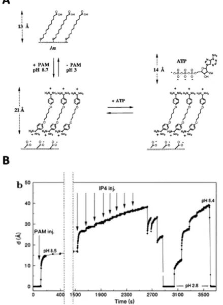
The first generation rSAMs was built up of symmetrical bolaamphiphiles, bisbenzamidines, α,ω-bis(4-amidinophenoxy(alkanes)) on mercaptoalkanoic acid modified gold. The first report in 1996 by Sellergren et. al. demonstrated self-assembly of pentamidine (PAM) (n = 5) on mercaptoundecanoic acid
Figure 6. A. Schematic illustration of reversible assembly of bisbenzamidine rSAMs on 11-mercaptoundecanoic acid (MUA) modified gold for recognition of inositol tetraphosphate (IP4). B. In situ ellipsometric thickness of pentamidine and IP4 assembled at pH 8.5, followed by pH adjustments with 0.1 M HCl and 0.1 M NaOH. Images adapted with permission from ref 68. American Chemical Society.
(MUA) (n = 10) modified gold surfaces at pH 8.5 (Figure 6). These formed rSAMs preferentially adsorbed inositol phosphates (IP) (inositol
tetraphosphate, Ka = 2.7 x 10
5
M-1
) and adenosine phosphates (adenosine triphosphate, Ka = 5.3 x 10
4
M-1
) with higher number of phosphate groups and
featured rapid restorability with acidification to pH 2.8.68
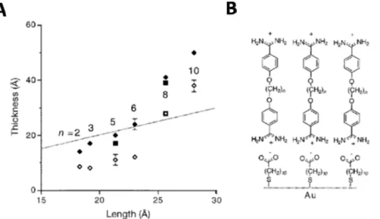
Elongation of the alkyl chain length in bisbenzamidines (n = 2 to 10) and improved molecular order of the underlying SAM layers with increasing chain length of the thiols improved molecular order and rinse stability of the formed
rSAMs (Figure 7).69
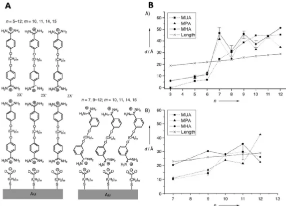
Interestingly, the thickness of the formed layers had an odd– even chain length dependence, with the odd-numbered alkyl chain of bisbenzamidines, n = 7, 9, 11 preferentially forming bilayers and the even-numbered alkyl chain of the bisbenzamidines, n = 6, 8, 10, 12 forming
monolayers (Figure 8).70
These formed rSAMs were applied to detect oligonucleotides71
and adsorbed gold nanoparticle72
in a fully reversible manner.
Figure 7. Rinse stability of bisbenzamidine, α,ω-bis(4-amidinophenoxy(alkanes,
n = 2-10) rSAMs. A. In situ ellipsometric thickness of the corresponding
bisbenzamidine assembled on MUA modified gold at pH 9 (black diamonds) and after pH 9 rinsing (open diamonds). B. Schematic illustration of α,ω-bis(4-amidinophenoxy(alkanes, n = 2-10) assembled on MUA modified gold. The dotted line represents calculated length of the corresponding bisbenzamidine. Images reused with permission from ref 69. Copyright 1999 John Wiley and Sons.
The success of the bisbenzamidine rSAMs led to further investigation on asymmetrical benzamidines, ω-R, α-4-amidinophenoxyoctane with different
terminal groups (R = OCH3, NO2 or CH3) on 16-mercaptohexadecanoic acid
(MHA) modified gold. These asymmetrical bolaamphiphiles formed well-ordered pH-responsive monolayers, analogous to the symmetrical
benzamidines. Mixing the amphiphiles ω-(NO2),α-(4-amidino-phenoxy)octane
and ω-(OCH3),α-(4-amidinophenoxy)octane, denoted by % NO2 in the
assembling solution formed layers with statistical incorporation of the amphiphiles (Figure 9).73
Figure 8. Odd even effect of para- or meta-substituted bisbenzamidine, α,ω-bis(3- or 4-amidinophenoxy(alkanes, n = 3-12)) rSAMs. A. Schematic illustration of α,ω-bis(3- or 4-amidinophenoxy(alkanes, n = 5-12)) assembled on mercaptoalkanoic acid (m=10 (MUA), 14 (MPA) or 15 (MHA)) modified gold. B. In situ ellispometric thickness of para- (top) or meta (bottom)-substituted bisbenzamidines assembled on MUA, MPA or MHA on gold after pH 9 rinsing. Images reused with permission from ref 70. Copyright 2004 John Wiley and Sons.
Figure 9. Mixed rSAMs on MHA modified gold. A. Schematic illustration of ω-R,α-(4-amidinophenoxyoctane) assembled on MHA modified gold and B.
Plot of IRAS integrated area of NO2 stretch (1347 cm-1) and OCH3 stretch
(1236 cm-1) of the formed layers as a function of % NO
2, ω-NO2
,α-(4-amidinophenoxy(octane) in ω-OCH3,α-(4-amidinophenoxy(octane) used in
STATE OF THE ART
Understanding and manipulating multivalent recognition processes and cell adhesion behaviour require advancements in biomimetic surfaces, specifically designed with long-range mobility and stimuli-responsiveness. 2D molecular assemblies made up of small molecules packed parallel to each other such as dynamic SAMs and SLBs, coupled with their inherent control over surface density of the bioactive ligands are especially attractive in this aspect. Here we highlight selected examples of 2D molecular assemblies on solid substrates with an emphasis on glycan and RGD functionalized surfaces for cell studies and multivalent recognition.
Supported Lipid Bilayers (SLBs)
Lipid bilayer is the major structural component of cellular membranes. Initial work to understand their properties led to the immobilization of the assembled layers onto solid supports to enhance stability and robustness. As surfaces for studying cellular membrane behaviour, apart from their lateral dynamics and structural similarity towards the cellular membranes, SLBs are non-fouling and easy to functionalize. Its major limitations towards applications outside of academic setting are its poor stability upon air exposure and fragility.17, 31
Attempts to improve these aspects include insertion
of polyethylene glycol (PEG)-conjugated lipids in the layers74
, assembly of
phospholipid monolayers on alkanethiol-SAMs75
, post-modification of
biotinylated SLBs with streptavidin76
and immobilization of the SLBs on
cholesteryl-PEG surfaces (Figure 10)77
. Most of these “air-stable” SLBs modifies the surface composition, which would interfere with surface interactions. Coupled with the complex preparation process and requirement of careful handling after fabrication, robust alternatives are highly desired.
Lateral mobility in the bioactive ligands modulates multivalent binding events due to translational adaptation of the ligands to the receptors. This long-range mobility is an important process in integrin-mediated cell adhesion for signaling downstream processes for cell growth and differentiation. The presence of lateral mobility of ligands on SLBs also uncovers interesting
aspects in multivalent binding.16-17, 28 For instance, Cremer and coworkers
reported that cholera toxin (CTB) binding affinity weakens at higher GM1
concentration in the phospholipid bilayers (Figure 11). With careful atomic
force microscopy (AFM) investigation, it was uncovered that GM1 formed
larger clusters at higher concentrations, which inhibited the binding of
pentameric CTB receptors.78 Another example by Guo and colleagues
demonstrated two distinct plateaus in the number of bounded E. coli as a function of mannose density in the SLBs. Correlating the amount of E. coli Figure 10. Air-stable and fluidic SLBs. A. SLBs formed by fusion of small unilamellar vesicles (SUVs) containing 0.5% Texas-Red 1,2-dihexadecanoyl-sn-glycero-3-phosphoethanolamine (TR-DHPE), 50% egg phophatidylcholine (EggPC) and 50% 1,2-dioleoyl-3-trimethyl ammonium propane (DOTAP) on cholesteryl-PEG/glass surfaces. B. FRAP results of the formed SLBs (bottom) and after air-drying and rehydration (top). Image adapted with permission from ref 77. Copyright 2008 American Chemical Society.
bounded with the number of mannose immobilized in the layers suggested a switch from monovalent to multivalent binding modes of E. Coli depending
on the surface density of mannose.79
Figure 11. Clustering of GM1 on supported phosphatidylcholine (POPC)
bilayers effect on binding affinity of cholera toxin B subunits (CTB) A.
Schematic illustration of CTB inhibition by GM1 clustering B. Apparent
equilibrium dissociation constants, KH for CTB-GM1 binding fitted using
Hill-Waud binding model vs the mol % of GM1 in the POPC bilayers C. Mol % of
GM1 in POPC bilayer and the apparent GM1 domain size as determined by
atomic force microscopy (AFM). Image adapted with permission from ref 78. Copyright 2007 American Chemical Society.
Another important feature of SLBs is its tunable lateral dynamics. Low melting temperature lipids such as 1,2-dioleoyl-sn-glycero-3-phosphocholine (DOPC) features SLBs with higher diffusion coefficient, while SLBs constitute of higher melting temperature lipids, 1,2-dipalmitoyl-sn-glycero-3-phosphocholine (DPPC) demonstrates a diffusion coefficient one order of magnitude lower (Figure 12B). This trait has been exploited to understand the influence of lateral dynamics in cell adhesion and differentiation.27, 29
For instance, Benette et. al, compared the cell size, cytoskeleton organization, actin flow and focal adhesion of adhered C2C12 cells on RGD functionalized DOPC, DPPC SLBs and SAMs on glass (Figure 12). On surfaces with higher mobility, the adhered cells demonstrated increase circularity, decrease cell area, decrease actin flow and decrease focal adhesion area. From these findings, the authors concluded that higher viscosity lead to higher force loading rate exposing the mechanosensitive proteins and activates downstream pathways.27
In an earlier example by Kocer et. al., hMSC adhesion demonstrated an opposing trend with larger adhered cell area on RGD-functionalized DOPC as compared to DPPC SLBs. With higher RGD density
Figure 12. Lateral mobility of RGD peptide on morphology of adhered cells. A. Schematic illustration of RGD functionalized SAMs on glass, DPPC SLBs and DOPC SLBs B. Diffusion coefficient of DOPC and DPPC SLBs based on FRAP. C. Cell morphology and focal adhesions in cells on RGD-functionalized DOPC, DPPC and SAMs respectively. Image adapted from ref 27. Copyright 2018 Proceedings of National Academy of Sciences of the United States of America.
and mobility of the layers, there is an increased in cell area directed osteogenic differentiation.29
Despite conflicting conclusions regarding cell adhesion behavior with viscosity, these results emphasize the importance of understanding the influence of lateral dynamics on cell adhesion and differentiation.
One of the creative approaches in stimuli-responsive lipid bilayers is the polymerization of diacetylene lipid films for direct colorimetric detection. Conjugated polydiacetylene backbone changes colour from blue to red upon increase in temperature or changes in pH due to the conformation change in the conjugated backbone. Functionalization of these layers with sialic acid at the terminal end induce a visible colour change from blue to red upon exposure to 100 HAU of influenza virus (Figure 13). Coupled with the visible absorption spectroscopy, quantification of the influenza viruses can be
achieved from ~30 to 80 HAU.40
Figure 13. Direct colorimetric detection of influenza virus using sialoside polymerized bilayer assembly. A. Schematic illustration of the sialoside polymerized bilayer assembly B. Blue (left) to red (right) colour response of the layer after exposure to 100 HAUs influenza virus (top) and visible absorption spectrum of the bilayer assembly before (solid line) and after virus incubation (dashed line) (bottom). Images adapted with permission from ref 40. Copyright 1993 American Association for the Advancement of Science.
Stimuli-Responsive Surfaces
Unlike SLBs, the thiol-gold based SAMs has limited long-range lateral mobility (diffusion coefficient of 10-18
cm2
s-1 at 60°C).26
In recent years, incorporating stimuli-responsive functionalities (light-responsive, electrical-responsive, chemical-responsive and host-guest chemistry) at the end groups of SAMs conceive dynamic SAMs, surfaces that response to external stimuli. Stimuli-responsive functionalities coupled with host-guest chemistry will be discussed in the host-guest chemistry chapter.
Light-Responsive
One of the most commonly used light-responsive functional group is azobenzene, which is often integrated with host-guest chemistry for light-controlled release of bioactive ligands (refer to host-guest chemistry). Azobenzene moieties undergo reversible E-Z isomerism with the transformation from E to Z configuration under UV light (340-380 nm) and the reversal from Z to E with visible light (450-490 nm). This photoisomerism property has been exploited to study the effect of carbohydrate orientation on protein and cell recognition and the reversal of cell adhesion using azobenzene derivatised SAMs. Weber et al. formed photoswitchable SAMs using an azobenzene mannoside derivative consisting of a thiol, an undecane chain, an oligoethylene glycol chain, azobenzene unit and a terminal α-D-mannose (Figure 14). When the formed SAM surface was illuminated with a light at
Figure 14. Photo-responsive azobenzene SAMs reverse E. Coli adhesion. A. Schematic illustration of E to Z isomerism of azobenzene controlling the orientation of the α-D-mannose. B. E. Coli adhesion to the azobenzeneα-D-mannose monolayer going through EZE isomerization cycles. Images reused with permission from ref 80. Copyright 2014 John Wiley and Sons.
365 nm, E to Z isomerism was induced, evidenced by the infrared
reflection-absorption spectroscopy (IRAS), with a band at 1240 cm-1
for
aryl-O(-mannosyl) vibration. With illumination at 450 nm, the band at 1240 cm-1
decreased indicating reversal of the mannose orientation. This change in the
peak area at 1240 cm-1
correlated well with the density of E. Coli adhered on the corresponding surfaces and strongly suggested the importance of glycan
orientation for optimal binding.80
A similar approach was undertaken by Zhang et al. using galactose and mannose-decorated azobenzene derivatives and similarly, the affinity of lectin (peanut agglutinin and concanavalin A) and cell (Hep-G2 and M2 macrophages) reduced with the Z-configured
SAMs.81
Liu et al. presented a mixed SAMs with GRGDS thiol with an azobenzene unit and PEG-terminated thiol for reversible cell adhesion. Adhesion of NIH 3T3 fibroblast on the SAMs was observed at E configuration of the azobenzene. Upon UV irradiation of the surface, the Z configuration of the azobenzene concealed the GRGDS within the cell resistant PEG layers and reduced cell adhesion.82
Figure 15. Photo-triggering of cell adhesion by caged cycled RGD peptides. A. Chemical structure of cyclo[RGD(DMNPB)fK] (DMNPB in red) attached to the surface through the triethylene glycol-spacer (green). B. Cell density on surfaces modified with cyclo[RGD- (DMNPB)fK] before and after irradiated at 351 nm for 10 min. Images reused with permission from ref 83. Copyright 2008 John Wiley and Sons.
Another light-responsive approach is to introduce photolabile caging group 3-(4,5-Dimethoxy-2-nitrophenyl)-2-butyl ester(DMNPB) to conceal the active ligand of integrin, pentapeptide cyclo(-Arg-Gly-Asp-d-Phe-Val-) (cyclo(RGDfK)) (Figure 15). Upon irradiation at 351 nm, the caging group, DMNPB was released and the bioactive peptide exposed. Consequently, the 3T3 fibroblasts recognized the bioactive peptide and cell adhesion on the surface increased 11 to 13 times depending on the duration of incubation of the cells.83
Electrical-Responsive
Mrksich and co-workers pioneered the work of electrochemical control of cell adhesion based on Diels-Alder cycloaddition. The cell resistant (inactive) layer was based on a SAM presenting hydroquinone groups within a background of
Figure 16. Electrochemical activation of hydroquinone for Diels-Alder-mediated immobilization of GRGDS-terminated cyclopentadiene (RGD-Cp) to switch on cell adhesion. Image reused with permission from ref 84. Copyright 2001 John Wiley and Sons.
penta(ethylene glycol) groups (Figure 16). When an electrical potential of 500 mV was applied for 10 s, the hydroquinone oxidized to quinone, which then reacted with GRGDS-terminated cyclopentadiene (RGD-Cp). Thus, immobilizing the active GRGDS on the surface and allowed the cell migration and growth in situ.84
Another approach to electrically control cell adhesion is by concealing the GRGDS motif within the oligo(ethyleneglycol) cell resistant layer using an applied negative potential. This electrically-switchable mixed SAMs comprised of oligopeptide (cysteine, three lysines and a terminal GRGDS) and ethylene glycol-terminated thiol. Under a negative potential (-0.4V), the charged lysine repeating units collapsed and concealed the recognition motif, GRGDS within the ethylene glycol cell-resistant layer. Reversing to open circuit conditions, the lysine repeating units restored to the extended conformation, the GRGDS exposed to the external environment and the adhesion of RAW 264.7 mouse
macrophage cells increased by 70 %.85
Chemical-Responsive
One interesting approach to reverse cell adhesion is via specific biomolecular exchange. The fructose/glucose-responsive layers were fabricated by grafting poly(hydroxyethyl methacrylate)(PHEMA)-graft-phenylboronic acid (PBA) polymer brush on a glass substrate and immobilizing RGD-poly(3-gluconamidopropyl methacrylamide)(PGAPMA) based on multivalent interactions between the PBA and cis-diol groups of the glycopolymers (Figure 17). Addition of 60 mM fructose or glucose to the substrate competed with the glycopolymers for the phenylboronic acid binding sites, freed the
RGD-conjugated glycopolymers and released the adhered MG63 cells.86
Figure 17. Natural small biomolecules triggered cell release based on multivalent PBA-cis-diol interactions. A. Schematic illustration of reversible cell adhesion on RGD-PGAPMA on PHEMA-graft-PBA layers by glucose
and fructose B. MG63 cell release from RGD-PGAPMA10 coupled
Glass-PBA400 at different times by incubation with 60 mM fructose (a) and glucose
(b). Images adapted with permission from ref 86. Copyright 2014 American Chemical Society.
HSurfaces based on Host-Guest Chemistry
Host-guest chemistry studies the selectivity of interactions between host and guest molecules. One example of host-guest pairs is the interaction of macrocycles (cucurbit[n]urils(CB[n]) and cyclodextrins(CD)) and small guest molecules (naphthol, adamantane, azobenzene and viologen). The number of repeating units and charge/hydrophobicity of the host cavity controls the selectivity and affinity of the guest molecules and this area of study has been extensively reviewed in literature.87-90
When stimuli-responsive guests such as azobenzene and methylviologen is used, the affinity of the guest molecules can be controlled via external cues such as light or electrical potential (Figure 18).87-90
Figure 18. Triggered release by host-guest affinity using: (A) competitive release of the first guest by the second guest based on higher binding affinity of the latter to the CD host; (B) switching from trans to cis-azobenzene with UV light reduces its fit to the CD host; (C) electrochemical reduction of methylviologen leads to weakening of the charge-transfer complex with naphthol inside the CB[8] cavity and (D) dual trans-azobenzene and methylviologen guest with CB[8] demonstrates dual-responsiveness. Image reused with permission from ref 90. Copyright 2016 Elsevier Ltd.
Through a combination of host and guest molecules, reversible cell adhesion had been achieved based on UV light-triggered trans to cis isomerism of
azobenzene with α-CD91
, electrochemical reduction of methyl viologen with CB[8]92
and competitive binding of guest molecules with higher binding affinity, 1-adamantane-amide-G5-RGES with the bioactive
1-naphthalene-amide-G5-RGDS in β-CD93
. A similar approach was used to reverse E. Coli adhesion with UV light via trans to cis azobenzene isomerism with β-CD host.94
One unique example is the coupling of host-guest chemistry with SLBs. In this work by Sankaran et. al., the methyl viologen was conjugated onto the SLBs using the reaction of maleimide with thiol. Consecutive assembly of CB[8] and mannose-terminated azobenzene allowed selective binding of E.
Coli on the mannose-terminated SLBs. Releasing of the bacteria was achieved
upon 360 nm UV irradiation that induced the cis isomerism of azobenzene
and freed the host molecules from the guest cavity.95
Figure 19. Light-triggered release of E. Coli based on host-guest chemistry on SLBs. A. Schematic illustration of step-wise assembly and UV-triggered release of mannose-terminated azobenzene from CB[8] immobilized on methylviologen SLBs. B. Detachment of E. Coli with 5 mins UV irradiation of the surface. Images adapted with permission from ref 95. Copyright 2015 John Wiley and Sons.
AIM OF THE THESIS
The two most well-established 2D system for immobilization of glycans and proteins are SAMs and SLBs. The advantages are the control over ligand density and orientation for unambiguous interaction studies. However, the former lacks lateral mobility, which is one of the most important aspects of lipid bilayers. Although SLBs is the model of choice for translational mobility, their fragility limited their applications outside of academic settings. Recent strategies of combining SAMs with dynamic covalent chemistry or supramolecular chemistry demonstrated control over surface properties but these layers do not demonstrate long-range fluidity of lipid bilayers. We have here summarized the pros and cons of a few classes of 2D molecular assemblies (Table 3).
Table 3. Advantages and disadvantages of 2D lipid bilayer mimics.
Assemblies Advantages Disadvantages SLBs Close packing of layers
Long-range lateral mobility (10-8
cm2
s-1
)
Poor stability upon air exposure, apart from few examples.
Complex preparation Lack of stimuli-responsiveness Dynamic SAMs Close packing of layers
Stimuli-responsive
Lack of long-range lateral mobility
Host-Guest Stimuli-responsive Lateral mobility unknown Lack close packing of ligands rSAMs Close packing of layers
Stimuli-responsive Long-range lateral mobility Stability upon exposure to air
The aforementioned challenges highlight the need for molecular architectures that combine robustness with the dynamic nature of cellular membranes. The concept of rSAMs offers a unique example in this regard. These are pH-switchable monolayers allowing reversible and ordered introduction of affinity reagents on surfaces. The principal layer building blocks consist of α-(4-amidinophenoxy)alkanes decorated at the ω-position with ligands. These spontaneously self-assemble on top of carboxylic acid terminated SAMs to form reversible homo- or mixed monolayers (rSAMs) that are tunable with respect to the nature of the head group, layer order and stability while featuring pH-responsiveness, dynamic nature of non-covalently build assemblies and rinse stability. Here we investigate rSAMs for biosensing of influenza viruses, understanding multivalent lectin-carbohydrate interactions and modulating cell adhesion.
SURFACE CHARACTERIZATION
TECHNIQUES
To gain insight on the stimuli-responsiveness and physical characteristic of rSAMs, several surface characterization techniques were used in parallel. Ellipsometry provided thickness of the formed layers both in real time and in air. Fourier transform infrared reflection adsorption spectroscopy (IRAS) provided information regarding the composition and molecular orientation of the formed layers. Lastly, AFM gave insight on the surface homogeneity and distribution of the amphiphiles. The former two techniques were subsequently used to establish trends in protein and virus adsorption with respect to the chemical composition of rSAMs. This chapter explains the basic principles and limitations of these key methods used during the course of this work.
Ellipsometry
Ellipsometry is an optical technique to deduce film thickness and optical properties of a material by measuring change in the polarization state of the light after reflection from a surface (Figure 20). When a linearly or circular
Figure 20. Measurement principle of ellipsometry. Image adapted with permission from ref 96. Copyright 2007 John Wiley and Sons.
polarized light is reflected by a surface, the parallel (p) and perpendicular (s) components of the reflected light changes. The amplitude ratio and the phase difference between the two components after reflection are represented by the ellipsometric angles, ψ and Δ.96
tan exp ⁄
where and correspond to the Fresnel coefficients of reflection, E stands
for the vectors of the electric field, Eip and Erp represents the incidence and
reflection of the p polarized light, while Eis and Ers represents the incidence and reflection of the s polarized light.96
From the experimental determined values, Δ and , the thickness and optical properties of the layers can be calculated by
fitting the data into an appropriate model.97
Experimental and fitting details are presented in the respective papers (Papers I-IV). The layers were assumed to be homogenous and distinct with no exchange occurring upon protein or virus exposure unless stated otherwise. As no or little change in ellipsometric thickness does not indicate no adsorption of the analytes, IRAS was often used in parallel to give a more accurate interpretation of the results.98
Infrared Reflection Absorption Spectroscopy (IRAS)
Infrared spectroscopy (IR) is a technique that measures absorption or emission of infrared radiation. This technique is especially useful for the identification
of functional groups in samples by relating the frequency, 1ṽ needed to
excite vibrating bonds according to Hooke’s Law (Figure 21).
where ṽ is wavenumber in cm-1
, k stands for the force constant in dynes cm-1
(a
measure of bond strength) and µ represents the reduced mass in gatom-1
. In practice, the peaks of the spectra are assigned based on established literature references.99
The line shape and the peak widths of IR spectra is sensitive to the changes in the local environment such as hydrogen bonding or packing of chain molecules. This property has been exploited to understand crystallinity of
molecular assemblies. For instance, the frequency of CH2 asym and sym mode
decreases with relation to the crystalline state of molecular assemblies. It is
well established that frequency below 2920 and 2850 cm-1
for the two modes respectively indicates crystallinity of alkyl chains in SAMs and polyethylene assemblies.100-101
When used in the reflection-absorption mode (IRAS), molecular orientation of the assemblies on reflective surfaces can be determined. In this technique, the source of radiation penetrates the sample at a high angle of incidence, θ before reflected from the substrate to ensure maximum intensity (Figure 22). Only components parallel to the plane of incidence can absorb radiation and give measurable adsorption. Based on these surface selection rules, when the alkanethiol is in all trans conformation and perpendicular to the surface, the
Figure 22. Schematic illustration of alkanethiol on gold in an IRAS experiment.
CH2 asym and sym stretchs are not parallel to the plane of incidence and will
not absorb the p-polarized light. As the alkyl chain tilts away from the surface
normal, the CH2 asym and sym stretchs become increasingly parallel to the
plane of incidence and spectra intensity increases. Based on these correlations, Porter et. al. deduced alkanethiol SAMs have an average tilt angle of 20-30°
by comparing the peak intensities of CH2 stretching modes of the layer spectra
to its isotropic solid.101-103
Atomic Force Microscopy (AFM)
AFM is a technique to obtain information regarding topography of the layers with atomic resolution. In a typical experimental set up, the AFM consists of a cantilever with a sharp tip at its end (Figure 23). As the tip is brought into close contact and scanned across the surface, the intermolecular forces between the tip and the surface lead to deflection of the cantilever. This deflection is measured by a laser spot that is focused and reflected from the top of the cantilever into a photodiode. This information can determine the movement in the cantilever with relation to the surface position and translated
into a topographical image of the surface.104
Figure 23. Schematic representation of AFM. Image reused from ref 104. Copyright 2010 Oxford University Press.
RESULTS AND DISCUSSIONS
The key objective of this work is to develop rSAMs into platforms suitable for biological applications such as biosensing and cell studies, analogous to SAMs but with lateral mobility and stimuli-responsiveness. Four papers are included in this thesis, 2 (Paper I and III) are published in peer-reviewed journals and 2 (Paper II and IV) are in manuscript form.
Amphiphiles Design and Synthesis
Designing SAMs for molecular recognition require an upright orientation of molecules in the assembly with the bioactive ligands facing the external environment. With the well-established alkanethiol SAMs, this was achieved by simply substituting the ω-position with the ligand of interest. Following a similar approach, Mucha et. al. synthesized a versatile azide-terminated α-(4-amidinophenoxy(decane)). The azide functionality produced the desired ligand-terminated amphiphile via a one-step conjugation with the
corresponding alkyne-terminated ligand.105
The downside of this preliminary design is the ester that links the hydrophobic core to the spacer. Esters are susceptible to hydrolysis and in the presence of the basic aqueous solution required for assembly, the stability of the amphiphile is a major concern. Inspired by the unique ether-linked lipid structure of archaebacteria and its ability to survive in harsh volcanic environment, the ester of the earlier amphiphile design was replaced by an ether as an attempt to improve stability.53
When designing the synthetic route to produce the desired azide 9, the key goal was to shorten the previous laborious synthetic procedure (10-step synthesis) and eliminate the use of protecting groups. With these factors in mind, the amidine azide 9 was prepared in five steps (Figure 24) by sequential Williamson ether synthesis followed by Pinner conversion and azide
substitution in an overall yield of 17 % (Paper I). Subsequent elongation of the ethylene glycol spacer from 2 to 6 repeating units allowed optimization of the spacer chain length with regards to the binding affinity. In this work, alkyne-sialic acid and alkyne-RGD were coupled to the ω-(azide),α-(4-amidinophenoxy(decane)) to produce the desired EX-SA and RGD 10 (Figure 25) for the recognition of lectin (Paper I and II), virus (Paper I) and cells (Paper IV).
For the development of functional biomaterials such as SAMs, filler amphiphiles are commonly mixed with the amphiphiles containing the bioactive ligands during layer formation to allow control over ligand surface density and improve packing of the layers with the insertion of filler amphiphiles between the ligand-terminated amphiphiles to reduce steric hindrance between the ligand head groups. The ideal filler molecule is inert towards non-specific interactions. As such, we utilized a common approach for SAMs formation by introducing repeating units of ethylene glycol at the
ω-position.106 A series of ω-(ethylene glycol),α-(4-amidinophenoxy)decane with 2
to 6 even repeating units of ethylene glycol, E2-6 was synthesized from Figure 24. Synthetic pathway of azide-terminated amphiphile 9. Refer to Paper I for synthetic details.
intermediate 7 with hydroxyl substitution and a final Pinner conversion (details of synthesis and characterization in Paper III).
Surface Fabrication and Characterization
Homogenous or mixed rSAMs were prepared by immersing a carboxylic acid- or sulfonic acid-functionalized gold or quartz surface in a pH 8 aqueous solution containing the amphiphiles (Figure 26). Statistical control of amphiphiles in the layers were achieved by adjusting the ratio of the amphiphiles in the assembling solution.
Figure 25. Amphiphiles design for fabricating rSAMs.
Figure 26. Schematic illustration of rSAMs formation. (i)alkanethiol (MHA, 4 mercaptobenzoic acid (MBA) and 10-mercaptodecanesulfonic acid (MDSA) modified gold placed in 50 µM mixed amphiphilic solution with the amphiphile structures detailed in figure 25. (ii) Ordered rSAMs formed after 18 hrs of incubation and rinsing with pH 8 HEPES buffer
Based on the immobilization protocol, 50 µM of ω-(ethylene glycol)2
,α-(4-amidinophenoxy)decane, E2 in pH 8 HEPES buffer formed crystalline-like monolayers with the amphiphiles tilted ca. 20° away from the surface normal on either MHA- or 4-mercaptobenzoic acid (MBA) modified gold as evidenced by ellipsometry, IRAS and AFM (Paper II and III). Mixing E2 with the sialic acid amphiphiles, E4-SA in the same conditions, surface compositions derived from IRAS and film thickness information (AFM, ellipsometry) suggested that the amphiphiles were statistically incorporated in the monolayer (Paper II). As evidenced from AFM and IRAS, the mixed assemblies retained crystalline-like order with the amphiphiles tilted 10-20° from the surface normal with the ligands orientated towards the external environment (Paper II).
Concerning rSAMs robustness and surface dynamics, in contrast to SLBs, ellipsometric and IRAS results suggested stability of the E2 and E4 rSAMs in buffer solution and upon exposure to air. The layers’ pH stability and responsiveness corresponded to the composition of the underlying SAMs layers. Titrating the E2 rSAMs assembled on MHA SAMs suggested rSAMs stability above pH 7 with complete removal of the layers at pH 4. With the rSAMs assembled on MBA SAMs, the layers were stable above pH 6, while retaining disassembly at pH 4 (Paper III). FRAP results further confirmed the long-range lateral mobility of the layers (Paper II).
Mechanistic Insights into Layer Formation
Layer formation with respect to the amphiphile molecular structure, concentration of amphiphile and pH of buffer used during assembly, duration of incubation and choice of oxoacids and molecular order of the underlying SAMs were investigated via ellipsometry and IRAS (Paper III).
Amphiphile molecular structure (Paper III). Layers formed with the
amphiphiles with 2 to 4 ethylene glycol repeating units, E2-E4 were orientated upright with an average tilt angle of 20-30°. The amphiphiles with increasing ethylene glycol repeating units, (E2<E4<E6) formed layers with decrease ellipsometric thickness, molecular order and stability.
Amphiphile concentration (Paper III and IV). Increase amphiphile
concentration (10-8
– 10-4
M) during assembly increased ellipsometric thickness of the resulting assembly (Paper III and IV). This suggested that the adsorption of benzamidine amphiphiles on oxoacid SAMs is likely to be similar to the
adsorption of cationic surfactants, CTA+
on the anionic surface of mica, where the thickness of layers increases with concentration and levels off in the range of the cmc.56
pH of buffer during assembly (Paper III). The limiting equilibrium
ellipsometric thickness was independent of pH between 7.4 to 9. After rinsing the layers, the decreased pH correlated with reduced ellipsometric thickness suggested that the protonation state of the carboxylic acid of the SAMs was crucial for layer stability but not the assembling process within the probed pH range.
Underlying SAMs layer (Paper III, IV and unreported data). Layer stability
improved with respect to enhanced molecular order of the MHA SAMs and decreased surface pKa of the underlying SAM layer (MHA>MBA>MDSA). MBA SAMs with lower surface pKa as compared to MHA SAMs due to lack of lateral hydrogen bonding between the carboxylic acids on the surface demonstrated layer stability down to pH 6.
Figure 27. Engineering functional rSAMs. Legends are described in Figure 25 and 26. Images are not drawn to scale.
Engineering Functional rSAMs
Herein rSAMs were functionalized with either sialic acid or RGD and investigated with regards to their lectin, virus or cell recognition. The unique lateral dynamics and stimuli responsiveness of rSAMs were compared to static SAMs or SLBs in selected cases.
Enhancement in multivalent interactions with viruses (Paper I). Here we
investigated if adaptability of rSAMs in displaying sialic acid enhanced multivalent interactions with hemagglutinin or influenza viruses. The sialic acid-terminated amphiphile, E2-SA formed sub-monolayers on MHA SAMs as evidenced in ellipsometry, IRAS and AFM. By comparing the sialic acid rSAMs binding with concanavalin A and human serum albumin, it was concluded that these surfaces demonstrated selective binding to hemagglutinin at the nM range. Masking the sialic acid binding sites of hemagglutinin with mucin, an epithelial glycoprotein abundant in sialic acids demonstrated a marked decrease in binding towards the surface and supported the view of selective hemagglutinin-sialic acid interactions.
With the success demonstration of selective glycan-protein interaction on the rSAMs, the layers were tested and found fM range affinity with H5N1 influenza viruses. By masking the sialic acid binding sites on influenza viruses with mucin, the adsorption of the viruses were effectively suppressed. To confirm the role of lateral dynamics in enhancing binding affinity, a static sialic acid SAM was carefully constructed and influenza viruses bound minimally to these static sialic acid SAMs within the probed concentration range. All in all, these findings strongly indicated that adaptability is effective in enhancing multivalent interactions and the importance of dynamic interfaces in enhancing affinity.
Despite the positive findings of adaptability in enhancing multivalent-based interactions, the spent layers exposed to viruses were not reversible with pH 2 rinsing. This indicated that the influenza viruses interacted with the underlying SAMs. To enhance stability of the layers towards external analytes, elongation of the alkyl chain or introduction of lateral hydrogen bonding in the rSAMs can be investigated in the future.
Influence of glycan density and spacer chain length on lectin binding (Paper I and II). Optimization of binding affinity of lectins to glycan functionalized