Can sugar be good for your oral
health?
Correlations between caries and levels of bound
monosaccharides in whole saliva.
Hanna Vikström
Kosovare Shala
Supervisors:
Claes Wickström - Associate professor
Henrik Berlin - Lecturer, specialist in Pediatric Dentistry
Master Thesis in Odontology (30 ECTS)
Malmö University
Program in Dentistry
Faculty of Odontology
Kan socker vara bra för din
orala hälsa?
Korrelationen mellan karies och bundna
monosackarider i saliven.
Hanna Vikström
Kosovare Shala
Handledare:
Claes Wickström - docent
Henrik Berlin - övertandläkare pedodonti
Masterarbete (30 hp)
Malmö högskola
Tandläkarprogrammet
Odontologiska fakulteten
Abstract
Introduction and aim: Caries development is affected by factors within bacteria and host.
But when different individuals are exposed to same levels of external risk factors, some individuals are still more susceptible to caries. One explanation could be different
glycosylation of salivary glycoproteins. In this pilot study, we investigated the difference in levels of the monosaccharides sialic acid, fucose and galactose between individuals with or without previous caries experience. We also aimed to investigate if plaque glycosidases could be a modifier of these glycoproteins.
Material and method: Two groups, with 10 subject in each, were included in this study. One
group had DMFT = 0 and the other DMFT ≥ 1. Saliva and plaque were collected and content of bound monosaccharides (sialic acid, fucose and galactose) and glycosidases (sialidase, α-fucosidase, β-galactosidase, α-glucosidase and N-acetylglucosaminidase) were detected using absorbance and fluoroscens respectively. Salivary flow rate was also measured.
Results: Content of both sialic acid and galactose were significantly higher in the group with
DMFT = 0, while the content of fucose did not differ significantly between the groups. No significant differences could be seen between the two groups (DMFT = 0 and DMFT ≥ 1) regarding any of the investigated glycosidases and salivary flow rate.
Conclusion: Higher levels of bound sialic acid and galactose were found in the group with
DMFT = 0 and the results indicate that these monosaccharides could be a possible marker for oral health. Larger longitudinal studies are needed to verify this correlation.
Sammanfattning
Introduktion och syfte: Kariesutveckling influeras av faktorer hos både värd och bakterier.
Men när olika individer exponeras för samma nivåer av externa riskfaktorer, är en del individer mer mottagliga för karies jämfört med andra. En förklaring skulle kunna vara olika glykosylering av glykoprotein i saliven. I denna pilotstudie undersökte vi skillnaden i nivåer av monosackariderna sialinsyra, fukos och galaktos hos personer som aldrig haft karies och personer som har/har haft karies. Syftet var även att undersöka om plackenzym kan vara en modifierare av nämnda glykoprotein.
Material och metod: Två grupper, med 10 individer i varje, inkluderades i studien. Ena
gruppen hade DMFT = 0 och den andra DMFT ≥ 1. Saliv och plack samlades och innehållet av bundna monosackarider (sialinsyra, fukos och galaktos) samt glykosidaser (sialidas, β-fukosidas, β-galaktosidas, α-glukosidas och N-acetylglukosaminidas) analyserades med en fluorometer. Även salivflödet kalkylerades.
Resultat: Innehållet av både sialinsyra och galaktos var signifikant högre i gruppen med
DMFT = 0, medan innehållet av fukos inte skilde sig åt signifikant mellan grupperna. Ingen signifikant skillnad kunde ses mellan de två grupperna avseende enzymaktivitet och
salivflöde.
Konklusion: Högre nivåer av bunden sialinsyra och galaktos fanns hos gruppen med DMFT
= 0. Resultaten indikerar att dessa monosackarider kan vara en möjlig markör för oral hälsa. Större longitudinella studier behövs för att verifiera sambandet.
Content
Introduction ... 1 Dental caries ... 1 Etiology ... 1 Progression ... 2 Fluoride ... 2 Saliva ... 2 Oral glycoproteins ... 2 Plaque ... 3Bacteria and their properties ... 3
Biofilm formation ... 4
Bacterial degradation of salivary glycoproteins ... 4
Glycosidases ... 4
Biological risk factors ... 5
Risk indicators in saliva ... 5
Present investigation ... 7
Aim ... 8
Hypotheses ... 8
Material and methods ... 9
Ethics ... 9
Study population ... 9
Sample collection ... 9
Levels of sialic acid, fucose and galactose in saliva (ELLA – enzyme-linked lectin-assay) ... 9
Bacteria cultivation ... 10
Glycosidase activity with fluoroscens ... 10
Statistics ... 10
Results ... 11
Sialic acid, fucose and galactose ... 11 Flow rate ... 11 Glycosidases ... 11 Discussion ... 13 Saliva ... 13 Glycosidase activity ... 14 Salivary flow ... 14 Study population ... 14 Ethics ... 15
Future outlook and putative clinical value ... 15
Conclusion ... 15
Acknowledgments ... 15
References ... 16
Attachment 1a: Ethics application ... 19
Attachment 1b: Affirmation of the ethics committee ... 22
Attachment 2: Information to participants ... 23
1
Introduction
Dental caries
Dental caries is a multifactorial disease caused by complex interactions between acid producing bacteria, fermentable carbohydrates and host factors. The disease is the most common reason for toothache and tooth loss. Significant factors associated with caries progression are high levels of cariogenic bacteria, frequent sugar intake, insufficient oral hygiene, low fluoride exposure and impaired saliva function. The risk of developing caries is also dependent on factors of socioeconomic and psychosocial origin (1).
WHO (World Health Organisation) has established goals concerning global caries prevalence. By 2020, 80 % of the 6-year-olds should be caries free. Sweden has not reached that goal, with 76 % caries free in 2014 (2). Prevalence of caries in children is an indicator for dental status in the whole population. The prevalence is decreasing among children and adolescents, and the condition was considerably more common before. Today the vast majority has a relatively good oral health with caries prevalence still decreasing. Of the Swedish 19-year-olds, 36 % were caries free in 2014. Corresponding number was 25 % in 2005. Distribution of the disease is skewed with a part of the population still constituting a group with higher risk (3).
Decayed, missed and filled teeth are registered and used as an index (DMFT-index) to describe the caries prevalence in an individual, or a population. On a population basis, DMFT-index can be used to compare caries prevalence in different groups (4).
Etiology
Caries development is a process where gradual demineralization of the tooth is possible due to acid production in plaque (1). Bacteria in the oral cavity digest molecules in our food and can produce acids due to metabolism of fermentable sugars. When sugar levels in plaque are high, the rate of acid production increases fast. Not all bacteria species tolerate such an acidic environment, leading to a shift in plaque composition to the advantage of more acid tolerant bacteria (5).
The oral environment constitutes equilibrium between bacterial products and oral fluids oversaturated with minerals and ability to buffer. If this equilibrium shifts, for example because of higher sugar intake or more frequent meals, the change in the environment causes a shift in plaque composition. This process is called the ecological plaque hypothesis (4). When the shift leads to a more acidic plaque, demineralization of teeth can occur. Demineralization starts at enamel surface, but if the process is allowed to progress for a prolonged time it can reach dentin and eventually the pulp (1).
If bacterial acid production decreases and mineral levels in the fluids and deposition of fluoride are high enough, re-mineralization can take place. The potential of re-mineralization is greatest when the lesion only involve enamel. Lesions involving dentin are difficult to arrest, especially if the lesion has resulted in a cavity making it impossible to keep bacteria away by tooth brushing and saliva clearance (1).
2
It takes about two hours to buffer the pH to a favorable level after food intake. Frequent intakes inhibit the saliva to buffer and attain an optimal pH-level before a new intake makes it drop again. This means that a high frequent intake is a risk factor for caries (1).
Progression
Progression of caries is generally a slow process. It takes in average more than eight years for a lesion to progress from the enamel to the dentin. In dentin, the degradation is faster and it takes about three years to incorporate half the thickness of the dentin (6). The lesion should therefore be detected and treated non-operatively before it progresses through the enamel. Operative treatment is the choice for dentin caries, but should be avoided if possible, since a restoration always decreases the sustainability of the tooth (7).
Fluoride
The major crystals of the hard tissues are hydroxyapatite, Ca10(PO4)6(OH)2. These crystals are
soluble and dissolution will occur if saliva is unsaturated in ions. Crystal solubility is greatly affected by pH in saliva. A low pH leads to combination of salivary PO43- and OH- with H+
from bacterial acids and thereby removing a proportion of PO43- and OH- from the solution.
This results in a non-saturated saliva and therefore a continued demineralization of hard tissue. Incorporation of fluoride into enamel results in a crystal less susceptible to
demineralization (1). Fluoride also has bacteriostatic effects, for example by inhibiting the intracellular enzyme enolase active in the acid-producing process (5).
Saliva
The oral cavity includes a complex ecosystem with constant interactions between
microorganisms and host mechanisms. Different components in saliva are protecting oral tissues against chemical and mechanical challenges. Saliva lubricate all surfaces to minimize friction, flush away bacteria and food debris, and prevent direct contact between tissues and noxious stimuli, toxins and trauma. Saliva has a buffering effect and is supersaturated with calcium and phosphate ions, protecting against demineralization of teeth caused by acidic food intake and/or bacteria produced acids (8). Antimicrobial properties of saliva have impact on bacteria colonizing oral tissues (9).
A thin acellular film consisting of salivary components covers all surfaces in the oral cavity. On the tooth surface this film is called the acquired enamel pellicle. Formation of the pellicle occurs through a selective adsorption of macromolecules to the oral surfaces and starts right after exposure to saliva (10). In an in vitro study phosphoproteins, such as acidic proline-rich-proteins (PRP), histatins and statherins have been shown among the initially adsorbed proline-rich-proteins on the tooth (11). In vivo studies show a more diverse composition of the initial pellicle, including glycoproteins like mucins (MUC5B and MUC7), amylase, cystatins, lysozyme and lactoferrin. These molecules determine which bacteria are able to adhere to the initial pellicle (12).
Oral glycoproteins
Glycoproteins constitute an important group of multifunctional salivary components. They consist of a protein core, to which oligosaccharides are attached in different patterns. Sialic acid and fucose are often terminal monosaccharides on glycoproteins, which means they can
3
influence the oral environment. Galactose is also of interest since it is often exposed when sialic acid or fucose are cleaved by enzymes (13).
Sialic acid, fucose and galactose
About 40 different derivatives are included in the family of sialic acids. They all consist of nine carbon atoms and have the amino acid group on the fifth carbon atom and carboxyl group on the first. Placement of the different groups gives the sialic acid its negative charge. The different types of sialic acids also differ in adherence to cells or molecules (13).
Sialic acid has a major effect on its surroundings because of its hydrophilic nature and negative charge. Since sialic acids are negatively charged and often located as terminal monosaccharides on glycoproteins, they will repel one another. This is one of the factors giving glycoproteins a stable configuration where the molecules slide on each other whilst holding water, resulting in one of the protecting features of saliva. Attraction and repulsion between cells and molecules caused by sialic acid makes the monosaccharide central to reconnaissance. The terminal position of sialic acid protects the underlying parts of glycoprotein from degradation caused by bacterial proteolytic activity (13).
Fucose is a deoxythexose that has important roles in for example leukocyte-endothelial adhesion and host-microbe interactions. In fucose containing glycan structures, fucose can exist as a terminal modification or serve as attachment for other sugars. In human glucans, fucose is most commonly linked α-1,6 to the reducing terminal β-N-acetylglucosamine (14). Galactose is a hexose that has multiple clinical roles in the living organism, for example, it regulates absorption of cholesterol. It is used to build up biologically functional
glycoconjugates but does rarely exist in free form. Galactose is an essential monosaccharide in the synthesis of IgG, lectins and vitamin K and B (15)
Plaque
Bacteria and their properties
Both gram-positive and gram-negative oral bacteria interact with different types of
glycoprotein in the oral cavity. Streptococci is a dominant bacteria group and have been found on every site in the mouth. It is divided in four groups; mutans, salivarius, anginosus and
mitis. Streptococcus mutans is described as a cariogenic bacterium because they are able to
produce acid from fermentable carbohydrates at a very rapid rate (5). S. mutans also have the ability to tolerate, survive and grow under acidic conditions, giving them a great advantage in an acidic plaque (5,16). The ability of S. mutans to attach to hydroxyapatite and form a
biofilm increases when sucrose is present. An addition of only 1 % sucrose in saliva is enough to increase these abilities (17).
The mitis group includes among others Streptococcus sanguinis and Streptococcus gordonii. They are both early colonizers on the tooth surface and both produce extracellular glucans to create an environment that favors a continued biofilm formation. The bacteria use special survival strategies; S. sanguinis cleaves sIgA with proteases and can thereby avoid agglutination, while S. gordonii binds to amylase in the saliva and uses it to break down starch. Two of the most common oral Streptococci are Streptococcus mitis and Streptococcus
4
Among the gram-positive rods, Actinomyces constitutes a big part of the microflora.
Actinomyces naeslundii is an early colonizer often found proximal between teeth. A different
gram-positive rod is Lactobacillus, which is particularly found on the tongue and teeth. Prevalence of Lactobacillus rises during the caries process because of constitutive acid
tolerance. Similar to S. mutans, this gives Lactobacillus potential to become cariogenic (5,16).
Biofilm formation
Formation of a biofilm is a dynamic process where bacteria constantly attach, grow and become replaced. The early colonizers adhere through weak van der Waals forces to
components in the pellicle. Adherence can then become irreversible by different receptors in the pellicle and complementary receptors – adhesins – on the bacterial cell surface (5). Adhesins are proteins recognizing specific receptors, often saccharides or oligosaccharides (18). With adhesins a particular bacterium can recognize specific monosaccharides, such as sialic acid. Bacteria without the capacity to adhere will be carried away by saliva. The adherence potential is therefore essential for the survival of bacteria (19).
When initial bacteria are adhered, they will multiply and create an extra cellular matrix. They will influence the local environment in different ways, such as consuming oxygen and
producing acids. This influence will determine which bacteria can grow in the developing biofilm. Bacteria species that adhere and continue the maturation of the biofilm are therefore controlled by both the local environment in plaque and adhesins on the bacteria cell surface (5).
Bacterial degradation of salivary glycoproteins
Endogenic sources for bacterial nutrition, such as carbohydrates and amino acids, are valuable to an expanding biofilm, since concentration of glucose in saliva is low between food intake (20). Oral bacterium expresses enzymes with the ability to break down different components in saliva, allowing bacteria to use the components as nutrition when the host is abstaining from food (21,22). The capacity of one bacterial species to break down a complex molecule is not enough even though they express different enzymes. To be able to degrade a glycoprotein, different bacteria species must cooperate (23). A broad repertory of bacteria results in a larger spectrum of different enzymes, which at least in the beginning of the biofilm formation gives an increased bacterial growth (24). In conclusion, the initial colonizers of the biofilm play a key role in formation of the environment for the growing plaque (23).
Glycosidases
Glycosidases are a group of enzymes cleaving monosaccharides in glycoproteins by
hydrolyzing the glycoside link between the monosaccharide and the remaining molecule. One example of a glycosidase is sialidase, cleaving sialic acid. The reason why bacteria have the ability to produce sialidase is that they live in close contact with more advanced and sialic acid producing species (13).
Enzyme activity is dominated by glucosidase and β-N-acetylglucosaminidase when plaque is isolated and cultivated on mucins, but β-galactosidase, β-N-acetylglucoseaminidase, sialidase and α-fucosidase are also active (25). Studies show that common bacteria in plaque have sialidase activity, see Table 1. Studies indicate that S. mitis express sialidase (26),
5
galactosidase (20), and some fucosidase (20) activity, while A. naeslundii express sialidase (24,27) and galactosidase (24).
Even though mucins are not the primary nutrition source for S. mutans, they are able to use amino acids and galactose from mucins to increase their growth (28). S. mutans show activity from galactosidase when cultivated on mucins (20) but have low fucosidase (20,24) and none sialidase activity (20,24,26). The proteolytic activity is also heightened when for example S.
mutans and S. mitis are growing on mucins (29), which illustrates the need for a larger
consortium for degradation of complex salivary molecules (23).
Biological risk factors
Many attempts have been made to define a risk factor for illness. A risk factor is defined as an environmental, behavioral, or biological factor in a given illness confirmed by longitudinal studies. If a risk factor is present in an individual the probability of a disease to occur is directly increased. Removal of the risk factor once the disease occurs does not always lead to a cure (30). Since longitudinal studies are expensive and take a long time to complete, other risk terms have been established, for example risk indicator. A risk indicator is a variable thought to be a risk factor according to cross-sectional studies. The indicator becomes a factor when confirmed by longitudinal studies (31).
Different risk indicators/factors for caries have been explored. Low socioeconomic status, intake of sugar containing beverages and candy, and S. mutans (32) are all significantly associated with caries. The risk of developing caries is about 32 times lower when these risk indicators/factors are not present (33).
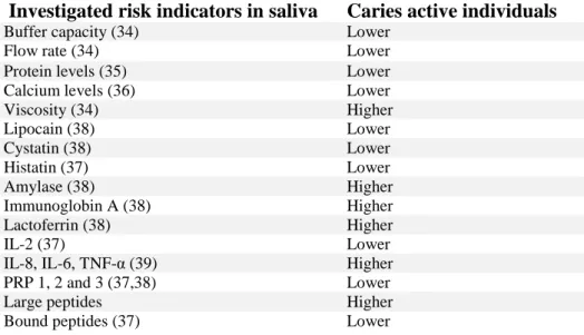
Risk indicators in saliva
Different features of saliva have been investigated as possible risk factors for caries. Studies have found correlation between caries and the quality and quantity of salivary components. See summary of risk indicators in Table 2.
The salivary flow rate and capacity to buffer have been shown to be higher in caries free children when compared to children with caries (34). The protein levels in saliva also seem to differ between the groups, with levels being almost twice as high in caries free individuals (35). Not only proteins, but also calcium levels are higher in a caries free group (36). In one study the saliva viscosity was higher in caries active individuals (34).
The amounts of antimicrobial molecules in saliva have been observed to differ between caries active and caries free individuals in numerous studies. Antibacterial molecules like lipocain, cystatin and histatin have been found in higher amounts in caries free individuals. These protease inhibitors might be a natural defense against caries (37,38). In contrast, levels of the
Sialidase Galactosidase Fucosidase
S. mutans - (20,24,26) + (20) - * (20, 24)
S. mitis + (26) + (20) + (20)
A.naeslundii +** (24,27) + (24) - (20)
+ Enzyme activity detected - Enzyme activity not detected
* Low or no fucosidase activity detected ** In the majority of the bacteria stems
6
sugar cleaving enzyme amylase are elevated in individuals with higher DMFT. Higher levels of immunoglobin A and lactoferrin have also been correlated to higher DMFT (38) and the individuals with more interleucin-2 (IL-2) have generally lower DMFT (37). There also seems to be higher levels of interleucin-8 (IL-8), interleucin-6 (IL-6) and tumor necrosis factor alpha (TNF-α) in patients with caries. These molecules are all part of a pro-inflammatory response (39).
Higher levels of acidic proline-rich proteins (PRP) have been observed in caries free
individuals (38). One study showed higher levels of PRP 1, 2 and 3 in the group with lower DMFT (38). The ability to penetrate dental plaque has been verified in low molecular weight glycoproteins. Salivary peptides from caries susceptible individuals seem to be larger than those in the caries free group. This could mean that the peptides have too high molecular weight to be able to affect the biofilm (40). Caries free individuals possess a higher numbers of peptides bound to the tooth surface instead (37).
Investigated risk indicators in saliva Caries active individuals
Buffer capacity (34) Lower
Flow rate (34) Lower
Protein levels (35) Lower Calcium levels (36) Lower
Viscosity (34) Higher Lipocain (38) Lower Cystatin (38) Lower Histatin (37) Lower Amylase (38) Higher Immunoglobin A (38) Higher Lactoferrin (38) Higher IL-2 (37) Lower
IL-8, IL-6, TNF-α (39) Higher PRP 1, 2 and 3 (37,38) Lower
Large peptides Higher
Bound peptides (37) Lower
Glycoproteins
Mucins are large glycoproteins lining the oral surfaces. Their role is to protect against
chemical and mechanical damage caused by for example bacterial toxins and food intake (41). One type of mucin, MUC5B, is able to prevent S. mutans from attaching through specific molecule characteristics according to one study (17). The glycoprotein does not however possess any bactericidal properties but presence of MUC5B can reduce the attachment and biofilm formation of S. mutans and therefore maintain bacteria in the planktonic phase. Levels of mucin could alter individuals’ susceptibility to caries and this could be a diagnostic marker for disease (17). In another study, lack of both MUC5B and another mucin, MUC7, was associated with higher DMFT-score (42). A third study showed different results, since levels of MUC5B were higher in a group with more caries. Instead, only MUC7 levels were lower in individuals with caries (43).
Table 2: Summary of different possible risk indicators in saliva. Higher or lower
7
Sialic acid, fucose and galactose
The content of sialic acid in minor gland saliva and whole saliva did not differ significantly between different age groups and between sexes in one study. The content of sialic acid appears stable between age 3 and 25 years (44). However, bacteria induced reduction of sialic acid, caused by high levels of bacteria and bacteria sialidases, has been demonstrated in patients with oral diseases like gingivitis and periodontitis (45). Higher levels of free salivary sialic acid have also been observed in children with congenital heart disease, where a
correlation between elevated levels of salivary sialic acid and caries could be drawn (46).
Present investigation
Salivary glycoproteins and their post-translational modifications could play a role in determining their protective features against dental caries. In our bachelor thesis, we
investigated the ability for the known acid tolerant bacterium S. mutans to adhere to the major oral mucin, MUC5B. Our results showed a significant increase in adherence when terminal sialic acid was eliminated from the molecule.
It would be valuable to have a tool to assess internal caries risk in dental practice. If saliva glycosylation and/or glycosidase activity was shown to be risk indicators for caries, risk assessment could be done with a saliva test. Previous studies have investigated the role of saliva molecules in caries, but to the authors’ knowledge, none with focus on glycoprotein glycosylation.
8
Aim
The aim of this study was to investigate if there are differences in levels of bound sialic acid, fucose and galactose in salivary glycoproteins between individuals with DMFT = 0 and DMFT ≥ 1. These monosaccharides can possibly affect oral bacteria’s ability to adhere and thereby influence caries incidence.
Since glycosidase activity can modify glycosylation profiles of glycoproteins we also aimed to investigate if the activity in supragingival dental plaque flora differ between the two groups. The studied glycosidases are sialidase, α-fucosidase, β-galactosidase, β-glucosidase and N-acetylglucosaminidase.
Hypotheses
H0a: There is no difference between individuals with DMFT = 0 and individuals with DMFT
≥ 1 regarding levels of bound salivary sialic acid, fucose and galactose in glycoproteins. H1a: Levels of bound salivary sialic acid, fucose and galactose in glycoproteins is higher in
individuals with DMFT = 0 compared to individuals with DMFT ≥ 1.
H0b: There is no difference between individuals with DMFT = 0 and individuals with DMFT
≥ 1 regarding investigated glycosidase activity in supragingival plaque.
H1b: Investigated glycosidase activity in supragingival plaque is lower in individuals with
9
Material and methods
EthicsEthics application (attachment 1a and b) was approved by the local Ethics Committee at the Faculty of Odontology, Malmö University (Dnr: STUD 3.5.2.-2016/928). All participants were informed about the study and the procedures both verbally and in written (attachment 2). The subjects were allowed to withdraw their consent at any time before, during or after
sampling. All subjects signed an informed-consent form (attachment 3) before start. No specific questions were asked regarding feelings about participating in the study, but the majority said it was a fun and quick experience.
Study population
For this study 20 individuals (16 women and 4 men) aged 22-40 years were recruited among dental students at the Faculty of Odontology, Malmö University. Inclusion criteria were healthy individuals with no use of medication at the time of examination. Exclusion criteria were use of tobacco or antibiotics the last month before sampling and no ongoing orthodontic treatment. The participants were not allowed to drink or eat one hour before sampling. The participants were asked to avoid tooth brushing 24 hours before sampling to make sure plaque was present.
Sample collection
Saliva and plaque samples were collected from subjects between 11:00 and 12:00. The subjects were characterized according to the DMFT index, into two groups: caries free (DMFT = 0) and caries susceptible (DMFT ≥ 1). Collection of unstimulated saliva was performed by direct draining into a saliva collection tube. The collection was timed to estimate flow rare. After collection, all samples were stored in -80° C until analysis. Plaque samples were collected from buccal and proximal surfaces of tooth 16-26 and 46-36 with one Quick Stick (DAB, Malmö, Sweden) for each jaw. The sticks were fitted into tubes containing 500 µl phosphate-buffered saline (0.15 M sodium chloride, 10 mM potassium phosphate, pH 7.2; PBS).
Levels of sialic acid, fucose and galactose in saliva (ELLA – enzyme-linked lectin-assay)
Whole saliva was coated on 96-well plates (3912, Falcon, Franklin Lakes, NJ, USA) and placed in a humid chamber over night at room temperature. The plates were then rinsed three times with PBS containing 0.05 % (v/v) Tween-20 (Bio-Rad, Hercules, CA, USA) to remove non-adherent molecules. The plates
were incubated for 1 h in a blocking solution of PBS containing containing 0.05 % (v/v) Tween-20 with 1 % BSA (Sigma Chemical Co., St Louis, MO, USA) to avoid unspecific adherence of lectins to uncoated well surfaces. The plates where then incubated for 1 h with four different horseradish peroxidase (HRP)-conjugated lectins
Monosaccharide Lectine
Sialic acid α(26) Sambucus nigra
Sialic acid α(23) Maackia amurensis
α-Fucose Ulex europaes I
Galactose Erythrina cristagalli
10
(see Table 3) diluted in blocking solution with 0,1 mM CaCl2. The plates were then rinsed
again with washing solution to remove unbound lectins. SigmaFast
O-fenylendiamine-dihydrocloride (OPD) (Sigma-Aldrich, St Louis, MO, USA) was used as substrate to visualize the lectins. The reactions were recorded in a fluorimeter, Fluostar Optima (BMG, Cary, NC, USA). Carbohydrate reactivity was expressed as absorbance at 450 nm.
Bacteria cultivation
The cultivable microflora in the sample was investigated by culturing on blood agar plates. 50 µl of the sample was serially diluted in PBS and 200 µl of three dilutions were plated on agar and distributed evenly using glass beads. Plates were incubated for 3 days in an aerobe environment containing 5% CO2 at 37°C. Colony-forming units (CFU) ranged between 3.0 x
109 and 4.5 x 10¹⁰ in the group with DMFT = 0 and between 3.4 x 10⁹ and 8.0 x 10¹⁰ in the
group with DMFT ≥ 1. The differences in CFU are minor and could therefore be disregarded as a factor for the results of glycosidase activity.
Glycosidase activity with fluoroscens
Glycosidase activity in the supragingival dental plaque was assayed using fluorogenic substrates in a 96-well microtitre plate (Nunc Maxisorb, Roskilde, Denmark). Dental plaque was collected in N-Tris[hydroxymethyl]methyl-2-aminoethane sulfonic acid (TES) buffer, pH 7.5, and mixed with 20 µl of each subtrate (see Table 4). Samples were incubated for 3h at 37°C and absorbance was recorded in a fluorimeter, Fluostar Optima (BMG, Cary, NC, USA), fitted with a 96-well plate reader, at excitation and emission wave length of 355 and 460 nm, respectively.
Statistics
Results were analysed using the Mann–Whitney U-test and a p-value < 5% was considered significant. Glycosidase Substrate Sialidase 4-Methylumbelliferyl-N-Acetyl-O-D-neuraminide β-galactosidase 4-Methylumbelliferyl-b-d-galactoside α-fucosidase 4-Methylumbelliferyl-a-L-fucoside N-acetylglucosaminidase 4-Methylumbelliferyl-N-Acetyl-b-D-glucosaminide β-glucosidase 4-Methylumbelliferyl-b-D-glucoside
11
Results
Saliva
Sialic acid, fucose and galactose
The aim was to investigate differences in levels of usual monosaccharides bound to salivary glycoprotein in
individuals with previous caries experience and individuals with no experience. Samples of whole saliva from 20 individuals were analyzed for levels of sialic acid, fucose and galactose with
enzyme-linked lectin-assay, recorded with a fluorometer.
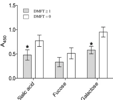
A glycosylation profile of bound monosaccharides in whole saliva was performed for each subject. Results were then divided in two groups; DMFT = 0 and DMFT ≥ 1. Average and standard deviation for the two groups and respective
monosaccharide are shown in figure 1.
Sialic acid (α(26) and α(23)) was detected using the lectins Sambucus nigra respective
Maackia amurensisk. The results (figure 1) showed a significantly higher (p = 0.043) content
of sialic acid in the group with DMFT = 0. Fucose was detected with the lectin Ulex europaes
I. Content of fucose did not differ significantly between the two groups. Galactose was
detected using the lectin Erythrina cristagalli, and the content was significantly higher (p = 0.012) in the group with DMFT = 0.
In summary, the content of both sialic acid and galactose were significantly higher in the group with DMFT = 0, while the content of fucose did not differ significantly between the groups.
Flow rate
In our study the average saliva rate was 0.47 ml/min in both groups and no significant difference was detected.
Glycosidases
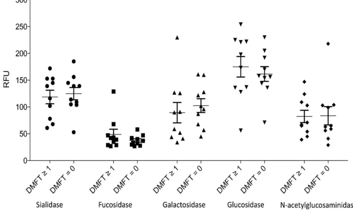
Bacterial glycosidase activity was investigated due to the possible influence on the
configuration of salivary glycoproteins. Plaque samples were collected from all 20 subjects and activity of usual glycoprotein degrading glycosidases were assayed using fluorogenic substrates. Chosen glycosidases were sialidase, α-fucosidase, β-galactosidase, β-glucosidase and N-acetylglucosaminidase since their corresponding monosaccharides are found on salivary glycoproteins.
Figure 1. Glycosylation profile of whole saliva in
groups with DMFT ≥ 1 and DMFT = 0 analyzed with ELLA and recorded with a fluorometer.
DMFT ≥ 1 DMFT = 0
12
The results are summarized in figure 2. Every plot represents the value of an individual subject and these are gathered in respective group (DMFT = 0 and DMFT ≥ 1) for each glycosidase. Values for glycosidase activity were overall stable and only a few subjects did differ from their group. No significant differences can be seen between the two groups (DMFT = 0 and DMFT ≥ 1) regarding any of the investigated glycosidases.
Figure 2. Five different glycosylases were detected using substrate for the specific enzymes. The
results were then recorded with a fluorometer. In the figure, activity for enzymes in the two groups, DMFT = 0 and DMFT ≥ 1, are illustrated.
13
Discussion
The main issue of this study is how glycosylation pattern of glycoproteins, with focus on sialic acid, fucose and galactose, might influence caries progression. This has, to the authors’ knowledge, never been investigated before which makes our results unique. In our study, lower levels of bound sialic acid and galactose were found in the group with caries experience (DMFT ≥ 1) compared to the group without caries (DMFT = 0). The results indicate that the investigated monosaccharides could be a possible risk indicator for caries.
In our bachelor thesis (47), we investigated the ability for S. mutans to adhere to the major oral mucin, MUC5B. Our results showed a significantly increased ability of adherence when terminal sialic acid was eliminated from the mucin. This indicates that lower levels of sialic acid could promote adherence of S. mutans earlier in the biofilm formation and consequently be a possible influencing factor for caries. With these results in mind we hypothesized that individuals with caries experience have qualitatively different glycosylation of salivary glycoproteins in comparison to individuals with no caries experience. We focused mainly on the monosaccharide sialic acid, but also fucose and galactose, as possible factors in caries susceptibility. We did not aim to investigate the saliva and bacterial glycosidases in subjects with active caries lesions, since the aim was to find potential internal risk indicator.
Saliva
When comparing levels of salivary bound sialic acid, fucose and galactose in the two groups, some differences were found. The caries free group had significantly higher levels of sialic acid and galactose. The results showed no significant difference regarding fucose levels. Sialic acid has major effects on its surroundings. One of the effects is giving glycoproteins a stable configuration leading to the protecting features of saliva. It also protects underlying parts of glycoproteins from degradation (13). These protecting features could explain why individuals with higher levels of sialic acid are more resistant to caries, since it is preventing bacteria from adhering and utilizing glycoproteins.
We did not specify the bacteria species in plaque samples and cannot draw any conclusion regarding specific associations between species and glycosidases. Earlier studies have
however investigated this relationship. S. mutans show galactosidase activity when cultivated on mucins, but express low levels of fucosidase and sialidase (20,24,26). S. mutans may consequently have difficulties utilizing glycoproteins with higher amount of sialic acid, resulting in disadvantage in a biofilm. If we specified the species in our study and levels of S.
mutans was higher in the group with DMFT ≥ 1, this could consequently depend on the lower
levels of sialic acid.
Content of sialic acid appears to be stable between the ages of 3 and 25 years, and the content in minor gland and whole saliva does not differ between different age groups and sexes, according to one study (44). If glycosylation levels of sialic acid and other monosaccharides remain stable over an individual’s life, levels could possibly be used as a caries risk indicator. Levels of PRPs have been shown to be increased in caries free groups (38,39). PRPs contains both sialic acid and galactose, and therefore this could be an additional explanation to why levels where higher in the group with DMFT = 0.
14
Glycosidase activity
In this study, we also aimed to investigate glycosidase activity in supragingival plaque. The chosen glycosidases were sialidase, α-fucosidase, β-galactosidase, β-glucosidase and N-acetylglucoseaminidase since their corresponding monosaccharides are found on salivary glycoproteins.
The results showed no differences between the studied groups regarding any of the
glycosidases. It seems like the null hypothesis (H0b) was correct, indicating no phenotypic
differences regarding the included glycosidases in the plaque of the two groups. Thus, glycosidase activity in this study does not seem to indicate any caries risk. According to our results the intrinsic genetic factors in salivary glycoproteins have greater importance than plaque glycosidase activity in caries susceptibility.
If the group with DMFT ≥ 1 showed higher levels of sialidases and galactosidases this could have been a possible explanation to the lower levels of bound sialic acid and galactose in glycoproteins. But since our results showed no differences regarding glycosidase activity between the groups it seems that some people have a different and more caries susceptible glycosylation profile.
One possible source of error in the investigation of glycosidase activity is the differences in the amount of collected plaque from each participant. To neutralize and control this
difference, cultivated bacteria were counted. CFU ranged between 3.0 x 10⁹ and 4.5 x 10¹⁰ in the group with DMFT = 0 and between 3.4 x 10⁹ and 8.0 x 10¹⁰ in the group with DMFT ≥ 1. Differences between the two groups were small, and it indicates that the numbers are not affecting the results of glycosidase activity.
CFU is a rough number of cells grown on the plate and the method is not flawless. The agar plate represents a special environment in which all bacteria species cannot grow, which means all bacteria are not included in the counting. The number of cells included in one CFU can also vary from one up to thousand cells (48). We still used this method for counting since it is the simplest and best lab procedure available.
Salivary flow
Saliva rate is significant for oral health, since a high flow rate enhances the ability for oral clearance, lubrication and buffering of pH level in the oral cavity. For stated reasons, low saliva rate has been associated with a higher caries susceptibility (34). In our study the salivary flow did not differ significantly between the two groups.
Since glycosidase activity and salivary flow rate did not differ between the two studied groups, the only detected difference was levels of sialic acid and galactose. This excludes glycosidases and flow rate as possible modifiers of the results.
Study population
The individuals recruited to our study are dental students, aged 22-40 years at the Faculty of Odontology. Age and education level do not reflect the society since the sample is not randomized, which could be a source of error in this study. However, it also means that the two compared groups have similar external background factors such as age and education. With more homogeneous groups the external factors are less likely to affect the results.
15
Since we did not have the resources to examine all subjects before sampling, they had to declare their own caries status. This could be a source of error, since the individuals might not have been aware of recently developed caries. All subjects were dental students with great awareness of caries disease, which makes their statement more likely to be correct.
All subjects were adults during sampling and we do not have the details of caries development and risk factors earlier in the individuals’ life. Therefore, we cannot determine a causal
relationship between monosaccharides in saliva and caries development. To be able to do that longitudinal prospective studies are required.
Ethics
The benefits of this study are greater for the society then for the participants, but the subjects were not put at any risk when participating. Participants could withdraw their consent at any time before, during and after sample collection. Subjects could have found it inconvenient not to drink or eat for one hour before sampling and not brushing their teeth for 24 h. In
conclusion, the risks with performing this study were very low when compared to the benefits.
Future outlook and putative clinical value
Our results indicate that individuals with more sialic acid and galactose bound to salivary glycoproteins could have an inherent protection against caries. If a longitudinal study could confirm that low levels of these, or other monosaccharides, are good indications for higher caries susceptibility, a saliva test could easily be used in the clinical situation to estimate caries risk. In order to develop such a test, studies must be designed to determine how low levels of sialic acid and/or galactose must be, for them to be considered risk factors.
With a solid and comprehendible method of risk assessment, population groups with a greater caries risk could be identified earlier and treated prophylactic to avoid cavities. If resources could be reallocated more effectively, it would gain both patients and society.
Conclusion
In our study, lower levels of bound sialic acid and galactose were found in the group with DMFT ≥ 1 compared to the group with DMFT = 0. Results indicate that these
monosaccharides could be a possible risk indicator for caries. No differences were seen regarding glycosidase activity and salivary flow rate between the groups. Larger longitudinal studies are needed to verify these correlations. If this study was repeated in a larger
longitudinal study, the results could lead to a more valid caries risk assessment and thus a more effective way to channel oral health resources.
Acknowledgments
We want to thank Ulrika Troedsson for the support and guidance during the laboratory trials, we would have been lost without you. We also want to thank our supervisors Claes
16
References
(1) Dental caries: the disease and its clinical management. 2. red. Oxford, UK; Ames, Iowa, USA: Blackwell Munksgaard; 2008.
(2) Socialstyrelsen. Karies hos barn och ungdomar - Epidemiologiska uppgifter för år 2014. 2015. (3) Socialstyrelsen. Karies hos barn och ungdomar. En lägesrapport för år 2008. Stockholm: Socialstyrelsen; 2010.
(4) World Health Organization. Oral health surveys: basic methods . 5th red. : World Health Organization; 2013.
(5) Marsh Philip. Oral microbiology. : Churchill Livingstone; 2009.
(6) Lith A., Lindstrand C., Grondahl H. G. Caries development in a young population managed by a restrictive attitude to radiography and operative intervention: II. A study at the surface level.
Dentomaxillofac.Radiol. 2002 Jul;31(4):232-239.
(7) Karies - diagnostik, riskbedömning och icke-invasiv behandling: en systematisk litteraturöversikt : december 2007. Stockholm: Statens beredning för medicinsk utvärdering (SBU); 2007.
(8) Dental caries: the disease and its clinical management. 2. red. Oxford, UK; Ames, Iowa, USA: Blackwell Munksgaard; 2008.
(9) Nanci Antonio. Ten Cate's oral histology: development, structure, and function. 8. red. St. Louis: Mosby Elsevier; 2013.
(10) Hay D. I. The adsorption of salivary proteins by hydroxyapatite and enamel. Arch.Oral Biol. 1967 Aug;12(8):937-946.
(11) Jensen J. L., Lamkin M. S., Oppenheim F. G. Adsorption of human salivary proteins to hydroxyapatite: a comparison between whole saliva and glandular salivary secretions. J.Dent.Res. 1992 Sep;71(9):1569-1576.
(12) The teeth and their environment: physical, chemical and biochemical influences. New York; Basel: Karger; 2006.
(13) Traving C., Schauer R. Structure, function and metabolism of sialic acids. Cell Mol.Life Sci. 1998 Dec;54(12):1330-1349.
(14) Becker D. J., Lowe J. B. Fucose: biosynthesis and biological function in mammals. Glycobiology 2003;13(7):41R-53R.
(15) Pomin Vitor H. Galactose : Structure and Function in Biology and Medicine. New York: Nova Science Publishers, Inc; 2014.
(16) Svensater G., Larsson U. B., Greif E. C., Cvitkovitch D. G., Hamilton I. R. Acid tolerance response and survival by oral bacteria. Oral Microbiol.Immunol. 1997 Oct;12(5):266-273. (17) Frenkel E. S., Ribbeck K. Salivary mucins protect surfaces from colonization by cariogenic bacteria. Appl.Environ.Microbiol. 2015 Jan;81(1):332-338.
(18) Nobbs A. H., Lamont R. J., Jenkinson H. F. Streptococcus adherence and colonization. Microbiol.Mol.Biol.Rev. 2009 Sep;73(3):407-50, Table of Contents.
(19) Kolenbrander P. E., Palmer R. J.,Jr, Rickard A. H., Jakubovics N. S., Chalmers N. I., Diaz P. I. Bacterial interactions and successions during plaque development. Periodontol.2000 2006;42:47-79. (20) van der Hoeven J. S., van den Kieboom C. W., Camp P. J. Utilization of mucin by oral
Streptococcus species. Antonie Van Leeuwenhoek 1990 Apr;57(3):165-172.
(21) Beighton D., Smith K., Hayday H. The growth of bacteria and the production of exoglycosidic enzymes in the dental plaque of macaque monkeys. Arch.Oral Biol. 1986;31(12):829-835.
(22) Beckers H. J., van der Hoeven J. S. Growth rates of Actinomyces viscosus and Streptococcus mutans during early colonization of tooth surfaces in gnotobiotic rats. Infect.Immun. 1982
Feb;35(2):583-587.
(23) Wickstrom C., Svensater G. Salivary gel-forming mucin MUC5B--a nutrient for dental plaque bacteria. Oral Microbiol.Immunol. 2008 Jun;23(3):177-182.
(24) Bradshaw D. J., Homer K. A., Marsh P. D., Beighton D. Metabolic cooperation in oral microbial communities during growth on mucin. Microbiology 1994 Dec;140 ( Pt 12)(Pt 12):3407-3412. (25) Wickstrom C., Svensater G. Salivary gel-forming mucin MUC5B--a nutrient for dental plaque bacteria. Oral Microbiol.Immunol. 2008 Jun;23(3):177-182.
17
(26) Beighton D., Whiley R. A. Sialidase activity of the "Streptococcus milleri group" and other viridans group streptococci. J.Clin.Microbiol. 1990 Jun;28(6):1431-1433.
(27) Moncla B. J., Braham P. Detection of sialidase (neuraminidase) activity in Actinomyces species by using 2'-(4-methylumbelliferyl)alpha-D-N-acetylneuraminic acid in a filter paper spot test. J.Clin.Microbiol. 1989 Jan;27(1):182-184.
(28) Mothey D., Buttaro B. A., Piggot P. J. Mucin can enhance growth, biofilm formation, and survival of Streptococcus mutans. FEMS Microbiol.Lett. 2014 Jan;350(2):161-167.
(29) Kindblom C., Davies J. R., Herzberg M. C., Svensater G., Wickstrom C. Salivary proteins promote proteolytic activity in Streptococcus mitis biovar 2 and Streptococcus mutans. Mol.Oral Microbiol. 2012 Oct;27(5):362-372.
(30) Burt B. A. Definitions of risk. J.Dent.Educ. 2001 Oct;65(10):1007-1008.
(31) Beck J. D. Risk revisited. Community Dent.Oral Epidemiol. 1998 Aug;26(4):220-225. (32) Pienihakkinen K., Jokela J., Alanen P. Assessment of caries risk in preschool children. Caries Res. 2004 Mar-Apr;38(2):156-162.
(33) Grindefjord M., Dahllof G., Nilsson B., Modeer T. Prediction of dental caries development in 1-year-old children. Caries Res. 1995;29(5):343-348.
(34) Animireddy D., Reddy Bekkem V. T., Vallala P., Kotha S. B., Ankireddy S., Mohammad N. Evaluation of pH, buffering capacity, viscosity and flow rate levels of saliva in caries-free, minimal caries and nursing caries children: An in vivo study. Contemp.Clin.Dent. 2014 Jul;5(3):324-328. (35) Hong S. W., Seo D. G., Baik J. E., Cho K., Yun C. H., Han S. H. Differential profiles of salivary proteins with affinity to Streptococcus mutans lipoteichoic acid in caries-free and caries-positive human subjects. Mol.Oral Microbiol. 2014 Oct;29(5):208-218.
(36) Tulunoglu O., Demirtas S., Tulunoglu I. Total antioxidant levels of saliva in children related to caries, age, and gender. Int.J.Paediatr.Dent. 2006 May;16(3):186-191.
(37) Vitorino R., Lobo M. J., Duarte J. R., Ferrer-Correia A. J., Domingues P. M., Amado F. M. The role of salivary peptides in dental caries. Biomed.Chromatogr. 2005 Apr;19(3):214-222.
(38) Vitorino R., de Morais Guedes S., Ferreira R., Lobo M. J., Duarte J., Ferrer-Correia A. J., et al. Two-dimensional electrophoresis study of in vitro pellicle formation and dental caries susceptibility. Eur.J.Oral Sci. 2006 Apr;114(2):147-153.
(39) Gornowicz A., Bielawska A., Bielawski K., Grabowska S. Z., Wojcicka A., Zalewska M., et al. Pro-inflammatory cytokines in saliva of adolescents with dental caries disease.
Ann.Agric.Environ.Med. 2012;19(4):711-716.
(40) Ayad M., Van Wuyckhuyse B. C., Minaguchi K., Raubertas R. F., Bedi G. S., Billings R. J., et al. The association of basic proline-rich peptides from human parotid gland secretions with caries
experience. J.Dent.Res. 2000 Apr;79(4):976-982.
(41) Derrien M., van Passel M. W., van de Bovenkamp J. H., Schipper R. G., de Vos W. M., Dekker J. Mucin-bacterial interactions in the human oral cavity and digestive tract. Gut Microbes 2010
Jul;1(4):254-268.
(42) Banderas-Tarabay J. A., Zacarias-D'Oleire I. G., Garduno-Estrada R., Aceves-Luna E., Gonzalez-Begne M. Electrophoretic analysis of whole saliva and prevalence of dental caries. A study in Mexican dental students. Arch.Med.Res. 2002 Sep-Oct;33(5):499-505.
(43) Gabryel-Porowska H., Gornowicz A., Bielawska A., Wojcicka A., Maciorkowska E., Grabowska S. Z., et al. Mucin levels in saliva of adolescents with dental caries. Med.Sci.Monit. 2014 Jan
18;20:72-77.
(44) Sonesson M., Ericson D., Kinnby B., Wickstrom C. Glycoprotein 340 and sialic acid in minor-gland and whole saliva of children, adolescents, and adults. Eur.J.Oral Sci. 2011 Dec;119(6):435-440. (45) Shetty P. K., Pattabiraman T. N. Salivary glycoproteins as indicators of oral diseases.
Indian.J.Clin.Biochem. 2004 Jan;19(1):97-101.
(46) Hegde A. M., Kavita R., Sushma K. S., Suchetha S. Salivary sialic acid levels and dental health in children with congenital heart disease. J.Clin.Pediatr.Dent. 2012 Spring;36(3):293-296.
(47) Vikström. H Shala K. Muciners sialinsyror och deras roll i bindning av Streptococcus mitis, Streptococcus mutans och Actinomyces naeslundii. 2015.
18
(48) Sutton Scott. Accuracy of Plate Counts. 2011(JOURNAL OF VALIDATION TECHNOLOGY) :43-46.
19
Attachment 1a: Ethics application
MAH / Odontologiska fakulteten Etikprövningskommittén
Datum: 2015-11-26
ANSÖKNINGSBLANKETT
ETISK PRÖVNING Projektets titel:
Sialinsyrainnehåll i helsaliv och enzymaktivitet i plack och dess inverkan på kariesaktivitet.
Studenter som genomför projektet. Adress och din e-post. Hanna Vikström, Delsjögatan 30, 217 65 Malmö hanna-vikstrom@hotmail.com Kosovare Shala, Enningervägen 6, 243 31 Höör kosovare.sh@hotmail.se Utbildningsnivå: Master
Handledarens namn, kompetens och e-post: Claes Wickström, universitetslektor oral biologi claes.wickstrom@mah.se
Henrik Berlin, övertandläkare pedodonti henrik.berlin@mah.se
Uppgifter om projektet
1. Sammanfattande beskrivning
Projektet handlar om att ta reda på om sialinsyrainnehållet i saliven och
enzymaktiviteten hos bakterier kan vara bidragande faktorer till att vissa patienter har mer karies än andra. Vi har tidigare i kandidatuppsatsen sett att S. mutans binder bättre till MUC5B som saknar sialinsyra och vill därför ta reda på om minskad mängd sialinsyra i saliven kan ha en påverkan på kariesaktiviteten kliniskt.
2. Ange primär vetenskaplig frågeställning
Har sialinsyramängden i saliven och enzymaktiviteten i placket någon effekt på kariesaktivitet?
20
3. Beskriv undersökningsprocedur, datainsamling och datas karaktär, hantering av data
Deltagarna rekryteras genom muntlig förfrågan i tandläkarutbildningen kurs 2 och 8 och möjlighet till intresseanmälan via mail ges vid samma tillfälle. Försökspersonerna delas upp i två grupper – de som någon gång haft manifest karies och de som aldrig har haft det. Försöket görs under ett besök. Deltagarna får inte äta eller dricka en timme före besöket, och ska inte ha borstat tänderna det senaste dygnet före besöket. Vilosaliv samlas under ca 5 minuter eller tills vi har samlat ihop 2 ml saliv. Bakterieprov tas genom att plack skrapas från deltagarens tänder. Salivprov och bakterieprov markeras med en kod som är kopplad till deltagarens personnummer. Ålder, kön och DMFT antecknas för varje individ. Personnummer sparas enbart i en kodlista som förvaras hos laborativ handledare, Claes Wickström. Enzymaktivitet i bakterieproverna undersöks direkt efter den kliniska provtagningen. Ihopsamlad saliv förvaras i frys på avdelningen för Oral Biologi tills alla prover samlats och undersöks sedan
gemensamt. Efter sammanställning av data förstörs kodlistan och alla prover.
4. Plats där datainsamling sker:
Salivprov och bakterieprover samlas och analyseras på avdelningen för Oral Biologi på Malmö Tandvårdshögskolan.
Uppgifter om population/material
1. Beskriv urvalsförfarande och rekrytering
Deltagarna är myndiga studenter på Malmö Tandvårdshögskola. Deltagarna rekryteras genom information om projektet och möjlighet till intresseanmälan. Individerna ska ha god
allmänhälsa och får inte stå på antibiotika eller ha avslutat en antibiotikakur den senaste månaden eftersom det skulle kunna påverka bakteriernas enzymaktivitet. Individer som regelbundet tillför kemikalier som t.ex. tobak (snusning och rökning) eller mediciner
exkluderas. Även deltagare med astma, diabetes och/eller tandställning exkluderas. Proverna sparas på avdelning för Oral Biologi, Malmö Högskola, i biobank 497.
2. Undersökningsmaterialets storlek, power analys:
Denna studie är en explorativ pilotstudie och därför ärpower analys inte applicerbart. Studien kommer att basera sig på 20-30 personer.
Information och samtycke
1. Beskriv procedur och innehåll i information vid tillfrågan om deltagande
Information om studien ges både muntligt och skriftligt till deltagaren minst en vecka före planerat besök.
2. Hur inhämtas samtycke?
Samtyckesblankett skrivs på före provtagning. Deltagarna kan när som helst innan, under eller efter undersökningen avbryta/avböja medverkan i studien. Prover och datauppgifter förstörs direkt om deltagaren avbryter studien.
3. Generell avvägning risk/nytta.
Nyttan för samhället är större än för individen som deltar i studien. I längden skulle resultaten av studien kunna innebära att man kan hitta fler biologiska riskfaktorer för karies, samtidigt som studien inte innebär några risker för någon.
21
1. Vilka risker och komplikationer för deltagarna kan uppstå i samband med undersökningen?
Risken är mycket låg avvägt mot nyttan. Deltagaren får inte äta/dricka en timme före besöket och kan tycka det är något obehagligt att lämna salivprovet. Ingen risk för skada.
2. Beskriv eventuell nytta som projektet kan innebära för deltagarna
I samband med information om studien kan nyfikenhet väckas avseende kariesproblematik, riskfaktorer och den forskning som bedrivs i området.
Redovisning av resultat
1. Hur kommer resultaten att offentliggöras?
Studien avslutas februari 2017. Resultaten kommer att offentliggöras i vår
masteruppsats i tabeller och diagram. De kommer även att analyseras i uppsatsen.
2. Hur säkerställs informanternas integritet när materialet publiceras?
De enda uppgifterna som kommer samlas in vid saliv- och bakterieprovtagning är ålder, kön och DMFT. Proverna kommer att kodas och koden kopplas till deltagarens personnummer. Lista med koder och personnummer kommer att bevaras inlåst på Claes Wickströms kontor. Kodlistan förstörs efter att studien är slutförd.
Obligatoriska bilagor
1. Skriftlig information till de som tillfrågas 2. Skriftlig samtyckesblankett enligt anvisningar 3. Undertecknat tillstånd från avdelningsföreståndare
22
23
Attachment 2: Information to participants
Malmö högskola / Odontologiska fakulteten Avdelningen för Oral biologi
Information till deltagare
Sialinsyrainnehåll i helsaliv och enzymaktivitet i plack och dess
inverkan på kariesaktivitet.
Bakgrund och syfte
Trots att den orala hälsan har förbättrats under de senaste decennierna är hål i tänderna (karies) fortfarande ett stort problem, inte minst bland ungdomar. Det finns olika orsaker till varför man får karies, t ex socker, småätande och brist på fluor. Men man vet inte helt säkert vad det är som gör att vissa människor har större risk än andra för att få karies.
Syftet med studien är att undersöka om det finns någon skillnad i saliven mellan personer som har/har haft karies och de som inte har/har haft det.
Förfrågan om deltagande
Du tillfrågas om deltagande i denna studie då du arbetar eller studerar på Malmö Tandvårdshögskola. Samtycke för att delta i studien ges genom att du fyller i en samtyckesblankett vid provtagningen.
Hur går studien till?
För att vi ska kunna utföra studien behöver vi salivprov och bakterieprov. Salivprovet tas genom att du lutar dig över en mugg och där spottar ut all saliv som bildas under ca 5 minuter. Bakterieprovet utförs genom att vi skrapar bort bakterier från en tand med hjälp av en plaststicka. Detta går fort och känns inte. Totalt kommer de båda
provtagningarna att ta ca 10 minuter.
Vid provtagningstillfället är det viktigt att du inte påverkar saliven eller bakterierna. Det innebär att du inte ska äta och/eller dricka en timme innan besöket. Du får heller inte ha rökt, snusat eller medicinerat med antibiotika den senaste månaden. För att det ska finnas tillräckligt med plack vid provtagningen ska du heller inte ha borstat tänderna den aktuella dagen.
Biobanksprover
Proverna kommer att ges en kod som är kopplad till ditt personnummer. Bakterieproverna kommer att analyseras direkt efter att de samlats in. Salivproverna kommer att frysas ner och analyseras när salivprov från samtliga deltagare har samlats in. Dessa prover kommer endast användas i denna studie. All hantering och analys av prover kommer ske på avdelningen för Oral Biologi på Malmö tandvårdshögskola.
24
Vilka är riskerna?
Det finns inga risker med att delta i studien.
Finns det fördelarna med deltagandet?
Det finns inga specifika fördelar för dig som deltagare, men studiens resultat skulle kunna leda till att vi bättre kan identifiera personer med ökad risk för karies.
Hantering av data och sekretess
De personuppgifter vi samlar in är: ålder, kön och om du har/har haft karies eller inte. Detta gör vi för att definiera undersökningsgruppen. Personuppgifterna kommer att behandlas enligt personuppgiftslagen (PuL), och hanteras så obehöriga inte kan ta del av dem. Personuppgiftsombud för Malmö Högskola är Hans Jonsson, 0708-65 52 89. Proverna kommer att registreras med en kod knuten till ditt personnummer. Koden kommer finnas i en kodlista. Ansvarig för den är Claes Wickström. Listan kommer att
hanteras så att inte obehöriga kan ta del av den. Resultatet från studien kommer att presenteras i en examensuppsats (masternivå) och kan komma att publiceras i en vetenskaplig tidskrift. Du kommer inte att kunna identifieras vid presentation av resultaten. Efter att studien har slutförts kommer proverna och listan med personnummer att förstöras.
Hur får jag information om studiens resultat?
Studien och resultaten kommer att publiceras på Malmö högskolas forskningsdatabas. Vid intresse kan du kontakta ansvariga så sänder vi dig en kopia på den färdiga uppsatsen.
Frivillighet
Det är frivilligt att vara med i studien och när som helst innan, under eller efter provtagningen kan du avbryta/avböja ditt deltagande utan särskild förklaring. Att avböja/avbryta deltagandet kommer inte att påverka dig på något sätt.
Om du inte längre vill vara med i studien får du maila eller ringa Claes Wickström (se uppgifter nedan). Du uppger då namn och personnummer. Alla uppgifter vi samlat om dig samt dina prover kommer då omedelbart att förstöras.
Med vänliga hälsningar,
Tandläkarstuderande Tandläkarstuderande
Hanna Vikström Kosovare Shala
hanna-vikstrom@hotmail.com kosovare.sh@hotmail.se Universitetslektor, Oral biologi Övertandläkare, Pedodonti
Claes Wickström Henrik Berlin
claes.wickstrom@mah.se henrik.berlin@mah.se Tel: 040-665 86 58 Tel: 040-665 84 88 Malmö Högskola
Odontologiska fakulteten
25
Attachment 3: Informed consent form
Malmö högskola / Odontologiska fakulteten Avdelningen för Oral Biologi
Samtyckesblankett för deltagare
Studie: Sialinsyrainnehåll i helsaliv och enzymaktivitet i plack och dess
inverkan på kariesaktivitet.
Jag har muntligt informerats om studien och tagit del av bifogad skriftlig information. Jag är medveten om att mina prover under studiens gång kommer sparas samt att mina personuppgifter kommer finnas i en kodlista. Jag är även medveten om att mitt deltagande är frivilligt och att jag när som helst och utan förklaring kan avbryta mitt deltagande.
Jag lämnar härmed mitt samtycke till att delta i ovanstående studie:
Datum:.…….………..……….………..………...… Deltagarens underskrift:…..………….………..……… Namnförtydligande:………. Inhämtare av samtycke:………...………..……… Datum:.……….……… Underskrift:……….………...………. Studieansvariga studenter: Hanna Vikström, tandläkarstuderande, Masternivå hanna-vikstrom@hotmail.com Kosovare Shala, tandläkarstuderande, Masternivå kosovare.sh@hotmail.se Studieansvariga handledare: Claes Wickström,
universitetslektor, Oral biologi
claes.wickstrom@mah.se Henrik Berlin,
övertandläkare, Pedodonti henrik.berlin@mah.se
Hantering av datauppgifter/PUL-ansvarig vid Malmö högskola:
Hans Jonsson, 0708-655289.
Malmö högskola, Odontologiska fakulteten, 206 05 Malmö, Tfn 040-6657000