Characterization of FtsH proteases in the
annual plant Arabidopsis thaliana
Harald Aigner
Department of Chemistry
Umeå University, Sweden Doctoral Thesis 2012
Characterization of FtsH proteases in the
annual plant Arabidopsis thaliana
Harald Aigner
Department of Chemistry
Umeå University, Sweden Doctoral Thesis 2012
Copyright © Harald Aigner ISBN: 978-91-7459-445-4
Tryck/Printed by: CMC KBC, Umeå University Umeå Sweden 2012
Organization Document type
UMEÅ UNIVERSITY DOCTORAL THESIS Department of Chemistry
SE-901 87 Umeå, Sweden Date of issue May 2012
Author
Harald Aigner
Title
Characterization of FtsH proteases in the annual plant Arabidopsis thaliana
Abstract
Background FtsH is an ATP-dependent membrane-bound metalloprotease. A. thaliana
contains 12 FtsH proteases localized in membranes of chloroplasts and mitochondria where they form homo- or hetero-hexameric complexes. FtsH11 – the main subject of this thesis – is located in the chloroplast envelope.
Methods
Field studies with A. thaliana to determine Darwinian fitness. A growth under outdoor conditions often allows discovering of phenotypes that are unascertainable in the controlled environment of growth chambers.
Proteomic methods to discover fragments of substrate proteins (limited proteolysis) and changes in the proteome of FtsH protease deficient mutants.
Results ftsh11 has increased amount of: RuBisCO activase, several Calvin cycle enzymes,
two enzymes involved in starch synthesis and some chaperons. Some of those enzymes have been identified as possible substrates of FtsH11. Under long photoperiods ftsh11 develops a chlorotic phenotype accompanied by decreasing NADP+/NADPH ratio and increase of ROS damaged proteins.
Conclusion Impaired functioning of the Calvin cycle in ftsh11 leads, during long
photoperiods, to shortage of NADP+,which causes over reduction of the electron transport chain and increased ROS production. ROS damages the photosynthetic proteins and induces chlorosis.
1
TABLE OF CONTENTS
1 List of papers 2 Abbreviations 3 Introduction 3.1 Proteases in plants3.2 The hydrolytic cleavage of peptide bonds 3.3 The FtsH proteases
3.4 FtsH proteases of Arabidopsis thaliana
3.4.1 The thylakoid FtsH protease complex consisting of FtsH1, FtsH2, FtsH5 and FtsH8
3.4.2 The chloroplast envelope located FtsH proteases 3.4.3 The mitochondrial FtsH proteases
4 Aim of this work
5 Key-methods used in my studies
5.1 Darwinian fitness and phenotype characterization 5.2 Proteomics
5.2.1 The gel-based approach
5.3.1.1 2-dimensional isoelectric focusing/SDS polyacrylamide gel electrophoresis (2D-IEF-PAGE)
5.3.1.2 2-dimensional Blue Native/SDS polyacrylamide gel electrophoresis (2D-BN-PAGE)
5.3.2 The chromatography approach (gel free proteomics) 5.3.2.1 MudPIT
5.3.2.2 COFRADIC 5.3.3 Quantitative proteomics 6 Results
6.1 Field studies on FtsH knock-out mutants 6.2 FtsH11
6.2.1 Phenotype of ftsh11
6.2.2 Proteomic Studies on ftsh11 6.2.2.1 Refraction-2D 6.2.2.2 Gel-free proteomics
2
6.4 Complex partner of FtsH11 6.5 Results on other FtsH proteases 6.5.1 Modeling approach 6.5.2 FtsH7 and FtsH9 6.5.3 FtsH3 and FtsH10 7 Conclusions 8 Outlook 9 Reference list 10 Acknowledgements
3
1 LIST OF PAPERS
This thesis is based on the following publications and manuscripts that will be referred to in the text by their numbers.
1. Wagner, R., Aigner, H., Pruzinska, A., Jankanpaa, H.J., Jansson, S. and Funk, C. (2011) Fitness analyses of Arabidopsis thaliana mutants depleted of FtsH metalloproteases and characterization of three FtsH6 deletion mutants exposed to high light stress, senescence and chilling. The New Phytologist, 191, 449-458.
2. Aigner, H., Wagner, R., Sjögren, L.L.E., Eubel, H., Millar, A. H., Clarke, A.K. and Funk, C. FtsH11 protease is required for Arabidopsis thaliana to adapt to growth in continuous light. Manuscript
3. Aigner, H., Plasman, K., Tsiatsiani, L., Lam, X.T., Wagner, R., Gevaert, K. and Funk, C. Searching for substrates of the metallo protease FtsH11 of Arabidopsis thaliana using N-terminal proteomics. Manuscript
4. Aigner, H., Lam, X.T., Wagner, R. and Funk, C. Comparison of hypothetical 3D-structures of Arabidopsis thaliana FtsH Proteases with the aim to predict FtsH complex formation. Manuscript
5. Wagner, R., Aigner, H. and Funk, C. (2012), FtsH proteases located in the plant chloroplast. Physiologia Plantarum, 145: 203–214.
4
2 ABBREVIATIONS
σ 32 heat-shock transcription factor sigma 32 2DE two-dimensional electrophoresis
AAA ATPases associated with a variety of cellular activities ADG ADP-glucose pyrophosphorylase
ADP adenosine diphosphate AOC allene oxide cyclase ATP adenosine triphosphate
BN Blue Native
COFRADIC combined fractional diagonal chromatography
Col0 columbia 0
DIGE differential gel electrophoresis E. coli Escherichia coli
FTSH filamentation temperature sensitive H gene FtsH filamentation temperature sensitive H protein
ftsh11 filamentation temperature sensitive H knock-out mutant GMO genetically modified organism
GS glutamine synthetase
GTP guanosine-5'-triphosphate HCF high chlorophyll fluorescence
HflC/HflK high frequency of lysogenization C/K HPLC high-performance liquid chromatography
Hsp heat-shock protein
ICAT isotope-coded affinity tag IEF isoelectric focusing IPG immobilized pH gradient
iTRAQ isobaric tag for relative and absolute quantitation
JA jasmonic acid
mRNA messenger RNA
MS mass spectroscopy
MudPIT multidimensional protein identification technology NADPH nicotinamide adenine dinucleotide phosphate PAGE polyacrylamide gel electrophoresis
5
PETC photosynthetic electron transport chain
pI isoelectric point
pKa acid dissociation constant PMF peptide mass fingerprint
PSBO photosystem II O
PSBP photosystem II P
RBL large subunit RuBisCO
RP reversed phase
RuBisCO ribulose-1,5-bisphosphate carboxylase oxygenase SAILAC stable isotope labeling by amino acids in cell culture SCX strong cationic exchange
SDS sodium dodecyl sulfate
SecYEG secretory YEG
SRH second region of homology SRP signal recognition particle
T-DNA transfer DNA
TI tetrahedral intermediate
6
3 INTRODUCTION
3.1 Proteases in Plants
Proteases are enzymes that by cleaving the peptide bond degrade other proteins. They are involved in many different physiological reactions ranging from unlimited proteolysis, where food- or storage-proteins are degraded into amino acids or damaged proteins are removed, to limited proteolysis in which only specific peptide bonds are cleaved according to different regulatory mechanisms (Feller, 2004). Enzymes of all kind can be activated or deactivated by limited proteolysis e.g. as part of signaling cascades were proteases regulate the function of kinases, receptors, adaptors and transcription factors (auf dem Keller et al. 2007). Therefore, it is probably not exaggerated to say that proteolysis is directly or indirectly involved in most cellular processes (Callis 1995).
Also in plants increasing numbers of cellular pathways are recognized to be regulated via proteolytic events. The MERPOS database (http://merops.sanger.ac.uk, Rawlings et al. 2004) lists 686 proteases and 184 supposable inactive homologs for the plant model organism
Arabidopsis thaliana. Additionally there are 1,300 genes of the ubiquitin/proteasome pathway
(Frugis and Chua 2002, Vierstra 2003).
3.2 The hydrolytic cleavage of peptide bonds
According to their active site proteases are divided into six different groups (metalloproteases, serine proteases, threonine proteases, cysteine proteases, aspartate proteases and glutamic acid proteases) and within these groups into clans and families of related proteases. Representatives from all groups except the glutamic acid proteases are present in Arabidopsis
thaliana (Schaller 2004).
Cleavage of the peptide bond is achieved by a nucleophilic attack (Barrett et al. 2003) of either an amino acid residue located in the active site (serine, cysteine and threonine proteases) or an activated water molecule (aspartic acid, metallo- and glutamic acid proteases; Figure 1).
7
Figure 1: Metalloproteases activate a water molecule that starts a nucleophilic attack on the
carbon of the peptide bond. A divalent cation, usually zinc, and a catalytic base (in the case of FtsH glutamate) activate the water molecule. The positive charged zinc cation promotes the migration of the carbon-oxygen double bond electrons to the oxygen. The water attaches to the carbonyl carbon of the substrate and forms a tetrahedral intermediate (TI). The TI decomposes by breaking the bond to the nitrogen. At the end the two newly formed peptides are released.
8
3.3 The FtsH proteases
FtsH proteases are membrane-bound, ATP-depended metallo proteases (Ito and Akiyama 2005), which can be found in prokaryotes (except Archaea) and in endosymbiotic derived organelles (chloroplasts and mitochondria) of eukaryotes. In Archaea a membrane-anchored Lon-protease appears to have overtaken the function of FtsH (Maupin-Furlow et al. 2000, Ruepp et al. 2000).
The name FtsH (filamentation temperature sensitive) is deduced from the growth behavior of the Escherichia coli (E. coli) mutant Y16 (Santos and De Almeida 1975) that was attributed to the FTSH gene. Later it turned out that Y16 harbored a second temperature-sensitive mutation, FTSI that actually was responsible for the thermo-sensitive filamentation (Begg et
al. 1992). Nevertheless, FtsH proteases turned out to be involved in several fundamental
biological processes. Among the known substrates of E. coli FtsH are a subunit of the F0 complex of the H+-ATPase (Akiyama et al. 1996) and SecY (Kihara et al. 1995), which
forms the translocon together with SecE and SecG. In general FtsH seems to degrade mainly membrane proteins, however, some soluble substrates e.g. the heat-shock transcription factor sigma 32 (σ32) (Herman et al. 1995, Tomoyasu et al. 1995), are also known. For σ32 it has been shown that its degradation is dependent on 2 chaperons, Hsp70 and Hsp40 (Blaszczak et
al. 1999).
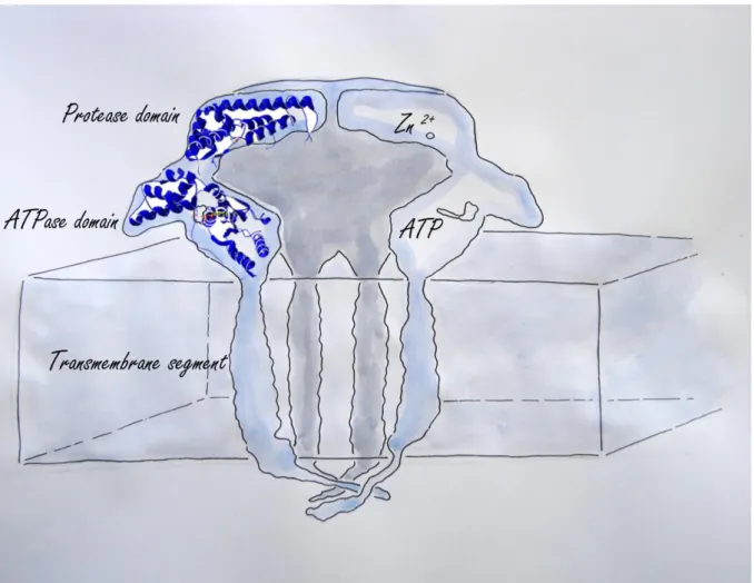
FtsH proteases are built of an N-terminal transmembrane segment and a C-terminal region containing an AAA-ATPase domain and a protease domain (Suno et al. 2006) (Figure 2). Due to the AAA domain, FtsH proteases are attributed to the family of AAA-proteins (ATPases associated with a variety of cellular activities (Patel and Latterich 1998) and like most members of this family they form hexameric complexes. AAA proteins are characterized by a highly conserved AAA cassette of about 200-250 residues, which contains the Walker A and B motifs necessary for nucleotide binding and hydrolysis and the so-called ‘second region of homology’ (SRH) that carries conserved arginine residues (“arginine finger”) important for oligomerization and nucleotide hydrolysis (Bieniossek et al. 2009, Ogura and Wilkinson 2001, Tomoyasu et al. 1993). From crystallographic studies it is known that the subunits of the FtsH complex are able to switch between an open and closed status. This is due to a structural change in the AAA domain driven by ATP hydrolysis. The conformational change leads to movement of a conserved hydrophobic area (FGV pore motif; Bieniossek et al. 2009) toward the inner proteolytic chamber. This region binds the substrate (Hinnerwisch et al. 2005, Park et al. 2005) and pulls it inside the proteolytic chamber.
9
Figure 2: Cross section through a hexameric FtsH complex. Each FtsH protease subunit is
built of a protease domain binding each a zinc ion, an ATPase domain necessary for transporting the substrate into the catalytic cave and a transmembrane segment with either one or two transmembrane helixes. The cave in the center is the site where substrate proteins are degraded.
The protease domain, C-terminal to the AAA domain, is categorized as zinc metallo-protease or zincin (Hooper 1994). The zinc ion is coordinated by three amino acids, which are the two histidine residues of the characteristic HEXXH motif and an aspartate. The glutamate (E) in the HEXXH motif serves as the catalytic base. The fourth coordination position of the zinc ion is taken by a water molecule (Figure 1). This activated water acts as a nucleophile that attacks the carbon of the peptide bond.
10
Figure 3: Bacterial FtsH hexamer assembled with another multimeric protein complex
composed of two highly homologous proteins HflC and HflK and the membrane insertase YidC (left). Homologs of HflC/HflK and the YidC can be found in plants; for the mitochondrial m-FtsH complex already an association with prohibitins (homologs of HflC/HflK) could be demonstrated (Piechota et al. 2010).
Newly synthesized membrane proteins incorporated into the membrane e.g. by the Sec translocon (right) have to be folded correctly in order to fulfill their function. If they cannot reach their native state they have to be removed. For this task biological membranes contain several proteolytic enzymes of which FtsH seems to be one of the more important.
Soluble proteins are also known to be substrates of FtsH. To degrade the soluble substrate σ32 (top) FtsH needs the assistance of the chaperones Dna70 and Dna40.
11
Besides functioning as protease, there is evidence that FtsH also acts as a molecular chaperone and is involved in protein assembly into and through the membrane (Akiyama et
al. 1994). Such ATP-dependent proteases with intrinsic chaperone activity are called
charonins (reviewed by Schumann 1999). The E. coli hexameric FtsH complex is part of a super-complex with molecular mass of around 1000 kDa, containing the proteins HflK and HflC (Saikawa et al. 2004) and YidC (van Bloois et al. 2008). HflK and HflC are homologs of prohibitins, which are membrane-bound chaperones (Nijtmans et al. 2000). Prohibitins have been shown to regulate membrane protein degradation by FtsH proteases (Steglich et al. 1999). YidC is a membrane insertase (Serek et al. 2004). Recently also in Arabidopsis mitochondria such a “High Molecular Weight Complex” consisting of FtsH and prohibitins has been observed (Piechota et al. 2010). In E. coli YidC is known to bind not only to FtsH, but also to the SecYEG translocon (Luirink et al. 2001), a physical interaction of FtsH and the SecYEG translocon might therefore exist. A close cooperation between FtsH and SecYEG during insertion of membrane proteins is possible, in which FtsH controls the quality of the newly inserted membrane proteins and additionally, by degrading SecY, also the amount of the newly inserted proteins.
3.4 FtsH proteases of Arabidopsis thaliana
The genome of Arabidopsis thaliana contains 12 genes coding for FtsH proteases (Garcia-Lorenzo et al. 2006). Based on their sequence identity several FtsH genes form homologous pairs pointing to recent gene duplication. Some FtsH proteases within these pairs have been shown to be able to substitute and complement each other in a complex, for example the FtsH 2/8 pair or the FtsH 1/5 pair, located in the thylakoid membrane of the plant chloroplast (Zaltsman et al. 2005, Zhang et al. 2010). Additionally to the twelve FtsH proteases
Arabidopsis contains five homologues, which not contain the zinc binding motif and therefore
probably are inactive (Sokolenko et al. 2002, Wagner et al. 2011a). Those proteins are termed FtsHi. Eight of the twelve FtsHs (FtsH 1, 2, 5-9, and 12), as well as the five inactive FtsHi are exclusively targeted to chloroplasts (Adam et al. 2006, Ferro et al. 2010); three FtsH proteases are located in mitochondria (FtsH 3, 4, and 10) (Janska et al. 2010). FtsH11 is double targeted to both organelles (Urantowka et al. 2005).
3.4.1 The thylakoid FtsH protease complex consisting of FtsH1, FtsH2, FtsH5 and FtsH8
In Arabidopsis the best studied FtsH proteases are the thylakoid located FtsH proteases, which were found to form a hetero-oligomeric hexameric complex (Zaltsman et al., 2005). Within
12
the complex the proteases are divided into two types (type A, FtsH1 and FtsH5; type B, FtsH2 and FtsH8, Garcia-Lorenzo et al. 2006, Zaltsman et al. 2005). The members of one type have sequence identity of about 90%, and can partly substitute each other in the complex. The identity between the members of type A and type B is close to 50%. Deletions of the higher expressed members of each type (FtsH5 and FtsH2) cause a variegated phenotype (Sakamoto
et al. 2003). Single mutants of the lower expressed subunits, FtsH1 and FtsH8, show no
phenotype. The best documented function of the thylakoid FtsH complex is the degradation of the photodamaged Photosystem II reaction center protein D1 (Bailey et al. 2002, Kato et al. 2009, Lindahl et al. 2000), but also other substrates have been reported for FtsH1, 2, 5 and 8, e.g. degradation of RieskeFeS (Ostersetzer and Adam 1997) in pea and regulation of cytochrome b6 levels in Chlamydomonas (Malnoe et al. 2011). A more detailed description of
the chloroplast located FtsH proteases can be found in our recent review (Wagner et al. 2011, Paper 5).
Because of its high sequence similarity also FtsH6 is grouped to the thylakoid FtsH proteases. However, neither a definite thylakoid location nor an accredited function is known for this protease. T-DNA knock-out mutants show no significant phenotype (Wagner et al. 2011b, Paper 1). Sequence analysis showed that FtsH6 homologous also exist in other plant species (Gracia et al., 2006). This evolutionary conservation indicates that FtsH6 is not just a non-sense remaining of gene duplication, but has a so far hidden task.
3.4.2 The chloroplast envelope located FtsH proteases
With four active and five inactive FtsH homologs (Ferro et al. 2010) the chloroplast envelope is the membrane compartment hosting most FtsH proteins. Of these proteases a function has only been reported for FtsH11 (Chen et al., 2006). We could show that FtsH11 knock-out mutants become continuous light sensitive plants (Paper 2) compared to the continuous light tolerant plant wild type. The earlier proposed heat intolerance (Chen et al. 2006) is part of this phenotype.
Not much is known about FtsH7, FtsH9 and FtsH12. FtsH7 and FtsH9 derived from recent gene duplication (Garcia et al. 2006), therefore it seems likely that these proteases have similar functions and they even might form a complex. Single deletion mutants of both proteases do not cause a phenotype.
FtsH12 does not have a close homolog in Arabidopsis, but due to their localization it might be possible that FtsH12 and FtsH11 have overlapping functions. Because of their differences in expression (Garcia et al. 2008) it seems unlikely that FtsH12 and FtsH11 form a complex,
13
however, performing different analyses we found that the amount of FtsH12 is increased in
ftsh11 (Paper 2 and 3), we therefore conclude that both proteases have one or more common
substrates. Unfortunately ftsh12 is embryo lethal, preventing detailed analysis of this mutant. The five inactive FtsHi most likely to not possess any protease activity, nevertheless they might be part of a proteolytic active FtsH complex. For FtsH2 it was shown that not all subunits of a proteolytic complex have to be active to maintain the function (Zhang et al. 2010). The expression data of some of the FtsHi and FtsH12 are similar furthermore some of the FtsHi knock-outs are also embryo defective, therefore one or several FtsHi might form a proteolytic complex with FtsH12.
3.4.3 The mitochondrial FtsH proteases
The mitochondrial FtsH proteases are divided in two groups (FtsH and i-FtsH). The m-FtsH proteases m-FtsH3 and m-FtsH10 are located in the inner envelope of mitochondria; their proteolytic domain faces the mitochondrial matrix (Kolodziejczak et al. 2002, Kwasniak et al. 2011). Single knock-outs of neither FtsH3 nor FtsH10 develop a phenotype under controlled conditions. Grown in the field reduced seed production and reddish leaves could be observed for ftsh10 (Wagner et al. 2011b). Recently it was shown that FtsH10 is associated with prohibitins similar to the yeast m-FtsH proteases (Piechota et al. 2010). Also the i-FtsH protease FtsH4 is located in the inner mitochondrial membrane, however, its active side faces the inter-membrane space (Gibala et al. 2009). Knock-out of FtsH4 causes altered leaf morphology at the late stage of rosette development when grown under short-day photoperiod (Kicia et al. 2010). In Arabidopsis FtsH11 is the closest homolog of FtsH4, it therefore is considered to be an i-FtsH too. The mitochondrial FtsH proteases have been reviewed recently in Kwasniak et al. (2011).
14
4 AIM OF THIS WORK
Higher plants like Arabidopsis thaliana contain multiple proteases of the FtsH family; especially the lower expressed members are not characterized very well. The aim of this thesis was to characterize the less studied members of the Arabidopsis FtsH protease family. After our field studies (Wagner et al., 2011b, paper1) FtsH11 became the main topic of this thesis. I wanted to identify the mechanism that causes the chlorotic phenotype of ftsh11. I assumed that a substrate is responsible for the phenotype therefore I used proteomic methods to detect accumulating proteins and their degradation products (peptides).
5 KEY-METHODS USED IN MY STUDIES
5.1 Darwinian fitness and phenotype characterization
Controlled laboratory growth conditions are essential for the reproduction of uniform plant materials. However, these conditions do not reflect the natural environment of plants. To find the real function of different proteins, it might be necessary to compare knock-out mutants with wild type plants under natural conditions. Umeå Plant Science Centre offers the unique possibility to perform field experiments with transgenic Arabidopsis plants. Darwinian fitness of mutants is measured by the amount of possible offspring produced by a plant grown under semi-natural field conditions (Frenkel et al. 2008; Wagner et al. 2011). T-DNA knock-out lines of the FtsH proteases were screened for homozygote knockout mutants. These lines were pre-grown in short day growth chambers. After one week they were transferred to the field where they produced seeds. Due to the permanently changing abiotic factors it is likely that a phenotype develops, which was not observed in controlled growth conditions resulting in a decrease seeds production. Since we work with GMOs (genetically modified organism) special precautions had to be taken. The fenced facility had to be isolated with plastic mats to prevent release of GMO material to the environment. To prevent insects from spreading pollen, the plants were grown under cages covered with transparent network. After the harvest, the entire material had to be removed from the field and was properly disposed.
5.2 Proteomics
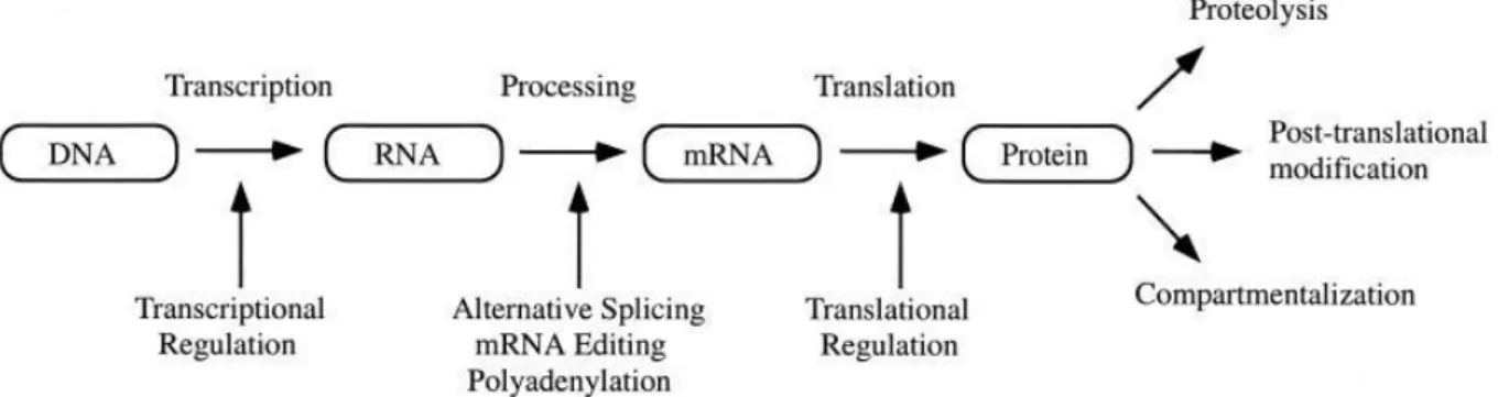
It has been shown that there often is little or no correlation between microarray data identifying mRNA abundance and the steady state protein levels (Gygi et al. 1999, Lian et al.
15
2002). This is due to several regulative steps that follow transcription ( Figure 4; Graves and Haystead 2002). In order to estimate the impact of a protein it is therefore necessary to know the amount, location (cell compartment), and the activity of this protein. In this work I used various proteomic methods to detect changes in the proteome of FtsH protease deficient mutants. Furthermore I searched for fragments of substrate proteins (limited proteolysis).
Figure 4: Mechanisms determining amount and impact of a protein (Graves and Haystead 2002)
5.2.1 The gel-based approach
To receive sufficient electrophoretic separation of a proteome usually two-dimensional electrophoresis (2DE) is applied; in the first dimension I separated proteins either according to their isoelectric point (isoelectric focusing) or according to the molecular mass of their native complexes (blue-native electrophoresis). In the second dimension co-migrating proteins were then separated according to their molecular mass.
5.2.1.1 2-dimensional isoelectric focusing/SDS polyacrylamide gel electrophoresis (2D-IEF-PAGE)
The principle of electrophoresis is the motion of charged particles (ions) in an electric field. Proteins contain amino acids with pKa values in a range from 3 to 12. The pKa value is the negative logarithm of the acid dissociation constant, it determines the pH at which the probability for a side chain to be charged is 50%. The charge of a protein therefore depends
16
on the pH of its surrounding medium. At a certain pH, the isoelectric point (pI), the number of positive and negatively charged residues will be equal, the protein will therefore not carry an electrical net-charge. In 2D-IEF-PAGE protein mixtures are loaded on immobilized pH gradient strips (IPG) and voltage is applied. The proteins are separated according to their pI, differences of 0.001 pI units are possible to detect (Issaq and Veenstra 2008). Ready-to-use immobilized pH gradient (IPG) strips guarantee high reproducibility of the separation. These IPGs are available in several different pH ranges and they are simple to use.
To separate the proteins in a second dimension, the IPGs are treated with SDS and applied on a polyacrylamide gel. SDS is an anionic detergent denaturing proteins by disrupting their non-covalent bonds and adding negative charge to them. By this treatment all proteins obtain the same mass-charge ratio that allows separation according to their molecular mass. Depending on the sample and on the staining method a 2D-IEF-PAGE will typically resolve up to 3000 spots (Lilley et al. 2002), which is by far lower than the expected number of proteins within a cell. The concentration of low abundant proteins might be too low to be visualized, or they might be hidden below very abundant proteins. Sub-fractionation of samples can help to capture lower abundant proteins. In my work I have biochemically isolated and analyzed mitochondria, chloroplasts, chloroplast stroma sample and chloroplast envelope membranes from Arabidopsis.
2D-IEF-PAGE has the advantage that it is quite simple to execute and does not require expensive equipment. However, using this method one should keep in mind that hydrophobic, extreme basic or extreme acidic proteins are not resolved well by IEF (1st dimension). Additionally, proteins smaller than 10 kDa or larger than 150 kDa are not resolved in the SDS PAGE (2nd dimension) (Kearney and Thibault 2003, Mann et al. 2001).
5.3.1.2 2-dimensional Blue Native/SDS polyacrylamide gel electrophoresis (2D-BN-PAGE)
In contrast to IEF, native gel electrophoresis allows the separation of hydrophobic proteins and additionally provides information about the composition of protein complexes. Blue Native PAGE is the most popular native gel electrophoretic method. In the first dimension the sample is solubilized with mild non-ionic detergents and receives negative charge due to addition of the dye Coomassie Blue G250. The intention of this method is to detach intact protein complexes from the surrounding membranes and separate them according to their size (Eubel et al. 2005). In the second dimension the protein complexes are disrupted and denatured by treatment with SDS and reducing agents like 2-mercaptoethanol. The complex
17
subunits are then resolved on a polyacrylamide gel as described above (3.3.2.1). Unfortunately single proteins are with this method insufficiently separated. 2D-BN-PAGE is therefore not suitable to resolve a complete proteome, but it can complement 2D-IEF-PAGE for the analysis of membrane proteins. It mainly is applied in research addressing large membrane complexes like the photosynthetic electron transport chain complexes of the thylakoid membrane, or the mitochondrial complexes of the respiratory electron transport chain.
To identify proteins of interest, gel pieces from 2D PAGE as well as from BN PAGE are excised and proteolytic digested, the resulting peptides are extracted and analyzed by mass spectrometry. The two most commonly used techniques for protein characterization are peptide mass fingerprinting- (PMF) and tandem-MS (mass spectrometry).
5.3.2 The chromatography approach (gel free proteomics)
While the gel-based proteomic method has the advantage to be able to separate complex samples (different splicing forms, not fully degraded breakdown products), it also has the above mentioned limitations. A gel-free approach is less impaired by those features and has higher protein coverage. It also can be largely automated and therefore is less time consuming than gel-based proteomics. However, it requires expensive equipment (e.g. tandem-MS and HPLC instruments). In contrast to gel-based proteomics the proteins are digested with a protease (e.g. trypsin) before their separation. The resulting peptide mix is then fractionated by reversed phase (RP) HPLC and the eluate is analyzed on-line in an electrospray-ionization (ESI) tandem-MS. However, in complex peptide mixtures co-elution is a limiting factor for the number of peptides that can be identified. A chromatographic peak is only available for a short time frame, in which an MS as well as several MS/MS spectra have to be acquired (Baggerman et al. 2005). Furthermore very abundant peptides can suppress the signal of less abundant ions (ion suppression) (Hirabayashi et al. 2007). Therefore, in the last years several techniques have been developed to overcome this limitation. The goal is to accomplish a better peptide separation and a reduced sample complexity. Two of those methods, which I have used, are MudPIT (multidimensional protein identification technology) and COFRADIC (combined fractional diagonal chromatography)
5.3.2.1 MudPIT
Increased separation of peptides can be accomplished by combining the RP-HPLC with a second liquid chromatography step. This technique is called MudPIT. It uses a strong cationic
18
exchange (SCX) column in front of a RP-column (Peng and Gygi 2001). The SCX-column in the first dimension binds positivly charged peptides (cations). Uncharged peptides pass through and bind to the RP-column; they are eluted with a gradient. Peptides bound to the SCX are stepwise eluted into the RP-column with increasing salt concentrations. An advantage of MudPIT is that the salt ions of the SCX-fractions, which would interfere with the MS analysis, are removed in the RP step (Lin et al. 2003).
5.3.2.2 COFRADIC
In this method two identical RP-HPLC peptide separations are executed, but before the second RP-HPLC separation a specific modification reaction (sorting step) is performed. Peptides that remain unchanged after the sorting step elute at identical positions in the two chromatographic separations, whereas the modified peptides segregate from the unchanged peptides and elute in either earlier or later fractions (Gevaert et al. 2007). The method can be used to select N- or C-terminal peptides or peptides containing certain amino acids e.g. methionine or cysteine. Furthermore the method can be used for sorting peptides carrying post translational modifications.
5.3.3. Quantitative proteomics
An approved gel-based quantitative proteomic method is DIGE (Differential Gel Electrophoresis). Highly sensitive fluorescent dyes are bound covalently to ε-amino groups of proteins. After labeling the samples (e.g. control and analyte) and an internal standard with different dyes, they are pooled and applied to one 2D gel, in this way gel-to-gel variation is eliminated. Gel images of each labeled sample are acquired on a multi wavelength scanner and analyzed with image analysis software. In my work I applied refraction-2D, a method identical to DIGE, but using different fluorescent labeling dyes.
Gel free quantitative methods compare the amount of peptides. Several labeling procedures have been developed (iTRAQ, ICAT, SILAC). These methods use the ability of mass spectrometry to distinguish between different isotopes. Peptides can contain isotopic labeled amino acids (SILAC) or can be covalently labeled with tags (iTRAQ, ICAT) containing different isotopes. In my work I used a labeling method, which is similar to iTRAQ, however, I used different isotopic labeling compounds.
19
6 RESULTS
6.1 Field studies on FtsH knock-out mutants
For our field studies we used single knock-out mutants of FtsH2, 3, 4, 5, 6, 7, 8, and 11, as well as a knock-down mutant of FtsH10 (Wagner et al., 2011b, Paper 1).
The mutants ftsh3, ftsh6, ftsh7 and ftsh8 did not show any phenotype, neither when grown indoor nor outdoor. FtsH3 as well as FtsH8 are known to form complexes with their homolog partners, respectively, that seem to be able to compensate for the loss of those proteins. FtsH7 and FtsH9 also form a homolog pair, however, neither function nor complex formation of these proteins is known so far. Three independent knock-out lines of ftsh6 were examined, neither of those showed a phenotype.
Ftsh2 and ftsh5 showed dwarf growth and the well-known variegated phenotype of indoor
plants was enhanced when grown in the field, so that almost no green leaf areas where left. In the end the plants did not survive.
Ftsh10 developed smaller rosettes with a reddish color. This phenotype was surprising as this
mutant did not display any phenotype under controlled growth conditions. FtsH10 seems to be important during the early phase of plant development, as plants, which were pre-grown in the greenhouse for a longer period did not develop any phenotype.
Ftsh4 plants grown in short day conditions develop morphological leaf disturbances at the age
of 8 weeks (Gibala et al. 2009). Ftsh4 grown under field conditions developed no phenotype (Aigner, Smakowska, Janska and Funk, unpublished result), probably due to the long daily photoperiod in this nordic climate, which led the plants flower much earlier, so that they did not reach the necessary age. FtsH4 might therefore be important during certain plant developmental stages.
Unexpected was the phenotype of ftsh11 grown in the field. The plants became chlorotic, showed dwarf growth and developed very few seeds with decreased germination rate. Ftsh11 develops no phenotype when grown under short day conditions at moderate temperatures. At the time of our field studies ftsh11 was known to develop chlorotic leaves when exposed to high temperatures (Chen et al. 2006); however, during our experiment temperatures at the field site usually were around 20oC during day time and never exceeded 30oC. As seedlings and young plants were described to be more vulnerable towards heat (Chen et al. 2006), we also tested ftsh11 pre-grown in short day conditions for 3 weeks, to challenge stronger, older plants with outdoor conditions. Also those plants developed chlorosis.
20
6.2 FtsH11
6.2.1 Phenotype of ftsh11
To investigate whether the permanently changing conditions or rather a certain abiotic stress induced the leaf bleaching, Arabidopsis wild type and ftsh11 plants were grown under controlled conditions and exposed to different light intensities, day lengths and temperatures. By evaluating these experiments we were able to classify the phenotype of ftsh11 as continuous light sensitive. Species like tomato, eggplant or cucumber exhibit such a phenotype even in their wild type state. Those continuous light intolerant species develop leaf injuries (chlorosis, necrosis) if transferred to continuous light, their leaves have been shown to have an increased sucrose content and a lower maximum rate of RuBisCO carboxylation. Increasing temperatures enhance the leaf damage whereas low temperatures can suppress the phenotype.
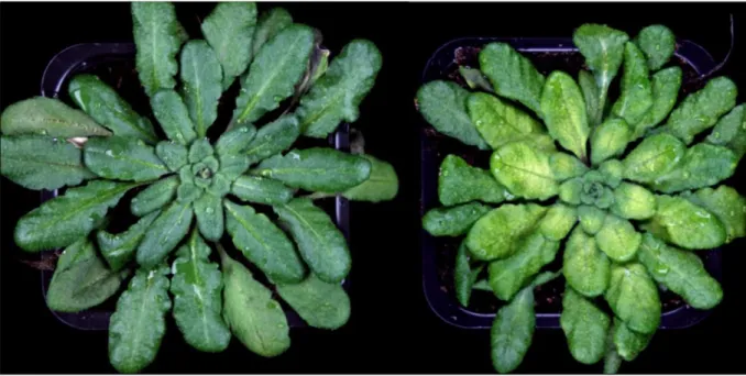
Figure 5: Seven week old Arabidopsis plants (wild type left, ftsh11 right) after three days of
continuous light. A decrease in chlorophyll content (chlorosis) is clearly visible in the mutant.
Ftsh11 developed strong chlorosis if transferred to continuous light at 22 °C. Increased
temperature enhanced the chlorophyll-loss, whereas lower temperatures decrease it. Heat stress performed in darkness did not cause chlorosis. A (weaker) phenotype could also be produced, when the photoperiod was extended from 8 h to 12 h or 16 h. At a photoperiod of
21
12 h the light intensity had to be raised from 100 to 250 µmol photons m-2 s-1 to induce chlorosis in ftsh11, indicating a synergetic effect of the light intensity. However, experiments with increased light intensities up to 500 µmol photons m-2 s-1 in short day (8 h) did not induce the phenotype. The necessity of extended light periods to develop chlorosis could indicate that a harmful factor accumulates during the light period, e.g. impaired carboxylation activity by RuBisCO (Pettersen et al. 2010, van Gestel et al. 2005) could lead to an accumulation of reduction equivalents (NADPH) and in further consequence to over-reduction of the photosynthetic electron transport chain (PETC). This in its turn would lead to increased ROS production and further to chlorosis. We therefore examined the protein damage caused by ROS in chloroplasts quantifying the amount of carbonylated proteins and detected increased protein carbonylation in ftsh11 if grown in long day conditions, but normal levels when grown in short day conditions.
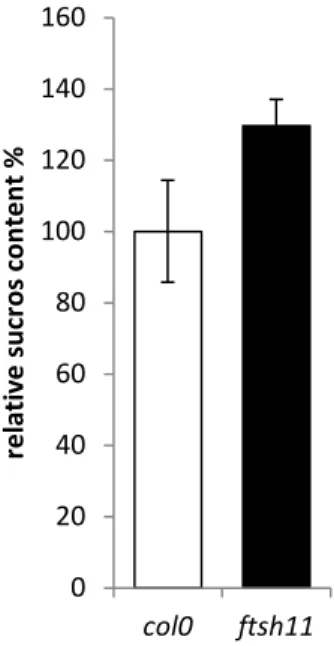
Some natural continuous light sensitive species accumulate increased levels of leaf sugars when grown under continuous light. We could show that ftsh11, grown in short day conditions, has a sucrose level that is equal to WT. After 24 hours continuous light (100 µmol photons m-2 s-1)the sucrose level in ftsh11 is increased by about 30 % relative to WT (Figure 6).
Figure 6: after 24 h continuous light is the sucrose content in ftsh11 leaves relative to wild
type (col0) increased.
0 20 40 60 80 100 120 140 160 col0 ftsh11 rel ati ve suc ros co n te n t %
22
Already in the middle of the last century continuous light injury has been suspected to be linked to the circadian clock (Highkin and Hanson 1954). However, ftsh11 shows no impairment of the circadian rhythm. The daily rhythm of seedling leaf movement and chlorophyll auto fluorescence in adult plants did not differ between ftsh11 and WT (data not shown).
We noticed a decrease of anthocyanin accumulation in ftsh11 plants exposed to continuous light. To determine whether the decreased anthocyanin accumulation is associated with the chlorotic phenotype, or if it can develop independently, we induced anthocyanin accumulation with cold temperatures in short day. Under those conditions ftsh11 accumulated less anthocyanin, but did not show chlorosis (Paper 4).
Electron microscopy images were taken of wild type and ftsh11 chloroplasts. No obvious differences in the chloroplast ultra-structure were detected in plants grown under normal growth conditions. Under continuous light, ftsh11 chloroplasts were reduced in size, had large grana stacks and only very few stroma lamellae. The stroma thylakoids seemed to be separated from the grana thylakoids, while the grana thylakoids seemed to lose structure; the plastoglobuli increased in number. The degree of damage and also the number of damaged chloroplasts increased with increasing temperatures (2012, Paper 2).
6.2.2 Proteomic studies on ftsh11
Ftsh11 develops chlorotic leaves suggesting a reduction of the photosynthetic complexes.
This hypothesis was confirmed by the electron microscopy images indicating changes of the chloroplast morphology, which inevitably were accompanied by severe changes of the proteome. Those differences, however, were most likely caused indirectly, by the damage induced due to increased ROS level and not directly by the absence of the FtsH11 protease. Therefore we decided to analyze unstressed ftsh11 grown under short-day conditions using the proteomic approach.
6.2.2.1 Refraction-2D
In three independent biological replicates chloroplasts from three to four week old ftsh11 and WT (Col0) plants were isolated and compared by refraction-2D. Eighteen protein spots showed changed intensity and could be identified by mass spectrometry (a table of all identified proteins can be found in Paper 2), in total eleven different proteins were shown to change in abundance; all of them had increased level in ftsh11. The large subunit of RuBisCO (RBL) and RuBisCO activase each were identified in three different spots. RBL is known to
23
appear in several spots on standard 2D gels (SWISS-2DPAGE, Sarazin et al. 2000). Some spots migrate in the low molecular weight range and seem to be break-down products. In my gels approximately 5 to 7 spots, with a molecular mass of the mature protein, form a so called charge-train. Charge-trains are different forms of a protein with variation in its pI, either caused by post-translational modifications or by reactions of charged amino acids (mainly lysine) with carbamic acid (a break-down product of urea).
RuBisCO activase, the other enzyme identified in three spots, is known to remove inhibitors from the active side of RuBisCO and thereby activates it (Stotz et al. 2011). However, in
ftsh11 I could neither determine increased RuBisCO activity nor an increase of activated
reactive sites per RuBisCO. Since RuBisCO is present in higher amounts in ftsh11 it seems that a part of the enzyme is non-functional or damaged.
Four other enzymes acting in the Calvin cycle were shown to be present in higher amounts in
ftsh11: phosphoglycerate kinase, glyceraldehyde-3-phosphate dehydrogenase, phosphoribulokinase and sedoheptulose-1,7-bisphosphatase. These proteins might be up-regulated in response to limited flux through the Calvin pathway.
Another protein increased in ftsh11 was the GTP-binding translation elongation factor (2 spots); a homolog of this protein in Zea mais has been shown to have a stabilizing effect on RuBisCO activase under high temperatures (Ristic et al. 2007). Furthermore the plastidic chaperones HSP70-1 and HSP70-2 and the proteins “aberrant growth and death 2” and GS2 (glutamine synthetase 2) had increased abundance in ftsh11 (table 1 in Paper 2).
Some of the above described proteins belong to the most abundant soluble proteins in the chloroplast stroma. To exclude the possibility that only the preparation method (harvest of intact chloroplasts) caused the observed differences in protein amount, I repeated the refraction-2D experiment with purified stroma sample. Fourteen protein spots with increased intensity could be identified, 11 of those corresponded to proteins that already were identified in the chloroplast refraction-2D experiments (or homologues of them with a very high sequence identity: Protein identification was performed by peptide mass fingerprinting, which cannot discriminate between proteins with high sequence identify with absolute certainty). Proteins that had not been observed in the total-chloroplast sample were chaperonin 60β, ADP-glucose pyrophosphorylase (ADG1) catalyses a prime regulatory step in the synthesis of starch in plants (Ballicora et al. 1998), and phosphoglucomutase (also involved in platidic starch synthesis (Kofler et al. 2000)). Additionally in the stroma sample 8 protein were identified with decreased amount in ftsh11 compared to WT. Five of them are known to be located in the thylakoid lumen, they are either part of the oxygen evolving complex of
24
Photosystem II (PSBO-1, PSBO-2 and PSBP-1) or have a function in assembly and maintenance of it (HCF136 and cyclophilin 38). The observed decrease might be caused by deletion of FtsH11 (we could observe a decrease of photosynthetic proteins in ftsh11 compared to WT, Paper 2) or might only be to the results of a higher contamination of lumen proteins in the wild type samples. The remaining three proteins with decreased amounts in
ftsh11 compared to WT were ferredoxin-NADPH oxidoreductase, beta-glucosidase 37 and the
33 kDa ribonucleoprotein.
As mentioned above FtsH11 is dual targeted to chloroplasts and mitochondria (Urantowka et
al. 2005), however, comparing the mitochondrial proteome of wild type and ftsh11, we were
not able to detect any significant differences.
6.2.2.2 Gel-free proteomics
The plastidic FtsH11 is located in the chloroplast envelope (Ferro et al. 2010, Paper 2), inserted by membrane-spanning helices. Proteins with hydrophobic, membrane-spanning domains are difficult to examine by IEF-2D electrophoresis, we therefore examined purified chloroplast envelope using gel-free protein quantification (Paper 3). N-hydroxy-succinimide propionate was used to label peptides of isolated chloroplast envelope. ftsh11 was labeled with propionate containing three 13C isotopes, the propionate label of the WT sample contained only 12C. With this method 1094 peptides were detected, 487 of those peptides (216 proteins) are listed in the chloroplast envelope database AT_CHLORO. As the samples did not contain equal amounts of envelope (due to varying contaminations with other compartments) the ratio had to be recalculated based on enzymes with known envelope localization. Using the corrected ratio we found that only the FtsH12 protease was significantly up-regulated in ftsh11. Several other proteins with increased amount in ftsh11 compared to wild type had to be discarded as they were (also listed by AT_CHLORO) not located in the chloroplast envelope. Their higher abundance in one of the samples was only caused due to different levels of contamination (mainly proteins located in tonoplast or ribosomal proteins). Three different peptides of FtsH12 were identified with an average increase of 1.98 relative to wild type. The increase of FtsH12 in ftsh11 was verified with specific antibodies directed against FtsH12 (Paper 2). I assume that FtsH11 is a protease involved in degrading damaged proteins from the chloroplast envelope. FtsH12 might, to some extent, take over this function, however, I do not believe that both proteases are part of the same complex, because they are expressed differently in plants (Garcia et al, 2008).
25
In a second approach to identify the FtsH11 substrate COFRADIC was applied on leaf extracts of ftsh11 and WT. Degradation products of proteins usually have a short lifetime and will be present only in low amounts. While a gel-free MS/MS approach can detect peptides in very low concentration, the high number of tryptic peptides will most likely repress the signal of interest. COFRADIC provides a way to separate peptides generated by trypsin from peptides containing original N-termini and neo-N-termini generated by the proteases present in the cell. Therefore one should be able to identify in the wild type leaf extract the N-terminal peptides generated by FtsH11, whereas in the ftsh11 sample this peptides should be missing. Growth conditions of Arabidopsis ftsh11 and WT plants were the same as described before (refraction-2D experiments); ftsh11 leaf extract was labeled with butyrate containing 4
12
C, butyrate to label the wild type sample contained 4 13C. 1305 peptides belonging to 763 proteins could be identified.
Seven peptides were only found in the wild type sample, these peptides can be the directly result from proteolytic events by the FtsH11 protease or the result of another protease that needs FtsH11 for its activation (Paper 3). Five of those proteins have been identified earlier in the refraction-2D experiment: three chaperones (two HSP70 and a chaperonin 60), RBL and ADP-glucose-pyrophosphorylase (ADG). The identification of RBL and ADG as a substrate is interesting, both could influence the activity of the Calvin cycle and so be the trigger of the chlorotic phenotype (Paper 2 and 3).
Eight neo-N-terminal peptides were found to only occur in the ftsh11 sample. Such peptides that only are detected in the protease knock-out mutant might be the result of another protease activity that is normally inhibited by FtsH11. The full list of these peptides can be found in Paper 3. Particularly interesting was the finding of two proteins (AOC1 and AOC2) involved in JA (jasmonic acid) biosynthesis (Schaller et al. 2008). JA is involved in regulation of stomata closure (Hossain et al. 2011). I could demonstrate that ftsh11 has decreased transpiration rate (Paper 3). The higher resistance of ftsh11 to drought might be a consequence of increased JA levels caused by impaired AOC degradation.
6.3 NADP+/NADPH ratio in ftsh11 is changed in continuous light
The amount of Calvin cycle enzymes was found to be increased in ftsh11 showing the endeavor of the plant to increase the activity of this cycle. In the photosynthetic process the Calvin cycle uses ATP and NADPH as energy source and reductions equivalents, which are generated during the light reaction from ADP and NADP+. Measurement of the NADP+/NADPH ratio in leaves from wild type and ftsh11 exposed to continuous light for 24
26
h showed a significant increase of the reduced form and a decrease of the oxidized form in
ftsh11 relative to wild type.
6.4 Complex partner of FtsH11
FtsH proteases are known to function in homo- (Akiyama 2009) or hetero-oligomeric hexamers (Adam et al. 2006), which, at least in E. coli and mitochondria, form high molecular weight super-complexes with prohibitins and YidC (van Bloois et al. 2008). Performing blue native gel electrophoresis on an enriched chloroplast envelope fraction, followed by immunodetection we could detect FtsH11 and FtsH12 in complexes with molecular mass of approximately 2000 kDa. Unfortunately the protein amount of these complexes was not sufficient to be identified by mass spectrometry. An enrichment of the complex using gel filtration was not successful.
6.5 Results on other FtsH proteases
6.5.1 Modeling approach
To investigate if other FtsH proteases are able to form hetero-oligomeric hexamers with their homologue partners I performed modeling studies. I observed that amino acids that hypothetically form contact surfaces between the subunits are better conserved than amino acids situated outwards of the complex. Therefore I assumed that amino acids at the contact surfaces are more protected from amino acid substitution, in order to not disturb the binding site. Based on the conservation of these contact surfaces I tried to predict complex formation of several FtsH proteases. ”Swiss Model Server” (http://swissmodel.expasy.org) was used to create model structures of all Arabidopsis FtsH proteases. As expected I could show that the contact surfaces of FtsH1 and FtsH5 as well as FtsH2 and FtsH8 have almost perfectly conserved contact surfaces; these proteases are known to substitute for each other in a complex (Zaltsman et al., 2005). The amino acid exchanges of these proteases are almost exclusively located at the outside of the complex or toward the proteolytic cave. Also FtsH subunits of a complex that have a lower sequence identity (e.g. FtsH2 and FtsH5) are conserved better at the contact surfaces, however the same is true for subunits that do not form complexes (e.g. FtsH2 and FtsH3). A prediction of complex-formation on basis of conserved contact surfaces was therefore not possible.
27
6.5.2 FtsH7 and FtsH9
Although the modeling approach did not deliver the expected results, I discovered an interesting detail about the amino acid substitution between the homologous pair FtsH7 and FtsH9. These two proteins are located in the chloroplast envelope. Their sequence has a very high identity, which attests recent gene duplication. Surprisingly, 36 % of all amino acids changes of those two proteins involve a serine. Alone in the part used for modeling (protease domain and ATPase domain, Paper 4) FtsH9 has 10 amino acid substitutes towards serine; several of those are predicted phosphorylation sides. The advantage of having two highly similar proteins in the same membrane system might therefore be explained by regulation via phosphorylation. Other homologous FtsH pairs did not show such a high serine exchange rate.
6.5.3 FtsH3 and FtsH10
Also FtsH3 and FtsH10 are proteases with high similarity. They are located in the inner membrane of mitochondria with their catalytic domain oriented toward the mitochondrial matrix (m-AAA proteases) (Arnold and Langer 2002). FtsH3 and 10 form hetero-oligomeric hexamers, but may also be able to form homo-hexameric complexes (Piechota et al. 2010). Superimposing their sequence I detected very few amino acid substitutions in the intra-complex surface areas of the monomers, whereas the outside of the intra-complex seemed to be less conserved (Paper 4).
DIGE experiments showed a reduction of porin in mitochondria of ftsh10 compared to wild type; furthermore two spots of succinyl-CoA ligase were changed in amount. The spot migrating in the acidic region was reduced, whereas the spot with more basic pI was increased in amount in ftsh10. No changes in protein amount were detected in ftsh3 compared to wild type. However, immunodetection indicated that in ftsh3 the amount of FtsH10 was increased (Piechota et al. 2010), indicating that FtsH10 is able to replace FtsH3. Our results support this hypothesis as we could neither find a phenotype of ftsh3 nor any up or down regulation of proteins in the proteome of ftsh3 using DIGE. In ftsh10, which only is a knock-down mutant, we also observed a phenotype when the plants were grown in the field (see 4.1 Field studies on FtsH knock-out mutants and Paper 1)
28
7 CONCLUSIONS
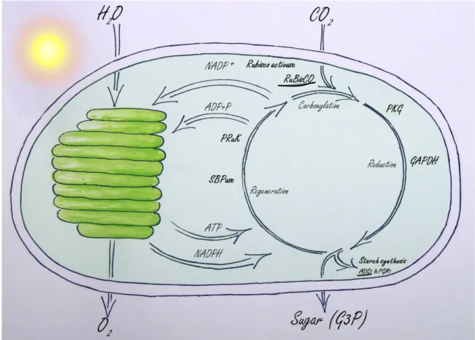
I showed that Arabidopsis ftsh11 develops a phenotype similar to natural continuous light sensitive plants. This makes the ftsh11 mutant to a valuable tool to study sensitivity to long photoperiods. Furthermore I can propose a model explaining the mechanism of photobleaching in ftsh11 (Figure 7). A slightly impaired Calvin cycle in ftsh11 leads during long photoperiods to an accumulation of NADPH and to a reduced amount of NADP+. NADP+ is the electron acceptor of Photosystem I and its shortage causes over reduction of the electron transport chain and increased ROS production. ROS damages the photosynthetic proteins and induces chlorosis. I also have identified several possible substrates of FtsH11, among those RBL and ADG. Both enzymes could if their function is impaired cause slow down of the Calvin cycle and in this manner be responsible for the observed phenotype.
Figure 7: Proposed mechanism leading to the chlorotic phenotype in ftsh11. In ftsh11 the
Calvin cycle (right) works at a slightly reduced level generating a shortage of NADP+, the electron acceptor of PSI. This causes over-reduction of the photosynthetic electron transport
29
chain in the thylakoids (left) and ROS production, which damages the photosynthetic apparatus. Enzymes written in bold were found at higher abundance in ftsh11. The large subunits of RuBisCO and ADG (underlined) were additionally detected to be a substrate for FtsH11 in the COFRADIC assay.
8 OUTLOOKS
In my work I was able to detect several possible substrates of FtsH11 that could explain the
ftsh11 phenotype. These substrates have to be confirmed with independent methods and the
proposed impact on the phenotype has to be assayed, e.g. RuBisCO activity after increased photoperiods or measurements of the reductions state of components of the PETC.
The identification of other proteins forming a complex with FtsH11 is of highest importance as well as the determination of the activity of such FtsH11 complexes in vivo. Using tandem-affinity purification to isolate tagged FtsH11-complexes will be an adequate tool. Protease activity could be assayed using activity-based probes that react with the catalytic site of proteases in a mechanism-dependent manner (van der Hoorn and Kaiser 2011).
Artificial light is an important tool in crop production especially in winter in the northern countries. For economic and technical reasons long light periods with relatively low light intensity are preferential used, however several important crop species e.g. eggplant (Murage
et al. 1997) and tomato (Demers et al.1998) do not grow well under such conditions. The
problems range from impaired carbon fixation rates (Van Gestel et al. 2005) to serious leaf injuries like chlorosis (Pettersen et al. 2010) and necrosis (Cushman and Tibbitts 1998). We will gain better understanding of intolerance to long photoperiods by resolving the mechanism that transfers Arabidopsis from a continuous light tolerant species to a continuous light intolerant ftsh11 plant. In the future my data and the ongoing research in our lab could lead to targeted breeding of continuous light tolerant crops with increased carbon fixation capacity under artificial light.
30
9 REFERENCE LIST
Adam, Z., Rudella, A. and van Wijk, K.J. (2006) Recent advances in the study of Clp, FtsH
and other proteases located in chloroplasts. Current opinion in plant biology, 9, 234-240.
Akiyama, Y. (2009) Quality control of cytoplasmic membrane proteins in Escherichia coli.
Journal of biochemistry, 146, 449-454.
Akiyama, Y., Kihara, A. and Ito, K. (1996) Subunit a of proton ATPase F0 sector is a
substrate of the FtsH protease in Escherichia coli. FEBS letters, 399, 26-28.
Akiyama, Y., Ogura, T. and Ito, K. (1994) Involvement of FtsH in protein assembly into
and through the membrane. I. Mutations that reduce retention efficiency of a cytoplasmic reporter. The Journal of biological chemistry, 269, 5218-5224.
Arnold, I. and Langer, T. (2002) Membrane protein degradation by AAA proteases in
mitochondria. Biochimica et biophysica acta, 1592, 89-96.
auf dem Keller, U., Doucet, A. and Overall, C.M. (2007) Protease research in the era of
systems biology. Biological chemistry, 388, 1159-1162.
Baggerman, G., Vierstraete, E., De Loof, A. and Schoofs, L. (2005) Gel-based versus
gel-free proteomics: a review. Combinatorial chemistry & high throughput screening, 8, 669-677.
Bailey, S., Thompson, E., Nixon, P.J., Horton, P., Mullineaux, C.W., Robinson, C. and Mann, N.H. (2002) A critical role for the Var2 FtsH homologue of Arabidopsis thaliana in
the photosystem II repair cycle in vivo. The Journal of biological chemistry, 277, 2006-2011.
Ballicora, M.A., Fu, Y., Nesbitt, N.M. and Preiss, J. (1998) ADP-Glucose
pyrophosphorylase from potato tubers. Site-directed mutagenesis studies of the regulatory sites. Plant physiology, 118, 265-274.
Barrett, A.J., Rawlings, N.D. and Woessner, J.F. The Handbook of Proteolytic Enzymes,
2nd ed. Academic Press, 2003. ISBN 0-12-079610-4
Begg, K.J., Tomoyasu, T., Donachie, W.D., Khattar, M., Niki, H., Yamanaka, K., Hiraga, S. and Ogura, T. (1992) Escherichia coli mutant Y16 is a double mutant carrying
thermosensitive ftsH and ftsI mutations. J Bacteriol 174: 2416-2417
Bieniossek, C., Niederhauser, B. and Baumann, U.M. (2009) The crystal structure of
apo-FtsH reveals domain movements necessary for substrate unfolding and translocation.
Proceedings of the National Academy of Sciences of the United States of America, 106,
31
Blaszczak, A., Georgopoulos, C. and Liberek, K. (1999) On the mechanism of
FtsH-dependent degradation of the sigma 32 transcriptional regulator of Escherichia coli and the role of the Dnak chaperone machine. Molecular microbiology, 31, 157-166.
Callis, J. (1995) Regulation of Protein Degradation. The Plant cell, 7, 845-857.
Chen, J., Burke, J.J., Velten, J. and Xin, Z. (2006) FtsH11 protease plays a critical role in
Arabidopsis thermotolerance. The Plant journal : for cell and molecular biology, 48, 73-84.
Cushman, K.E. and Tibbitts, T.W. (1998) The role of ethylene in the development of
constant-light injury of potato and tomato. Journal of the American Society for Horticultural
Science. American Society for Horticultural Science, 123, 239-245.
Demers, D.A., Dorais, M., Wien, C.H. and Gosselin, A. (1998) Effects of supplemental
light duration on
greenhouse tomato (Lycopersicon esculentum Mill.) plants and fruit yields. Sci. Hortic. 74, 295–306
Eubel, H., Braun, H.P. and Millar, A.H. (2005) Blue-native PAGE in plants: a tool in
analysis of protein-protein interactions. Plant methods, 1, 11.
Ferro, M., Brugiere, S., Salvi, D., Seigneurin-Berny, D., Court, M., Moyet, L., Ramus, C., Miras, S., Mellal, M., Le Gall, S., Kieffer-Jaquinod, S., Bruley, C., Garin, J., Joyard, J., Masselon, C. and Rolland, N. (2010) AT_CHLORO, a comprehensive chloroplast
proteome database with subplastidial localization and curated information on envelope proteins. Molecular & cellular proteomics : MCP, 9, 1063-1084.
Feller, U. (2004) Proteolysis. In: Noodén LD, editor. Plant cell death processes. San Diego:
Academic Press. p. 107-123.
Frugis, G. and Chua, N.H. (2002) Ubiquitin-mediated proteolysis in plant hormone signal
transduction. Trends in cell biology, 12, 308-311.
Garcia-Lorenzo, M., Pruzinska, A. and Funk C (2008) ATP-dependent Proteases in the
Plant Chloroplast. In E Kutejova, ed, ATP-dependent proteases, Research Signpost, Kerala,
India, pp 145-176
Garcia-Lorenzo, M., Sjodin, A., Jansson, S. and Funk, C. (2006) Protease gene families in
Populus and Arabidopsis. BMC plant biology, 6, 30.
Gevaert, K., Impens, F., Van Damme, P., Ghesquiere, B., Hanoulle, X. and Vandekerckhove, J. (2007) Applications of diagonal chromatography for proteome-wide
32
Gibala, M., Kicia, M., Sakamoto, W., Gola, E.M., Kubrakiewicz, J., Smakowska, E. and Janska, H. (2009) The lack of mitochondrial AtFtsH4 protease alters Arabidopsis leaf
morphology at the late stage of rosette development under short-day photoperiod. The Plant
journal : for cell and molecular biology, 59, 685-699.
Graves, P.R. and Haystead, T.A. (2002) Molecular biologist's guide to proteomics.
Microbiology and molecular biology reviews : MMBR, 66, 39-63; table of contents.
Gygi, S.P., Rochon, Y., Franza, B.R. and Aebersold, R. (1999) Correlation between protein
and mRNA abundance in yeast. Molecular and cellular biology, 19, 1720-1730.
Herman, C., Thevenet, D., D'Ari, R. and Bouloc, P. (1995) Degradation of sigma 32, the
heat shock regulator in Escherichia coli, is governed by HflB. Proceedings of the National
Academy of Sciences of the United States of America, 92, 3516-3520.
Highkin, H.R. and Hanson, J.B. (1954) Possible Interaction between Light-dark Cycles and
Endogenous Daily Rhythms on the Growth of Tomato Plants. Plant physiology, 29, 301-302.
Hinnerwisch, J., Fenton, W.A., Furtak, K.J., Farr, G.W. and Horwich, A.L. (2005)
Loops in the central channel of ClpA chaperone mediate protein binding, unfolding, and translocation. Cell, 121, 1029-1041.
Hirabayashi, A., Ishimaru, M., Manri, N., Yokosuka, T. and Hanzawa, H. (2007)
Detection of potential ion suppression for peptide analysis in nanoflow liquid chromatography/mass spectrometry. Rapid communications in mass spectrometry : RCM, 21, 2860-2866.
Hooper, N.M. (1994) Families of zinc metalloproteases. FEBS letters, 354, 1-6.
Hossain, M.A., Munemasa, S., Uraji, M., Nakamura, Y., Mori, I.C. and Murata, Y.
(2011) Involvement of Endogenous Abscisic Acid in Methyl Jasmonate-Induced Stomatal Closure in Arabidopsis. Plant physiology, 156, 430-438.
Issaq, H. and Veenstra, T. (2008) Two-dimensional polyacrylamide gel electrophoresis
(2D-PAGE): advances and perspectives. BioTechniques, 44, 697-698, 700.
Ito, K. and Akiyama, Y. (2005) Cellular functions, mechanism of action, and regulation of
FtsH protease. Annual review of microbiology, 59, 211-231.
Janska, H., Piechota, J. and Kwasniak, M. (2010) ATP-dependent proteases in biogenesis
and maintenance of plant mitochondria. Biochimica et biophysica acta, 1797, 1071-1075.
Kato, Y., Miura, E., Ido, K., Ifuku, K. and Sakamoto, W. (2009) The variegated mutants
lacking chloroplastic FtsHs are defective in D1 degradation and accumulate reactive oxygen species. Plant physiology, 151, 1790-1801.