Institutionen för kvinnors och barns hälsa Biomedicinska analytikerprogrammet
Examensarbete 15 hp
RETROSPECTIVE SEROLOGICAL AND VIROLOGICAL SURVEY OF INFLUENZA D VIRUS AMONG CATTLE IN SWEDEN
Evelina Ahlgren
Supervisor: Siamak Zohari
Abstract
Respiratory diseases in cattle can cause economic losses due to the decreased dairy and meat production. Virus is the main reason for these diseases. Symptoms can be fever, cough and nasal discharge.
Influenza are a group of viruses belonged in the Ortomyxoviridae family. The big influenza groups are influenza A, B and C. The viruses can cause respiratory signs, and mammals can be affected. Recently a new influenza virus was found in the United States. The influenza virus was found in swine, but the natural host was later considered to be cattle. The virus was named influenza D. Different studies worldwide have confirmed the virus in a variety of regions. Antibodies have also been reported.
In this study, virologic and serologic methods were used to detect if influenza D circulates among cattle in Sweden. The serologic method performed was indirect ELISA. Serum and milk samples were investigated in the ELISA method. For the virologic detection a real-time RT-PCR was made, with a variety of study material.
Antibodies against influenza D were found in both serum and milk samples. No virus was found in the real-time RT-PCR. In Sweden the animal keeping is different compared to several other nations. For instance, the conditions of health and hygiene are better in Sweden, this may be an important cause of a system more resistant against spreading of infections. Influenza D could be more common in Sweden, but in that case further researches are needed to determine the prevalence.
Keywords
Introduction
Common respiratory diseases in cattle can cause major economic losses. When cattle are infected, beef and dairy production may significantly decrease. Infected dairy cattle cannot produce the same amount of milk as healthy ones. Also, cattle intended for meat production will not be able to grow as rapidly as uninfected cattle will. Virologic pathogens alone, or together with other pathogens as bacteria or parasites, can result in respiratory diseases. The viruses can be Bovine respiratory syncytial virus (BRSV) belonging to the Orthopneumovirus genus, Bovine adenoviruses, Bovine coronavirus (BCoV), Bovine parainfluenza virus 3 (PIV-3) or Bovine herpes virus 1 (BHV1). Bacteria able to infect cattle are, for instance Pasteurella
multocida, Mannheima haemolytica, Salmonella dublin, Haemophilus somnus, Arcanobacterium pyogenes and Mycoplasma spp. (M. bovis and M. dispar)1. Bovine
respiratory disease (BRD) can cause clinical symptoms like cough, fever, nasal discharge, a respiratory rate of more than 40 breaths per minute and lung sounds. The infections can cause mortality, but this is not common [1].
Influenza viruses can cause respiratory diseases. There are three different main ways for influenza to be spread. The first one is by a direct transmission when mucus from an infected individual comes in contact with the mouth, nose or eyes of another individual. The second way the virus can spread is more commonly known as the airborne route. This way is when aerosols, such as coughing, spitting or sneezing, are inhaled from an infected individual. The third way influenza can spread is by contact through nose, eye or hand-to-mouth, from direct contact with an infected individual or a contaminated surface. Clinical symptoms of influenza are fever, cough, sneezing, runny nose, nasal congestion, headache, muscle pains, and irritated eyes. The replication of influenza can only happen in living cells. To cause an infection and replicate, the influenza virus has to bind to a cell and then enter the
cell to deliver its genome. Then a production of new viral copies starts. Influenza viruses consist of segmented negative-strand RNA and belong to the Ortomyxoviridae family. The big influenza virus have for long been divided in three genus; influenza A, B and C. Limited subtypes of influenza A infect mammals and are usually found in waterfowl. Influenza A and B consist of eight genomic segments. Both influenza A and B have two surface glycoproteins, the surface glycoproteins are neuraminidase (NA) and hemagglutinin (HA). Influenza B is commonly found in humans and to gather with influenza A, is a part of the human seasonal influenza epidemics. Influenza C consists of seven genomic segments and has the surface glycoprotein hemagglutinin-esterase-fusion (HEF) protein. Influenza C can give clinical respiratory signs and fever. The virus infects humans, usually in the lower ages.
In 2011 in Oklahoma, United States of America (USA), a new influenza virus was found in swine. The virus was 50 % homological to the human influenza C, which could be seen after sequencing. Equally as in influenza C, the new virus had a genome consisting of seven negative sense single-strand RNA segments. The seven genome segments encode for 9 proteins, including the surface glycoprotein HEF. Polymerases and other proteins are also encoded, like Polymerase basic 1 (PB1), Polymerase basic 2 (PB2), Polymerase 3 (P3), matrix proteins (M1 and CM2), nonstructural proteins (NS1 and NEP) and nucleoprotein (NP). The virus was named D/swine/Oklahoma/1334/2011 (D/OK) and gave influenza-like symptoms. The natural host reservoir for the virus was later confirmed to be cattle; of samples screened for BRD with reverse transcription- PCR (RT-PCR) about 18% tested positive for influenza D in a study carried out in USA [2, 3]. The results of RT-PCR suggested a new influenza virus, influenza D. The virus main reservoir was cattle but could also be isolated from swine and may spread to other mammals causing infection, and perhaps even to humans. This gave a reason to investigate and analyze samples from cattle that may have influenza D [4].
Influenza D was first identified in cattle in the United States in 2013, later the virus was isolated in China in 2014 and after that, a screening for influenza D in France was made. The screening in France was part of a survey that tested for typical pathogens in respiratory diseases. In the screening the study materials used were nasal swabs, trans-tracheal aspiration liquids and bovine lung fragments. Archived samples stored from 2010-2013, 25 samples per year, were tested and also 34 samples from the period during January to March 2014. Of those samples six were proved to be positive [5]. The discovery of influenza D in China in 2014, was the result of a pilot survey. The pilot survey was made to see if influenza D circulated in China. The pilot survey started when influenza D was found in the United States in 2013. In China three influenza D viruses were identified and they were genetically homogeneous at a high level to the influenza viruses isolated in the United States. With the pilot survey in China the virus was confirmed in Asia and that gave a conclusion that the bovine influenza viruses evolve slowly in the world [6].
The natural reservoir for influenza D is cattle, but the virus has also been identified in several different animals. Influenza D virus or antibodies against influenza D has been detected in swine, sheep, goat, and camelids. Humans working with these animals have in some regions shown antibodies against influenza D. Influenza D has been reported from USA, China, Japan, Mexico, France, Ireland, Italy and Luxembourg. Antibodies have been found in Canada and several countries in Africa. The epidemiology for influenza D is largely unknown, and there is therefore in interest to see if it is a potential zoonotic disease [7, 8]. The serological method, indirect Enzyme-Linked Immunosorbent Assay (ELISA), can be used to screen for influenza D antibodies. In indirect ELISA a surface-attached antigen is placed in 96-well plates to enable antibodies to bind. A substrate is added to the wells to bring a colour change or a light signal if antibodies are detected. The colour change or signal
correlates to the original amount of antibodies from the samples being tested. To stop the reaction a stop solution is added [9].
Real-time RT-PCR is commonly used to detect influenza viruses. A set up for the different types, A, B, C and D was made in Germany. The aim was to do a large-scale screening of samples, which generated in 4033 swine samples from 707 farms collected from twelve countries in Europe. The samples were from pigs with signs of respiratory disease. The study was made to give a better understanding of the viruses’ epidemiology. For the screening, a tetraplex RT-PCR was validated to target influenza A, B, C and D. They used AG-PathIDTM
One-Step RT-PCR Kit (Ambion), and it was performed on a BioRad CFX96 real-time PCR where thermocycling conditions were optimized by temperature and adapting annealing time. For influenza A, B, C and D the most optimal cycling conditions were '10 minutes 45°C, 10 minutes 95°C, 42 cycles each of 15 seconds 95°C , 20 seconds 55°C and 30 seconds 72°C'. The real-time RT-PCR set up could also be used for cattle samples, to detect influenza D, which was made in this study [10].
The principal for real-time RT-PCR is to analyze gene expression by fluorescence
resonance energy transfer where fluorogenic primers are used combined with the traditional RT-PCR. Real-time RT-PCR is also known as Two-step quantitative real-time RT-PCR (RT-qPCR). During the PCR reaction, the fluorescence intensity can be followed in real-time. Working with molecular detection methods can be divided in several steps. The first step is to isolate the nucleic acids (DNA/RNA) from target cells or tissue, in case of RNA viruses such as influenza D virus the second step is to convert the viral RNA into cDNA by
reverse-transcription, the third step is when PCR primers that are gene-specific modified amplify from the cDNA a segment that is of interest and then the reaction is followed in real-time [11]. The aim of the study was to determine the presence and provide the evidence of exposure to influenza D among cattle populations in Sweden. The methods used were indirect ELISA
performed on serum and milk samples, and real-time RT-PCR with a variety of materials for example nasal swabs. This study used Swedish cattle samples and was performed at the National Veterinary Institute, Uppsala, Sweden.
Materials and methods
Study materials
A retrospective serosurvey in cattle was carried out in spring 2019; 1093 serum samples collected between 2016 and 2019 were selected among samples originally collected within the Swedish surveillance program for infectious bovine rhinotracheitis (IBR) and bovine viral diarrhea virus (BVDV). The serum samples were analyzed for the presence of antibodies against influenza D. In addition, to evaluate the contemporary situation of influenza D in cattle populations in Sweden, from February to April 2019, bulk milk samples from 472 randomly selected dairy herds were collected and analyzed for the presence of antibodies against influenza D. Serum and bulk tank milk samples were collected from various age groups and breeds of animals, and farms that derived these samples were located in different parts of the country.
For the molecular screening, clinical materials from cattle’s showing respiratory symptoms, submitted to the National Veterinary Institute (SVA), Uppsala, Sweden, during the period November 2018 to March 2019 were used. In total, 352 samples were tested with the real-time RT-PCR for the presence of influenza D.
Ethics
Samples used in this study were submitted to the National Veterinary Institute for diagnostic purposes and were collected by licensed veterinarians. The types of samples collected
conducted according to the protocol for diagnostics performed during the course of a disease investigation approved by Swedish board of agriculture.
IDV-ELISA
Serum and bulk tank milk samples were analyzed for the presence of antibodies against influenza D using an inhouse indirect ELISA. Briefly, the 96-well plates, consisting of Nunc polysorp (batch 149830), were coated with a whole virus antigen French strain
(D/bovine/France/5920/2014) that was from a cell culture, prepared 161214 as well as the control antigen. The antigens were diluted 1:2000 with coating buffer (0.05 M Sodium carbonate-bicarbonate buffer, pH 9.6. 4.29 g/l of Na2CO3 10 H2O and 2.93 g/l of NaHCO3). In one column the control antigen dilution was added and in the rest of the eleven columns the IDV-antigen, 100 µl per well. After the distribution, the plates were incubated in +4°C over-night. A washing and diluting buffer (PBS 0,05 % (v/v) Tween-20) was made from
concentrate (Svanova Washing Buffer, 20x) diluted in deionized water. The plates were washed twice in washing buffer with 300 µl per well. As for blocking, 300 µl washing buffer was added to the wells and incubated in room temperature for one hour. Serum samples were diluted 1:50 in a U-shaped low binding plate with PBS-Tween. Control serum (Negative 2018-02-05, Low positive Internal control 2018-02-05, Positive 2018-02-05) were diluted in the same way as the samples, 1:50. The controls were added to the first two columns with a multi-channel pipette, 100 µl per well. The samples were added to the rest of the columns, 100 µl per well, then incubated in +37°C for one hour. The plates were washed three times in washing buffer, 300 µl per well and per wash. The conjugate (Mouse anti-bovine IgG (clone 2:2) HRP, Svanova BRSV kit) was diluted 1:2 in PBS-Tween. To the plates, 100 µl of the conjugate was added and then incubated one hour in +37°C. After the incubation the plates were washed four times in washing buffer, 300 µl per well. TMB substrate (Svanova) was
added, 100 µl per well, and incubated for 10 minutes in room temperature and then 50 µl stop solution (Svanova Stop Solution, 10 % H2SO4) was added. The plates were measured at 450 nm to get the optical density (OD). To calculate the PP-values (Positive Percent values) the Corrected ODs (COD) had to be calculated.
COD= ODvirus antigen – ODcontrol antigen
PP-value= (CODsample/CODpositive control) * 100
Real-time RT-PCR
Samples that had been analyzed in the diagnostics for respiratory diseases in cattle were used. A primer/probe mix was prepared to target the PB1-gene, the influenza gene that is most
conserved. For 100 µl primer/probe mix 1,22 µl of forward primer dilution, 1,22 µl of reverse primer dilution, 0,41 µl of probe dilution and 97,15 µl nuclease-free water were mixed
together. Forward primer (Eurofins Genomics) was diluted with 536 µl nuclease-free water (Sigma) for the concentration 100 pmol/µl. Reverse primer (Eurofins Genomics) was diluted with 491 µl nuclease-free water (Sigma) for the concentration 100 pmol/µl. The probe (Eurofins Genomics) was diluted in a dilution buffer (10 mM Tris-HCl; pH 8; 1 mM EDTA) for the concentration 100 pmol/µl. The nucleotide-sequences for the primers and probe were:
Primer/Probe Nucleotide sequence
C/OK Forward 5'-GCT GTT TGC AAG TTG ATG GG-3'
C/OK Reverse 5'-TGA AAG CAG GTA ACT CCA
AGG-3'
C/OK Probe 5'-TTC AGG CAA GCA CCC GTA GGA
TT-3'
Master mix for one sample was 7,5 µl 2X RT-PCR Buffer, 4,9 µl primer/probe mix and 25X RT-PCR Enzyme mix. The total master mix volume per well was 13 µl. Samples or controls were added to the wells, a volume of 2 µl which gave the total reaction volume 15 µl. The plates were centrifuged for one minute before the real-time RT-PCR could start. The real-time RT-PCR instrument used was C1000TM Thermal Cycler CFX96TM Real-Time System (BIO
RAD). Temperatures performed were 45°C 10 minutes, 95°C 10 minutes, 95°C 15 seconds and 60° 45 seconds, and this for 47 cycles. Fluorescence data was collected in the last step of 60°C using FAM filter.
Results
To detect if influenza D circulates among cattle in Sweden, serologic and virologic analyzes were made. The aim of this study was to answer the question if the virus could be found in Sweden, or if antibodies could be detected.
IDV-ELISA
The results of the samples tested with indirect ELISA were considered positive or negative. Table 1 shows the serum samples results from 2016 to 2019, with the total number of tested animals, the number of positive samples and the percent of positives in relation to the total. In table 2 the serum samples from 2016 to 2019 are divided into columns by the bovine
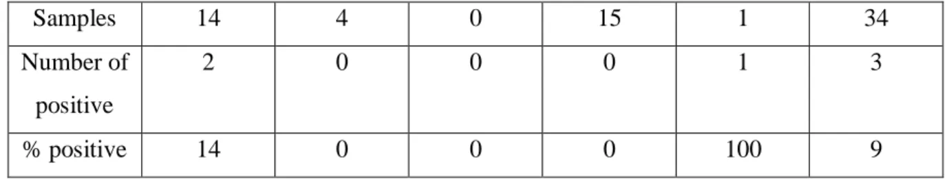
respiratory diseases they have been screened for. The results in these table show how many of the samples were positive for influenza D. The different diseases were IBR, BVDV, bovine leukemia virus (BLV), bovine parainfluenza 3 virus (BPI3V) and bluetongue virus (BTV). Not all the diseases were represented each year.
Milk samples were only tested from 2019 and were collected from milk tanks. Of 472 milk samples, 147 were positive (31%). In table 6 the milk sample results can be seen.
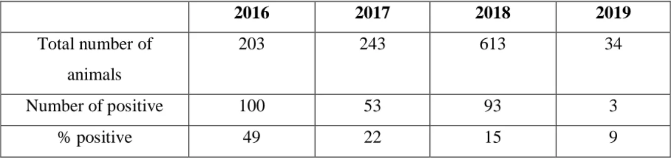
Table 1. Data compiled from serum samples from 2016 to 2019. The number of samples per year and how many were positive. The samples were tested with ELISA.
2016 2017 2018 2019 Total number of animals 203 243 613 34 Number of positive 100 53 93 3 % positive 49 22 15 9
Table 2. Data from samples collected in 2016, 2017, 2018 and 2019, which were tested for respiratory diseases. The samples were screened for IBR, BVDV, BLV, BPI3V and BTV.
2016 IBR BVDV BLV BPI3V BTV Total
Samples 150 23 30 203 Number of positive 100 0 0 100 % positive 66 0 0 49 2017 Samples 131 56 13 43 243 Number of positive 39 0 10 4 53 % positive 30 0 77 9 22 2018 Samples 229 224 99 29 32 613 Number of positive 36 19 30 0 8 93 % positive 16 8 30 0 25 15 2019
Samples 14 4 0 15 1 34 Number of positive 2 0 0 0 1 3 % positive 14 0 0 0 100 9 Real-time RT-PCR
All of the samples tested were negative, both for the PB1 gene and the nucleoprotein. The virus was not found in any of the 352 samples. The positive controls were positive, as can be seen in figure 1. The controls were a dilution series, and five were used in the real-time RT-PCR.
Figure 1. Five curves of the positive controls used in the real-time RT-PCR for influenza D. All samples, 352, were negative.
Discussion
Diseases in the respiratory tracts in cattle is a large problem worldwide. The respiratory diseases can cause major economic losses, both in the industry of dairy and beef. Cattle with bovine respiratory disease (BRD) can have coinfections such as virologic or bacteriologic.
The new influenza virus, influenza D, have showed significant role in BRD. Influenza D has for instance been reported to cause BRD together with the pathogen Mannheimia
haemolytica, giving a coinfection. The cattle in these situations have had respiratory
symptoms. Influenza D has also been detected in healthy cattle, with no reported symptoms. Which leads to a reflection if influenza D needs an immunosuppressed individual or another pathogen, to cause an infection with clinical signs. The virus can spread with aerosols and by direct contact [12]. Cattle is the primary host for influenza D, but the virus has also been detected in other mammals. Swine, goat, sheep and camelids are a few animals that the virus has been discovered in or have antibodies against it. Farmers working with these animals have showed antibodies, which gives the question if the influenza D virus may be zoonotic?
Influenza D varies with different lineages, and has been identified in the United States,
Europe, Africa and Asia. The geographic distribution is global. In 2016 a study in Japan could identify influenza D among cattle. The study material for the survey was 28 serum samples collected from healthy female cattle in one heard. The method used was Hemagglutinin inhibition test (HI-test), using the viruses D/swine/Oklahoma/1334/2011 (D/OK) and D/bovine/Nebraska/9-5/2012 (D/NE) with heterologous antigenicity. The assay was also completed with samples treated with receptor-destroying enzyme (Denka: RDE II). Of the 28 samples, eight were positive for the both of the influenza D viruses with antibodies against them. HI-test was also conducted on several serum samples from different regions in Japan, which had a similar result like in the studied herd. Serum samples proved positive, were positive for both D/OK and D/NE. The influenza D virus circulated therefore among cattle in a variety of regions in Japan [13].
Epidemics in cattle can spread in different ways, one important risk factor is movement of cattle. In Switzerland every cattle movement has to be reported into a database, the Swiss cattle movement database (Tierverkehrsdatenbank). By reporting movements, epidemics can
decrease with the knowledge of which herds are infected. Healthy cattle can in this case be held away from the infected. Switzerland is free from many severe diseases, for instance IBR [14]. With good animal keeping, diseases do not have the same possibility to spread. Animal keeping in Sweden differ compared to many other countries. Sweden has a law about animal protection, with terms in environment, hygiene, controls and more2. Animal keeping in
Sweden is in general better than in many other nations, therefore it was interesting to see if influenza D had a different prevalence.
The aim of the present study was to investigate the presence of influenza D among cattle populations in Sweden. Through the analysis of serum and bulk tank milk samples the
seroprevalence and distribution of influenza D seropositive farms in Sweden between 2016 to 2019 could be revealed. The serosurvey carried out in this study showed the presence of seropositive cattle in all years tested, with a total positivity rate of 23% (249 positives out of 1093 samples), where the positivity rates varied across the years (15-49%). This strongly suggests that influenza D viruses have circulated for at least three years in Sweden. In a study presented by Ferguson et al. positive samples were found among samples collected as early as in 2004 [3], suggesting that the influenza D virus has been circulating in cattle populations in some parts of the world for more than a decade without being noticed.
The number of samples differed in years and between the serologic and virologic methods, due to the accessibility. Antibodies were detected with ELISA, but no virus was found with real-time RT-PCR. The results of the serum samples from 2016 to 2019 showed no significant difference between which disease the samples had earlier been screened for. There were differences from year to year. The reason why the positive results in antibody detection differ in years, could be that different farms and different locations had sent in the samples.
2
Influenza D antibodies may be a part of a herd in one part of Sweden, and not in another. We do not know how long antibodies stay in the host, because the influenza D virus is not studied enough, a wider knowledge is required. The discovery of antibodies against influenza D indicates that the new influenza is circulating among cattle in Sweden. Virus was not isolated, but the number of samples was only 352 in that method. With a wider spectrum of samples, influenza D virus may be found in Sweden. Antibodies exist, therefore virus should also exist. There can be a geographic variance in Sweden with influenza D. Cattle keeping is more common in the southern parts of Sweden, the probability is higher to find the virus here than in the north. The south is also more exposed for pathogens coming from Europe with the link to Denmark. Animal keeping in Denmark is not the same as is Sweden.
Indirect ELISA was used to detect antibodies for influenza D in cattle serum samples, the method has its advantages and disadvantages. The sensitivity of ELISA is high, and higher than in another serological test named HI-test. The HI-test was a common method in similar studies. In a HI-test erythrocytes are used, where sialic acid receptors situated on the surface of the erythrocytes can bind to surface glycoprotein in influenza virus. The glycoprotein is HEF, and the binding creates a network with virus particles and interconnected erythrocytes. Advantages with HI-test are that it is relatively inexpensive, is completed within hours and do not require the same techniques as molecular tests. The disadvantages are that it is less
sensitive than ELISA and is a subjective method. ELISA is an objective method and enables less difference depending on the one who performs the procedure [15, 16]. To eventually find the influenza D virus the method real-time RT-PCR was used but could not detect any virus in the samples tested in this study. If the outcome of the PCR had been positive in samples, sequencing by Next generation sequencing (NGS) could have been made to check which lineages and subtypes the viruses had.
Differences with welfare of cattle and other animals may be a cause of different outcome by if virus is isolated in different nations. How the farmers work with the animals, if export or import are current and the climate could be vital factors. The ability to do these kinds of researches is also a significant factor. Based on the preliminary results presented in this study, research efforts will be directed towards a better understanding of the epidemiology of
influenza D infection dynamics among cattle population in Sweden, which in many aspects differs from that in continental Europe.
Acknowledgements
Many thanks to my supervisor Siamak Zohari for helping me with this project, and for
coming up with the idea of this study. Thanks to Katarina Näslund and Fereshteh Banihashem for the help with materials and methods, and the National Veterinary Institute. Thanks also to Jean-Francois Valarcher and Ignacio Alvarez at SLU.
References
[1] Tortorelli G, Carrillo Gaeta N, Mendonça Ribeiro BL et al. Evaluation of mollicutes microorganisms in respiratory disease of cattle and their relationship to clinical signs. J Vet
Intern Med, 2017;31:1215-20.
[2] Collin EA, Sheng Z, Lang Y et al. Cocirculation of two distinct genetic and antigenic lineages of proposed influenza D virus in cattle. J Virol, 2015;89:1036-42.
[3] Ferguson L, Eckard L, Epperson WB et al. Influenza D virus infection in Mississippi beef cattle. Virology, 2015;486:28-34.
[4] Hause BM, Collin EA, Liu R et al. Characterization of a novel influenza virus in cattle and swine: proposal for a new genus in the Orthomyxoviridae family. mBio, 2014;5:e00031-14. [5] Ducatez MF, Pelletier C, Meyer G. Influenza D virus in cattle, France, 2011-2014. Emerg
Infect Dis, 2015;21:368-71.
[6] Jiang WM, Wang SC, Peng C et al. Identification of a potential novel type of influenza virus in Bovine in China. Virus Genes, 2014;49:493-6.
[7] Asha K, Kumar B. Emerging influenza D virus threat: what we know so far! J Clin Med, 2019, e-published.
[8] Salem E, Cook EAJ, Lbacha HA et al. Serologic evidence for influenza C and D among ruminants and camelids, Africa, 1991-2015. Emerg Infect Dis, 2017;23:1556-9
[9] Lin AV. Indirect ELISA. Methods Mol Biol, 2015;1318:51-9.
[10] Henritzi D, Hoffmann B, Wacheck S et al. A newly developed tetraplex real-time RT-PCR for simultaneous screening of influenza virus types A, B, C and D. Influenza Other
Respir Viruses, 2019;13:71-82.
[11] Wagner EM. Monitoring gene expression: quantitative real-time rt-PCR. Methods Mol
[12] Zhang X, Outlaw C, Olivier AK et al. Pathogenesis of co-infections of influenza D virus and Mannheimia haemolytica in cattle. Vet Microbiol, 2019;231:246-53.
[13] Murakami S, Endoh M, Kobayashi T et al. Influenza D virus infection in herd of cattle, Japan. Emerg Infect Dis, 2016;22:1517-9.
[14] Hässig M, Meier AB, Braun U et al. Cattle movement as a risk factor for epidemics.
Schweiz Arch Tierheilkd, 2015;157:441-8.
[15] Assaf R, Montpetit C, Marsolais G. Serology of bovine parainfluenza virus type 3: comparison of the enzyme linked immunosorbent assay and hemagglutination inhibition. Can
J Comp Med, 1983;47:140-2.
[16] Pedersen JC. Hemagglutination-inhibition assay for influenza virus subtype identification and the detection and quantitation of serum antibodies to influenza virus. Methods Mol Biol, 2014;1161:11-25.