Research
Retinal injuries from handheld lasers:
An updated report
2015:54
Authors: Jörgen Thaung Cesar Lopes Stefan Löfgren
SSM perspective Background
The safety limits that exist for human exposure of laser radiation are essential to reduce the risk of injuries. These values, however, give very little information on what tissue damages that may be expected at various elevated exposure levels. Similarly, the Swedish Radiation Safety Authority (SSM) has very little information on how such tissue damage is related to the impairment of the vision. This type of relation-ship between an imaginary exposure and a subsequent disability is very useful in the risk assessments that are made in the authority’s supervi-sion activities. Also, the damage’s evolvement over time is information that the authority can make use of in risk assessments.
Objective
The purpose of this study was to investigate what dose of laser radia-tion, in terms of intensity and exposure time, may be associated with eye damages. The study has been limited to exposures of laser radiation from commercial handheld lasers. Of particular interest has been to search for data that clarify the dose-response relationships for functional dis-abilities that persist more than 6 months.
Results
The study shows that long-term vision loss can occur as a result of expo-sure from freely available handheld lasers at close range. The injury may occur before a normal person is able to respond by closing the eyelid. A minor such damage is transient within a few days. It is also likely that such a visible injury to the retina becomes functional, i.e. prevents read-ing skills. The use of RiskRatio is introduced, which describes how many times the actual exposure exceeded the exposure limits. Also, a compari-son with severity of injury is made.
Need for further research
There is a need for further research on damage mechanisms and their dynamics, treatment of laser damages, long-term permanent laser damages, and on the effect of visual aids and refractive errors in laser pointer retinal damages. Finally, it is proposed investigate in which mag-nitude infrared leaking lasers can influence the development of retinal damages.
Project information
Contact person SSM: Martin Lindgren Reference: SSM2015-3531
2015:54
Authors: Jörgen Thaung1, Cesar Lopes2, Stefan Löfgren3
1 Dept. Ophthalmology, University of Gothenburg, Mölndal, Sweden 2 Command and Control Systems, FOI, Linköping, Sweden
3 St. Erik Eye Hospital, Karolinska Institutet, Stockholm, Sweden
Retinal injuries from handheld lasers:
An updated report
This report concerns a study which has been conducted for the Swedish Radiation Safety Authority, SSM. The conclusions and view-points presented in the report are those of the author/authors and do not necessarily coincide with those of the SSM.
Content
1. Introduction and background ... 2
1.1. Short summary of previous SSM report, 2013 ... 3
2. Reported retinal injuries from handheld lasers ... 5
3. Analysis of case exposures ... 7
3.1. Exposure data and exposure limits ... 7
3.2. Calculation of risk ratio ... 9
3.3. Comparing exposure data with retinal damage ... 10
4. Evolution and repair of retinal damage ... 13
4.1. Timeline of identifiable organic damage ... 13
4.2. Damage mechanisms ... 16
5. What to do during or after a laser exposure ... 18
6. Future research ... 19
New proposals for future research ... 20
7. Conclusions ... 21
8. References ... 22
Appendix ... 25
A.1 Case histories ... 25
1. Introduction and background
Handheld lasers, or high powered laser pointers, continue to cause eye injuries around the world. In 2013 the Swedish Radiation Safety Authority (SSM)
published a report on eye injuries from handheld lasers (Löfgren et al. 2013). The purpose of the SSM study was to:
• “Increase the knowledge about retinal damage from commercial non-military laser pointers”. • “Aid the regulating bodies in establishing a better basis for protective measures against laser pointer exposures”. • “Provide a background for risk assessments of permanent functional visual impairment”.
In the current report we continue to analyse the reported cases for, mostly, the past 2-3 years. The primary interest of this report, as in the earlier SSM report, is continuous wave handheld laser exposures in the waveband 400-1400 nm, and with exposure duration from 0.05 to 1 second.
The continuous wave, emitted by the handheld lasers, is still dominated by the green (532 nm) wavelength. However, the technology for production of blue (multiple wavelengths) and red (multiple wavelengths) is also well established (Figure 1.1a). The quality of the lasers is varying and they are often mislabelled (Galang et al. 2010, Hadler et al. 2013) with output powers above their
specification, and inadvertently emitting wavelengths in the near-infrared. Not surprisingly the optical power of the handheld lasers, compared to those in our previous report, has increased as exemplified in the case reports (Chapter 2 and Appendix A.1). This means that the number of eye injuries with a severe outcome has increased.
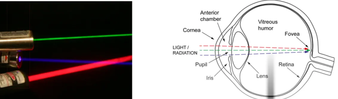
The eye is the body part most vulnerable to laser radiation, due to the focusing ability of the eye optics (Figure 1.1b). The optics of the eye concentrates the optical power approximately 100 000 times before it reaches the retina. Therefore, the injuries to the eye are more severe than to the skin.
Figure 1.1. (a) Example of commercially available blue, green and red handheld lasers
(Photo: Lars-Göran Heimdal, permission from FOI), and (b) light is focused, by the eye optics, to a small spot on the retina (Löfgren et al. 2013).
In the previous SSM report we analysed 34 cases and only a few of them had confirmed retinal injuries. In this report the number of cases with confirmed retinal injuries have increased. If the availability of the handheld lasers continues to increase, along with higher optical power and lower cost, we expect that the number of severe eye injuries will continue to grow. Also, the distance from which the lasers can be used to disrupt various societal functions, such as air traffic (pilots) and policing will also increase.
Almost all the reported cases involve young children and teenagers. There is also a huge overrepresentation by males. Often the lasers are sold as toys and
purchased by a relative as a gift to a child.
Earlier reports originated almost exclusively from developed countries. This situation has now changed and more and more reports originate from developing countries. The problem is worldwide and hence there is a need to educate and inform the societies of this danger.
1.1. Short summary of previous SSM report, 2013
• The optics of the eye focus light and radiation in the wavelength band of 400 to 1400 nm into an image on the retina. The same optics protect the eye from harmful radiation such as ultraviolet radiation and far infrared radiation. • The pupil and the eyelid regulate the amount of light entering the eye. The eyelid can close in ~0.16 seconds. • The visual system is most sensitive to green light. This means that a green laser is perceived several times stronger than for instance a red laser having the same optical power.
The eye has, thus, its own defence against strong and harmful radiation.
Unfortunately, modern handheld lasers have reached optical powers high enough to overload the ocular defence mechanisms with severe retinal damage as result. The best and mostly used methods to detect retinal damage is examination with an ophthalmic microscope and with OCT (optical coherence tomography) which creates a cross-section image of the retinal layers. Developments in hardware and software make the OCT instruments increasingly powerful, resulting in high-resolution imaging of retinal laser damage.
An eye healthy person, never exposed to laser radiation, can have slight
deviations in the retina with no functional significance. Caution should therefore be used when an identifiable retinal damage is correlated to laser exposure. In addition, there is no good correlation between the retinal damage and functional deficit.
There is currently no proven effective medical treatment for retinal laser damage, but various drugs have been used to reduce retinal swelling and anti-oxidants to minimize photochemical damage.
Exposure data from 34 reported cases were analysed and compared to existing MPE values. All the reported exposures occurred at a short distance, less than a meter, to the laser. Cases were reported for green, red, and infrared-A lasers with only a few causing retinal damage:
• Red laser, ≤5 mW, caused temporary loss of central vision • Green laser, ≤5 mW, disrupted the retinal pigment epithelia (RPE) with choroidal infarction • Green laser, ≤7 mW, caused visible damage to the RPE • Green laser, ≤20 mW, viewed for more than one second caused enlarging lesion, retinal edema and haemorrhage • A case involving infrared radiation-A (<5 mW, 825 – 880 nm) resulted in retinal edema and focal retinal detachment.
Areas where research are needed were also outlined: for example, damage mechanisms and treatment of laser damage. A database on laser exposure and damage would also benefit the society as has been proven in the USA.
2. Reported retinal injuries from handheld lasers
The use of lasers by laboratories, industry, armed forces, health care sector, and so on continues to grow. In these cases, means are taken to keep accidents at a low level. Another area is entertainment such as light shows where there have been many retinal eye injuries to spectators over the years (Löfgren et al. 2013, McHatton 2015, Jeon et al. 2014, Aras et al. 2009).The main focus in this report is the misuse of handheld lasers leading to eye injuries. Handheld lasers usually emit light with many available wavelengths (colours), from violet/blue to green, yellow, and red. They can also emit near infrared radiation. The optical power emitted by the handheld lasers has increased over the years and is expected to continue to do so. It is therefore not a surprise that there is an increased severity of the injuries.
Handheld lasers are easily accessible and have been used to blind drivers, pilots, law enforcement personnel, in terrorist attacks among others. (Lopes et al. 2010, Löfgren et al. 2013). Lasers are often inappropriately sold or used as toys and many cause severe retinal eye injuries in children.
In this report we summarize the medical case histories of retinal injuries caused by commercially available handheld lasers during the recent few years. Children, mostly young males, still dominate among the cases. There are also cases where military handheld lasers are used inappropriately or where civilian handheld lasers are misused among military personnel. The misuse of handheld lasers is now a worldwide problem and reports of retinal eye injuries in developing countries are now being published. There is an increase in reported cases with severe retinal injury. The increasing occurrence of retinal damage together with significant functional loss might be one of the reasons why ophthalmologists tend to publish more details of the injuries.
As expected it is difficult to collect all relevant information after a retinal injury. In the SSM report 2013:30 (Löfgren et al. 2013) the difficulties were identified as: • Problems in getting reliable information from the victim • Identification of the laser • The distance to the laser • The divergence of the laser beam • A medical evaluation of the injury by ophthalmic professionals familiar with retinal laser injuries
The reader should be aware of these difficulties when reading the case reports. The recent reports are more detailed than previously and in some cases include measurements of the laser output power. This is very important as it is well known that the pointers are often not correctly labelled (Galang et al. 2010, Hadler et al. 2013).
The reports are all available in the open literature. Only cases involving
commercially available handheld lasers are used in this report (cases involving military pulsed lasers are omitted). The term Visual Acuity (VA) is used to follow how a retinal injury affects vision. Normal VA with a Snellen chart is usually defined as 1.0 or higher, depending on age. We have converted any ratio VA values to decimal values. The case reports clearly show that VA can improve with time, weeks to months after an injury. On the other hand, there are cases where no improvement is seen or even a continued worsening over weeks. A total of 47 new cases are described in the Appendix A.1. A few new case reports are excluded due to poor description of the laser exposure and the eye damage. We have indicated the publication countries to illustrate that retinal injuries from handheld lasers is a worldwide problem. In Table 2.1 we present one case to exemplify the method of presenting the 47 cases.
Table 2.1. Case 3 – A 17-year-old male with self-inflicted laser eye (Dhrami-Gavazi et al. 2015) Laser power Laser Wavelength Distance Exposure duration
102 - 105 mW (measured)
663 nm Centimetres 20 - 25 seconds
Year/country Examination time frame
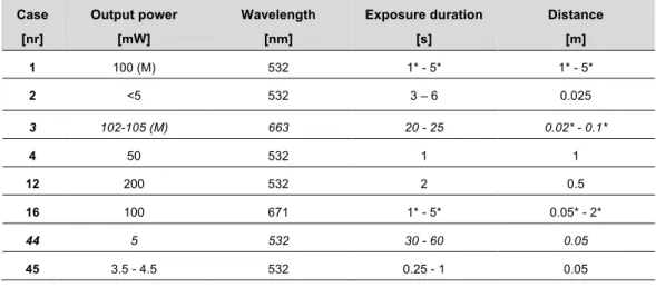
Symptom/injury Remarks
2015/USA First: Four days after incident.
Last: 3 months later
Central macular injury. Curvilinear bands of dense hyperreflectivity.
Yellowish (borders), grey –green (centres) excavated lesions. Near normalization of the acute changes after three months
The evolution of the injury was followed during three months with multimodal imaging. OCT imaging identified rapid evolution of hyperreflectivity areas (vertical curvilinear bands). Visual field testing indicated a persistent central scotoma.
There was a decreased central vision of the right eye, VA 0.2 which improved after three months to 0.67.
This case is well documented and the laser output power was measured. The distance to the laser and exposure duration are two key parameters that usually are difficult to obtain. Further, many cases include no information about the laser. Nevertheless, the quality of the reported cases has increased since our first
3. Analysis of case exposures
The objective of this chapter is to compare estimations of described laser exposure, in the case histories, with the established maximum limit of safe laser exposure. Furthermore, the objective is to compare these exposure data with the description of the resulting visual impairment using a four level graded scale. A measure is calculated showing how much higher the actual exposure was
compared to the maximal permitted exposure limit. Details on the calculations are described in the previous report (Löfgren et al. 2013).
3.1. Exposure data and exposure limits
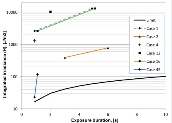
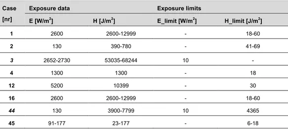
Available exposure data were identified in the 47 listed case histories (see Appendix A.1). Only one case (Case 4) had sufficient data to make a fully accepted analysis. In order to include more cases some assumptions were necessary, and lacking data were estimated when possible. The validity of these estimations are discussed in the text. The eight cases that were included in the analysis are listed in Table 3.1. Estimated data are marked with an asterisk (*), and data that were measured are marked with a letter M. Case 3 and 44 have exposure durations which actually falls far outside the scope of this report (0.05 - 1 s) but are left for comparison purposes (data in italic).
Table 3.1. The following eight cases were included in the analysis ( * = estimated data, M = measured). Case [nr] Output power [mW] Wavelength [nm] Exposure duration [s] Distance [m] 1 100 (M) 532 1* - 5* 1* - 5* 2 <5 532 3 – 6 0.025 3 102-105 (M) 663 20 - 25 0.02* - 0.1* 4 50 532 1 1 12 200 532 2 0.5 16 100 671 1* - 5* 0.05* - 2* 44 5 532 30 - 60 0.05 45 3.5 - 4.5 532 0.25 - 1 0.05
The limits for a safe exposure in each case were derived using the ICNIRP guidelines (2013). After identification of the exposure data listed in Table 3.1 the following equations are applicable:
Hlimit = 18*t0,75*CE [J/m2] 5µs < t < 10s (Eq 3.1)
Hlimit = 100*CB [J/m2] 10 ≤ t < 100s (Eq 3.2)
Elimit = 10 [W/m2] 10 ≤ t < 100s (Eq 3.3)
CE = 1 (point sources) (Eq 3.4)
CB = 1 400 ≤ λ < 450nm (Eq 3.5)
CB = 100.02(λ -450) 450 ≤ λ ≤600nm (Eq 3.6)
The three equations (3.1 – 3.3) are applied in the following way: when the exposure duration (t) is shorter than 10 seconds, only photothermal limits has to be considered, see equation 3.1, Hlimit (integrated irradiance). In cases with longer
exposure durations (case 3, and 44) both the photothermal limit using equation 3.2 and the photochemical limit using equation 3.3 (Irradiance, Elimit) have to be
considered. Note that the photochemical limits are only calculated for wavelengths (λ) in the range of 400 to 600 nm.
By identification of the parameters included in the equations it is possible to validate the assumptions that were made in the exposure data set (table 3.1):
• For all distances up to 5 m the laser beams are assumed to have a smaller diameter than the 7 mm limiting aperture. This means that uncertainties in distance (centimetres or few meters) are not crucial and gives no reason for exclusion (case 1, 3, or 16). • Estimation of exposure duration can be more problematic. Data for case 1, 16, and 45 should be used with some caution. Since the intervals are relatively small the uncertainties are deemed acceptable. Still, it is important to remember that the exposure limits are changing with the exposure duration when the duration is shorter than 10s.
When the exposure distance was specified as “centimetres” the distance was estimated to be in a distance range of 2 to 10 cm. If the exposure duration was given as “few seconds” a range of 1 to 5 seconds was used in the analysis. When the duration was given as “less than a second” the range of 0.25 to 1 s was used. All calculations in this report assume an intrabeam viewing situation (worst case) with a collimated laser beam smaller than 7 mm. This may not be true in all cases.
The exposure limits are given in Joule per square metre (Hlimit), or Watt per
square metre (Elimit) in the corneal plane. Corresponding exposure irradiances
were calculated for the eight cases and listed together with the limits in Table A.2.1 in the Appendix. In some cases, the laser output power and/or exposure durations were given in intervals. For these cases the irradiances were also calculated as minimum and maximum values and presented as a range. The six cases with exposure durations shorter than 10 seconds are plotted in Figure 3.1 together with the exposure limit. The two cases with longer exposure durations are excluded in Figure 3.1 since they must meet both photothermal and
photochemical limits, which means that they cannot be presented in a single diagram. Furthermore, as these longer exposure durations are longer than the 0.005 to 1 second delimitation in this report, additional diagrams are not produced. Anyway, all data are found in the Appendix (Table A.2.1).
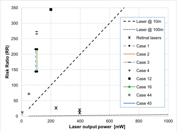
Figure 3.1. Irradiance data of the six cases with exposure duration less than 10 s. Single
point data are shown with symbols, and ranges of case exposures are shown with lines that connect the minimum and maximum data points. The solid black line is showing the exposure limit according to the ICNIRP guidelines (2013).
3.2. Calculation of risk ratio
In the previous report we examined how many times the actual exposure exceeded the exposure limits by calculating the quotient of the two. This ratio was called “Risk Ratio, RR. As a comparison, RR values were also calculated for a fictive laser exposure (t=0.25 s) using a typical handheld laser (thin beam, no optics, “top hat” distribution, and 1 mrad divergence) at two different distances. Furthermore, an estimation of the expected RR values from three different ophthalmic lasers were used as a comparison. The same evaluation is here repeated with the new set of cases presented in this report (see Figure 3.2).
10 100 1000 10000 0 2 4 6 8 10 Int egr at ed ir ra dia nc e (H ), [J /m 2] Exposure duration, [s] Limit Case 1 Case 2 Case 4 Case 12 Case 16 Case 45
Figure 3.2. Plots of Risk Ratios (RR) calculated as the ratio between estimated exposure
data and exposure limits. Data are show for the eight cases, and for a fictive case using a handheld laser at two distances (10 and 100 m, beam divergence = 1 mrad, “top hat” distribution, t=0.25 s). Additionally, data from three ophthalmic lasers used for retinal treatment are shown in the graph.
Analysis of the data shows that all exposures in the eight cases exceeded the exposure limit meaning that RR>1. Four cases show RR values higher than 100, and the highest estimated exposure among the eight cases was 344 times the safety limit. Case 45 have the lowest RR (4) and case 2, 44, and 45 have all RR values below 15, see left-bottom corner of figure 3.2. The lasers for retinal treatment have values between about 10 and 30. For those cases (1, 2, 3, 16, and 45) where the exposure data were given in larger ranges, matching ranges of RR values were calculated.
3.3. Comparing exposure data with retinal damage
To analyse the possible link between calculated RR and reported eye injuries, as described in the case histories, the relevant information was summed in Table 3.2. An attempt was also made to grade the reported injuries into “severity of injury” (SI) according to the grading scale presented in the official Journal of the
European Union (2010). A similar comparison is found in our previous report where the eye injuries were classified using the four level grade scale (see Table 3.3). The grading of SI was based on the information of visual acuity given in the case histories (Appendix A.1), and is not built on any statistical evaluation.
0 50 100 150 200 250 300 350 0 200 400 600 800 1000 Ri sk Ra ti o ( RR)
Laser output power [mW]
Laser @ 10m Laser @ 100m Retinal lasers Case 1 Case 2 Case 3 Case 4 Case 12 Case 16 Case 44 Case 45
Table 3.2. Comparison of Risk Ratio (RR) and severity of injury (SI) in eight analysed cases.
Case RR SI Short description
1 144-216 3 Central scotoma in left eye
2 10-11 2-3 Paracentral scotoma in left eye. (Normal to subnormal VA)
3 265-273 3 Central macular injury in right eye. (Subnormal VA)
4 72 2-3 Central blind spot in let eye that disappeared even though OCT showed a small defect. (Subnormal VA)
12 344 2-3 Foveal lesion in right eye. (Normal to subnormal VA)
16 216 3 Central scotoma in both eyes. (Subnormal VA)
44 13 2-3 Macular lesion in both eyes. (Normal to subnormal VA)
45 4-6 2-3 Metamorhopsia in right eye. Blurred vision improved over time. Visible change in retinal layers after 6 months and subnormal VA.
Table 3.3. “Severity of injury” grading scale for eye injuries (Journal of the European Union. 2010). Grade Description
1 Temporary pain in eye without need for treatment
2 Temporary loss of sight
3 Partial loss of sight. Permanent loss of sight (one eye)
4 Permanent loss of sight (both eyes)
It seems difficult to find any obvious relation between RR, retinal damage, and SI in the eight analysed cases since all of them is estimated to fall within grade 3, or possibly in grade 2 since we do not have access to the visual status before laser exposure. In our previous report, the case exposures had significantly lower corneal irradiances and RR values, and it was more obvious to identify SI
relations between different RR values. There, 25 of the 27 analysed cases had RR values below 20, and in the cases with RR<10 many cases were graded as SI 1 or 2, indicating a safety margin for permanent damage of about 10. There were also two cases with RR values of about 30 which resulted in retinal detachments. A comparison with surgical retinal lasers with estimated RR values in the range of 10 to 30 indicated that a permanent retinal damage will theoretically occur for RR values exceeding 10.
In this report we found much larger RR values for the analysed cases and the described eye injuries are more severe. However, none of the injuries fell into SI
grade 4 with Permanent loss in both eyes, also none of the cases were graded as SI 1 - Temporary pain in eye without need for treatment. Most of the cases were graded in between 2 and 3, and in these cases a range of 2-3 was specified. This made it difficult to find any clear relationship between exposure level and SI. Several subjects had a best corrected visual acuity that we considered to be subnormal compared to the expected visual acuity in relation to their age. Since we have little knowledge of the visual status before the exposure it was difficult to grade what impact the laser had on the visual performance.
In the case with highest RR (case 12, RR 344) there was only a marginal loss of vision (VA=0.63 right eye), and at the examination 18 months later the visual acuity was found to be 1.0 in both eyes. One explanation can be that the retinal damage occurred “pericentrally” and not in the most central part of the fovea – responsible for the highest VA. The most likely explanation is that the exposure duration (2 s) was overestimated due to aversion responses.
4.
Evolution and repair of retinal damage
Tissue repair is organised along two routes, either as repair of damaged cells, or removal of cells that are damaged beyond repair. The latter can occur either via necrosis (unplanned cell disintegration) or via apoptosis (pre-programmed cell death). The necrosis pathway induces secondary damage to adjacent cells, while the apoptosis pathway results in a cleaner removal of dead cells. Necrosis is more prominent in supra-threshold laser damage while apoptosis dominates in low power laser exposure but also occurs in the peripheral zone around a necrosis region.
The scenario is laser damage to the fovea, the central part of the retina where the visual acuity is highest. Laser damage can occur wherever a strong laser hits the retina but in reality, most documented cases exhibit damage in the foveal region. This is not surprising since most exposures are self-inflicted and the individuals look straight into the laser beam.
4.1. Timeline of identifiable organic damage
The case reports during the last years describe OCT findings more frequently than in previous years, and the documentation of the rise and fall of retinal damage is improving. There is still a lack of data on the exposures, especially regarding the true power of the lasers. In most cases, no power measurements were made with the handheld lasers that were apprehended.
The information from the case reports in this review report is consolidated to create a timeline overview of the identifiable organic damages:
Days
The most common early visible changes in the retina are white-yellowish spots seen in the fovea with ophthalmoscopy. Retinal pigment epithelial damage is seen as hyperfluorescence with fluorescein angiography and hypoautofluorescence. Tissue edema and disruption is observed with OCT in any level of the retina but preferentially in the outer retinal layers, including the pigment epithelium. OCT reveals vertical curvilinear bands of hyperreflectivity originating in the interface of pigment epithelium and photoreceptors, extending and deviating horizontally in the axon layer of the photoreceptors (see Figure 4.1). In cases with very high powered laser early preretinal or intraretinal haemorrhage or even macular hole may occur (see Figure 4.2). Depressed visual acuity and relative scotoma
(localized visual field defect) occur instantaneously or within 1-2 days, depending on the laser power or cumulative exposure.
Weeks
The initial acute damage phase is followed by repair resulting in resolution of edema and a trend of normalisation of the tissue disruption. Tissue atrophy may develop, seen as a white (non-pigmented) and/or dark (hyperpigmented) region with ophthalmoscopy, and absence or patchiness of specific retinal layers viewed
in OCT images. Improvement of the visual disturbance occurs unless secondary retinal damage develops or the cell death is too advanced. An example of the latter is when the pigment epithelial cells do not repair sufficiently, the overlying photoreceptor cells will degrade permanently.
Months
The damage repair continues over months, most often in parallel with tissue atro-phy. A minority of the documented cases recovered to normal visual acuity (Figure 4.3). Secondary eye disease may develop, such as choroidal
neovascularization, macula pucker (thickening and traction of the inner surface membrane of the retina), macular hole, cystic retinal edema and retinal layering or detachment. Some secondary eye disease may repair spontaneously but many cases need surgical treatment.
Years
The laser tissue damage reaches a quiet phase with no further visual deterioration. There are a few anecdotal reports where very late on-set retinal disease are
speculatively linked to a previous laser exposure, but the evidence for a cause-effect relationship is weak.
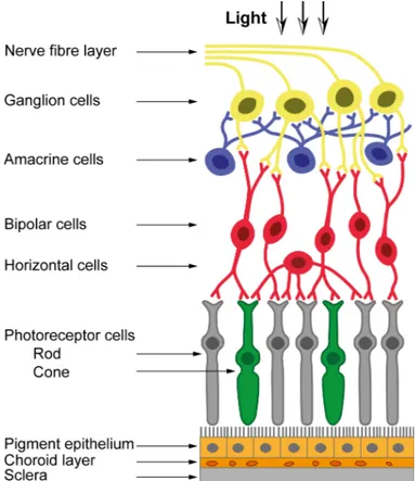
Figure 4.1. Schematic of the retinal layers. The bottom layer, sclera, is the white outer
layer of the eye, while the top layer is a nerve fibre layer facing the inside of the eye. Laser energy delivered during short exposures is predominantly absorbed in the pigment epithelium, with thermal injury outwards to the choroid and inwards to the neural cell layers. Illustration by J Thaung.
Figure 4.2 Retinal hole after self-inflicted exposure to a strong blue handheld laser,
labelled 1000 mW. (A) Infrared autoflourescence photo of left central retina. The middle horizontal line indicates the position of the cross-section image in B. (B) OCT image exhibiting full thickness retinal hole in the fovea. (C) Colour photo indicating foveal damage. Reprinted from The Lancet, Petrou et al., 2014; 383: 1780, with permission from Elsevier.
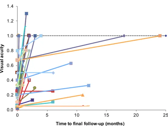
Visual acuity data in the case reports are used to indicate the functional outcome after the laser exposure (see Figure 4.3). A weakness of the representation is that many patients were followed for a surprisingly short time, even cases with severe visual impairment. Also astonishing is the fact that some patients declined offered surgical treatment for their severe retinal disease.
The average final visual acuity outcome for the case reports in the Appendix A.1 is 0.63 (blue laser), 0.59 (green laser) and 0.44 (red laser). Unfortunately, the numbers cannot be used to determine which wavelength is most harmful because the optical power is unknown in most of the cases.
Figure 4.3 Outcome of visual acuity after exposure to handheld lasers. The dotted line at
y = 1.0 indicates normal visual acuity. The data include case reports with visual acuity at both presentation and final follow-up.
4.2. Damage mechanisms
Lasers damage tissue through three separate mechanisms: photothermal, photo-chemical and photomechanical. The bulk of knowledge on cellular and
subcellular retinal damage mechanism comes from other types of laser than handheld lasers, for instance medical ophthalmic lasers. Therefore, the references for this paragraph are not limited to the inclusion criteria set for the case reports. It seems that the focus for the retinal damage is similar for all three types of damage mechanisms, namely the RPE region, with varying degree of extension into the neuroretina (Pocock et al. 2014).
Photothermal damage
Thermal injury is the dominating mechanism for retinal damage with the laser exposure parameters covered in this report. The laser energy is absorbed by various pigments or chromophores in the tissue, causing an increase in
temperature ultimately leading to protein coagulation and cell death. The three typical colours emitted by handheld laser are blue, green and red, plus the invisible infrared radiation that often is leaking through green lasers with inappropriate filters. All these wavebands will mainly absorb in the RPE, with little wavelength dependency for short exposure times. The thermal build-up in the RPE will dissipate outwards to the choroid and inwards to the neuroretinal layers, causing a spread of the damage zone. Variability in melanin pigment
0.0 0.2 0.4 0.6 0.8 1.0 1.2 1.4 0 5 10 15 20 25 Visual acuity
density in the RPE introduces a risk for hotspots within the laser beam area where localized damage might exceed the expected degree of injury (Sramek et al. 2009). The melanin pigment content in the human RPE varies both with age and topographic region of the retina, but the melanin level in the central retina is less dependent on age (Schmidt and Peisch, 1986). RPE melanin content is similar in Caucasians and Afro-Americans (Weiter et al. 1986). The main near-infrared absorber is the RPE. Longer wavelength infrared radiation can be absorbed by water and blood in the eye and retina but these wavelengths are not emitted by handheld lasers and thus irrelevant for this report (Lund et al., 2005, McHugh et al., 1995, van den Berg and Spekreijse, 1997).
Photochemical damage
Photochemical injury occurs when the laser energy absorbed by the tissue is converted into chemical reactions leading to cell death. Current safety limits exclude photochemical injury at shorter exposure times (<10 s), but the ratio of photochemical versus thermal injury increases with increasing exposure time (Sliney 2000, Denton et al. 2011) and the photochemical component is
increasingly stronger for light wavebands shorter than 550 nm (Lund et al., 2005). In contrast, the photochemical injury during longer laser exposures exhibits wavelength dependency. Chromophores in the photoreceptors exhibit differential sensitivity to light colour (Grimm et al. 2000).
Photomechanical damage
This occurs when laser energy is deposited in the tissue during very short exposures, which happens with lasers emitting very short and intense pulses. Photomechanical damage is not relevant with the exposures included in this report (0.05 to 1 s).
Robertson et al. (2000 and 2005) hypothesized that green handheld lasers are more harmful than red lasers, based on their findings with in vivo exposures with green and red laser in humans. A weakness in the conclusion is that the laser spectral output is not described in the publications and hence there might have been leaking infrared radiation in the green laser.
5. What to do during or after a laser exposure
Many employers have written advice on how employees might avoid eye damage or secondary damages during a laser exposure incident. Most of these advice focus on avoidance of the laser beam, securing the vehicle (air, land and sea), use of laser safety goggles, and alerting the authorities. Transport companies also puts emphasis on the important risks with the temporary dazzling which might lead to lost control of the vehicle. In a WHO report (WHO 1998) the following advice can be read:“Although the risk of permanent eye injury from a laser pointer may be small, a person receiving even a transient eye exposure will experience a bright flash, a dazzling effect, which is likely to cause distraction and temporary loss of vision in the affected eye and possibly after-images. The time taken to recover from these effects will vary for different people and will also be dependent on the ambient light level at the time of exposure. Medical attention should only be sought if after-images persist for hours, or if a disturbance in reading vision is apparent.”
St. Erik Eye Hospital in Stockholm Sweden has established guidelines for the general public in Stockholm, based on the experience of not having had a single case with serious retinal damage from handheld lasers throughout the years. The aim for the guidelines is to calm the public by explaining that exposure from handheld laser at distance by another person rarely is dangerous for the eyes. Within this scope, an eye examination is only required if there are lasting eye symptoms. On the other hand, we expect that people with severe visual disturbance will be very frightened and seek help at an emergency room, whatever guidelines there are. Some Swedish employer organizations have introduced these guidelines into their own advice to employees.
The number of laser exposures to commercial aircrafts in the USA is increasing according to the US Federal Aviation Administration (FAA). Luckily, there are few or no cases with permanent eye damage in commercial pilots. The FAA spokesperson Alison Duquette said “The FAA is unaware of any US commercial pilot who has suffered permanent eye damage as a result of exposure to laser light in the cockpit.” The March 19 2015 statement was made in response to an inquiry by LaserPointerSafety.com.
6. Future research
The previous report (Löfgren et al., 2013) proposed a number of research topics to be considered in future work. The six topics were:
• National database • Photochemical effects • Effects of visual aids • Treatment of laser pointer retinal damage • Secondary injuries • Long-term or permanent functional deficit
The database was proposed since we lack national patient data that could be useful in order to extend the dataset found from cases reported in scientific literature. By introducing a reporting scheme, we could ensure that vital information is not lost in the communication between patient, eye doctor, and researcher.
There seems to be a lack of data on photochemical effects after continuous wave laser exposure in the subsecond range, especially with interacting wavelengths or interacting damage mechanisms such as combined photothermal and
photochemical effects.
We have not found any discussion about the effect of visual aids during laser exposure. Positive and negative lenses will result in different irradiances at the cornea. The effect is smaller than a factor of 2, but may not be negligible in all exposure situations.
There is a lack of data on which treatment is effective and safe. This situation is at least partly linked to the problem of identifying the exact damage mechanisms. Besides the risk of eye injury there is also the risk of secondary injuries due to a strong disability glare caused by the (visible) laser. Subconscious reflex responses may result in actions that may lead to traffic accidents or other situations that can result in multiple casualties.
There is a need for studies on long-term or permanent functional deficit after exposure to laser pointers. Structural damage can be assessed by any model system, from in vitro to in vivo, from animals to humans, and the advances in the OCT technique have made this method one of the most suitable for identifying sub-clinical retinal structural damage. The resolution enhancement achieved by adaptive optics (AO) can further help in pinpointing subtle changes that common ophthalmic instruments cannot detect.
New proposals for future research
In addition to the previously proposed topics for future research we have added the following topic.
Infrared radiation in handheld lasers
It is known that many lasers simultaneously emit radiation in more than one wavelength. In most green handheld lasers, the radiated spectrum consists of at least three peaks, 532, 808, and 1064nm. Even if they are equipped with optical filters designed to block the infrared radiation, there may still be a significant leakage. How lasers can be mislabelled for this reason, and how strong the infrared radiation can be, have been discussed in recent papers (Galang et al. 2010, Hadler et al. 2013). Since lasers with leaking infrared are frequently found it is important to communicate and analyse the problem. Optical measurements must include spectral detection, a requirement that is costlier than standard power meters. Galang et al (2010) introduced a simple way of detecting the presence of infrared leakage but we also need to develop simple methods like the one
presented by Hadler et al (2013) to get reliable data for exposure calculations. A proposed study is to investigate in which magnitude these infrared leaking lasers can influence the calculation of the Risk Ratio.
7. Conclusions
The main focus in this report is the misuse of handheld lasers leading to eye injuries. The energy emitted by the handheld lasers has increased over the years and is expected to continue to do so. It is therefore not a surprise that an increase in the severity of the injuries was found.
We have summarized the medical case histories of retinal injuries caused by commercially available handheld lasers during the recent few years, thus updating our previous SSM report (Löfgren et al. 2013). Children, mostly young males, still dominate among the cases and we see an increase in reported cases with severe retinal injury. The increasing occurrence of visible retinal damage together with significant functional loss might be one of the reasons why ophthalmologists tend to publish more details of the injuries.
Eight of the 47 cases were described with sufficient data to be included in an analysis of the estimated laser exposure. We examined how much the actual exposure in each case exceeded the exposure limits by calculating the ratio of the two (“Risk Ratio”, RR). Four cases showed RR values higher than 100, and the highest estimated exposure among the eight cases was 344 times the safety limit. The RR data was also compared to the eye injuries using a grading scale
presented by the European Union. Visual acuity data in the case reports were used to indicate the functional outcome after laser exposure. Most of the cases were in between grade 2 and 3, where grade 3 means: Partial loss of sight.
Permanent loss of sight (one eye).
There is still a lack of data on the exposures, especially regarding the true power of the lasers. In most cases, no optical power measurements were made with the handheld lasers that were apprehended. True exposure duration is also an
important parameter that is lacking in many cases. Even when the laser exposure was reported to last for several seconds, it may have been reduced by aversion responses.
In addition to our previously proposed topics for future research we have added our interest for investigating to what magnitude the leakage of near-infrared radiation in green lasers can influence the relative risk (RR).
8. References
ALSULAIMAN, S. M., ALRUSHOOD, A. A., ALMASAUD, J., ALZAAIDI, S., ALZAHRANI, Y., AREVALO, J. F., GHAZI, N. G., ABBOUD, E. B., NOWILATY, S. R. & AL-AMRY, M. 2014. High-power handheld blue laser-induced maculopathy: the results of the King Khaled Eye Specialist Hospital Collaborative Retina Study Group. Ophthalmology, 121, 566-572. e1.
ARAS, C., KOYLUOGLU, N., HASHEMINIA, A. & AKAYDIN, Ö. 2009. Inadvertent laser‐induced retinal injury following a recreational laser show. Clinical &
experimental ophthalmology, 37, 529-530.
BHAVSAR, K. V., WILSON, D., MARGOLIS, R., JUDSON, P., BARBAZETTO, I., FREUND, K. B. & CUNNINGHAM, E. T. 2015. Multimodal imaging in handheld laser-induced maculopathy. American journal of ophthalmology, 159, 227-231. e2.
DENTON, M. L., CLARK III, C., NOOJIN, G. D., ESTLACK, L. E., SCHENK, A. C., BURNEY, C. W., ROCKWELL, B. A. & THOMAS, R. J. Characterizing temperature-dependent photo-oxidation to explain the abrupt transition from thermal to non-thermal laser damage mechanisms at 413 nm. SPIE BiOS, 2011. International Society for Optics and Photonics, 78970K-78970K-7.
DHOOT, D. S., XU, D. & SRIVASTAVA, S. 2014. High-powered laser pointer injury resulting in macular hole formation. The Journal of pediatrics, 164, 668-668. e1. DHRAMI-GAVAZI, E., LEE, W., BALARATNASINGAM, C., KAYSERMAN, L.,
YANNUZZI, L. A. & FREUND, K. B. 2015. Multimodal imaging
documentation of rapid evolution of retinal changes in handheld laser-induced maculopathy. International Journal of Retina and Vitreous, 1, 1-7.
DIRANI, A., CHELALA, E., FADLALLAH, A., ANTONIOS, R. & CHERFAN, G. 2013. Bilateral macular injury from a green laser pointer. Clinical
ophthalmology (Auckland, NZ), 7, 2127.
FARAGGI, E., GERSTMAN, B. S. & SUN, J. 2005. Biophysical effects of pulsed lasers in the retina and other tissues containing strongly absorbing particles: shockwave and explosive bubble generation. Journal of biomedical optics, 10, 064029-064029-10.
FARAJPOUR, R., AHOUR, M., FARAJPOUR, S., BAGHERI, M. & ASADOLLAHI, M. 2015. A Case of Retinal Damage Caused by Green Laser Pointer. Advances
in Ophthalmology & Visual System, 2, 00063.
GALANG, J., RESTELLI, A., HAGLEY, E. W. & CLARK, C. W. 2010. A green laser pointer hazard. NIST Technical Note 1668. National Institute of Standards and Technology. U.S Dept. of Commerce.
GRIMM, C., WENZEL, A., HAFEZI, F., YU, S., REDMOND, T. M. & REMÉ, C. E. 2000. Protection of Rpe65-deficient mice identifies rhodopsin as a mediator of light-induced retinal degeneration. Nature genetics, 25, 63-66.
HADLER, J., TOBARES, E. & DOWELL, M. 2013. Random testing reveals excessive power in commercial laser pointers. Journal of Laser Applications, 25, 032007. ICNIRP, STUCK, B., SCHULMEISTER, K., SLINEY, D., CESARINI, J., THOMAS,
R., GREINERT, R. & SÖDERBERG, P. 2013. ICNIRP Guidelines on limits of exposure to laser radiation of wavelengths between 180 and 1000 µm. Health
Phys, 105, 271-295.
JEON, S. & LEE, W. K. 2014. inner retinal damage after exposure to green diode laser during a laser show. Clinical ophthalmology (Auckland, NZ), 8, 2467.
KEUNEN, J., DELBECQ, A., CRUYSBERG, J., VAN MEURS, J., GAN, I. & BERENDSCHOT, T. 2013. [Laser pointers are not toys; eye injury with permanent loss of visual acuity]. Nederlands tijdschrift voor geneeskunde, 158, A7813-A7813.
KHEDR, Y. A. & KHEDR, A. H. 2014. Photoblepharokeratoconjunctivitis caused by invisible infrared radiation emitted from a green laser pointer. BMJ case reports, 2014, bcr2013202747.
LALLY, D. R. & DUKER, J. S. 2014. Foveal injury from a red laser pointer. JAMA
ophthalmology, 132, 297-297.
LEE, G. D., BAUMAL, C. R., LALLY, D., PITCHER, J. D., VANDER, J. & DUKER, J. S. 2014. Retinal injury after inadvertent handheld laser exposure. Retina, 34, 2388-2396.
LEE, G. D. & LALLY, D. 2015. Laser Pointer Retinal Injures. Retina Today.
LIM, M. E., SUELZER, J., MOORTHY, R. S. & VEMURI, G. 2014. Thermal macular injury from a 154 mW green laser pointer. Journal of American Association for
Pediatric Ophthalmology and Strabismus, 18, 612-614.
LOPES, C., SVENSSON, S. & ÖHGREN, J. 2010. Starka handhållna lasrar - Hot och utvecklingsmöjligheter. FOI-R-3036-SE.
LUND, D. J., EDSALL, P. & STUCK, B. E. Wavelength dependence of laser-induced retinal injury. Biomedical Optics 2005, 2005. International Society for Optics and Photonics, 383-393.
LÖFGREN, S., THAUNG, J. & LOPEZ, C. 2013. Laser pointers and Eye injuries - An analysis of reported cases. SSM 2013:30 ISSN: 2000-0456.
MCHATTON, R. 2015. How safe are laser shows? Photonics spectra. MCHUGH, D., ENGLAND, C., VAN DER ZYPEN, E., MARSHALL, J.,
FANKHAUSER, F. & FANKHAUSER-KWASNIESKA, S. 1995. Irradiation of rabbit retina with diode and Nd: YAG lasers. British journal of ophthalmology, 79, 672-677.
NOBLE, C. & BLICE, J. 2015. Permanent retinal injury from recreational laser pointer.
Military medicine, 180, e378-e380.
OFFICIAL 2010. Journal of the European Union. Legislation. ISSN 1725-2555. OJ L22 26.1.
PETROU, P., PATWARY, S., BANERJEE, P. J. & KIRKBY, G. R. 2014. Bilateral macular hole from a handheld laser pointer. The Lancet, 383, 1780.
POCOCK, G. M., OLIVER, J. W., SPECHT, C. S., ESTEP, J. S., NOOJIN, G. D., SCHUSTER, K. & ROCKWELL, B. A. 2014. High-Resolution In Vivo Imaging of Regimes of Laser Damage to the Primate Retina. Journal of ophthalmology, 2014.
RAOOF, N., CHAN, T., ROGERS, N., ABDULLAH, W., HAQ, I., KELLY, S. & QUHILL, F. 2014. ‘Toy’laser macular burns in children. Eye, 28, 231-234. RIVERS, B. & CRAWFORD, C. 2014. Chorioretinal Injury Caused by Presumed Laser
Dazzler. Surgery Curr Res, 4, 2161-1076.1000191.
ROBERTSON, D. M., LIM, T. H., SALOMAO, D. R., LINK, T. P., ROWE, R. L. & MCLAREN, J. W. 2000. Laser pointers and the human eye: a clinicopathologic study. Archives of ophthalmology, 118, 1686-1691.
ROBERTSON, D. M., MCLAREN, J. W., SALOMAO, D. R. & LINK, T. P. 2005. Retinopathy from a green laser pointer: a clinicopathologic study. Archives of
ophthalmology, 123, 629-633.
ROSSI, F., PINI, R., MENABUONI, L., MENCUCCI, R., MENCHINI, U.,
process following laser welding of the cornea. Journal of biomedical optics, 10, 024004-0240047.
RUSU, I., SHERMAN, J., GALLEGO-PINAZO, R., LAM, M. & FREUND, K. B. 2013. Spectral-domain optical coherence tomography and fundus autoflourescence findings in a case of laser pointer-induced maculopathy. Retinal cases and brief
reports, 7, 371-375.
SCHMIDT, S. & PEISCH, R. 1986. Melanin concentration in normal human retinal pigment epithelium. Regional variation and age-related reduction. Investigative
ophthalmology & visual science, 27, 1063-1067.
SHENOY, R., BIALASIEWICZ, A. A., BANDARA, A. & ISAAC, R. 2015. Retinal Damage from Laser Pointer Misuse–Case Series from the Military Sector in Oman. Middle East African journal of ophthalmology, 22, 399.
SLINEY, D. H. Overview of the range of mechanisms. Laser-Induced Damage in Optical Materials: 1999, 2000. International Society for Optics and Photonics, 2-19. SRAMEK, C., PAULUS, Y., NOMOTO, H., HUIE, P., BROWN, J. & PALANKER, D.
2009. Dynamics of retinal photocoagulation and rupture. Journal of biomedical
optics, 14, 034007-034007-13.
VAN DEN BERG, T. J. & SPEKREIJSE, H. 1997. Near infrared light absorption in the human eye media. Vision research, 37, 249-253.
WEITER, J., DELORI, F., WING, G. & FITCH, K. 1986. Retinal pigment epithelial lipofuscin and melanin and choroidal melanin in human eyes. Investigative
ophthalmology & visual science, 27, 145-152.
WENG, C., BAUMAL, C., ALBINI, T. & BERROCAL, A. 2015. Self-induced laser maculopathy in an adolescent boy utilizing a mirror. Ophthalmic surgery, lasers
& imaging retina, 46, 485-488.
VUKICEVIC, M., GIN, T. & KEEL, S. 2014. Laser Pointer Retinal Injury: A Case Report. Australian Orthoptic Journal, 46, 1.
XU, M. & LIN, T. 2014. Bilateral central scotomas following laser pointer misuse.
Canadian Medical Association. Journal, 186, E341.
YING, W., OUYANG, P. & TANG, L. 2014. Handheld Laser Pointer Maybe a Dangerous “Toy”: A Case of Maculopathy from DPSS Red Handheld Laser Pointer. J Clin Exp Ophthalmol, 5, 2.
YIU, G., ITTY, S. & TOTH, C. A. 2014. Ocular safety of recreational lasers. JAMA
Appendix
A.1 Case histories
Case 1 – A 12-year-old male with self-inflicted laser eye injury (Weng et al. 2015)
Laser power Laser Wavelength
Distance Exposure duration
100 mW (measured)
532 nm Few meters few seconds
Year/country Examination
time frame Symptom/injury Remarks
2015/USA First: Unknown
Last: 4 weeks after the incident.
Central
scotoma in the left eye retina. RPE disruptions in the fovea, etc.
Healing of the central scotoma four weeks after the injury. Laser reflected from a mirror
Visual acuity (VA) improved from 0.33 to 0.8, for the injured left eye, in four weeks.
Case 2 – A 16-year-old male with self-inflicted laser eye injury (Farajpour et al. 2015)
Laser power Laser Wavelength Distance Exposure duration <5 mW 532 nm 25 mm 3-6 seconds Year/country Examination time frame Symptom/injury Remarks
2015/Iran First: After one month
Last: After 2 months
Paracentral scotoma in the left eye retina. Foveal lesions. Focal disruption of segment junction of photoreceptor. No changes in VA.
Case 3 – A 17-year-old male with self-inflicted laser eye (Dhrami-Gavazi et al. 2015)
Laser power Laser
Wavelength Distance Exposure duration
102 - 105 mW (measured)
663 nm Centimetres 20 - 25 seconds
Year/country Examination
time frame Symptom/injury Remarks
2015/USA First: Four days after incident. Last: 3 months later Central macular injury. Curvilinear bands of dense hyper reflectivity. Yellowish (borders), grey – green (centres) excavated lesions. Near normalization of the acute changes after three
months
The evolution of the injury was followed during three months with multimodal imaging. OCT imaging identified rapid evolution of hyperreflectivity areas (vertical curvilinear bands). Visual field testing indicated a persistent central scotoma.
There was decreased central vision of the right eye, VA 0.2 which improved after three months to 0.67.
Case 4 – A 13-year-old male with self-inflicted laser eye injury (Noble et al. 2015)
Laser power Laser Wavelength
Distance Exposure duration
50 mW 532 nm One meter 1 second
Year/country Examination time frame
Symptom/injury Remarks
2015/USA First: after one week Last: after 6 months Small disruption of RPE, superior and temporal to the fovea. A central blind spot in the left eye immediately after the
exposure.
Uncorrected VA was 0.67 which change to 0.8 with a pinhole for the left eye. Further, amsler grid test showed a central visual field defect.
There was no change after 6 months, but after 2 years the blind spot disappeared even though OCT showed a small defect.
Case 5 – An 18-year-old female deliberately gazed into three different lasers, several occasions (Bhavsar et al. 2015)
Laser power Laser
Wavelength Distance Exposure duration
100 mW (green) 50 mW (red) 50 mW (purple) 532 nm 635(?) nm ~405 nm Centimetres Unknown Year/country Examination time frame Symptom/injury Remarks
2015/USA First: 3 weeks after the incident. Last: 5 weeks later. Macula and superior peripheral retina with bilateral streaks and vertical hyper reflective bands. Lamellar macular hole (OD).
The injury in the OD developed to a macular hole. RPE alteration in the superior periphery for both eyes.
Both eyes affected with blurred vision for three weeks. VA (corrected): 0.4 OD and 0.5 OS.
The VA changed to 0.13 OD and 0.1 OS after four weeks. After 5 weeks the VA was: 0.13 OD and 0.2 OS.
Case 6 – A 11–year-old male with self-inflicted laser eye injury (Bhavsar et al. 2015)
Laser power Laser Wavelength
Distance Exposure duration
Unknown 532 nm Centimetres Unknown
Year/country Examination time frame
Symptom/injury Remarks
2015/USA First: Unknown
Last: 1 month after the incident.
Superior macula with yellow foveal lesion and outer retinal streaks (OS).
OD, no lesions. After one month; resolution of the yellow foveal lesion with RPE changes
First evaluation showed VA (corrected): 1.0 OD and hand motions OS. After one month VA for OS was counting fingers at a distance of 1.5 meters.
Case 7 – A 14-year-old male with peer-inflicted laser eye injury (Bhavsar et al. 2015)
Laser power Laser
Wavelength Distance Exposure duration
Unknown Unknown Unknown Unknown
Year/country Examination time frame
Symptom/injury Remarks
2015/USA First: after 5 weeks
Last: 1 month after the first examination.
Ellipsoid loss and bilateral focal foveal lesion. No streaks. Changes in RPE, perifoveal, greater in left than right eye.
VA (corrected): 0.4 OD and 0.29 OS five weeks after the incident. VA (corrected) one month later: 1.0 OD and 0.67 OS.
Case 8 – A 27–year-old male soldier with peer-inflicted laser eye injury (Shenoy et al. 2015)
Laser power Laser Wavelength
Distance Exposure duration
Unknown Blue-green Centimetres 5-10 seconds
Year/country Examination time frame
Symptom/injury Remarks
2015/Oman First: One day after the incident Last: 1 month after the incident occurred
Central scotoma, left eye and subhyaloid haemorrhage (3/2 disc diameter) which was resolved one month after the incident After images, severe phototopia, headache and blurred vision one day after the incident
VA for the affected left eye was 0.1 (refraction of −0.50–0.25 × 90°) one day after the incident. One month later the retinal layers showed irregularity and there were no vision improvements for the left eye.
Case 9 – A 28–year-old male soldier with peer-inflicted laser eye injury (Shenoy et al. 2015)
Laser power Laser
Wavelength Distance Exposure duration
Unknown Blue-green Centimetres 5-10 seconds
Year/country Examination time frame
Symptom/injury Remarks
2015/Oman First: One week after the incident Last: No follow up as the patient did not appear
Central scotoma, right eye and subhyaloid haemorrhage covering the foveal area (premacular with 1 disc diameter) Decreased vision. Case 9 and 10 was a contest between two soldiers to see who could last longest (see case 10).
VA for the affected right eye was 0.02 (refraction of plano −0.25 × 85).
Case 10 – A 28–year-old male soldier with peer-inflicted laser eye injury (Shenoy et al. 2015)
Laser power Laser Wavelength
Distance Exposure duration
Unknown Unknown Centimetres Unknown
Year/country Examination time frame Symptom/injury Remarks 2015/Oman First: Immediately Last: unknown Central scotoma, left eye with a full thickness macular hole.
The macular hole showed cystoid changes at the edges with increased reflectivity in the hole.
VA for the affected left eye was 0.02 (refraction of −0.25 D sphere).
Case 11 – A 15–year-old male with self-inflicted laser eye injury (Vukicevic et al. 2014)
Laser power Laser Wavelength
Distance Exposure duration
Unknown 532 nm ~Centimetres -
Year/country Examination
time frame Symptom/injury Remarks
2015/Australia First: Three weeks after the incident
Last: 9 weeks after the incident.
Only a small and pale sub-macular lesion at the fovea.
Decreased vision and central blur in the right eye. The central blur had shifted to the right 9 weeks after the incident.
VA were 0.67, right eye, and 1.2, left eye at the first examination.
VA 0.8, right eye, 9 weeks after the incident. Only a small focal defect was observed in the sub-foveal RPE along with a track of decreased reflectivity.
Case 12 – A 10–year-old male with self-inflicted laser eye injury (Lee et al. 2014)
Laser power Laser Wavelength Distance Exposure duration 200 mW 532 nm ~50 cm 2 seconds Year/country Examination time frame Symptom/injury Remarks
2014/USA First: Four days after the incident Last: 18 months later
Foveal lesion in the right eye, pigment loss. Hyperreflectivity from RPE to ELM. Reflections from a mirror.
VA: 0.67, right eye, and 1.0, left eye.
Left eye showed no lesion after two weeks. At the last examination the VA was 1.0 for both eyes. Though, a pericentral scotoma in the right eye was left.
Case 13 – A 9–year-old male with self-inflicted laser eye injury (Lee et al. 2014)
Laser power Laser
Wavelength Distance Exposure duration
121 mW
(measured) 654 nm ~50 cm At several times with duration of minutes.
Year/country Examination time frame
Symptom/injury Remarks
2014/USA First: Four days after the
incidents
Yellow foveal lesions and RPE disruption.
Reflections from a mirror. No ocular pain. No
Last: 12 months
later radiating from the fovea. Permanent damage of photoreceptors. retinal haemorrhage or intra-retinal fluid.
VA: counting fingers at 1 m distance for both eyes.
VA (best corrected) was 0.1 after six months with central scotomas and central RPE clumping and pigment loss. VA continued to improve and twelve months after the incident it was 0.2, right eye, and 0.29, left eye. A focal loss of the subfoveal ellipsoid band was observed.
Case 14 – A 6–year-old Asian male with self-inflicted laser eye injury (Lee et al. 2014)
Laser power Laser
Wavelength Distance Exposure duration
5 mW
(measured) 532 ±10 nm Centimetres Several times, duration unknown
Year/country Examination
time frame Symptom/injury Remarks
2014/USA First: Unknown
Last: 11 months after first visit
Yellow foveal lesions and RPE disruption. radiating from the fovea (right eye). Left eye had Yellow juxtafoveal spots with RPE clumping. Central yellow deposit in the fovea, pigment loss in both eyes remained 11 months after first visit. Also the right eye showed subretinal hyperreflective area and disruption of outer photoreceptor ellipsoid.
VA: counting fingers, right eye and 1.0 for the left eye. VA improved to 0.2, right eye, 11 months later.
Case 15 – A 9–year-old male with peer-inflicted laser eye injury (Yiu et al. 2014)
Laser power Laser Wavelength
Distance Exposure duration
1250 mW 445 nm ~Cm - meter Several times,
duration unknown
Year/country Examination time frame
Symptom/injury Remarks
2014/USA First: Unknown
Last: 2 months after the incident
Vision loss (bilateral). Macula haemorrhages, both eyes. Suggestion of only superficial retinal vessels damage. No damage to RPE was observed. Improved vision within 2 months and as well preretinal haemorrhages in both eyes.
VA: 0.16, right eye, and 0.2, left eye.
VA improved to 1.0, left eye, one week after the incident and 0.8, right eye, after two months.
Case 16 – A 12–year-old Asian male with self-inflicted laser eye injury (Ying et al. 2014)
Laser power Laser Wavelength
Distance Exposure duration
100 mW 671 nm Cm to meters seconds
Year/country Examination
time frame Symptom/injury Remarks
2014/China First: Few days after the incident (when vision decreased) Last: 4 weeks later. Blurry vision, central scotoma. Foveal lesion (greyish) and leakage. Disrupted RPE layer. Reflections from a mirror. Both eyes affected. He was wearing glasses. Development of injury a few days after the incident.
VA (best corrected): 0.1 for the both eyes.
VA improved to 0.4, right eye, and 0.5, left eye, 4 weeks later.
Case 17 – A 12–year-old male with self-inflicted laser eye injury (Xu et al. 2014)
Laser power Laser Wavelength
Distance Exposure duration
48 mW (measured)
Year/country Examination
time frame Symptom/injury Remarks
2014/Canada First: Three days after the incident. Last: 4 months later
Central blind spots (bilateral). Both maculae had foveal and parafoveal pigment changes. Fovea damage; RPE and photoreceptors. Scarring after 6 weeks which showed no improvement at last examination.
VA (Snellen): 0.8, right eye, and 1.0, left eye. After 4 months’ vision returned to normal.
Case 18 – A 9–year-old male with self-inflicted laser eye injury (Raoof et al. 2014)
Laser power Laser
Wavelength Distance Exposure duration
57 mW 42 mW 72 mW 405 nm 532 nm 650 nm Centimetres Unknown Year/country Examination time frame Symptom/injury Remarks
2014/UK First: one day
after the incident. Last: 9 months later
The left eye showed a vitlliform-like maculopathy which lasted for three days
24 vision loss without any pain. Foveal RPE changes remained after last examination.
VA (Snellen): 0.5, right eye, and 0.4, left eye. The right eye had before the incident a VA of 1.2. VA was 0.63, 9 months later.
Case 19 – A 11–year-old male with self-inflicted laser eye injury (Raoof et al. 2014)
Laser power Laser
Wavelength Distance Exposure duration
- - - -
Year/country Examination
time frame Symptom/injury Remarks
2014/UK First: Unknown.
Last: 8 weeks after first visit
Macular lesions which left sub-foveal RPE
Decreased vison (bilateral).
changes 8 weeks later.
VA (Snellen): 0.8, both eyes after the incident. 8 weeks later VA were 0.5, right eye, and 0.4, left eye.
Case 20 – A 15–year-old male with self-inflicted laser eye injury (Petrou et al. 2014)
Laser power Laser Wavelength Distance Exposure duration 1000 mW 474 nm Centimetres Unknown Year/country Examination time frame Symptom/injury Remarks
2014/UK First: Three
weeks after the incident. Last: March 2012 Macular holes, both eyes. Decreased central vison (bilateral) immediately as he looked directly into the laser and continuing negative development over three weeks.
VA: 0.33, both eyes after the incident.
No vision improvement was observed at last examination.
Case 21 – A 13–year-old male with self-inflicted laser eye injury (Lim et al. 2014)
Laser power Laser
Wavelength Distance Exposure duration
154 mW (measured)
536 (measured) Unknown Unknown
Year/country Examination time frame
Symptom/injury Remarks
2014/USA First: four weeks after the incident. Last: One year later.
Central scotoma, left eye with decreased vision. RPE with a hypopigmented area centrally. Reflections from a mirror before hitting the eye. Suggestion of a thermal injury.
VA: 0.2, left eye, and 0.8, right eye.
Case 22 – A 13–year-old male with self-inflicted laser eye injury (Keunen 2014)
Laser power Laser Wavelength
Distance Exposure duration
125 mW
(measured) Unknown 30 cm ∼30 seconds
Year/country Examination time frame
Symptom/injury Remarks
2014/Netherlands First: One month after the incident. Last: 11 months later.
Central scotoma in the right eye and pigmented scar in the fovea. Left eye had a scar in the fovea.
Decreased vision. The scars were unchanged 11 months after the first contact and there was no change in vision.
VA: 1.2, left eye, and 0.05, right eye.
No change in VA 11 months after the first control.
This publication described two more cases, 9 and 12-year-old males with self-inflicted injuries, but the data is not detailed enough to be used.
Case 23 – A 40+–year-old female with peer-inflicted laser eye injury (Khedr et al. 2014)
Laser power Laser
Wavelength Distance Exposure duration
<200 mW 532 nm±10 1 - 2 meters 15 - 30 minutes, twice
Year/country Examination time frame
Symptom/injury Remarks
2014/Egypt First: two days after the incident. Last: 10 days after the incident.
Photoblepharoker atoconjunctivitis due to NIR radiation. First degree skin burn with corneal opacification.
The green laser had no NIR-filter. The visual impairment began less than 24 h after the incident. No retinal damage.
VA: Only hand movements, both eyes.
Two days after treatment VA was 0.2, right eye and 0.3, left eye. After another two days VA was 0.5, right eye. And 0.6, left eye.