AKAP-ANCHORED CALCINEURIN, PKA, AND NFAT COUPLE NEURONAL L-TYPE CALCIUM CHANNELS TO TRANSCRIPTIONAL SIGNALING
by
JONATHAN GEORGE MURPHY
B.S., University of Oregon, 2004
A thesis submitted to the
Faculty of the Graduate School of the
University of Colorado in partial fulfillment
of the requirements for the degree of
Doctor of Philosophy
Neuroscience Program
ii
This thesis for the Doctor of Philosophy degree by
Jonathan George Murphy
has been approved for the
Neuroscience Program
By
William A. Sather, Chair
K. Ulrich Bayer
Diego Restrepo
Sukumar Vijayaraghavan
Qinghong Zhang Mark L. Dell’Acqua, Advisor
iii Murphy, Jonathan George (Ph.D., Neuroscience)
AKAP-anchored Calcineurin, PKA, and NFAT Couple Neuronal L-type Calcium Channels to Transcriptional Signaling
Thesis directed by Professor Mark L. Dell’Acqua.
ABSTRACT
In neurons, L-type voltage-gated Ca2+ channels (LTCC) couple electrical activity to gene expression that is necessary for long lasting changes in synaptic strength. A-kinase anchoring protein (AKAP) 79/150 targets cAMP-dependent protein kinase A (PKA) and the Ca2+-dependent phosphatase calcineurin (CaN) to the LTCC, conferring bidirectional regulation of phosphorylation upon the channel. Additionally, AKAP anchoring of CaN to the LTCC is required for signaling to the transcription factor, nuclear factor of activated T-cells (NFAT). However, the mechanisms by which CaN and PKA work in concert to coordinate NFAT signaling within the local environment of the LTCC remain poorly understood. In this thesis, I show that AKAP79/150 anchoring of both CaN and PKA is required for efficient NFAT signaling. AKAP79/150 and NFAT interact with CaN through a conserved (P/I)xIxIT motif. For efficient NFAT signaling to occur, CaN must shuttle between the AKAP scaffold and NFAT. Mutations modifying the affinity of CaN for the AKAP79/150 anchoring site disrupts NFAT signaling due to a loss of balance between CaN recruitment to the LTCC and its ability to dynamically release to activate NFAT. I show here that the affinity of the AKAP79/150 PxIxIT motif is precisely tuned for efficient NFAT signaling within hippocampal neurons. Further, though AKAP79/150 targets PKA to the LTCC, it is not understood how this regulates transcriptional signaling. Opposite to
iv
global inhibition of PKA activity, genetic deletion of the PKA anchoring site of AKAP79/150 disrupts CaN-NFAT signaling. Loss of PKA from the AKAP79/150-LTCC complex results in a reduction of basal LTCC phosphorylation and Ca2+ influx. Reduced LTCC activity in the absence of AKAP-anchored PKA impairs depolarization triggered NFAT movement to the nucleus and activation of transcription. Finally, I present data supporting a role for AKAP79/150 recruitment of NFAT to the LTCC nano-domain in an arrangement that may confer specificity upon the LTCC for transcriptional signaling. These findings support a model wherein basal activity of AKAP79/150-anchored PKA opposes CaN to preserve LTCC phosphorylation, thereby sustaining LTCC activation of CaN-NFAT signaling to the neuronal nucleus.
The form and content of this abstract are approved. I support its publication. Approved: Mark L. Dell’Acqua
v
TABLE OF CONTENTS CHAPTER
I. INTRODUCTION 1
Hippocampal synaptic plasticity as a cellular and molecular basis for learning and memory storage 2
Activity-dependent regulation of hippocampal synaptic plasticity 4
Voltage-gated Ca2+ channel structure and function 6
Neuronal CaV1 isoforms and their subcellular distribution 9
Excitation-transcription coupling and L-type Ca2+ channel -dependent plasticity 9
NFAT signaling and function in neurons 10
Regulation of CaV1 by PKA phosphorylation 14
Regulation of hippocampal synaptic plasticity by the postsynaptic scaffolding protein AKAP79/150 16
II. PRECISELY TUNED DYNAMICS OF AKAP79/150-ANCHORED CAN ALLOWS FOR EFFICIENT Ca2+-CAN-NFAT SIGNALING 20
Introduction 20
Structural insights into the CaN-AKAP interaction 24
Studies of the CaN-AKAP79 complex in solution 25
Systematic analysis of the sequence determinants for the CaN-AKAP complex 28
Regulation of NFAT signaling by dynamics of AKAP79 -anchored CaN 29
Rationale for continued studies of the CaN-AKAP complex 35
Results 35
vi
The high affinity AKAP79 variant PPAIIIT does not couple Ca2+ influx to NFAT-dependent transcription in neurons 37
Discussion 39
III. PRECISELY TUNED DYNAMICS OF AKAP79/150-ANCHORED CAN ALLOWS FOR EFFICIENT Ca2+-CAN-NFAT SIGNALING 45
Introduction 45
Results 47
Characterization of a novel PKA-anchoring deficient
AKAP150PKA mouse model 47
AKAP79/150 anchoring localizes PKA and CaN to neuronal
dendritic spines 51
AKAP79/150 anchoring of both PKA and CaN is required for NFAT translocation to the neuronal nucleus 54 AKAP79/150 anchoring of PKA and CaN is required for NFAT translocation out of dendritic spines 61 AKAP79/150 anchoring of CaN and PKA is required for NFAT-dependent transcription 61 AKAP79/150 anchored CaN and PKA control basal
phosphorylation of CaV1.2 62 Neuronal LTCC Ca2+ influx is inhibited by loss of AKAP79/150
-PKA anchoring 65
Discussion 71
IV. AKAP79/150 RECRUITS NFAT TO THE LTCC NANO-DOMAIN 76
Introduction 76
Results 82
The AKAP79 LZ domain is required for depolarization-triggered NFAT movement to the nucleus in hippocampal neurons 82 The AKAP79/150 LZ domain is required for NFAT-dependent
vii
The AKAP79 LZ domain influences AKAP association with the
C-terminus of CaV1.2 85
The AKAP79 LZ domain is not required for regulation of LTCC activity 87
AKAP79 and NFAT co-assemble through a LZ interaction 89
Discussion 91
V. BROAD DISCUSSION AND ONGOING RESEARCH 94
Discussion 94
Ongoing Research 98
VI. EXPERIMENTAL PROCEDURES 103
Animal care and use 103
Mammalian cDNA constructs 103
Mutant mouse genotyping and husbandry 103
Immunoprecipitation and immunoblotting 104
Live-cell FRET microscopy in tsA-201 cells 105
Quantification of live-cell FRET microscopy in tsA-201 Cells 106
Transfection of primary rodent hippocampal neurons 107
Quantification of AKAP79/150, CaNA, and PKARII spine enrichment 107
NFATc3-EGFP translocation in hippocampal neurons 108
Measurement of NFAT transcriptional reporter expression 109
Phospho-Cav1.2 western blots 110
Generation of AKAP150ΔPKA knock-in mice 111
viii
Fluorescence microscopy and quantitative image analysis of
fixed-cells 112
Ca2+ imaging in hippocampal neurons 113
ix
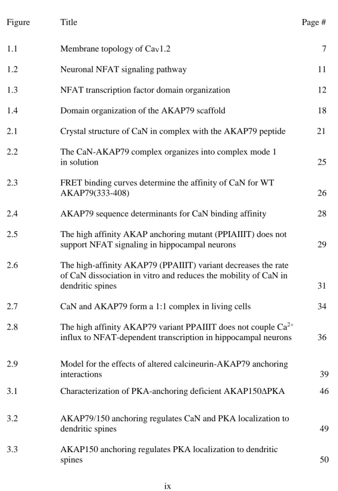
LIST OF FIGURES
Figure Title Page #
1.1 Membrane topology of CaV1.2 7
1.2 Neuronal NFAT signaling pathway 11
1.3 NFAT transcription factor domain organization 12 1.4 Domain organization of the AKAP79 scaffold 18 2.1 Crystal structure of CaN in complex with the AKAP79 peptide 21 2.2 The CaN-AKAP79 complex organizes into complex mode 1
in solution 25
2.3 FRET binding curves determine the affinity of CaN for WT
AKAP79(333-408) 26
2.4 AKAP79 sequence determinants for CaN binding affinity 28 2.5 The high affinity AKAP anchoring mutant (PPIAIIIT) does not
support NFAT signaling in hippocampal neurons 29 2.6 The high-affinity AKAP79 (PPAIIIT) variant decreases the rate
of CaN dissociation in vitro and reduces the mobility of CaN in
dendritic spines 31
2.7 CaN and AKAP79 form a 1:1 complex in living cells 34 2.8 The high affinity AKAP79 variant PPAIIIT does not couple Ca2+
influx to NFAT-dependent transcription in hippocampal neurons 36
2.9 Model for the effects of altered calcineurin-AKAP79 anchoring
interactions 39
3.1 Characterization of PKA-anchoring deficient AKAP150PKA 46
3.2 AKAP79/150 anchoring regulates CaN and PKA localization to
dendritic spines 49
3.3 AKAP150 anchoring regulates PKA localization to dendritic
x
3.4 AKAP79/150 anchoring of both CaN and PKA regulates depolarization triggered NFAT movement to the nucleus in
hippocampal neurons 52
3.5 AKAP79/150 anchoring of both CaN and PKA regulates NFAT movement to the nucleus in response to L-type Ca2+ channel
opening driven by spontaneous neuronal activity 55 3.6 AKAP79/150 anchoring of both CaN and PKA is required for
depolarization triggered NFAT translocation from dendritic
Spines 56
3.7 AKAP79/150 anchoring of both CaN and PKA is necessary for
NFAT-dependent transcription 58
3.8 Anchoring of both CaN and PKA to AKAP79/150 regulates basal phosphorylation of CaV1.2 59 3.9 LTCC Ca2+ signals are reduced in the absence of AKAP79/150
-PKA anchoring 62
3.10 LTCC Ca2+ signals imaged with GCaMP6f are reduced in the absence of AKAP79/150-PKA anchoring 65 3.11 Model for regulation of LTCC activity and nuclear signaling by
AKAP79/150-anchored PKA 66
4.1 Organization of the leucine zipper domains of AKAP79 and
CaV1.2 72
4.2 AKAP79LZ does not rescue AKAP150 shRNAi mediated loss of NFAT translocation to the nucleus 73 4.3 The AKAP79 LZ domain is required for depolarization triggered
NFAT movement to the nucleus in hippocampal neurons 75 4.4 The AKAP79 LZ is required for NFAT-dependent transcription 78 4.5 The AKAP79 LZ domain influences AKAP association with the
C-terminus of CaV1.2 81
4.6 The AKAP79 LZ domain is not required for regulation of LTCC
activity 82
xi
4.8 X-ray structure of the Fos/Jun heterodimer interaction with
NFAT and DNA 84
4.9 The NFAT LZ domain facilitates formation of the AKAP79
-NFAT complex 85
5.1 AKAP79/150 couples the LTCC to NFAT transcriptional
xii
LIST OF ABBREVIATIONS
AEBSF 4-(2-Aminoethyl) benzenesulfonyl fluoride hydrochloride AKAP A kinase anchoring protein
AMPAR α-amino-3-hydroxy-5-methyl-4-isoxazolepropionic acid receptor ANOVA analysis of variance
BAPTA 1,2-bis(o-aminophenoxy) ethane-N,N,N',N'-tetraacetic acid
BayK Bay K 8644
BSA bovine serum albumin
CaV voltage-dependent calcium channel
CaV1 voltage-dependent calcium channel, L-type CaV1.1-4 1S, 1C, 1E, and 1F subunits of CaV1 family cAMP cyclic adenosine monophosphate
CaM calmodulin
CaMKII Ca2+/calmodulin dependent protein kinase II CaNA calcineurin, A subunit
CaNB calcineurin, B subunit CaN calcineurin holoenzyme
cDNA complementary deoxyribonucleic acid CFP cyan fluorescent protein
CK I casein kinase I
D/D dimerization/docking domain of cAMP dependent protein kinase A
DIV days in vitro
xiii
ECL enhanced chemiluminescence substrate EDTA ethylenediaminetetraacetic acid
EGTA ethylene glycol tetraacetic acid EPSP excitatory postsynaptic potential FBS fetal bovine serum
FRAP fluorescence recovery after photobleaching FRET Förster resonance energy transfer
FRETc bleed-through corrected FRET FRETeff FRET efficiency
GCaMP green genetically encoded calcium indicator GFP green fluorescent protein
GluA1-4 AMPAR subunits 1, 2, 3, or 4 GSK3 glycogen synthase kinase 3 beta HEK-293 human embryonic kidney 293 cells HRP horse radish peroxidase
IP immunoprecipitation
IP3R inositol triphosphate receptor JNK c-jun N-terminal kinase
Kd/i equilibrium dissociation or inhibition constants Kon/off rate of association or rate of dissociation
L-LTP late LTP
LTCC L-type calcium channel LTD long term depression
xiv LTP long term potentiation
LZ leucine zipper
MAGUK membrane associated guanylate kinase domain MAPK mitogen activated protein kinase
mCh mCherry fluorescent protein MDCK Madin-Darby canine kidney cells mGluR metabotropic glutamate receptor
NB Neurobasal medium
NFATc nuclear factor of activated T-cells
NFATn nuclear resident NFAT-interacting transcription factors NIH National Institutes of Health
Nim nimodipine
NMDAR N-methyl-D-aspartate receptor GluN1-4 NMDAR subunits 1, 2, 3, or 4 PBS phosphate buffered saline PCR polymerase chain reaction
PDZ post synaptic density, Drosophila disc large tumor suppressor, zona occludens-1 protein
PIX AKAP79 PxIxIT motif
PKA cAMP-dependent kinase A
PKA RI/II regulatory subunits of PKA PKA C catalytic subunit of PKA PSD postsynaptic density
xv
RGECO-1 red genetically encoded calcium indicator for optical imaging SAP97 synapse associated protein 97
Sch Schaffer collaterall axons of CA3 pyramidal neurons SEM standard error of the mean
SH3 SRC homology 3 domain
shRNAi short hairpin interfering RNA
TTX tetrodotoxin
VGCC voltage gated calcium channel
WT wild-type
1 CHAPTER I INTRODUCTION
Despite decades of intense study, the underlying biology of learning and memory in the brain remains largely unclear. Considerable effort in the study of learning and memory has focused on an anatomically small structure within the limbic system known as the hippocampus. The importance of this structure is well described in human subjects with focal hippocampal lesions as they have profound deficits in the ability to recall new memories (Bartsch et al., 2010; Zola-Morgan and Squire, 1993). Further support for the hippocampus as a critical brain structure in cognition is evidenced by hippocampal abnormalities in human subjects with developmental and neurodegenerative diseases including Down’s syndrome, Alzheimer’s disease, epilepsy, and schizophrenia (Chang and Lowenstein, 2003; Harrison, 2004; Hyman et al., 1984; Isacson et al., 2002).
The hippocampus receives numerous excitatory projections both from the cerebral cortex and from within the hippocampus itself. As a nexus of integration, these electrochemical signals modify neuronal connections and circuit activity within the principal cell subfields of the hippocampus. On the cellular level, integration of synaptic information occurs in the dendrites of pyramidal cells, the principal excitatory neurons of the hippocampus. The highly branched dendritic arbor of a hippocampal neuron is densely arrayed with small biochemically isolated compartments called dendritic spines, each of which forms a single glutamatergic synapse with the axon terminal of a presynaptic neuron. As a result of their biochemical isolation from the parent dendrite, spines can independently
2
undergo an activity-dependent increase or decrease in strength after responding to a given neurotransmitter release event in a process known as synaptic plasticity (Nimchinsky et al., 2002). This widespread phenomenon occurs at glutamatergic hippocampal synapses and is bi-directional in nature, after which synapses may either respond more strongly to neurotransmitter, as in long-term potentiation (LTP), or more weakly, as in long term depression (LTD) (Malenka and Bear, 2004). Long-term persistence of LTP and LTD requires the concomitant activation of glutamate receptors, intracellular kinases and phosphatases, and the transcription and translation of plasticity related gene products. Initiation of intracellular signaling pathways that direct transcription of plasticity related genes is particularly dependent on L-type voltage-gated Ca2+ channels (LTCC). LTCC activity is critical for excitation-transcription coupling to a variety of signaling pathways. In particular, we have focused much of our attention on the role of LTCCs in coupling to nuclear factor of activated T-cells (NFAT) transcription factor signaling. However, little is known about the spatial and temporal dynamics of LTCC coupling to NFAT, nor is it clear how local kinase and phosphatase activity affects LTCC activity within subcellular compartments of hippocampal neurons. Herein, I will describe how recruitment of protein kinase A (PKA) and the serine-threonine phosphatase calcineurin (CaN) to the LTCC by the postsynaptic scaffolding protein AKAP79/150 allows for precise spatial and temporal control of LTCC activity and NFAT signaling.
3
Hippocampal synaptic plasticity as a cellular and molecular basis for learning and memory storage
Synaptic plasticity is now a well-established mechanism that provides a cellular and molecular basis for learning and memory in the brain. It was Santiago Ramon y Cajal who, in 1984 delivered the prestigious Croonian Lecture, where he proposed that plasticity of neuronal connections may be the substrates of memory. This declaration was incredibly prescient, however, because it was not until 1973 that neurophysiological experiments in the hippocampus of anesthetized rabbits first formally described long lasting synaptic plasticity (Bliss and Lomo, 1973). The hippocampus remains a favorite brain region for the study of the cellular basis of learning and memory. The organization of the hippocampus has lent itself to intense study not by chance but because of the visually prominent and experimentally robust tri-synaptic circuitry of the dentate granule cell-CA3-CA1 path. In particular, much effort has focused on a class of synapse in the hippocampus in which Schaffer collateral (Sch) axons of CA3 pyramidal neurons synapse with the dendrites of CA1 pyramidal neurons, the principal output neurons of the hippocampus. The Sch-CA1 synapse undergoes two stages of postsynaptic plasticity characterized temporally and typically defined by a dependence upon new protein synthesis. Rapid NMDAR-induced increases or decreases in synaptic strength (LTP or LTD lasting less than 1hr) involve local covalent modifications and trafficking of glutamate receptors in dendritic spines of CA1 pyramidal neurons (Esteban et al., 2003; Luscher et al., 1999; Mulkey et al., 1994; Yang et al., 2008). Long-lasting forms of postsynaptic plasticity (e.g. late LTP (L-LTP)) have been demonstrated to last for hours in vitro and up to a year in vivo (Abraham et al., 2002). Thus, L-LTP has received most of the attention as a potential molecular and
4
cellular mechanism for learning and memory. L-LTP is dependent upon covalent modifications at active synapses and within the nucleus where phosphorylation regulates transcription factor activity, gene expression and protein synthesis. LTCC are critical for coupling neuronal activity to induction of gene expression in L-LTP and will be discussed in more detail below.
Activity-dependent regulation of hippocampal synaptic plasticity
Excitatory hippocampal pyramidal neurons undergo activity-dependent changes in synaptic strength through the coincident detection of both depolarization and presynaptic glutamate release. Glutamatergic synaptic transmission and modulation occurs through a broad family of glutamate receptors that are both ionotropic and metabotropic in nature. However, though metabotropic and ionotropic kainate glutamate receptors play important roles in synaptic plasticity and disease (Gladding et al., 2009; Lerma and Marques, 2013), the contribution of the α-amino-3-hydroxy-5-methyl-4-isoxazolepropionic acid receptors (AMPAR) and the n-methyl-d-aspartate receptors (NMDAR) to synaptic plasticity are more clearly defined and will be focused on here.
Fast synaptic transmission in excitatory neurons is initiated when presynaptic axon terminals release glutamate into the synaptic cleft. Glutamate released into the synaptic cleft diffuses to the postsynaptic membrane and is detected by AMPAR on postsynaptic dendritic spines. The majority of AMPARs are found in a paired-pairs configuration composed of a heterotetramer of two GluA2 subunits and either two GluA1 or GluA3 subunits, with the subunit composition conferring distinct biophysical properties to the ion channel (Kauer and Malenka, 2006). Upon binding of glutamate to two or more AMPAR
5
subunits, the ion channel pore opens, permitting flow of primarily Na+ ions into the postsynaptic neuron causing a net depolarization of the postsynaptic membrane. The GluR2 subunit is responsible for controlling the ionic permeability of the AMPAR as seen in tetrameric assemblies that lack GluR2, which are permeable to Ca2+. Ca2+-permeable AMPARs may have important implications for synaptic plasticity and neuronal injury (Liu and Zukin, 2007).
The mechanism primarily responsible for induction of synaptic plasticity occurs through a second class of ionotropic glutamate receptor, the n-methyl-d-aspartate receptor (NMDAR). NMDARs differ from AMPAR-type glutamate receptors in many ways including subunit composition, agonist binding, ion permeation, and mechanism of activation. Like AMPARs, NMDARs are composed of a paired-pair tetramer consisting of two obligatory GluN1 subunits and either two GluN2A or GluN2B subunits. NMDARs are unique in the glutamate receptor family due to the requirement for binding of both glutamate to GluN2 and glycine or serine to GluN1. The NMDAR plays a critical role in the induction of synaptic plasticity because it acts as a molecular device controlling the coincidence detection of both glutamate and depolarization. In a resting neuron, Ca2+ conductance through the NMDAR is blocked by extracellular Mg2+. NMDAR coincidence detection occurs when repeated presynaptic glutamate release drives AMPAR-mediated depolarization of the postsynaptic neuron, relieving the Mg2+ block, and permitting Ca2+ influx through the NMDAR. A developmental switch occurs in early post-natal mammals in which NMDARs, initially composed of predominantly GluN1-GluN2B subunits become diluted by an increase in GluN2A subunit expression to include many more GluN1-GluN2A tetramers (Liu et al., 2004). This GluN1-GluN2A-GluN2B switch creates a critical period
6
during development for plasticity induction because GluN2A subunits have slower inactivation kinetics causing greater Ca2+ influx and stronger LTP (Flint et al, 1997, Barth and Malenka, 2001, Lu et al 2007). This observation of the importance of NMDAR-dependent plasticity supports the idea that learning plasticity is more readily achieved in young vs. old animals.
Ca2+ influx through the NMDAR causes a postsynaptic rise in Ca2+ within the dendritic spines of excitatory neurons, a necessary step for initiation of synaptic plasticity. At developmentally mature synapses, LTP is favored by activation of various kinases, with a steep requirement for Ca2+/calmodulin dependent kinase II (CaMKII) and also involves PKA and protein kinase C (PKC) among others (Malenka and Nicoll, 1999). LTP leads to numerous signaling cascades, many of which are still poorly understood. However, the principal design of these signaling networks is to facilitate phosphorylation and insertion of additional AMPAR into the postsynaptic plasma membrane and growth of the dendritic spine, leading to enhancement of synaptic strength. On the other hand, LTD or synaptic weakening, is also critically dependent on NMDAR Ca2+ influx, but can also be triggered through mGluRs. Though LTD is even less well understood than LTP, a long standing tenet involves the activity of phosphatases such as CaN and PP1 that dephosphorylate substrates in the postsynaptic spine. The primary target of these phosphatases are AMPARs, which are removed from the postsynaptic membrane during LTD, causing a weakening synaptic strength (Malenka and Bear, 2004).
7
Voltage-gated Ca2+ channel structure and function
In excitable cells, the voltage gated Ca2+ channel family is indispensable for an array of cellular processes including muscle contraction, insulin secretion, neurotransmitter release, and transcriptional regulation (Catterall, 2011). VGCCs are encoded by 10 genes that are divided into three evolutionarily distinct clades and are classified as CaV1 (CaV 1.1-4), CaV2 (CaV2.1-3), and CaV3 (CaV3.1-3). The CaV1 clade are referred to herein as L-type while CaV2 include P/Q-, N-, and R-type channel subtypes, and the CaV3 family are commonly referred to as T-type channels. VGCC isotypes are characterized by amino acid divergence in the -subunit and are defined by the range of voltage detection, ion selectivity, and sensitivity to drugs. All VGCCs are composed of three obligatory subunits Figure 1.1 Membrane topology of CaV1.2. The LTCC is composed of three obligatory
subunits: the pore-forming 1, intracellular , and predominantly extracellular 2. The
-subunit consists of four repeated domain modules (I-IV) containing 6 transmembrane segment (S1-S6), intracellular loops, and a long intracellular C-terminal tail. LTCC kinetics and open channel probability are influenced by the associated intracellular and the transmembrane 2 subunits.
8
including the pore forming subunit and the accessory subunits and 2 (Figure 1.1). The pore forming -subunit is composed of four homologous tethered domains (I-IV) each of which contains six transmembrane segments (S1-S6). VGCCs sense changes in membrane polarization by the twisting outward movement of four positively charged S4 segments causing channel opening. The four plasma membrane re-entrant S5-S6 loops form the selectivity filters of VGCCs while the larger I-II, II-III, and III-IV domain-intervening loops carry out distinct functional roles in the regulation of channel activity. Of particular interest to the studies described below are the LTCCs and they are defined by their selective blockade by dihydropyridines and their relatively depolarized range of voltage activation.
LTCCs are regulated to great and not entirely understood process of protein-protein interactions that occur in the intracellular loops and tails of the channel. CaMKII interacts with CaV1.2 primarily at its N and C-terminal intracellular tails and with the -subunit. The CaMKII interaction is thought to facilitate voltage and Ca2+ dependent facilitation through enhancement of channel open probability likely through a phosphorylation mechanism (Grueter et al., 2006; Hudmon et al., 2005; Lee et al., 2006). In LTCCs, loop I-II contains the alpha-interacting domain that binds to the subunit. -subunit binding alters the kinetics of activation and inactivation while increasing the current of CaV1.2 type LTCCs (Walker and De Waard, 1998). In addition, an ER retention signal has been identified in loop I-II that -subunit binding masks, promoting -subunit trafficking to the plasma membrane (Bichet et al., 2000). Growing evidence suggests that the II-III loop of CaV1.2 may serve as a protein-protein interaction site for SNARE proteins including Sx1A, SNAP-25, SNAP-23, syntaxin 2, and Syt1 (Arien et al., 2003; Cho et al., 2005; Coppola et al.,
9
2001; Ji et al., 2002; Wiser et al., 1999). This has important implications for LTCC regulation of exocytosis of synaptic plasticity related cargoes in neurons. However, the portion of the LTCC most often studied and implicated in regulation of channel activity is the intracellular C-terminal tail and its regulation by PKA phosphorylation with be discussed further below.
Neuronal CaV1 isoforms and their subcellular distribution
Two isotypes of LTCCs, CaV1.2 (or 1C) and CaV1.3 (or 1D) are expressed throughout the hippocampus. Both CaV1.2 and 1.3 expression can be detected in hippocampal neuron soma, dendrites, and in spines. However, it has been reported that CaV1.2 outnumbers CaV1.3 by a ratio of 4:1 in the hippocampus (Hell et al., 1993). Thus, it is most likely that CaV1.2 is the predominant LTCC isoform contributing to hippocampal LTCC current. While CaV1.2 knockout mice show deficits in hippocampal function (Moosmang et al., 2005), CaV1.3 knockout mice are normal when challenged with NMDAR-dependent and NMDAR-independent forms of hippocampal LTP (Clark et al., 2003). However, I cannot exclude a minor role for Cav1.3 in this work because CaV1.2 and 1.3 channels are not easily pharmacologically isolated nor is the potential regulation of CaV1.3 by AKAP79/150 well understood. At the cellular level, CaV1.2 is expressed in spines and dendritic shafts of CA1 pyramidal neurons of the hippocampus where they are poised to respond to rapid changes in membrane potential due to back propagating action potentials of excitatory postsynaptic potentials (EPSP) (Obermair et al., 2004).
10
Excitation-transcription coupling and L-type Ca2+ channel-dependent plasticity
LTCCs are voltage-activated either by membrane depolarization resulting from back propagating action potentials or from excitatory postsynaptic potentials initiated by NMDA and AMPARs localized within dendritic spines. LTCCs are involved in local Ca2+ signaling during synaptic plasticity (Yasuda et al., 2003), but also play an important role in the induction of a gene expression component of L-LTP that is independent of the NMDAR (Deisseroth et al., 2003; Grover and Teyler, 1990). LTCCs couple excitation to activation of transcription factors including CREB, MEF, and NFAT through kinases and phosphatases including, but not limited to, CaMKs, ERK, PKA, and CaN (Bading et al., 1993; Bito et al., 1996; Dolmetsch et al., 2001; Graef et al., 1999; Hall et al., 2007; Impey et al., 1996, 1998; Mermelstein et al., 2000; Murphy et al., 1991; Oliveria et al., 2012). The importance of CaV1.2 in learning and memory has been described in forebrain specific CaV1.2 knockout mice in which NMDAR-independent and protein synthesis dependent L-LTP is impaired. Furthermore, these mice show a defect in CREB activation and various forms of working memory (Moosmang et al 2005; Langweiser et al 2010).
NFAT signaling and function in neurons
Long lasting forms of synaptic plasticity require changes in the expression of various genes involved in plasticity related processes. Mediating transcriptional responses in neurons are a variety of transcription factors that become activated during changing neuronal activity states that include the nuclear factor of activated T-cells (NFAT) family of transcription factors.
11
The NFAT family is encoded by five gene products: NFATc1-4, and NFAT5. NFATc1-4 are monomeric, require activation by Ca2+/CaN signaling (Clipstone and Crabtree, 1992), and interact weakly with DNA. Conversely, NFAT5 exists as a dimer, interacts stably with DNA, and does not respond to Ca2+ signaling. Because this dissertation addresses the role of Ca2+ signaling in synaptic plasticity, I will focus here on the NFATc1-4 members of this transcription factor family.
In neurons, NFAT signaling is initiated when LTCCs open in response to AMPAR and NMDAR mediated membrane depolarization. An abundant population of CaM molecules are tethered to the LTCC intracellular loops and tails that sense Ca2+ influx and Figure 1.2 Neuronal NFAT signaling pathway. In neurons, LTCCs can couple electrochemical activity to transcription through a Ca2+- dependent signaling cascade resulting in NFAT transcription factor activation. Ca2+influx through the LTCC binds apoCaM, causing the Ca2+-CaM-dependent stimulation of CaN activation. Activated CaN dephosphorylates NFATc family members exposing a nuclear localization signal that results in nuclear import. Once in the nucleus, NFATc requires an association with NFATn transcription factors to efficiently initiate transcription of NFAT target genes. Nuclear kinases that include Dyrk1, Gsk3b, CK, and PKA rephosphorylate NFATc to regulate nuclear export.
12
cause the Ca2+/CaM-dependent activation of CaN. CaN then dephosphorylates NFATc proteins. NFAT dephosphorylation causes a conformational change revealing nuclear localization sequences (NLS) and masking nuclear export sequences (NES) resulting in nuclear import (Okamura et al., 2000). NFATc are subsequently exported after rephosphorylation by various kinases such as PKA, dual specificity tyrosine phosphorylation related kinase 1 (Dyrk1), casein kinase (CK), glycogen synthase kinase 3 (GSK3), and mitogen activated protein kinase (MAPK) family members (Beals et al., 1997; Chow et al., 1997; Gómez del Arco et al., 2000; Okamura et al., 2004; Zhu et al., 1998) (Figure 1.2). As described above, NFATc proteins have intrinsically weak DNA binding ability, therefore, to effectively interact with DNA, NFATc cooperate with other transcription factors that are referred to as NFATn. NFATn identified in neurons include AP-1, MEF2, and Sox2 transcription factors (Crabtree and Olson 2002). This is an important feature of NFAT signaling because it allows for the coincident integration of convergent signaling cascades.
Full length transcripts of the NFATc transcription factors generate relatively large proteins that range in size from 925 to 1075 amino acids in length. The NFATc homologues share a common domain organization consisting of unique N- and C-terminal transactivation domains, a complex regulatory domain, and a Rel homology region (RHR) involved in DNA and protein-protein interactions (Figure 1.3). The N- and C-terminal transactivation domains of the NFATc proteins are important for recruitment of transcriptional coactivators when bound to DNA (Luo et al., 1996). Association of NFATc family members with histone acetyltransferases such as p300 and CBP facilitate assembly of RNA polymerase at NFAT target genes (García-Rodríguez and Rao, 1998).
13
Transactivation domain phosphorylation further regulates NFATc activity in the nucleus (Ortega-Pérez et al., 2005). The regulatory domain of NFATc proteins contains both the PxIxIT and LxVP motifs that are required for efficient CaN binding and dephosphorylation. The PxIxIT motif, first identified in the NFAT proteins, has a relatively low affinity for CaN. This feature is important because it maintains sensitivity to cellular signals and prevents constitutive NFAT activity (Aramburu et al., 2000). A second critically important CaN docking site, the LxVP motif, is thought to facilitate association of substrate with the active site of CaN to ultimately drive catalysis (Grigoriu et al., 2013).
NFATc1-4 and their splice isoforms are expressed throughout the brain, including the olfactory bulb, hypothalamus, cerebral cortex, brainstem, and hippocampus (Bradley et al., 2005; Vihma et al., 2008). Because NFATc1-c4 are all expressed in the brain and exhibit considerable functional redundancy, determining a precise role for each NFATc Figure 1.3 NFAT transcription factor domain organization. All NFATc isoforms share a common domain architecture consisting of an N-terminal activation domain, a complex regulatory domain that confers Ca2+ regulation by calcineurin and an abundance of putative phosphorylation sites that regulate nuclear shuttling. The DNA binding domain or Rel homology region (RHR) contains the AP-1 transcription factor association domain and a putative leucine zipper conserved among all NFATc isoforms. The NFATc C-terminal region is also associated with transactivation functions in the nucleus. CaN interacts with the PxIxIT and LxVP motifs of NFATc isoforms to facilitate the efficient dephosphorylation of the serine-rich gatekeeper regions (SRR) and serine-proline repeat motifs (SP) to expose the nuclear localization signal (NLS) to the nuclear import machinery.
14
isoform remains a difficult task. Gene-knockout mice that lack individual NFAT proteins show mild deficits while combinatorial knockouts are lethal due to disorganized development of the vasculature and nervous system. Knockout of the NFATc2, c3 and c4 isoforms results in defective axon outgrowth in the embryonic brain suggesting that NFAT signaling is required for axon guidance during development (Graef et al 2003). It remains to be seen if conditional forebrain-specific knockouts could reveal the importance of NFAT signaling in mature animals. In cardiac myocytes, NFATc3 down-regulates the expression of the 1 subunit of large conductance Ca2+-activated K+ (BK) channels causing impaired Ca2+ sensitivity of the channels during hypertension and exacerbating smooth muscle contractility (Nieves-Cintron et al 2007). This type of regulation of BK channels has not been examined in detail in neurons but could have important implications for CaN/NFAT signaling in regulation of neuronal excitability and synaptic plasticity because LTCCs have been shown to couple locally to BK channels in neuronal nano-domains (Liu et al., 2004). The role of NFAT signaling in mature neuronal circuits is still unknown and because the genes regulated by the NFATc have primarily been determined outside the nervous system, only a handful of neuronal transcriptional targets of NFATc proteins have been identified. However, the few transcriptional targets that have been identified offer some intriguing potential interactions with known synaptic plasticity mechanisms and include brain derived neurotrophic factor (BDNF) (Groth and Mermelstein, 2003; Groth et al., 2007), inositol triphosphate receptors (IP3R) (Carafoli et al., 1999; Genazzani et al., 1999; Graef et al., 1999), M-type K+ channels (Zhang and Shapiro, 2012), and Down syndrome critical region gene 1 (DSCR1), a negative regulator of CaN (Cano et al., 2005).
15
Regulation of CaV1 by protein kinase A phosphorylation
PKA is composed of two catalytic subunits and a constitutively dimerized regulatory subunit that is held together by a four helix bundle known as the dimerization/docking (D/D) domain (Taylor et al., 2013). In the classical view, the catalytic subunits of PKA are inactive in the absence of cAMP due to allosteric inhibition by the regulatory subunits. When cAMP levels rise, due to GPCR activation or stimulation of Ca2+-sensitive ACs, cAMP binds to the regulatory domains causing a conformational change that relieves the allosteric inhibition and releases active catalytic subunits. Though PKA catalytic activity is enhanced by cAMP, the notion that PKA is inactive in the absence of cAMP is being challenged (Smith et al., 2013). PKA is more likely to reside within a spectrum of activity depending upon subcellular localization and local cAMP concentration.
The importance of PKA phosphorylation of LTCCs was first implicated in the heart where adrenergic stimulation was shown to enhance a slow inward Ca2+ current (Reuter, 1967). It was later demonstrated that cAMP and the cAMP dependent kinase PKA were involved in transduction of the -adrenergic stimulus to Ca2+ current enhancement (Osterrieder et al., 1982; Reuter, 1974; Trautwein et al., 1982; Tsien, 1973; Tsien et al., 1972). In addition to -adrenergic signaling there is evidence that basal PKA phosphorylation of LTCCs is required for their response to membrane voltage (Armstrong and Eckert, 1987; Ono and Fozzard, 1992). Additionally, endogenous activation of PKA was shown to potentiate the current elicited in heterologous cells expressing the full length isoform of CaV1.2, further supporting the role of direct PKA phosphorylation on enhancement of channel currents (Yoshida et al., 1992). Furthermore, one primary
16
phosphorylation site in the C-terminal tail of CaV1.2 was proposed to mediate the effects of PKA, Ser1928 (De Jongh et al., 1996). This was demonstrated when mutation of Ser1928 to alanine reduced the current enhancement caused by PKA activation using a non-hydrolysable form of cAMP by 80% (Gao et al. 1997). For some time it was widely accepted that Ser1928 was the primary site for PKA phosphorylation (Davare et al., 2001; Hall et al., 2006; Hulme et al., 2006; Kamp and Hell, 2000). However, controversy has clouded our understanding of which sites are truly critical for PKA enhancement of LTCC activity (Weiss et al 2013). Recent work has examined the requirement for Ser1928 in PKA mediated LTCC current enhancement by expressing a CaV1.2 Ser1928Ala mutant in either tsA-201 cells, virally transduced ventricular myocytes, or in dissociated mutant mouse myocytes. All of these studies suggested that Ser1928 is not required for -adrenergic enhancement of LTCC currents in non-neuronal cells (Lemke T et al 2008; Fuller MD et al 2010). Alternative conflicting lines of evidence suggest that another PKA site, Ser1700 is either not required (Yang et al. 2013) or alternatively, critical (Fuller MD et al 2010; Fu et al 2013) for -adrenergic enhancement of LTCC activity in heart. It is safe to say that the mechanism for PKA enhancement of LTCC activity is a mystery in the heart and will require more work to reconcile the ever expanding amount of conflicting evidence. The relevance of Ser1700 and Ser1928 in regulation of LTCC activity in the brain is largely unexplored. In opposition to PKAs role in stimulating LTCC activity, Ca2+-mediated feedback regulation of LTCCs occurs through a CaM/CaN-dependent mechanism (Peterson et al., 1999). In neurons, this mechanism depends on local anchoring to the LTCC nano-domain and will be discussed in more detail in chapter III.
17
Regulation of hippocampal synaptic plasticity by the postsynaptic scaffolding protein AKAP79/150
The biochemical pathways that underlie synaptic plasticity are under profound second messenger regulation through reversible biochemical reactions that act as chemical switches controlling neuronal signaling outcomes. Of particular importance in neuronal signaling is phosphorylation. The addition of phosphate groups by protein kinases causes rapid and reversible changes in protein function. Protein kinases are activated by second messengers that include Ca2+, (cyclic adenosine monophosphate (cAMP), and/or phospholipids to catalyze transfer of phosphate groups from adenosine triphosphate to serine, threonine, or tyrosine residues on substrate proteins. Conversely, phosphates are removed by phosphatases either constitutively or in a Ca2+-dependent manner. The degree of ensemble target protein phosphorylation thus reflects the balance between competing actions of kinase and phosphatase signaling pathways. Because kinases and phosphatases comprise a significant percentage of total neuronal protein, local regulation of the spatial and temporal specificity of phosphorylation is required. Enzyme-scaffold complexes are critical to many signaling pathways acting as nodes for integration of complex cellular signals by providing local enrichment of enzyme, substrate, and regulatory elements in distinct subcellular compartments (Good et al., 2011; Pawson and Scott, 2010; Zeke et al., 2009).
Though not appreciated at the time, MAP2 was the first member of a large family of scaffolding proteins known as A-kinase Anchoring Proteins (AKAPs) shown to interact with the RII regulatory subunit of the PKA holoenzyme (Theurkauf and Vallee, 1982).. AKAPs interact with PKA at nanomolar affinity through a 14-18 amino acid amphipathic
18
alpha helix that is buried within a hydrophobic groove formed by the D/D domain. (Carr et al., 1991; Kinderman et al., 2006; Newlon et al., 2001; Sarma et al., 2010; Scott et al., 1990). While the majority of AKAPs are specific for the RII subunit of PKA, there are also RI specific AKAPs and those that bind both RI and RII known as “dual specific” AKAPs (Huang et al., 1997). In addition to PKA, AKAPs often interact with other signaling enzymes including protein phosphatases, other kinases, phosphodiesterases (PDE), and andenylyl cyclases (AC) to form multi-enzyme signaling complexes that confer spatial and temporal precision in compartmentalized subcellular signaling through identifiable targeting motifs (Smith et al., 2006a). Within the complex cellular architecture of neurons, local signaling is likely to be of utmost importance. In postsynaptic spines of excitatory neurons, an electron dense structure known as the post synaptic density (PSD) is localized in close apposition to the synaptic cleft where a network of scaffolding and adapter Figure 1.4. Domain organization of the AKAP79 scaffold. The AKAP79 postsynaptic scaffold has a modular domain structure regulating interactions with many postsynaptic components involved in synaptic plasticity. An N-terminal polybasic region is known to interact with membrane phospholipids, F-actin, adenylyl cyclases, CaM, and PKC (some not shown). Secondary interactions with AMPAR and NMDAR are mediated by PSD-95 and SAP-97 binding to the MAGUK domain. The PxIxIT motif interacts with the CaNA subunit to mediate CaN holoenzyme anchoring. The classical amphipathic a-helix common to all AKAPs anchors PKA RII regulatory subunits to AKAP79/150. At the distal C-terminus is a leucine zipper motif that confers CaV1.2 binding through a complementary leucine zipper in the C-terminus of CaV1.2.
19
proteins, receptors, voltage sensing ion channels and signaling molecules are poised to respond to local synaptic activity. In this structural network, local scaffolding in multi-protein complexes allows for spatial precision and temporal efficiency in regulation of phosphorylation by kinases and phosphatases.
The A-kinase anchoring protein 79/150 (AKAP79/150) (note: AKAP79 is the human form while AKAP150 is the rodent orthologue) (also referred to as AKAP5) is localized within the PSD of excitatory neurons of the hippocampus through interactions with plasma membrane bound phospholipids (Gomez et al., 2002), the actin cytoskeleton (Colledge et al., 2000), and MAGUK adapter proteins (e.g. PSD-95 & SAP97) (Carr et al., 1992; Robertson et al., 2009). AKAP79/150 anchors the regulatory subunit (RII) of PKA and the catalytic subunit of the phosphatase CaN (CaNA) where they form an opposed regulatory loop in a microdomain able to respond to their respective Ca2+-stimulated effectors cAMP and Ca2+-CaM to modify local substrates (Dell’Acqua et al 2002; Coghlan et al 1995; Oliveria et al 2003). It has become clear that AKAP79/150 is a key regulator of the molecular constituents of the PSD from experimental evidence describing primary and secondary molecular interactions with PKC, CaM, ACs, MAGUKs, glutamate receptors, the cytoskeleton, various ion channels, and plasma membrane lipids.
20 CHAPTER II
PRECISELY TUNED DYNAMICS OF AKAP79/150-ANCHORED CAN ALLOWS FOR EFFICIENT Ca2+-CAN-NFAT SIGNALING1
Introduction
CaN is a serine/threonine protein phosphatase containing a ~60 kDa catalytic subunit (CaNA) and a ~19 kD regulatory subunit containing Ca2+ binding EF-hands (CaNB) (Aramburu et al., 2000). The Ca2+ sensing protein calmodulin translates intracellular Ca2+ signals into CaN activation through a high affinity interaction that causes displacement of an autoinhibitory segment from the CaN catalytic site. CaN signaling is important for cardiac development and pathophysiology, for nervous-system development, and for some of the plastic changes in neurons that are believed to underlie learning and memory (Groth et al., 2003; Heineke and Molkentin, 2006; Hogan et al., 2003; Molkentin and Dorn, 2001; Zeng et al., 2001). CaN-NFAT signaling has been intensely studied for its role in immune-cell activation and as the target of the immunosuppressive drugs cyclosporine A and FK506 (Macian, 2005). These studies led to the identification of a recognition sequence for CaN that is shared by many CaN substrates and that was first described as having the consensus sequence PxIxIT (Aramburu et al., 1998). A high-affinity version of the PxIxIT sequence, PVIVIT, was selected from a randomized peptide library (Aramburu et al., 1999) and used to define the structural basis of substrate recognition by CaN (Li et al., 2004, 2007). CaN recognition sites display considerable
1 This chapter has been modified with permission from Li, H., Pink, M.D., Murphy, J.G., Stein, A.,
Dell’Acqua, M.L, Hogan, P.G. Balanced Interactions of Calcineurin with AKAP79 regulate Ca2+
21
natural variation in their sequences and their affinities for CaN (Li et al., 2007, 2011). The recognition sites of NFAT proteins (PRIEIT, PSIRIT and PSIQIT) display an intermediate Kd of ~25 μM for CaN, whereas the sequences PQIIIS in human TRESK and PVIAVN in yeast Hph1 bind CaN with Kds of 5 μM and 250 μM, respectively. The strength of individual CaN-substrate interactions is an important parameter for intracellular signaling, and, in particular, increasing the strength of a calcineurin-substrate interaction above that of the wild-type proteins can lead to disordered signaling (Aramburu et al., 1999; Li et al., 2011; Roy and Cyert, 2009; Roy et al., 2007).
As introduced above, enzyme-scaffold interactions play a critical functional role in physically assembling the components of many signaling pathways. AKAP79/150 anchors the kinase PKA, the phosphatase CaN, and other proteins and targets them to a variety of locations in the plasma membrane (Coghlan et al., 1995; Logue and Scott, 2010). AKAP79/150 is of particular interest because it balances the opposing effects of calcineurin and PKA on neuronal voltage-gated LTCCs (Oliveria et al., 2007) and coordinates the activities of calcineurin, PKA and PKC on a number of other channels and receptors (Hall et al., 2007; Hoshi et al., 2003; Lin et al., 2011; Liu et al., 2004; Pawson and Scott, 2010; Sanderson and Dell’Acqua, 2011; Tavalin et al., 2002; Tunquist et al., 2008). In rat hippocampal neurons, NFAT signaling is initiated by the local elevation of Ca2+ concentration near L-type Ca2+ channels (Graef et al., 1999; Oliveria et al., 2007). RNA interference (RNAi)-mediated depletion of endogenous AKAP150, eliminates NFAT activation in neurons, which, however, can be rescued by expression of human AKAP79 (Oliveria et al., 2007). A conserved CaN-binding site has been mapped in AKAP79 (Figure 2.1A), and activation, despite abundant CaN elsewhere in the cytoplasm,
22
23
deletion of this anchoring sequence eliminates the ability of AKAP79 to rescue NFAT Figure 2.1. Crystal structure of CaN in complex with the AKAP79 peptide. A) Schematic of human AKAP79 showing the location of the CaN PxIxIT-like anchoring motif and sequence alignment relative to the PVIVIT peptide. Numbering for AKAP79 and the PVIVIT peptide are shown above and below the sequences respectively. B) Three dimensional shaded ribbon diagram depicting the overall structure of the CaNA (brown)-calcineurin B (magenta) heterodimer in complex with AKAP79 peptide. The key interacting residues of AKAP79 are labeled in stick form at the surface of CaNA. C) A 2F0-Fc map contoured at 1s describes the electron density which defines the Ile340-Ile342 stretch at the core of the CaN-anchoring motif of AKAP79. D) Overlaid ribbon diagram of the AKAP79 PIAIIIT containing peptide with the previously solved PVIVIT crystal structure(Li et al., 2007) demonstrates conservation of the CaN-AKAP79 contact mode. Slight backbone flexion of AKAP79 is visible due to Ile338 occupying the “proline pocket” of CaNA. Core residues are shown as in “B”. E) The H-bond between Thr343 of AKAP79 and Asn330 of CaNA recapitulates a thermodynamically critical contact in the CaN-PVIVIT structure. F) Side-by-side comparison of the CaN–AKAP79 structure (left) and CaN–PVIVIT structure (right) viewed from the ‘proline pocket’ defined in the CaN– PVIVIT structure. Calcineurin is shown in surface representation with residues in direct contact with the peptides colored orange, and the peptides are displayed in stick representation. Figure adapted from (Li et al., 2012).
24
emphasizing the local nature of CaN-NFAT signaling and the importance of CaN scaffolding by AKAP79 in these cells (Dell’Acqua et al., 2002; Oliveria et al., 2007).
Building off of work that defined the structural basis for the CaN-PVIVIT interaction (Li et al., 2007), our colleagues Dr. Patrick Hogan and Dr. Huiming Li undertook X-ray crystallographic studies to examine the structure, affinity, and kinetics of the CaN-AKAP79 interaction in a fruitful collaboration with our lab (Li et al., 2012). Here I will describe the results of their efforts and the work of Matthew Pink Ph.D., a former graduate student in the Dell’Acqua lab, as an introduction to the results I gathered as a co-author on the final manuscript.
Structural insights into the AKAP interaction. The crystallized CaN-AKAP79 complex bears one AKAP PIAIIIT peptide sandwiched between two CaNA-CaNB heterodimers in an asymmetric arrangement. Just as in the CaN-PVIVIT structure that describes consensus CaN substrate recognition (Li et al., 2007), the AKAP79 peptide is in closer apposition to one of the two CaN heterodimers in the crystal (Figure 2.1B). The 2 Å resolution of the crystal structure determined by Li and Hogan clearly defined the electron density of mutual surfaces of the CaN-AKAP79 interaction (Figure 2.1C). In complex, the AKAP peptide assumes an extended -strand configuration along -strand 14 of CaNA while seven H-bonds position CaNA in contact with residues Ala339 and Thr345 of AKAP79 (Figure 2.1B-D). In an arrangement identical to that seen in the CaN-PVIVIT complex, Ile340 and Ile342 of AKAP79 make hydrophobic contacts within the trough between -strand 11 and 14 of CaNA (Figure 2.1C), while Thr343 of AKAP79 forms an H-bond with Asn330 of CaNA (Figure 2.1E). Interestingly however, Ile338 of AKAP79 takes the position of the proline residue in the CaN-PVIVIT complex and to
25
approximately the same depth within the structure of CaNA (Figure 2.1F), causing a slight displacement of the backbone at Ile338 of AKAP79 away from the CaN surface when compared to PVIVIT (Figure 2.1D). Based on the structure obtained by Li and Hogan, the N-terminal flanking region of the AKAP79 peptide is poorly defined while the C-terminal residues form weak van der Waals contacts with the surface of CaNA. The poor interactions outside the PxIxIT motif of AKAP79 are consistent with the idea (Li et al., 2007) that regions outside the core recognition peptide of CaNA interacting proteins contribute only weakly to the Gbinding and may subserve the interaction though residing in a low energy configuration.
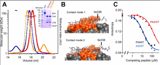
Studies of the CaN-AKAP79 complex in solution. Armed with the crystal structure of the CaN-AKAP79 complex, our collaborators further defined the stoichiometry and contact mode in vitro through solution binding experiments. Using size exclusion chromatography coupled with multi-angle light scattering (SEC-MALS) demonstrated a clear shift in the migration of the CaN peak upon binding an AKAP79 fragment (333-408). This shift corresponded to a ~8 kDa increase in mass suggestive of a 1:1 complex between AKAP79 (333-408) and CaN (Figure 2.2A). The asymmetric arrangement of the CaN-AKAP79 crystal structure offered two possible modes of contact between the PIAIIIT motif of AKAP79 and CaN in a 1:1 complex (Figure 2.2B). Contact mode 1 buries 1332 Å2 of surface area and makes seven backbone H-bonds while contact mode 2 buries 820 Å2 and makes four backbone H-bonds. One prominent difference between the two contact modes is whether Ile338 is buried within the “proline pocket” of the CaN binding surface or not (Figure 2.2B). Mutation of Ile338 to alanine reduced the affinity of the PIAIIIT for CaN substantially in a competitive binding assay suggesting that
26
Ile338 is important for CaN binding favoring contact mode 1. Further, although both contact modes 1 and 2 in the crystal structure suggested that Pro337 did not play an important role in the CaN-AKAP79 (PIAIIIT) interaction, the significant energetic contribution of Pro6 in the PVIVIT structure led to our collaborators to determine what effect mutation of Pro337 to alanine had on binding. Interestingly, although Pro337 might appear at first glance to potentially be important in the PxIxIT motif of CaN binding partners, it had no impact on CaN-AKAP affinity (Figure2.2C) suggesting that Ile338 of AKAP79 is functionally homologous to Pro6 in PVIVIT by interacting with the “proline pocket” of CaN. Taken together, the SEC-MALS and competitive binding data establish that CaN binding at the IAIIIT site, in solution, gives a 1:1 complex, and support the structural model depicted in Figures 2.1B and 2.1F.
Figure 2.2. The CaN-AKAP79 complex organizes into contact mode 1 in solution. A) SEC-MALS analysis of CaN alone (red), CaN–AKAP complex (orange) and BSA standard (blue). The corresponding SDS-polyacrylamide gel is shown (inset). AU, arbitrary units. (b) Alternative 1:1 complexes based on the two calcineurin molecules in the crystal asymmetric unit. (c) Competition of the indicated GST-AKAP79333–348 fusion proteins with fluorescent PVIVIT peptide for binding to calcineurin. The estimated Kis are AIAIIIT, 3.7 μM; PIAIIIT, 3.9 μM; PAAIIIT, 47 μM. Total competitor concentration (not free concentration) is plotted. Figure adapted from (Li et al., 2012)
27
Our lab has previously shown that the IAIIIT anchoring site of AKAP79/150 is essential for NFAT signaling in hippocampal neurons because it underlies recruitment of CaN to AKAP79 complexes adjacent to the L-type Ca2+ channel (Oliveria et al., 2007). Recruitment of CaN to AKAP-scaffolded complexes in neurons will depend on the concentration of free CaN and on the Kd of the binding interaction. To obtain a direct estimate of CaN-AKAP79 Kd, our collaborators made quantitative measurements of FRET Figure 2.3. FRET binding curves determine the affinity of CaN for WT AKAP79 (333-408).A) CaN binding to AKAP79333–408 was monitored by stopped-flow FRET measurements. The observed time course of binding at 22 °C is shown for four CaN concentrations. B) A plot of plateau FRET intensities against CaN concentrations. C) The association rate constant kon estimated from the data in “A” based on a model of reversible binding to a single class of sites. D) Dissociation of the CaN–AKAP79333–408 complex at 22 °C. In this AKAP79333–408 protein, the RIIα-anchoring site was replaced by the AKAP–in silico (AKAP-IS) sequence (Alto et al., 2003; Gold et al., 2006). Data shown are representative of two experiments. AU, arbitrary units. Figure adapted from (Li et al., 2012)
28
between an ATTO425-AKAP79 (333–408) donor and rhodamine-CaN acceptor. Titration with increasing concentrations of CaN yielded a binding curve at room temperature that could be fitted with Kd = 0.4 µM (Figure 2.3A, B; Table 2.1), supporting the Kd they estimated independently from kinetic measurements, ~0.3 µM (Figure 2.3C, D). Thus, our collaborators concluded that interaction at the IAIIIT site can effectively recruit CaN in neurons if the free CaN concentration is in the high hundreds of nanomolar to low micromolar range. The rapid dissociation of CaN-AKAP79 peptide complex is particularly relevant to the transmission of intracellular signals. Even though AKAP79 and NFAT utilize the same surface to dock on CaN, recruitment of CaN to AKAP79 need not be in conflict with productive signaling because of the rapid equilibration between free CaN and AKAP-bound CaN. Experiments reported below establish that dissociation at physiological temperature is even more rapid than in the experiment of Figure 2.3.
Systematic analysis of the sequence determinants for the CaN-AKAP complex. Again, the new knowledge of the CaN–AKAP79 structure gave our collaborators an opportunity to investigate the effect of systematically varying the affinity of this enzyme-scaffold interaction. Building on earlier studies performed by Li and Hogan that defined the CaN–PVIVIT structure and the rules that connect changes in the sequence of CaN recognition sites to variations in affinity for substrate (Li et al., 2007), they designed a small panel of AKAP79 anchoring peptides whose affinities for CaN span a wide range. Informed by the crystal structure of the CaN-AKAP79 complex, our colleagues replaced amino acids I338P, T343A, or both. For the remainder of this chapter, WT and variant AKAP proteins are referred to by 7mer sequences to facilitate comparison with the variations introduced. Competitive binding experiments in which these unlabeled AKAP
29
variants, as GST fusion proteins, were used to displace labeled PVIVIT demonstrated that the relative affinities were PPAIIIT > PIAIIIT > PPAIIIA > PIAIIIA, covering more than a 100-fold range in Kd (Figure 2.4A,B; Table 2.1). Notably, the PPAIIIT variant, in which Ile338 was substituted with Pro, bound ~4-fold more strongly to CaN than WT AKAP79. Regulation of NFAT signaling by dynamics of AKAP79-anchored CaN. In collaboration with Li and Hogan, a former graduate student in the Dell’Acqua lab, Matthew Pink Ph.D., explored the effect of varying the affinity of CaN-AKAP79 interactions on activation of the CaN-dependent NFAT signaling pathway in hippocampal neurons. Full-length AKAP79 proteins with either the WT PIAIIIT anchoring sequence or one of the mutated sequences PPAIIIT (high affinity), PPAIIIA (intermediate affinity), and PIAIIIA (low affinity) were cloned and expressed in rat hippocampal neurons. NFAT signaling was stimulated by using a high extracellular K+ depolarization (Figure 2.5A), a stimulus protocol that activates LTCC signaling to CaN and triggers NFAT Figure 2.4. AKAP79 sequence determinants for CaN binding affinity. A) Recombinant GST, GST-tagged wild-type AKAP79333–408 (PIAIIIT, lane 2), and the CaN-anchoring site variants PIAIIIA, PPAIIIA and PPAIIIT of AKAP79333-408 (lanes 3–5) were analyzed by SDS-PAGE and stained with Coomassie Brilliant Blue. B) Kis estimated in a competitive binding assay are PPAIIIT, 0.08 μM; wild type, 0.36 μM; PPAIIIA, 12 μM; and PIAIIIA, 39 μM. Total competitor concentration (not free concentration) is plotted. Data shown are representative of three experiments. Figure adapted from (Li et al., 2012).
30
Figure 2.5. The high affinity AKAP anchoring mutant (PPAIIIT) does not support NFAT signaling in hippocampal neurons.A) KCl stimulus protocol previously shown to activate L-type Ca2+-channel signaling through calcineurin in hippocampal neurons (Graef et al., 1999; Oliveria et al., 2007). B) Summed intensity projection images of neuronal cell bodies and proximal dendrites in non-stimulated (NS) cultures and in cultures fixed at the indicated times after KCl stimulation. Transfection with control plasmid, AKAP150 RNAi plasmid and RNAi-resistant AKAP79 expression plasmids is indicated. The paired images show YFP or AKAP-YFP (white), DAPI-stained nuclei (blue) and endogenous NFAT (red). Scale bar, 10 μm. C) Time course of NFAT nuclear import after KCl stimulation, from experiments as in “B”, quantified as nucleus-to-cytoplasm mean fluorescence intensity ratios (Oliveria et al., 2007). Each point represents n = 12–25 neurons, and in each case the data have been normalized to the value for non-stimulated cultures (t = 0 min). Statistical comparisons were by one-way ANOVA with a Bonferroni post-hoc test, *P < 0.05 and **P < 0.01 compared to AKAP79WT rescue. Error bars, s.e.m. Figure adapted from (Li et al., 2012)
31
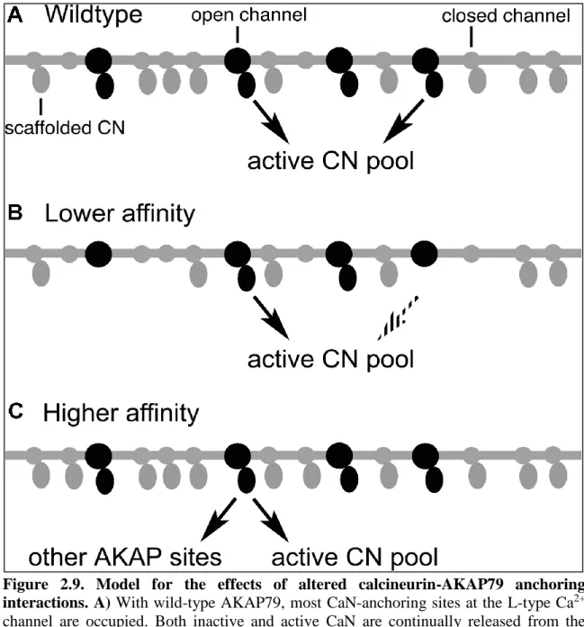
nuclear translocation and NFAT-dependent gene expression (Graef et al., 1999; Oliveria et al., 2007). Endogenous NFATc4 was localized immunocytochemically and quantified as the nucleus to cytoplasm ratio of fluorescence intensity. As shown previously, knockdown of endogenous AKAP150 causes a loss of NFAT nuclear translocation that can be rescued by RNAi insensitive human AKAP79 (Figure 2.5B and C). While intermediate affinity AKAP79 (PPAIIIA) rescued NFATc4 nuclear translocation as efficiently as WT AKAP79, the lowest affinity AKAP79 (PIAIIIA) rescued less efficiently (Figure 2.5B and C). Previous work has shown that AKAP79 lacking the CaN-anchoring site does not rescue CN-NFAT signaling (Oliveria et al., 2007). Interestingly, the highest affinity AKAP79 (PPAIIIT) mutant was similarly unable to rescue signaling (Figure 2.5D and E).
One interpretation for the failure of the high affinity mutant AKAP79 sequence PPAIIIT to allow signaling is that Ca2+-CaM/CaN is no longer able to release from the AKAP79 scaffold to bind to NFAT after activation. Li and Hogan compared dissociation of calcineurin from its complexes with wild-type AKAP79 (333–408) (PIAIIIT) and with the PPAIIIT variant in stopped-flow FRET experiments at 36 °C (Figure 2.6A and B). Preformed CaN–AKAP79 complex was mixed with an excess of unlabeled PVIVIT peptide to prevent rebinding of calcineurin to AKAP79, and complex dissociation was monitored as the decrease in the FRET signal. The dissociation of calcineurin from wild-type AKAP79 was rapid, with the mean dissociation rate 9.96 s–1 (range 8.84–11.84 s–1, n = 4), corresponding to a half-time of ~70 ms (Figure 2.6A). This result implied that calcineurin can rapidly expose its NFAT-binding surface even if that surface is initially engaged in a physiological complex with wild-type AKAP79. Dissociation of the higher-affinity calcineurin–PPAIIIT AKAP79 complex was appreciably slower, with mean
32
Figure 2.6. The high-affinity AKAP79 (PPAIIIT) variant decreases the rate of CaN dissociation in vitro and reduces the mobility of CaN in dendritic spines.
33
dissociation rate 4.24 s–1 (range 3.90–4.55 s–1, n = 4) and corresponding half-time ~163 ms Figure 2.6. The high-affinity AKAP79 (PPAIIIT) variant decreases the rate of CaN dissociation in vitro and reduces the mobility of CaN in dendritic spines. A, B) Dissociation of calcineurin from wild-type AKAP79333–408 “A” and its I338P variant “B” at 36 °C. AU, arbitrary units. C) Time-lapse images of wild-type AKAP79-YFP fluorescence recovery in single dendritic spines of rat hippocampal neurons in culture at the indicated times after photobleaching. A pre-bleach image (t = −10 s) is shown for comparison. D) YFP fluorescence recovery in neurons cotransfected with CaNA–YFP and wild-type AKAP79-CFP or AKAP79-CFP variants as indicated. CFP fluorescence is not shown. E, F) Percent YFP fluorescence recovery plotted against time for the FRAP experiments illustrated in c and “D” and for similar experiments with the other AKAP79 variants PIAIIIA and ΔPIX. In the experiment with CN-YFP alone, transfection with the RNAi plasmid was omitted. Max percent recovery (mobile fraction) values stated in the text were calculated from the curves in “E” and “F” by fitting a single exponential function. Statistical P values stated in the text were determined by one-way ANOVA with a Bonferroni post-hoc test (n = 10–28 neurons). Error bars, s.e.m.