DISSERTATION
5-HYDROXYMETHYLCYTOSINE AND ENDONUCLEASE G AS REGULATORS OF HOMOLOGOUS RECOMBINATION
Submitted by Crystal M. Vander Zanden
Department of Biochemistry and Molecular Biology
In partial fulfillment of the requirements For the Degree of Doctor of Philosophy
Colorado State University Fort Collins, Colorado
Summer 2017 Doctoral Committee: Advisor: P. Shing Ho Olve Peersen Santiago Di Pietro Nick Fisk
Copyright by Crystal M. Vander Zanden 2017 All Rights Reserved
ii ABSTRACT
5-HYDROXYMETHYLCYTOSINE AND ENDONUCLEASE G AS REGULATORS OF HOMOLOGOUS RECOMBINATION
Homologous recombination (HR) is a necessary biological process for all living organisms, and it is especially important for repairing damaged DNA. Improper HR results in DNA damage-related diseases, notably increased likelihood of cancer when HR regulators, such as the human BRCA1 gene, are impaired. HR is also a tool for biotechnology, giving scientists the power to easily delete or mutate genes and study the effects of those modifications. Recently, the epigenetically modified nucleotide 5-hydroxymethylcytosine (5hmC) was found to regulate
vertebrate HR via interaction with the protein endonuclease G (EndoG). In this dissertation, I use biochemical/biophysical methods to elucidate the interaction between 5hmC and EndoG, thus
working towards understanding their roles as regulators of recombination. I find that 5hmC forms
a unique hydrogen bond to stabilize Holliday junctions, the four-stranded DNA intermediate in HR. 5hmC also induces a global structure change to the junction, increasing protein access to the
junction crossover and providing potential for either direct or indirect readout of 5hmC. Further
connecting EndoG with recombination, we present the first evidence that EndoG preferentially binds and cleaves Holliday junction DNA, implicating a role for EndoG as a resolvase. I demonstrate that EndoG recognizes 5hmC in the junction context and observe unique cleavage
products from EndoG interaction with 5hmC-junctions. These results suggest that EndoG may
have a previously unrecognized junction resolvase function and, in this way, play a more direct role in recombination than simply creating double-stranded breaks in duplex DNA to initiate the
iii
HR mechanism. Finally, I present a unique structural feature of vertebrate EndoG that we hypothesize is the basis for 5hmC recognition. I present the structure of mouse EndoG and
propose that a two amino acid deletion, conserved in vertebrate EndoG sequences, is associated with unraveling of an α-helix. This structural perturbation positions amino acid side chains to confer 5hmC-sensing ability to all vertebrate EndoG. I expect that these deletion mutations and resulting structural effects co-evolved with the appearance of 5hmC in vertebrate genomes to give
EndoG an additional function of recognizing 5hmC in the cell. Overall this work is building onto
iv
ACKNOWLEDGEMENTS
The first person deserving of acknowledgement for the construction of this dissertation is my advisor – Dr. P. Shing Ho. Shing has been an incredible source of knowledge throughout this process, improving my understanding of a variety of topics ranging from quantum mechanics to grammar mechanics of the English language. He has been instrumental to my growth as a scientist. He encouraged me to take on a new project, attempt difficult experiments, write a grant, and be the teaching assistant for the Physical Biochemistry course, even when I was skeptical of my own abilities. I will be forever grateful for the training I received during my PhD, as I believe it has left me well prepared for my career ahead.
I want to thank my Student Advisory Committee: Dr. Olve Peersen, Dr. Santiago Di Pietro, and Dr. Nick Fisk. Olve was especially helpful in answering all of my questions about various biochemistry topics, especially kinetics. I am lucky to have the fortune of earning my PhD in such a supportive and nurturing scientific environment. I have received some form of assistance from probably every single lab in the BMB department, and I appreciate all of their kindnesses.
My students have taught me that I have a strong passion for education, which has been one of the most important lessons I’ve learned during my PhD. They all deserve
acknowledgement for every moment of patience they had for me as I struggled through learning how to effectively communicate. Thank you to the students in Physical Biochemistry (Fall 2012 and 2013), LIFE 203 (Spring 2013), and the various students I tutored during my time here; you have inspired me to pursue a career as an educator. I also appreciate the work of my trainees in
v
the lab: Amanda, Rhea Kay, Alex, Colleen, and Ethan. You kept me on my toes with your challenging questions and improved my science.
I have learned an incredible amount from my fellow lab mates Megan, Matt, Melissa, Rhea Kay, and Anna-Carin. You have been the best friends and coworkers I could have hoped for. You were always willing to talk out experiments and data with me, and you never hesitated to offer gestures of compassion when I was having a difficult time. I especially appreciate Matt who has been a friend to me since we met at recruitment weekend; he was always able to make me laugh and was there to offer support in times of struggle. I appreciate the strong friendships I’ve formed with others in and outside of the department. You inspire me with your strength, and I’m grateful for the bravery you showed to share your stories and triumphs with me.
Finally I want to thank my family, for they have been by my side the longest. I’m eternally grateful to all of my parents (Yvette & Allan, and Paul & Patty) for being supportive and providing the environment that got me here today. You taught me how to work hard, and I hope someday I can repay your sacrifices. Nicole has always been my best friend and ally, and she offers unconditional love in spite of my selfish time sacrifices towards my own education. You are an incredible person, and I’m proud to call you my sister. To AJ, Justin, Bryanna, and Joy – thank you so much for letting me be a part of your life. It’s amazing to watch you grow up, and I appreciate the weekly phone calls more than you can know. Thank you Nik for honestly thinking my science is cool; I’m not sure you realize how special that makes you. You have been incredibly supportive of my career and aspirations. You’re amazing.
vi TABLE OF CONTENTS Abstract...ii Acknowledgements...iv List of Tables...viii List of Figures...ix Chapter 1: Introduction……...1
1.1 Significance and Background...1
1.2 Aims of this Project...6
References...9
Chapter 2: Determining Thermodynamic Properties of Molecular Interactions from Single Crystal Studies ...15
2.1 Introduction...15
2.2 Structure-Energy Relationships of Biological Halogen Bonds...16
2.2.1 Quantitative conformer analysis by crystallographic occupancy titration....25
2.2.2 Estimating energy of Cl-bonds...28
2.2.3 Estimating energies of F-bonds, Br-bonds, and I-bonds...31
2.3 Concluding Remarks...36
2.3.1 Funding...37
References...38
Chapter 3: Effect of Hydroxymethylcytosine on the Structure and Stability of Holliday Junctions ...41
vii
3.2 Methods...45
3.3 Results and Discussion...47
3.3.1 5hmC and 5mC Modifications are Structurally Accommodated in the Holliday Junction Core...47
3.3.2 Hydroxymethyl Rotamers in the 5hmC junction...54
3.3.3 Energetic Effects of Hydroxymethyl and Methyl Substituents in Solution..54
3.4 Conclusions...64
3.4.1 Funding...69
References...70
Chapter 4: Vertebrate Endonuclease G Preferentially Cleaves Holliday Junctions and has a Distinct Structure to Recognize 5-Hydroxymethylcytosine...77
4.1 Introduction...78
4.2 Methods...81
4.3 Results and Discussion...86
4.3.1 EndoG Preferentially Binds and Cuts Holliday Junction DNA...87
4.3.2 Structure of Mammalian EndoG Confers Sequence Specificity...92
4.3.3 Mouse EndoG is computationally predicted to favorably bind 5hmC...101
4.4 Conclusions...106
4.4.1 Funding...109
References...110
viii
LIST OF TABLES
TABLE 2.1- LIST OF DNA CONSTRUCTS TO STUDY X-BONDING INTERACTIONS...21 TABLE 2.2- THERMODYNAMICS OF MELTING X- AND H-BONDED DNA JUNCTIONS
...35 TABLE 3.1- CRYSTALLOGRAPHIC PARAMETERS AND REFINEMENT STATISTICS
FOR G5HMCC AND G5MCC CORE HOLLIDAY JUNCTION STRUCTURES...49
TABLE 3.2- STRUCTURAL PARAMETERS OF GCC, G5MCC, AND G5HMCC CORE
HOLLIDAY JUNCTIONS...51 TABLE 3.3- TORSION ANGLES RELATING ATOMS C6, C5, C5A, O5 OF THE 5HMC
BASES...56 TABLE 3.4- MELTING TEMPERATURES (TM) AND MELTING ENTHALPIES (ΔHM)
MEASURED BY DSC OF GCC, G5MCC, AND G5HMCC CORE DNA
CONSTRUCTS IN SOLUTION...60 TABLE 3.5- THERMODYNAMIC STABILIZATION OF G5MCC AND G5HMCC JUNCTION
CORES RELATIVE TO THE GCC JUNCTION CORE...63 TABLE 4.1- SEQUENCES OF DUPLEX AND JUNCTION CONSTRUCTS...82 TABLE 4.2- CRYSTALLOGRAPHIC PARAMETERS AND REFINEMENT STATISTICS
FOR MOUSE ENDOG H138A...93 TABLE 4.3- RMSD AND TM SCORES FOR ENDOG STRUCTURES...97 TABLE 4.4- MINIMIZATION ENERGY OF MOUSE ENDOG OR C. ELEGANS CPS-6....105
ix
LIST OF FIGURES
FIGURE 1.1- MODEL FOR 5hmC AND ENDOG IN HOMOLOGOUS RECOMBINATION...5
FIGURE 2.1- HALOGEN POLARIZATION AND HALOGEN BONDING...18 FIGURE 2.2- MOLECULAR INTERACTIONS THAT STABILIZE DNA JUNCTIONS...19 FIGURE 2.3- H-BOND AND X-BOND DRIVEN ISOMERIZATION OF DNA JUNCTIONS
...22 FIGURE 2.4- ELECTRON DENSITY MAPS OF CHLORINATED BASE PAIRS IN THE
CL1J AND CL2J DNA JUNCTIONS...24 FIGURE 2.5- OCCUPANCY TITRATION AND BACKGROUND CORRECTION FOR CL2J
JUNCTION...27 FIGURE 2.6- OCCUPANCY TITRATIONS OF CL1J AND CL2J DNA CONSTRUCTS...29 FIGURE 2.7- DIFFERENTIAL SCANNING CALORIMETRY (DSC) TRACES FOR
MELTING OF BR2J CONSTRUCT AS A FUNCTION OF DNA
CONCENTRATION. ...34 FIGURE 3.1- COMPARISON OF THE STRUCTURES OF
5-HYDROXYMETHYLCYTOSINE (5HMC) AND 5-METHYLCYTOSINE (5MC)
IN DNA HOLLIDAY JUNCTIONS...50 FIGURE 3.2- STRUCTURES OF GCC, G5MCC, AND G5HMCC TRINUCLEOTIDE CORES OF
DNA JUNCTIONS...53 FIGURE 3.3- ENERGIES FOR ROTATIONAL ISOMERS (ROTAMERS) OF AN ISOLATED 5-HYDROXYMETHYLCYTOSINE...55
x
FIGURE 3.4- COMPARISON OF H-BOND ENERGIES (ΔEH-BOND) AND ROTAMER
ENERGIES (ΔEROTAMER)...57
FIGURE 3.5- REPRESENTATIVE DSC MELTING PROFILE...59 FIGURE 3.6- NORMALIZED TEMPERATURE FACTORS OF DNA JUNCTIONS FOR THE
GCC, G5MCC, AND G5HMCC STRUCTURES...65
FIGURE 4.1- ACTIVITY OF ENDOG ON JUNCTION AND DUPLEX DNA CONTAINING
5HMC...88
FIGURE 4.2- ENDOG CLEAVAGE PRODUCTS...89 FIGURE 4.3- BINDING OF INACTIVE ENDOG-H138A TO JUNCTION AND DUPLEX
DNA CONTAINING 5HMC...91
FIGURE 4.4- EXPRESSION AND PURIFICATION OF ENDOG-H138A-MBP...94 FIGURE 4.5- MOUSE AND C. ELEGANS ENDOG HOMOLOGUES HAVE CONSERVED
STRUCTURE AND DNA POSITIONING...96 FIGURE 4.6- EUKARYOTIC ENDOG HOMOLOGUE SEQUENCE ALIGNMENT...99 FIGURE 4.7- SPECIFIC CONTACTS BETWEEN MOUSE ENDOG AND
NUCLEOBASES...100 FIGURE 4.8- ELECTRON DENSITY INDICATES PRESENCE OF C110-C110 DISULFIDE
BOND...102 FIGURE 4.9- MINIMIZED STRUCTURES OF MOUSE ENDOG BOUND TO DNA
SEQUENCES...104 FIGURE 4.10- STRUCTURES OF ENDOG AND POTENTIAL DNA SUBSTRATES...108
1 CHAPTER 1
INTRODUCTION
1.1 Significance and Background
Homologous recombination (HR) is the process of exchanging genetic information between two separate pieces of DNA, and it is a natural phenomenon in all living organisms and viruses. HR is used in the cell to repair damaged DNA1, thus preventing DNA lesions from
incorrectly replicating and potentially becoming cancerous. In meiosis, DNA undergoes HR to recombine genetic information to generate offspring diversity during reproduction2. HR is also
implicated in the restart of stalled DNA replication forks3. When DNA replication is erroneous,
the cell invokes HR to remove the incorrect sequence and replace it with the proper nucleotides. Finally, HR is naturally involved in horizontal gene transfer as one organism transfers portions of its genetic information to another4.
Deficiencies in HR cause diseases in humans. One classic example is related to the breast cancer related genes BRCA1 and BRCA2. The protein products of these genes are
responsible for promoting proper HR, and when they are mutated or deleted the result is a higher likelihood of cancer 5. Another regulator of recombination is the protein RecQ and its absence
results in excessive cellular HR, leading to cancer-related diseases such as Bloom’s syndrome, Werner’s syndrome, and Rothmund-Thomson syndrome6,7. Insufficient recombination during
meiosis results in incorrect chromosome separation in sex cells, ultimately leading to Down syndrome8.
2
Aside from promoting healthy cellular behavior, HR is also a useful tool to recombine genetic information in laboratory research. Jack Szostak first developed HR technology using plasmids to induce recombination in yeast9. In 2007, Mario Capecchi, Martin Evans and Oliver
Smithies won the Nobel Prize in Physiology or Medicine for the development of mouse
embryonic gene targeting technology via HR10. HR is commonly used in the lab to introduce a
genetic change, and gives scientist the power to study the effects of particular gene mutations, deletions, and insertions. HR is an essential biological process and a powerful tool for scientific research, so it is crucial that we gain a complete understanding of HR regulation in the cell. In this dissertation, I present original structural and biophysical studies that clarify the roles of the epigenetic DNA marker 5-hydroxymethylcytosine (5hmC) and the enzyme Endonuclease G
(EndoG) in HR.
Mechanistically, HR is initiated by a double-strand break on one of the participating DNA duplexes. The 5’ ends of the broken strands are degraded, and one of the remaining 3’ ends invades an unbroken homologous double stranded DNA partner. The strands of the homologous DNA partner separate and instead cross over to anneal with the respective single strand ends of the broken partner. The DNA forms a 4-stranded intermediate structure, the Holliday junction11, which enables the crossover and exchange of genetic information. In a
process called branch migration, DNA nucleotides break away from their original partners and instead cross over to pair with their homologous neighbor. Branch migration will continue to move the Holliday junction through the needed amount of base pairs until it has reached its destination to complete the desired task (gene transfer, lesion repair, etc.). Branch migration halts, and DNA-cutting proteins (resolvases) will bind and resolve the junction crossover back into two separate pieces of DNA.12
3
Holliday junctions exist in two main conformations: open-X and stacked-X
structures13,14. The junction takes the open-X structure as it migrates through the DNA sequence
during HR. The open-X junction is not thermodynamically stable, and the only published crystal structures of the open-X junction are with assisted stabilization from a bound protein. This structure is, however, more optimized for junction migration because it is able to isoenergetically break and form new base pairs. In contrast, the stacked-X junction is topologically trapped, but quite thermodynamically stable with key structural stabilization elements determined via crystallography15. The stacked-X junction is stabilized by essential hydrogen bonds at the
junction crossover between cytosine amines and oxygens in the phosphate backbone. The necessity for these hydrogen bonds incurs sequence specificity at the core of the junction – sequences with a RYC trinucleotide (where R is a purine, Y is a pyrimidine) junction core are the most stable16. The biological interplay between open-X and stacked-X junctions is not fully
understood, but one thought is that stacked-X junctions provide a stable substrate for protein binding17. The hypothesis is that the open-X junction will migrate through the desired crossover
region until an RYC motif passes into the junction. The stabilizing RYC core will convert the junction to a stacked-X conformation, thus halting migration and providing a stable substrate for resolvases to bind. Cytosines are commonly subjected to epigenetic modification, and so one question is whether epigenetically modified cytosines are also able to stabilize a stacked-X junction core? This is particularly interesting in light of 5hmC, the recently rediscovered
epigenetic modification that has been linked to recombination.
5hmC is an epigenetic modifier present on up to one percent of cytosines in the
mammalian genome18. Although 5hmC DNA is not present in all eukaryotes, it seems to be a
4
oxidative conversion from 5-methylcytosine (5mC) via the ten-eleven translocation (Tet) family
of proteins22. The Tet enzymes also catalyze further oxidation to formyl- and carboxyl-cytosines,
which can be returned to canonical cytosine via removal of fully oxidized species23. In 2009, 5hmC was rediscovered as an important mammalian epigenetic modification when Heintz et al.
found significant levels of 5hmC present in Purkinje neurons24. Breakthroughs in sequencing
technology have revealed 5hmC is present at varying concentrations in a plethora of tissue types25,
even dynamically appearing in DNA to play a role in embryonic development26,27. Aside from
regulating Purkinje neuron development, 5hmC has since been implicated in a variety of functions
including 5-methylcytosine metabolism28, transcription regulation26,29,30, and DNA
recombination. The recombination role was first suggested after a high number of
hydroxymethylated cytosines were found in G/C rich recombination hotspots, suggesting the
5hmC was perhaps a marker for recombination26. Robertson et al. established a more direct link
when they discovered that 5hmC promotes recombination via interaction with the protein EndoG31.
The current model is that EndoG specifically binds the sequence 5’-GGGG5hm
CCAG-3’/5’-CTGGCCCC-3’ to create double-strand breaks that will initiate homologous recombination (Fig. 1.1). This finding was quite unexpected as EndoG was previously only thought to bind and cleave G/C rich regions with no particular sequence specificity.
EndoG was first discovered in mammals as a non-specific endonuclease with a preference to cleave G/C rich DNA sequences32–34. Later, EndoG was found as a conserved
protein in all eukaryotes35 and determined to function via a NHN motif found in many
nucleases36,37. Mitochondrial EndoG is thought to promote mtDNA replication by cleaving
RNA/DNA hybrids to generate primers for replication39, although this function is still debated.
5
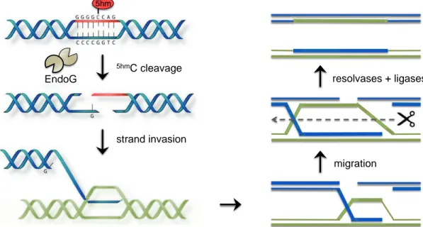
Figure 1.1: Model for 5hmC and EndoG in homologous recombination. As originally proposed
by Robertson et al., EndoG generates double stranded breaks via recognition of 5hmC in a
sequence specific context. Double strand breaks promote strand invasion into a homologous DNA neighbor, which initiates the junction crossover. The Holliday junction migrates through the desired length of DNA, and resolvases cleave the junction back into separate DNA strands. Broken and damages strands are finally repaired with ligases and polymerases. In this
dissertation we present a structure of mouse EndoG that accounts for 5hmC recognition. We also
propose an additional role for EndoG and 5hmC in recombination that invokes Holliday junction
recognition.
Adapted from Robertson et al., Nucleic Acids Research, 2014; 42(21): p. 13280-13293. migration migration resolvases + ligases 5hmC cleavage strand invasion EndoG 5hm resolvases + ligases strand invasion 5hm
6
are trafficked to the nucleus to degrade DNA during apoptosis40, recognizing chromatin as a
substrate41. Under non-apoptotic conditions, however, a basal level of EndoG is found regularly
present in the nucleus42. EndoG has been implicated in several recombination scenarios
including Herpes Simplex Virus43, myeloid/lymphoid leukemia break point clusters44,
immunoglobulins45, transfected plasmid recombination46, and genome maintenance through
HR47. The assumption is that EndoG binds to G/C rich double stranded DNA and creates double
stranded breaks to initiate these recombination events.
The work of Robertson et al. was the first instance of EndoG recognizing a specific sequence feature, and the first discovery of interactions between 5hmC and EndoG. 5hmC is only
present in vertebrates while EndoG is conserved in all eukaryotes, which begs the question of whether vertebrate EndoG has a unique structural feature to specifically recognize 5hmC?
Furthermore, despite the thematic appearance of EndoG in DNA recombination, it is unknown whether EndoG recognizes Holliday junctions as a DNA substrate. Finally, we wanted to
explore how 5hmC might impact Holliday junctions to serve as a structural or energetic marker for
recombination, and whether EndoG recognizes 5hmC in the junction context.
1.2 Aims of this Project
In this work, we aim to understand how 5hmC promotes recombination via EndoG by
asking these specific questions:
• Does 5hmC impact Holliday junction structure and stability and can it serve as a marker in
HR?
• Does EndoG cleave Holliday junctions as a method to promote recombination and does EndoG recognize 5hmC in the junction context?
7
• Is there a structural feature conserved among vertebrates that allows EndoG to recognize
5hmC DNA?
The first objective was to establish the Holliday junction as a system to study energies of molecular interactions at the junction core. This analysis would allow direct comparison of the structure and stability of C- vs. 5hmC-stabilized Holliday junctions. Furthermore, I provide
evidence of continuity between crystallographic structure and solution state energies for the Holliday junction system. In chapter 2, I discuss Holliday junction cores stabilized by halogen bonds (X-bonds), weak intermolecular interactions analogous to hydrogen bonds. The X-bond is a short electrostatic interaction between a negative acceptor and the positive crown of a polarized covalently bound halogen. In this case the positive halogen crown on a halogenated uracil acts as the donor, replacing the positive amine of a cytosine that typically stabilizes junctions. In this work I use crystallography and differential scanning calorimetry (DSC) to measure the energy of the X-bond stabilized junction compared to the H-bond stabilized junction. Furthermore we isolate specific energies that describe the relative H-bond and X-bond strengths. We show that the junction crystal structures are representative of the solution state energies determined by DSC. The methodology established for halogen bonds in this chapter is shown to be applicable to probe epigenetic effects on junction stability.
In chapter 3, we ask how 5hmC impacts junction structure and stability. We present the
crystal structure of a junction with 5hmC in the core, showing that 5hmC forms specific H-bonds to
replace the typical bonds formed by canonical cytosine at the core. 5hmC also confers global structure shape and broadens the angle relating the two arms of the junction. We apply the differential scanning calorimetry method established in chapter 2 to learn about how 5hmC
8
thermodynamically impacts junction stability. We find that 5hmC is more enthalpically stable,
although it suffers an entropic penalty and, as a result, the net change in free energy is very small compared to C-stabilized junctions. We conclude that 5hmC incorporation into junctions is stable,
and potentially provides a mechanism for protein recognition via either direct or indirect readout. The next question was whether EndoG recognizes junctions to promote recombination, and whether 5hmC plays a role in that recognition. In chapter 4, we find that EndoG does indeed
recognize junctions, and they are a preferred substrate over duplex DNA. 5hmC is not needed for
junction recognition, but does induce a unique cleavage profile. These results together suggest a role for 5hmC as a marker in EndoG-mediated recombination. Finally, we present the crystal
structure of mouse EndoG and discover a helix to loop transition relative to invertebrate EndoG structures; we believe this structural change is the source of vertebrate EndoG preference for
5hmC DNA. The loop structure is caused by a two amino acid deletion that is conserved for all
vertebrate species, suggesting all vertebrates are capable of recognizing 5hmC via EndoG. 5hmC is
only found in vertebrate species, thus we propose that EndoG co-evolved to accommodate the presence of 5hmC.
Overall this work supplies necessary information to understand how 5hmC and EndoG
work together to promote DNA recombination and expands on the original model presented by Robertson et al. This exciting avenue of research elucidates a new role for both EndoG and 5hmC
and is pioneering an unexplored aspect of HR. A mechanistic understanding of this process is the foundation to correct erroneous HR in disease and develop better biotechnology. We work towards a complete understanding of 5hmC as a marker for HR with hopes to eventually
9
REFERENCES
(1) Orr-Weaver, T. L., and Szostak, J. W. (1983) Yeast recombination: the association between double-strand gap repair and crossing-over. Proc. Natl. Acad. Sci. U. S. A. 80, 4417–4421. (2) Szostak, J. W., Orr-Weaver, T. L., Rothstein, R. J., and Stahl, F. W. (1983) The double-strand-break repair model for recombination. Cell 33, 25–35.
(3) Petermann, E., and Helleday, T. (2010) Pathways of mammalian replication fork restart. Nat. Publ. Gr. 11, 683–687.
(4) Cromie, G. A. (2009) Phylogenetic ubiquity and shuffling of the bacterial RecBCD and AddAB recombination complexes. J. Bacteriol. 191, 5076–5084.
(5) McCabe, N., Turner, N. C., Lord, C. J., Kluzek, K., Białkowska, A., Swift, S., Giavara, S., O’Connor, M. J., Tutt, A. N., Zdzienicka, M. Z., Smith, G. C. M., and Ashworth, A. (2006) Deficiency in the repair of DNA damage by homologous recombination and sensitivity to poly(ADP-ribose) polymerase inhibition. Cancer Res. 66, 8109–8115.
(6) Bugreev, D. V, Yu, X., Egelman, E. H., and Mazin, A. V. (2007) Novel pro- and anti-recombination activities of the Bloom’s syndrome helicase. Genes {&} Dev. 21, 3085–3094. (7) Hu, Y., Raynard, S., Sehorn, M. G., Lu, X., Bussen, W., Zheng, L., Stark, J. M., Barnes, E. L., Chi, P., Janscak, P., Jasin, M., Vogel, H., Sung, P., and Luo, G. (2007) RECQL5/Recql5 helicase regulates homologous recombination and suppresses tumor formation via disruption of Rad51 presynaptic filaments. Genes Dev. 21, 3073–3084.
(8) Lamb, N. A., Yu, K., Shaffer, J., Feingold, E., and Sherman, S. (2005) Association between Maternal Age and Meiotic Recombination for Trisomy 21. Am J Hum Genet 76, 91–99.
10
(9) Orr-Weaver, T. L., Szostak, J. W., and Rothstein, R. J. (1981) Yeast transformation: a model system for the study of recombination. Proc. Natl. Acad. Sci. U. S. A. 78, 6354–8.
(10) (2007) The Nobel Prize in Physiology or Medicine 2007. Nobel Media AB.
(11) Holliday, R. (1964) A mechanism for gene conversion in fungi. Genet. Res. 5, 282–304. (12) Haber, J. E. (2014) Genome Stability - DNA repair and recombination (Scholl, S., Ed.) 1st ed. Garland Science, New York.
(13) Lilley, D. M. J. (2000) Structures of helical junctions in nucleic acids. Q. Rev. Biophys. 33, 109–159.
(14) Hays, F. A., Schirf, V., Ho, P. S., and Demeler, B. (2006) Solution formation of Holliday junctions in inverted-repeat DNA sequences. Biochemistry 45, 2467–2471.
(15) Eichman, B. F., Vargason, J. M., Mooers, B. H. M., and Ho, P. S. (2000) The Holliday junction in an inverted repeat DNA sequence: Sequence effects on the structure of four-way junctions. Proc. Natl. Acad. Sci. 97, 3971–3976.
(16) Voth, A. R., Hays, F. a, and Ho, P. S. (2007) Directing macromolecular conformation through halogen bonds. Proc. Natl. Acad. Sci. U. S. A. 104, 6188–93.
(17) Khuu, P. A., Voth, A. R., Hays, F. A., and Ho, P. S. (2006) The stacked-X DNA Holliday junction and protein recognition. J. Mol. Recognit. 19, 234–42.
(18) Szwagierczak, A., Bultmann, S., Schmidt, C. S., Spada, F., and Leonhardt, H. (2010) Sensitive enzymatic quantification of 5-hydroxymethylcytosine in genomic DNA. Nucleic Acids Res. 38, e181.
(19) Delatte, B., Wang, F., Ngoc, L. V., Collignon, E., Bonvin, E., Deplus, R., Calonne, E., Hassabi, B., Putmans, P., Awe, S., Wetzel, C., Kreher, J., Soin, R., Creppe, C., Limbach, P. A., Gueydan, C., Kruys, V., Brehm, A., Minakhina, S., Defrance, M., Steward, R., and Fuks, F.
11
(2016) Transcriptome-wide distribution and function of RNA hydroxymethylcytosine. Science (80-. ). 351, 282–285.
(20) Diotel, N., Yohann, M., Coumailleau, P., Gueguen, M.-M., Serandour, A. A., Salbert, G., and Olivier, K. (2017) 5-Hydroxymethylcytosine Marks Postmitotic Neural Cells in the Adult and Developing Vertebrate Central Nervous System. J. Comp. Neurol. 525, 478–497.
(21) Raddatz, G., Guzzardo, P. M., Olova, N., Rosado, M., and Rampp, M. (2013) Dnmt2-dependent methylomes lack defined DNA methylation patterns. Proc. Natl. Acad. Sci. 110, 8627–8631.
(22) Tahiliani, M., Koh, K. P., Shen, Y., Pastor, W. A., Bandukwala, H., Brudno, Y., Agarwal, S., Iyer, L. M., Liu, D. R., Aravind, L., and Rao, A. (2009) Conversion of methylcytosine to 5-hydroxymethylcytosine in mammalian DNA by MLL partner TET1. Science 324, 930–5. (23) Ito, S., Shen, L., Dai, Q., Wu, S. C., Collins, L. B., Swenberg, J. A., He, C., and Zhang, Y. (2011) Tet proteins can convert 5-methylcytosine to 5-formylcytosine and 5-carboxylcytosine. Science 333, 1300–3.
(24) Kriaucionis, S., and Heintz, N. (2009) The nuclear DNA base 5-hydroxymethylcytosine is present in Purkinje neurons and the brain. Science 324, 929–30.
(25) Li, W., and Liu, M. (2011) Distribution of 5-hydroxymethylcytosine in different human tissues. J. Nucleic Acids 2011, 1–5.
(26) Stroud, H., Feng, S., Morey Kinney, S., Pradhan, S., and Jacobsen, S. E. (2011)
5-Hydroxymethylcytosine is associated with enhancers and gene bodies in human embryonic stem cells. Genome Biol. 12, R54.
(27) Ruzov, A., Tsenkina, Y., Serio, A., Dudnakova, T., Fletcher, J., Bai, Y., Chebotareva, T., Pells, S., Hannoun, Z., Sullivan, G., Chandran, S., Hay, D. C., Bradley, M., Wilmut, I., and De
12
Sousa, P. (2011) Lineage-specific distribution of high levels of genomic 5-hydroxymethylcytosine in mammalian development. Cell Res. 21, 1332–1342.
(28) Guo, J. U., Su, Y., Zhong, C., Ming, G., and Song, H. (2011) Hydroxylation of
5-Methylcytosine by TET1 Promotes Active DNA Demethylation in the Adult Brain. Cell 145, 423–434.
(29) Robertson, J., Robertson, A. B., and Klungland, A. (2011) The presence of
5-hydroxymethylcytosine at the gene promoter and not in the gene body negatively regulates gene expression. Biochem. Biophys. Res. Commun. 411, 40–43.
(30) Wen, L., Li, X., Yan, L., Tan, Y., Li, R., Zhao, Y., Wang, Y., Xie, J., Zhang, Y., Song, C., Yu, M., Liu, X., Zhu, P., Li, X., Hou, Y., Guo, H., Wu, X., He, C., Li, R., Tang, F., and Qiao, J. (2014) Whole-genome analysis of 5-hydroxymethylcytosine and 5-methylcytosine at base resolution in the human brain 15, 1–17.
(31) Robertson, A. B., Robertson, J., Fusser, M., and Klungland, A. (2014) Endonuclease G preferentially cleaves 5-hydroxymethylcytosine-modified DNA creating a substrate for recombination. Nucleic Acids Res. 42, 13280–13293.
(32) Ruiz-carrilio, A., and Renaud, J. (1987) Endonuclease G: a (dG)n* (dC)n-specific DNase from higher eukaryotes 6, 401–407.
(33) Cummings, O. W., King, C., Holden, J. A., and Low, L. (1987) Purification and
Characterization of the Potent Endonuclease in Extracts of Bovine Heart Mitochondria. J. Biol. Chem. 262, 2005–2015.
(34) Cote, J., Renaud, J., and Ruiz-Carrillo, A. (1989) Recognition of ( dG )n. ( dC )n Sequences by Endonuclease G 264, 3301–3310.
13
(35) Schäfer, P., Scholz, S. R., Gimadutdinow, O., Cymerman, I. A., Bujnicki, J. M., Ruiz-Carrillo, A., Pingoud, A., and Meiss, G. (2004) Structural and functional characterization of mitochondrial EndoG, a sugar non-specific nuclease which plays an important role during apoptosis. J. Mol. Biol. 338, 217–28.
(36) Wu, S.-L., Li, C.-C., Chen, J.-C., Chen, Y.-J., Lin, C.-T., Ho, T.-Y., and Hsiang, C.-Y. (2009) Mutagenesis identifies the critical amino acid residues of human endonuclease G involved in catalysis, magnesium coordination, and substrate specificity. J. Biomed. Sci. 16, 1– 14.
(37) Kieper, J., Lauber, C., Gimadutdinow, O., Urbaǹska, A., Cymerman, I., Ghosh, M., Szczesny, B., and Meiss, G. (2010) Production and characterization of recombinant protein preparations of Endonuclease G-homologs from yeast, C. elegans and humans. Protein Expr. Purif. 73, 99–106.
(38) Low, R. L. (2003) Mitochondrial Endonuclease G function in apoptosis and mtDNA metabolism: a historical perspective. Mitochondrion 2, 225–236.
(39) Côté, J., and Ruiz-Carrillo, A. (1993) Primers for Mitochondrial DNA Replication Generated by Endonuclease G. Science (80-. ). 261, 765–769.
(40) Parrish, J., Li, L., Klotz, K., Ledwich, D., Wang, X., and Xue, D. (2001) Mitochondrial endonuclease G is important for apoptosis in C. elegans. Nature 412, 90–94.
(41) Widlak, P., Li, L. Y., Wang, X., and Garrard, W. T. (2001) Action of recombinant human apoptotic endonuclease G on naked DNA and chromatin substrates: cooperation with
exonuclease and DNase I. J. Biol. Chem. 276, 48404–9.
(42) Gerschenson, M., Houmiel, K. L., and Low, R. L. (1995) Endonuclease G from mammalian nuclei is identical to the major endonuclease of mitochondria. Nucleic Acids Res. 23, 88–97.
14
(43) Huang, K.-J., Ku, C.-C., and Lehman, I. R. (2006) Endonuclease G: A role for the enzyme in recombination and cellular proliferation. Proc. Natl. Acad. Sci. 103, 8995–9000.
(44) Gole, B., Baumann, C., Mian, E., Ireno, C. I., and Wiesmüller, L. (2014) Endonuclease G initiates DNA rearrangements at the MLL breakpoint cluster upon replication stress. Oncogene. (45) Zan, H., Zhang, J., Al-Qahtani, A., Pone, E. J., White, C. A., Lee, D., Yel, L., Mai, T., and Casali, P. (2011) Endonuclease G plays a role in immunoglobulin class switch DNA
recombination by introducing double-strand breaks in switch regions. Mol. Immunol. 48, 610– 622.
(46) Misic, V., El-Mogy, M., Geng, S., and Haj-Ahmad, Y. (2016) Effect of endonuclease G depletion on plasmid DNA uptake and levels of homologous recombination in hela cells. Mol. Biol. 50, 252–261.
(47) Büttner, S., Carmona-Gutierrez, D., Vitale, I., Castedo, M., Ruli, D., Eisenberg, T., Kroemer, G., and Madeo, F. (2007) Depletion of endonuclease G selectively kills polyploid cells. Cell Cycle 6, 1072–1076.
15 CHAPTER 2
DETERMINING THERMODYNAMIC PROPERTIES OF MOLECULAR INTERACTIONS FROM SINGLE CRYSTAL STUDIES1
The concept of single crystals of macromolecules as thermodynamic systems is not a common one. However, it should be possible to derive thermodynamic properties from single crystal structures, if the process of crystallization follows thermodynamic rules. We review here an example of how the stabilizing potentials of molecular interactions can be measured from studying the properties of DNA crystals. In this example, we describe an assay based on the four-stranded DNA junction to determine the stabilizing potentials of halogen bonds, a class of electrostatic interactions, analogous to hydrogen bonds, that are becoming increasing recognized as important for conferring specificity in protein–ligand complexes. The system demonstrates how crystallographic studies, when coupled with calorimetric methods, allow the geometries at the atomic level to be directly correlated with the stabilizing energies of molecular interactions. The approach can be generally applied to study the effects of DNA sequence and modifications of the thermodynamic stability of the Holliday junction and, by inference, on recombination and recombination dependent processes.
2.1 Introduction
‘‘A picture is worth a thousand words’’. Although the origin of this phrase remains unresolved (being variously attributed to a Japanese philosopher or as an old Chinese proverb, commonly ascribed to Confucius), it has become the mantra in macromolecular
crystallography—the ‘‘picture’’ or structure of a protein or nucleic acid, or complex among or
1Previously published as: “Determining thermodynamic properties of molecular interactions from single crystal
16
between them can tell us so much about function. Of course, the absolute requirement of a crystal in crystallography immediately limits the picture to be a static one, at least for those parts that we can see (parts that are truly dynamic are invisible to our X-ray vision). This has lead to the general perception that one cannot learn anything about thermodynamics from
crystallographic studies on single crystals. A crystal is a solid that is assembled by regularly repeating the same molecule over- and-over again into a well-defined crystal lattice. This does not mean, however, that every molecule or even the atoms of the molecule are identical in every way, or even held entirely static. We review here an example of an approach to quantitatively determine the energies of halogen bonds, exploiting the isomerization of a DNA junction in the assay. As a result, we show that indeed crystallography can tell us much about the energies of molecular inter- actions that are important for the assembly and stability of nucleic acids. 2.2 Structure-energy relationships of biological halogen bonds
In this case study, we will characterize the stabilizing potentials of halogen bonds and their correlations with specific geometries in a biological system. Halogen bonds (X-bonds), formerly known as charge-transfer bonds1, are analogous to H-bonds2 in that they are directional,
primarily electrostatically driven molecular interactions that help to define the specificity of ligands against their protein targets3–8 and to drive macromolecular conformation9. The X-bond
is formed when the electropositive crown of a polarizable halogen (as a consequence of
depopulating the pz-atomic orbital when forming a covalent σ-bond to, for example, a carbon10)
interacts with an electron-rich acceptor, such as an oxygen, nitrogen, or sulfur. The stabilizing potential of the X-bond depends on the degree of polarization of the halogen (I > Br > Cl > F), the electron withdrawing ability of the molecule that is halogenated, the electronegative potential of the acceptor atom, and the distance and angle of approach of the acceptor to the halogen (Fig.
17
2.1). In order to develop empirical models that can be accurately applied to predict the
electrostatic behavior of halogens, including the existence of X-bonds, we must determine how the energies of interaction are related to the geometry of the interacting atoms. For this assay, we will describe how a Holliday junction construct (Fig. 2.2) can be designed to determine the geometries of X-bonds from single crystal studies, and their associated energies of stabilization relative to a competing H-bond in crystals9,11 and in solution11,12.
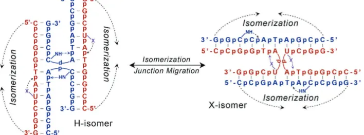
For this set of studies, we developed a DNA Holliday junction as the model system to compete an X-bond against an H-bond in defining the stability and the isomer conformation (conformer) of the four-stranded complex. The form of the Holliday junction under physiological salt concentrations is the stacked-X form13, in which the four helical arms are paired-up and
stacked to form essentially two near continuous columns of standard B-form DNA, interrupted by the over of one strand from each duplex to the adjacent duplex (Fig. 2.2). The cross-over strands form a tight U-turn, which topologically locks the junction in place. The cross-cross-over is stabilized by a set of intrastrand interactions localized at the N6Py7C8 trinucleotide core of the
general sequence motif 5’- CCG3Pu4N5N6Py7C8GG-3’ (where Py is a pyrimidine (C or T), Pu is
a purine (G or A), and G3Pu4N5 are nucleotides that maintain the inverted repeat symmetry of the
overall sequence)14,15. In particular, an H-bond from the cytosine of C
8 to the phosphate of Py7 is
essential, while an electrostatic interaction (including an H-bond) from Py7 to N6 is important for
bending the phosphodeoxyribose backbone into the U-turn of the crossing strands and specifying the sequence dependent stability of the junction in the crystal (Fig. 2.2)14,16 and in solution17.
"&!
Figure 2.1: Halogen polarization and halogen bonding. Halogen bonds (X-bonds) can be ascribed as the generation of a positive crown (a !-hole) resulting from the formation of a covalent (!) bond and the subsequent depopulation of the halogen’s pZ-orbital10. The !-hole is pronounced
with larger, more polarizable halogens (F < Cl < Br < I), and is enhanced when the halogen is attached to more electron withdrawing groups (in this case, a uracil base). Looking from the halogen down the axis of the C-X bond, electrostatic potential is shown mapped onto the surface of each halogenated uracil using a color scale ranging from + 25 kcal/mol (blue) to -25 kcal/mol (red)3.
"'!
Figure 2.2: Molecular interactions that stabilize DNA junctions. DNA Holliday junctions in inverted repeat sequences of the type 5’-CCG3Pu4N5N6Py7C8GG-3’ are stabilized by electrostatic
interactions. In particular, an H-bond from C8 to the preceding phosphate is essential, while an
H-bond or X-bond from Py7 to its preceding phosphate group is important for forcing the DNA
20
An important component of these crystals in relationship to the X-bond assay is that the lattice is stabilized primarily by end-to-end stacking of the stacked B-DNA duplex arms of the junctions, forming essentially a continuous sheet of DNAs in the b–c plane of the C2 unit cell14.
The interactions between sheets are mediated by cations that bridge across the backbones of the DNAs. Consequently, the important intrastrand interactions that stabilize the junction are well away from the lattice interactions, while the lattice interactions are largely independent of DNA sequence, as long as the terminal base pairs remain the same. Thus, this study assumes that the lattice interactions will be identical for all constructs, and that the population of conformations observed in the crystals will reflect their distributions in solution (i.e., there will be no crystal dependent discrimination among different conformers).
For the current assay, a junction was constructed from two different, non-inverted repeat DNA strands, with both strands including the H-bond that is essential at the C8 position. The strands
differ primarily at the Py7 nucleotide (and the associated Pu3), with the standard C7 at this
position to provide an H-bond, while the complementary strand places a halogenated uracil base (XU) at this position to potentially form an X-bond to the DNA backbone9,11, where X is any of
the common halogens (F, Cl, Br, or I, Table 2.1). In this mixed sequence construct, the junction can adopt one of two conformers, one that is stabilized by the H-bond (H-isomer) and the alternative that is stabilized by the X-bond (X-isomer) (Fig. 2.3). Aside from these stabilizing interactions, the two isomers are completely isoenergetic, so in effect, the isomer form is
determined explicitly by the relative strengths of the two competing interactions. For example, if the two competing interactions had the same energy, the population would occupy both isomers equally; however, if the two energies differ significantly, then the junction will be seen in one form or the other, or as a weighted average of the two forms.
21
Table 2.1: List of DNA constructs to study X-bonding interactions. The DNA constructs are named according to the number of halogenated uracil (XU) containing strands (X-strand) relative to cytosine bonding strands (strands) that form a junction. The potential X-bonding and H-bonding nucleotides (italics) results in defined molar ratios of X-H-bonding halogenated uracil and H-bonding cytosine bases (X:H).
DNA construct Sequences X:H
F2J H-Strand: (CCGGTACCGG)X-Strand: (CCGGTAFUCGG)22 2:2
Cl1J H-Strand: (CCGGTACCGG)2
X-Strand: (CCGGTAClUCGG + CCGGTAUCGG) 1:2
Cl2J H-Strand: (CCGGTACCGG)2
X-Strand: (CCGGTAClUCGG)2
2:2
Br1J H-Strand: (CCGGTACCGG)2
X-Strand: (CCGGTABrUCGG + CCGGTAUCGG) 1:2
Br2J H-Strand: (CCGGTACCGG)2
X-Strand: (CCGGTABrUCGG)
2
2:2
I1J H-Strand: (CCGGTACCGG)2
X-Strand: (CCGGTIlUCGG + CCGGTAUCGG) 1:2
I2J H-Strand: (CCGGTACCGG)2
X-Strand: (CCGGTAIUCGG)
2
2:2
H2J H-Strand: (CCGGTACCGG)2
X-Strand: (CCGGTAUCGG)2
((!
Figure 2.3: H-bond and X-bond driven isomerization of DNA junctions. A junction to assay the energy of an X-bond relative to a competing H-bond was constructed from two DNA sequences, one that contains a cytosine at the Py7 position, while the complementary strand contains a
halogenated uracil (XU
7) at this position. The competing interactions direct the junction to adopt
either the isomer form that is stabilized by standard H-bonds (H-isomer) or by X-bonds (X-isomer). Aside from these specific interactions, the H- and X-isomers are isoenergetic, allowing free isomerization between the two forms, thus creating an energetic competition between the H-bond and X-H-bond for control of the overall junction conformation.
23
An example of an X-bond that is nearly identical in its stability to the competing H-bond is that of the chlorine X-bond (Cl-bond)11. Chlorine is intermediate in its polarizability and,
therefore, is expect to be intermediate in its X-bonding potential. The position of the chlorine, either on the inside crossing strand (labeled ClU
7) or outside strand (labeled ClU’7, where the
prime is used to indicate that this is at the outside strand position) specifies whether the junction is in the X- or H-isomer form, respectively. Thus, the isomeric form, and the stabilizing
interaction, can be determined crystallographically by ClU
7 in the X- or ClU’7 in the H-isomer. In
doing so, we can estimate the difference in energy between the Cl-bond and the competing H-bond by analyzing the distribution of isomeric forms, since only the Py7 of the trinucleotide core
is varied.
For this analysis, we can define the difference in energy of the X- vs H-isomer form (ΔEIsoX–IsoH) according to Eq. (1)—this energy is not explicitly that of the molecular interactions.
Eq. (1) ∆𝐸!"#$!!"#$ = −𝑅𝑇 ln %!"#$%!"#$
Again, the assumption here is that the population distribution of isomers in the crystal mirrors the distribution in solution, since we do not expect any differences in the crystal lattice energies.
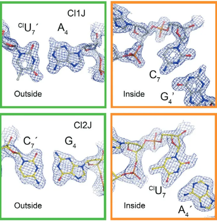
The isomer form (H- or X-isomer) can be distinguished in the crystal structures of the two constructs by analyzing the electron density maps from the single crystal structures at the Py7 nucleotide positions of the outside and the inside strands, along with the Pu4 position of the
complementary strands of the junction. In this analysis, the structures are initially refined with ambiguous base pairs at the Py7•Pu4 positions (essentially as U•A base pairs). The 2Fo-Fc density (Fig. 2.4) and Fo-Fc difference density maps are then analyzed to determine whether there is residual positive density ~1.5 Å from the C5 carbon of the Py7 pyrimidine base (evidence
(+!
Figure 2.4: Electron density maps of chlorinated base pairs in the Cl1J and Cl2J DNA junctions. 2Fo-Fc electron density maps are shown for the potentially chlorinated nucleotides and their base pairs, where 100% H-isomer would have the halogen completely occupying the outside position (green box) and 100% isomer would have it in the inside position (orange box) forming an X-bond and stabilizing the overall structure. The maps show more chlorine density on the outside for the Cl1J structure (one X-bond competing against two H-bonds) than for the Cl2J structure (two X-bonds and two H-bonds).
25
amino group of a guanine base). The presence of the excess density at Py7 and the absence of
excess density at Pu4 would indicate that this is a ClU7•A4 base pair, while the absence of excess density at Py7 and presence of density at Pu4 would indicate that this is a C7•G4 base pair. The
X-isomer would place the ClU of the ClU
7•A4 base pair at the inside crossing strand, while the H-isomer places the halogenated uracil on the outside strand. The structures can then be refined as fully either the X- or H-isomer form, and the electron density maps reanalyzed to determine whether there is residual positive or negative difference density, indicating a mixture of isomers in the crystal structure.
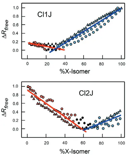
We designed two DNA constructs in which one potential X-bond competes against two H-bond (Cl1J construct) or two X-bonds competes against two H-bonds (Cl2J construct). Analysis of the electron density maps indicated that the Cl1J construct was primarily H-isomer, while the Cl2J junction was primarily X-isomer. This suggested that the Cl-bond is only slightly more stabilizing than the H-bond, out competing the H-bond in a one-to-one competition, but not capable of winning in a one-to-two competition.
2.2.1 Quantitative conformer analysis by crystallographic occupancy titration
In order to estimate the actual stabilization energy of the Cl- bond relative to the H-bond, we need to accurately quantify the percent composition of each isomer through an occupancy titration procedure11. The initial step for this quantitation is to refine the structure of the junction
using a model containing all the nucleotides that are common to both conformers, but with the four Py7•Pu4 base pairs that are unique to each isomer left as ambiguous (i.e., as unhalogenated U•A base pairs). Once the structure of this ambiguous model has been fully refined, with solvent added around the common nucleotides and maximum-likelihood convergence, the next step is to apply isomer-specific modeling and attempt to determine the percent of each isomer in the
($!
overall composition of the structure, which is the percent occupancy of the X- and H-isomers (%IsoX and %IsoH) in Eq. (1).
To determine %IsoX and %IsoH of a construct, the re! ned ambiguous structure is used to construct two models, one with the halogenated uracils of the XU
7•A4 base pairs at the inside crossing strand (X-isomer) and the other with the halogenated uracils on the outside strand (H-isomer). One approach to calculating the %IsoX and %IsoH at this point might be to re! ne the partial occupancies of the all atoms in the Py7•Pu4 base pairs, or of just the halogens and the N2 amino of the purines in each model; however, we found that this approach resulted in very large variations in occupancies, and with results that were often times incomprehensible (for example, where the sum of the occupancies for the two models were signi! cant higher or lower than 100%). We, therefore, needed to develop a more robust approach to determining the %IsoX and %IsoH for the DNA junction system.
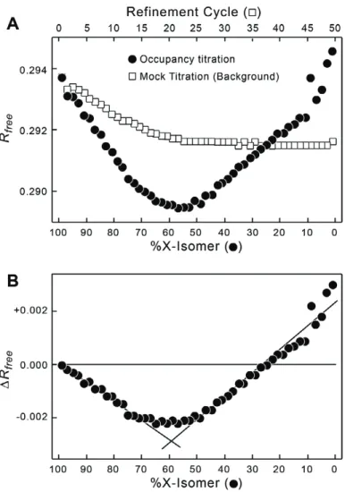
Our alternative approach makes the assumption that the sum of %IsoX and %IsoH will be 100% (this is a standard coupled occupancy analysis), and that the true ratio of conformers will be re" ected in the statistics of the crystallographic re! nement, in particular the Rfree value18. In
this analysis, a combined model that includes the two isomer forms in their entirety are re! ned, starting with the occupancies of all atoms, including solvent, of the X-isomer component set at 99% and those of the H-isomer set at 1%. The combined model is fully re! ned and the Rfree
-value recorded (Fig. 2.5). This re! ned model is then used in the next step, with the occupancy of the X-isomer reduced by 2% to 97% and the H-isomer increased accordingly, and the new composite model re! ned. This ‘‘titration’’ of the occupancy is repeated until we reach the end point of 1% X- and 99% H-isomers (Fig. 2.5A). To account for any effects of the re! nement process on Rfree, an equivalent number of re! nements was performed on a 99% X- and 1% H-
(%!
Figure 2.5: Occupancy titration and background correction for Cl2J junction. A. The occupancy titration (closed circles) and mock titration (open squares). For the occupancy titration, the DNA construct starts with 99% of the junction in the X-isomer and 1% as the H-isomer (Fig. 2.3), with the % X-isomer reduced by 2% and H-isomer increased by 2% at each titration point. The
composite isomer models are refined and the Rfree values recorded for each titration point. To
account for the effect of refinement on the Rfree values, the initial model is refined without
changing the 99% X-isomer/1% H-isomer compositions for the same number of refinement cycles as the actual occupancy titration. This mock titration serves as the background associated with the effect of the refinement process on Rfree. B. Background corrected occupancy titration.
The Rfree values from the mock background titration are subtracted from the occupancy titration
data in A. The resulting minimum !Rfree at each %X-isomer indicates the optimum contribution
of the X-isomer to the structure in the crystal. The linear regions of the data on both sides of the minimum are fit by linear regression analysis (lines), and the intersection of the two fitted lines is used to quantify the X-isomer to H-isomer ratio.
28
isomer model, but with no change in the actual occupancies. The Rfree values from this ‘‘mock’’
titration subtracted from the experimental values (Fig. 2.5B). To account for any potential
hysteretic effects, the titration was repeated in the opposite direction starting with 99% H- isomer
and decreasing the occupancy to 1% H-isomer (Fig. 2.6). The results for the chlorinated
junctions show clear minima for Rfree-values of both sets of titrations when plotted as a function
of % isomer, with minimum sitting below 50% isomer for the Cl1J and above 50% X-isomer for the Cl2J constructs, consistent with what was observed in the electron density maps.
To determine the actual observed %X-isomer (%IsoXobs), the linear portions of the
titration data on either side of the minima were fitted with simple linear models using the
KaleidaGraph program, and solving for the point of intersection for the equations of the two lines on either side of the Rfree minimum (Fig. 2.6). The corresponding %H-isomer observed
(%IsoHobs) was taken as 100%–%IsoXobs. The uncertainty in each %IsoXobs was estimated by
propagating the errors on the slopes and y-intercepts of the linear equations used to fit the titration curves.
2.2.2 Estimating energy of Cl-bonds
With the %IsoXobs and %IsoHobs now determined, the ΔEIsoX–IsoH can be estimated for
each DNA junction11 according to Eq. (1). From ΔE
IsoX–IsoH and the ratio of Cl-bonds to H-bonds
in the Cl1J and Cl2J constructs, we derived an approach to explicitly determine the absolute energies of the Cl-bond and the competing H-bond. This analysis starts with the assumption that the Cl-bonds in both constructs are nearly identical in structure and energy.
The structures are indeed nearly identical (Cl•••O distances differ by <2.5% and C–Cl•••O angles differ by ~4% from each other). We can, therefore, define the observed ΔEIsoX–IsoH in
('!
Figure 2.6: Occupancy titrations of Cl1J and Cl2J DNA constructs. The titrations show minima for !Rfree values as a function of % X-isomer (note: the % X-isomer scale of this figure is
reversed and the !Rfree values normalized between 0 and 1 relative to that in Fig. 2.5). The %
X-isomer at the !Rfree minima of the two isomers of were determined by least squares fitting of the
linear portions of each titration curve (red symbols represent % X-isomer values < !Rfree
minimum shown as, blue symbols for those > minimum, and black symbols for values not used in the calculations). The actual % X-isomer value of each construct was calculated from the slopes and y-intercepts of the two lines. The titrations repeated in the ascending (from 0% to 100% X-isomer, circles) and descending (from 100% to 0%, triangles) directions.
30
terms of the contributions of Cl-bonds and H-bonds for the Cl1J (ΔEIsoX–IsoH(Cl1J), Eq. (2)) and
Cl2J (ΔEIsoX–IsoH(Cl2J), Eq. (3)) constructs.
Eq. (2) ∆𝐸!"#$!!"#$(!"!!) = 𝐸!"!!"#$− 2𝐸!!!"#$
Eq. (3) ∆𝐸!"#$!!"#$(!"!!) = 2𝐸!"!!"#$− 2𝐸!!!"#$
It is now a simple matter to solve for the two unknowns (ECl-bond and EH-bond) using the
two simultaneous Eqs. (2) and (3). At this point, you recognize the flaw in this simple setup, which is that %IsoXobs likely does not reflect the actual % X-isomer for the singly chlorinated
junction in the Cl1J construct. That is because the junction, when annealed from two H-bonding strands, one potential Cl-bond forming ClU
7 containing strand, and one nonhalogenated U7
containing strand would statistically form three different species: the completely unchlorinated junction that can only form two H- bonds (H2J), the fully chlorinated junction that is identical to Cl2J, and the singly chlorinated Cl1J junction, in ratios of 1:1:2. If we normalize the statistical ratios, we see that the H2J:Cl1J:Cl2J components contribute in 0.25:0.5:0.25 fractional ratios to the overall percent of X-isomer observed (%IsoXobs) for the Cl1J construct Eq. (4). The resulting
contributions to the observed energy difference between the X- and H-isomers will also be defined by these fractional ratios Eq. (5).
Eq. (4) %𝐼𝑠𝑜𝑋!"# = 0.25 %𝑋!"!! + 0.5 %𝑋!"!! + 0.25 %𝑋!!!
Eq. (5) ∆𝐸!"#$!!"#$(!"!!) = 0.25 ∆𝐸!"#$!!"#$(!"!!) + 0.5 ∆𝐸!"#$!!"#$(!"!!) + 0.25 ∆𝐸!"#$!!"#$(!!!)
= 0.25 2𝐸!"!!"#!− 2𝐸!!!"#$ + 0.5 𝐸!"!!"#$ − 2𝐸!!!"#$ + 0.25 0 − 2𝐸!!!"#$ Eq. (5) reduces to exactly Eq. (2), which means that we were in fact correct in assuming that values for ECl-bond and EH-bond could be solved using the two simultaneous equations of 2 and
31
3. The resulting values for ECl-bond and EH-bond of -0.79 ± 0.12 kcal/mol and -0.64 ± 0.07 kcal/mol,
respectively, indicate that the Cl-bond is very similar in energy to that of the competing H-bond, yet slightly more negative as expected from analysis of the electron density maps.
2.2.3 Estimating energies of F-bonds, Br-bonds, and I-bonds
With the energy of the H-bond determined, we can use the %IsoXobs/%IsoHobs and
resulting ΔEIsoX-IsoH for each of the other halogens to determine their X-bonding potential. Using
the equivalent of Eq. (1), the stabilizing energies were estimated to be -0.52 ± 0.06 kcal/mol for the F-bond, -2.28 ± 0.11 kcal/mol for the Br-bond in the Br1J construct, and at least -2.1
kcal/mol for the I-bond in the I1J construct11. We could not estimate the energy of the shorter
Br-bond of Br2J nor the I-Br-bond in I2J, because their %IsoXobs was ≥95% or, within the error of the
method, essentially entirely X-isomer9,11. To determine these values, and as a validation of the
assumptions and energies from the crystallographic assay, we developed a differential scanning calorimetry (DSC) method to determine the Br-bond and I-bond energies in solution from the Br2J and I2J constructs.
The DSC assay compares the thermodynamics of melting the DNA constructs in their four-stranded junction form relative to their duplex forms. The assumption is that this
comparison eliminates the contributions of base stacking along the DNA helical arms, and localizes the observed differences in energy between junction and duplex (ΔEJ-D) to the
stabilizing interactions at the N6Py7C8-trinucleotide core of the junction. The ΔEJ-D for the H-
bonded H2J junction construct is subtracted from the ΔEJ-D for the Br2J and I2J junctions, which
further eliminates the contributions from the N6 and C8 nucleotides of the core, localizing the
differences in the observed energies to specifically the interactions at Py7. Since the H2J does not
32
attributed specifically to the Br-bond and I-bond of the Br2J and I2J constructs. We recognize that the halogens may also affect the base stacking interactions at Py7; however, the halogens in
these structures extend beyond the neighboring stacks and, therefore, should not be a dominant factor. In addition, these two large halogens are also known to be hydrophobic. To estimate the contribution of solvent effects on the solution-state energies, we calculated the solvent free energies (SFE) from the solvent accessible surfaces and atomic solvation parameters19–21 for each
halogen in models built for the X- and H-isomeric forms9,11. The resulting SFE indicates that
burying the bromine or iodine into their respective junctions would contribute on average -0.4 kcal/mol to the stabilization of the X-isomers11. Thus, the DSC assay should provide a
reasonable estimate for the thermodynamic terms responsible for the stabilizing potentials of the Br-bond and I-bond in solution.
The DSC melting profiles were measured using a TA Instruments Nano DSC with the pressure held constant at 3.0 atm; thus, the energy measured is the enthalpies of melting (ΔHm).
Each DNA sample was run against buffer in a heating cycle from 0°C to 90°C at a scanning rate of 1°C/min with an equilibrium time of 900 s. The analyses were repeated at least three times for each sample. Data were analyzed using the NanoAnalyze software from TA Instruments (TA Instruments, New Castle, DE) in the same manner as previously published12, with the best fit
determined according to the standard deviation of the fit.
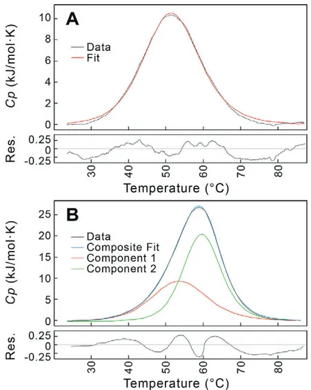
Each DNA construct was analyzed at multiple DNA concentrations. At low DNA concentrations, DSC profiles were fit using a standard single-component, two-state model for a duplex melting to single-stranded DNA (Fig. 2.7A). As the DNA concentration increased, the profiles became broader and shifted to higher melting temperature (Tm), indicating the formation of the
33
of sequence in solution12,17. The high concentration profiles were best fit applying a
two-component, two-state model (Fig. 2.7B). The lower Tm component was similar to that of the
DNA duplex, indicating that, for these constructs the duplex and junction forms coexist, and the equilibrium between the forms is slow17 compared to the rate of melting. The ΔH
m for melting
the duplex form of each construct was taken as the average ΔHm for DNA concentrations from
15 to 20 µM along the low temperature component from the two component analysis of data at [DNA] >100 µM (Table 2.2). The ΔHm of the junction form of each construct was taken as the
average of the higher temperature component of the analyses.
The ΔHm at the associated Tm allowed calculation of the ΔSm at the Tm according to the
Eq. (6), assuming that ΔGm = 0 at the melting temperature. All ΔHm and ΔSm were extrapolated to
a reference temperature of 25°C (ΔH25C and ΔS25C) using the heat capacities for each melting
profile. This then allowed the calculation of ΔG25C.
Eq. (6) ∆𝑆! = ∆!!!
!
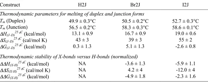
The energy differences between the junction and duplex forms at the reference temperature (ΔH J-D25°C, ΔSJ-D25°C, ΔGJ-D25°C) were simply calculated by subtracting the associated energy values of
the duplex from the junction forms, normalized to molar concentrations of duplex. For each construct analyzed (H2J, Br2J, and I2J), the trinucleotide core of the junction was seen to be highly stabilizing in terms of the enthalpy, but these interactions resulted in significant loss of entropy, as one would expect from the concept of enthalpy–entropy compensation.
When the ΔHJ-D25°C, ΔSJ-D25°C, ΔGJ-D25°C of the non-X-bonding H2J construct are
*+!
Figure 2.7: Differential scanning calorimetry (DSC) traces for melting of Br2J construct as a function of DNA concentration. A. Duplex melting at low DNA concentration. The Br2J
construct, annealed at a concentration of 40 µM, is melted and the heat capacity under constant 3 atm pressure (Cp) monitored as a function of temperature (dotted black line). The DSC trace can
be fit to a single component, two-state transition model (red curve), with a melting temperature (Tm) at 51.7°C and !Hm = 42.2 kJ/mol (!Hm values are normalized per mole of DNA duplex).
This was interpreted as the melting of the DNA as a duplex to single-strands. The residual (Res.) between the data and the fitted curve is shown in the panel below. B. Duplex and junction melting at high DNA concentration. The DSC trace of the Br2J construct, annealed at a concentration of 300 µM, is best fit by a two component, two-state transition model. The first component of this model (red curve) has a Tm = 53.8°C and !Hm = 43.5 kJ/mol, and therefore
was interpreted as the melting of the duplex DNA. The second component (green curve) has a higher Tm = 59.8°C and !Hm = 65.3 kcal/mol, and was interpreted as the melting of the junction
form of the DNA. The residual (Res.) between the data and the composite of the two component fitted curves (blue curve) is shown in the panel below.
35
Table 2.2: Thermodynamics of melting X- and H-bonded DNA junctions. The melting of each DNA construct was monitored by DSC for the melting temperature (Tm) and the enthalpy of
melting (ΔHm), with the entropy of melting (ΔSm) calculated from ΔHm and the Tm. The low Tm
parameters were assigned to the duplex and high Tm component to the junction forms of the
DNA, and each parameter normalized to the molar concentration of duplex DNA. The melting enthalpy and entropy are extrapolated to the reference temperature of 25°C applying the heat capacity of each construct to determine the difference in enthalpy (ΔH(J–D)25°C) and entropy (ΔS(J– D)25°C) between the junction and duplex forms, and the associated Gibb’s free energy (ΔG(J– D)25°C). The parameters for the stability (ΔΔH(X-H)25°C, ΔΔS(X-H)25°C, and ΔΔG(X-H)25°C) of the
X-bonds in the Br2J and I2J constructs were determined by subtracting the melting parameters for each from those of H2J.
Construct H2J Br2J I2J
Thermodynamic parameters for melting of duplex and junction forms
Tm (Duplex) 49.9 ± 0.3°C 50.5 ± 0.2°C 52.7 ± 0.3°C
Tm (Junction) 56.5 ± 0.2°C 58.3 ± 0.3°C 58.6 ± 0.1°C
ΔH(J–D)25°C (kcal/mol) 13.1 ± 0.9 16.7 ± 0.9 19.0 ± 0.6
ΔS(J–D)25°C (cal/mol K) 43 ± 3 39 ± 3 55 ± 2
ΔG(J–D)25°C (kcal/mol) 0.3 ± 1.3 5.1 ± 1.3 -2.6 ± 0.8
Thermodynamic stability of X-bonds versus H-bonds (normalized)
ΔΔH(X-H)25°C (kcal/mol) NA -3.6 ± 1.3 -5.9 ± 1.1
ΔΔS(X-H)25°C (cal/mol K) NA 4.2 ± 4 -12.0 ± 4