Faculty of Veterinary Medicine and Animal Science
Relaxin as a tool for pregnancy diagnosis
in alpacas
Evaluation of the point-of-care test FASTest Relaxin
Relaxin som verktyg för dräktighetsdiagnostik hos
al-packor
– utvärdering av snabbtestet FASTest RelaxinLinnea Lindgren Kero
Uppsala
2019
Relaxin as a tool for pregnancy diagnosis in
al-pacas
–
Evaluation of the point-of-care test FASTest Relaxin
Relaxin som verktyg för dräktighetsdiagnostik hos
al-packor
– utvärdering av snabbtestet FASTest RelaxinLinnea Lindgren Kero
Supervisor: Jane Morrell, Department of Clinical Sciences
Assistant Supervisor: Claire E. Whitehead, Camelid Veterinary Services Examiner: Bodil Ström Holst, Department of Clinical Sciences
Degree Project in Veterinary Medicine
Credits: 30
Level: Second cycle, A2E Course code: EX0869
Course coordination department: Department of Clinical Sciences
Place of publication: Uppsala Year of publication: 2019
Online publication: https://stud.epsilon.slu.se
Cover illustration: Linnea Lindgren Kero
Key words: alpaca, relaxin, pregnancy diagnostics, point of care test, FASTest Relaxin Nyckelord: alpacka, relaxin, dräktighetsdiagnostik, snabbtest, FASTest Relaxin
Sveriges lantbruksuniversitet
Swedish University of Agricultural Sciences
Faculty of Veterinary Medicine and Animal Science Department of Clinical Sciences
SUMMARY
The alpaca is a South American camelid that originates from the Andes. Their popularity and numbers in Sweden and other western countries have increased over the last few decades and they are kept as pets and for their fleece (fiber).
Their reproduction differs from other ruminants in that they are induced ovulators, have a long gestational period, very rarely have twins and are older when they are first mated. Pregnancy losses are also common in alpacas, especially early pregnancy losses, with 10-50% of the losses occurring during the first two months of pregnancy. Therefore, alpacas have a poor breeding performance which makes advances in breeding slow. It is estimated that only half of the al-pacas produce offspring each year. Because of this, an easily accessible, accurate and user-friendly tool for pregnancy diagnostics is crucial to be able to mate the female again during the season, thereby increasing the number of offspring produced by each alpaca and enabling the producer to reach breeding goals more effectively.
The most commonly used methods for diagnosing pregnancies are observing the females’ be-haviour towards males, ultrasonography and progesterone concentrations in plasma. Ultraso-nography is considered as an accurate method, but as many veterinarians either lack the knowledge or equipment to perform it, it is still not a readily available tool for alpaca breeders. Measuring progesterone concentration or observing the females’ behaviour towards males are not entirely reliable since they are not specific for pregnancy.
Relaxin, on the other hand, is a pregnancy-specific hormone produced in the utero-fetal-pla-cental unit. Relaxin concentration in blood is used in pregnancy diagnostics for several species, including dogs and cats. Two scientific papers described the relaxin concentration in pregnant and non-pregnant alpaca females and showed a significant difference between them.
In this study, the point-of-care test FASTest Relaxin, developed for dogs and cats, was evalu-ated as a tool in pregnancy diagnosis in alpacas. In total, 18 female alpacas were included in this study, which was conducted in the United Kingdom; 12 were 61-90 days pregnant and 6 were non-pregnant. The pregnancies were confirmed by ultrasonography on the same day as the blood samples were collected. The blood was centrifuged and the plasma was used in the FASTest Relaxin test, according to the test instructions, within 4 hours. Later, plasma was sent for quantitative analysis in a laboratory.
All the results from FASTest Relaxin were negative even though the results from quantitative analysis showed levels of relaxin similar to those of dogs and cats. The conclusion is, therefore, that FASTest Relaxin does not work as a tool for diagnosing pregnancy in alpacas. In contrast, the quantitative analysis showed a clear difference in relaxin concentrations between pregnant and non-pregnant females.
CONTENT
INTRODUCTION ... 1
LITERATURE REVIEW ... 2
General information about alpackas ... 2
Reproduction ... 2 Antibodies ... 4 Relaxin ... 4 Dog ... 5 Cat ... 5 Alpacas ... 6
Lateral flow test (LAT) ... 6
MATERIAL AND METHODS ... 9
Animal material ... 9 Fast test ... 9 Method ... 9 RESULTS ... 10 DISCUSSION ... 11 CONCLUSIONS ... 12 POPULÄRVETENSKAPLIG SAMMANFATTNING ... 13 REFERENCES ... 15
1 INTRODUCTION
The number of alpacas has rapidly increased in Sweden over the last decade. They are kept as pets and for their fleece. The first animals were imported in the nineties and in 2013 the Swedish Alpaca Association (Svenska Alpackaföreningen, 2018a) estimated that there were approxi-mately 1500-2000 animals in Sweden.
The reproduction of alpacas differs from other small ruminants in that they have a longer ges-tational period, rarely have more than one offspring per pregnancy and are older when they are first mated. Therefore, they have a low reproductive performance and advances in breeding are slow. A reliable/accurate, easy-to-use and readily available pregnancy diagnostic tool is crucial to determine accurately if a female has not been successfully mated or has experienced early pregnancy loss. In this way the female can be mated again in the same season or early in the next one, and the males´ fertility rate can be evaluated more accurately. Animal welfare of pregnant females is also dependent on reliable pregnancy diagnosis in order to be able to give them the right feed and care, and to detect reproductive health issues at an early stage. Currently, the most commonly used methods for diagnosing pregnancy are to observe the females’ behav-iour towards males, ultrasonography and measuring progesterone concentrations in plasma. Progesterone is not specific for pregnancy and does not indicate a living foetus. Ultrasonogra-phy is considered as an accurate method but since a lot of veterinarians either lack the knowledge or equipment to perform it in alpacas, it is still not a readily available tool for most alpaca breeders. Unlike progesterone, relaxin is specific for pregnancy and indicates a living foetus. Point-of-care tests for relaxin, to be used by the veterinarian at the clinic, have been developed for dogs and cats. No such test currently exists for alpacas and very few laboratories in the world analyse relaxin from alpaca. Therefore, it is not frequently used for pregnancy diagnosis in this species.
The aim of this study was to investigate if the point-of-care test for relaxin, FASTest Relaxin developed for dogs and cats, could also be used as a tool for diagnosing pregnancy in alpacas.
2 LITERATURE REVIEW
General information about alpackas
Camelids are normally divided into two groups, Old World Camelids and New World Camelids also known as South American Camelids (SAC). The one-humped camel/Dromedary, Bactrian camel and the wild Bactrian camel are the Old World Camelids. The South American Camelids are divided into the genus llama, consisting of the llama and the guanaco, and the genus vicugna consisting of the alpaca and the vicuña. The subject of how to classify the SACs was much debated before DNA-analysis but the relationships have now been established. Camelids started to evolve in North America over 82 million years ago, and were separated from what would become the Ruminantia 65 million years ago when they were single-stomached animals. The future Old World Camelids left North America for Asia approximately 2.6-0.78 million years ago. The SACs reached South America 3 million years ago (Fowler, 2011).
Both the British and Swedish alpaca associations consider that alpacas can be maintained out-side all year around if they have sufficient protection against wind and rain. Alpacas are flock animals and should not be kept alone but are husbanded in herds ranging from 2 animals up to several hundred (British Alpaca Society, 2018; Svenska Alpackaföreningen, 2018b) . In South America they are usually kept in the Andes above the treeline, called altiplano (Alpaca owners association, 2018; Brown, 2000).
Reproduction
There are no studies describing in detail the morphological, functional and endocrinological changes in female alpacas at puberty. Most of the females are sexually active at 12-14 months of age. Ovarian follicles over 5mm start to develop at 10 months of age (Novoa, 1970). The time of onset of puberty depends on the environment and the animal’s nutritional status. Around 60% of the adult body weight, or 33kg, is required for the onset of puberty (Smith et al., 1994). The exact timing of the mating season is dependent on the location and how the animals are kept. In Peru the mating season is between January and April when males and females are kept together. If males and females are kept separated but are allowed to mate regularly, e.g. once a month, both genders are sexually active during the whole year. During the mating season, both in the presence and absence of males, non-pregnant females may be in oestrus for up to 36 days, with anoestrus lasting a maximum of 48 hours. Alpacas differ from most domesticated livestock species in that they are induced ovulators and do not exhibit clear signs of oestrus (Sanmartin et al., 1968). Ovulation is induced when the penis penetrates the vagina and cervix and semen is deposited in the uterus but is not induced by the male merely riding the female. There is no connection between ovulation and the duration of the mating, which normally lasts for around 10-50 minutes (Fernandez-Baca et al., 1970). In the presence of a dominant follicle of the right size during mating, a surge of luteinising hormone (LH), will induce ovulation within 26-42 hours (Sumar, 1988). Normally, one oocyte will be released during ovulation, but approxi-mately 10% of ovulations will be multiple (Sanmartin et al., 1968).
3
The gestational period is 342-350 days, with implantation of the embryo taking place at 20-22 days after mating (Sanmartin et al., 1968; Bravo, 1994). A corpus luteum is required to maintain the pregnancy throughout the gestational period (Sumar, 1988).
Unlike other ruminants, the placenta of alpacas is epitheliochorial and lacks cotyledons. Instead there are half-circle domed projections or folds in chorionic epithelium acting as placentomes and matching depressions in the uterine mucosa (Steven et al., 1980).
Approximately 50% of adult alpacas do not produce an offspring each year even if the fertili-sation rate is 80% three days after mating. Early pregnancy losses are common, with 10-50% loss occurring during the first 60 days of pregnancy, but the cause is not clear (Fernandez-Baca et al., 1970; Knight et al., 1995). One possible reason might be that the right uterine horn is incapable of carrying a foetus to term. Although both ovaries ovulate equally, embryos in the right uterine horn rarely survive more than 30 days, and at most 87 days (Fernandez-Baca et al., 1979). One study found that 50.4% of 928 pregnancies were supported by a corpus luteum in the right ovary, suggesting that many pregnancies in the left uterine horn are also lost. A corpus luteum in the right ovary seems to regress faster than one in the left ovary, which might be due to a more potent luteolytic substance which an embryo in the right horn cannot mitigate (Fernandez- Baca et al., 1979) .
The most commonly used pregnancy diagnostic, and normally the first one to be used, is to observe the females’ behaviour towards males. A pregnant female will show non-receptive be-haviour and thereby reject the male. According to one study, 84% were diagnosed correctly at 70 days of pregnancy, and at 125 days 88% were correctly diagnosed (Alarcón et al., 1990). Rejection of the male is not a pregnancy specific behaviour, but rather indicates the presence of a corpus luteum. For example, a female might have a corpus luteum that has not yet regressed because of early pregnancy loss. A pregnant female might also accept the male if he is aggres-sive and she is submisaggres-sive. Likewise an inexperienced male might be frightened by a dominant female. External signs of pregnancy are not visible in the alpaca even in late pregnancy (Brown, 2000; Whitehead, 2017). Rectal palpation may be used by a skilled veterinarian as early as day 35, but is more accurate after 45-50 days (Alarcón et al., 1990). Another study found that a foetus was visible by rectal ultrasonography nine days after mating, which was later confirmed with ultrasonography and by measuring progesterone concentrations at day 23 and 34 (Parra-guez et al., 1997). Pregnancy diagnosed at an early stage should be confirmed again after 60 days of pregnancy because of the high number of early pregnancy losses. Transabdominal ul-trasonography has a high accuracy when used at 80 days of pregnancy but the accuracy drops further into the pregnancy (Whitehead, 2017).
Hormonal analysis of progesterone and oestrone sulphate can be used as a tool in pregnancy diagnostics. Concentrations of progesterone exceeding 6 nmol/liter indicate the presence of a corpus luteum and possibly a pregnancy. However, other factors than pregnancy can be the cause for elevated progesterone concentrations, such as spontaneous ovulation (Alarcón et al., 1990).
4
Oestrone sulphate, like relaxin, is a direct hormonal marker for pregnancy, as it is produced by the foetal placental unit and the trophoblast cells, and it is considered to indicate a viable preg-nancy. The concentration peaks twice during pregnancy, first at day 21-27 after mating and again during the last 60 days. It is a useful tool, but the interpretation requires knowledge of the correct breeding dates (Bravo et al., 1996).
Antibodies
All camelids have three types of IgG: IgG1, IgG2 and IgG3. IgG1 has the same structure as in other mammals, i.e. two heavy and two light chains, whereas IgG2 and IgG3 consist only of two heavy chains. Furthermore, IgG2 lacks the CH1 domain and has instead a long hinge re-gion. Together IgG2 and IgG3 represent approximately 75% of the camelids´ immuno-globu-lins. Even if IgG2 and IgG3 lack light chains they can still attach to most substrates, especially enzymes. Studies have shown that this is because they are convex and fit together well with the active concave surface on enzymes (Tizard, 2013).
Relaxin
The hormone relaxin was discovered in the 1920s when Hisaw gave serum from pregnant guinea pigs to unmated ones and saw that the pelvic symphysis relaxed. A later study by Fevold, Hisaw and Meyer also found that the pelvic symphysis of guinea pigs was relaxed when given extract from porcine ovaries (Bergfelt et al., 2014). The largest quantities of relaxin are pro-duced in the placenta, corpus luteum and uterus in conjunction with pregnancy in most species, but which of these sites is responsible for the greatest production differs between species. Re-laxin is also produced in small amounts in the prostate gland, the heart and the kidneys of males. It is considered to be a multifunctional endocrine and paracrine hormone which plays several roles in female and male reproduction, such as acting as a neuropeptide in CNS, as a vasodilator, as a cardiac stimulant in the cardiovascular system and as an anti-fibrotic agent (Sherwood, 2004; Bathgate et al., 2013). The nomenclature differs between species. In humans three types of relaxin have been found: Relaxin-1 (H1), Relaxin-2 (H2) and Relaxin-3 (H3). Human Re-laxin-2 is produced in connection with pregnancy (Bathgate et al., 2013). In this article, relaxin means the type that is equivalent to human Relaxin-2.
In general, the structure of relaxin is a 6kDa heterodimeric polypeptide consisting of one A-chain of 24 amino acids and a B-A-chain of 29 amino acids, joined together with two disulphide bridges (SS-bonds) with a disulphide loop in the A-chain, analogous to the one in insulin. Re-laxin is believed to be part of the larger family of reRe-laxin-like peptides in the insulin superfam-ily. The structure, size and two-chain construction of relaxin seems to be similar among many species, unlike the primary structure which is not well-conserved in the approximately 25 spe-cies studied to date (Sherwood, 2004).
Understanding the evolution of the relaxin peptides has proved difficult. A large variation in the amino acid sequence can be seen between closely related species, while in non-related spe-cies there is very similar amino acid sequence. A relaxin sequence has been found in non-ver-tebrates, but no gene has been found in birds or ruminants (Ruminantia) (Wilkinson et al.,
5
2005). For example, cDNA and peptide sequence from the tunicate Ciona intestinalis and por-cine relaxin show almost 100% similarity (Georges & Schwabe, 1999). Sequence analysis of relaxin from the two whales Balaenoptera acutostrata and Balaenoptera edeni show a differ-ence of 3 amino acids between the two species. However, the sequdiffer-ence only differed on one position between Balaenoptera edeni and the pig (Schwabe et al., 1989).
Searches in Pubmed, Web of Science and Google Scholar databases did not reveal any studies describing either the primary-, secondary- or tertiary structure of relaxin in alpacas or other camelids, or the genetic sequence coding for relaxin.
One study investigated preprorelaxin from the ovaries and utero-placental tissue in the one-humped camel (Camelus dromedarus). The cDNA was obtained by extracting mRNA from the tissue material and performing RT-PCR and RACE-PCR. The results show a 600 base pair code for preprorelaxin, resulting in a protein consisting of 199 amino acids. The preprorelaxin from the one-humped camel showed the most homology with porcine (74.6%) and equine (65.4%) variants, but less with canine (54.5%) and feline (48.3%). In the study, immunohistochemistry on the tissue material was also made, using polyclonal anti-relaxin antiserum R6 from rabbits for detection of immunoreactive relaxin and monoclonal antibodies to detect immunoreactive cytokeratin. The ovaries and uteroplacental tissue were both sources of relaxin in the pregnant one-humped camel (Hombach-Klonisch et al., 2000).
The differences in the primary structure prevented the development of an immunoassay for relaxin, because porcine relaxin lacks the amino acids tyrosine and histidine which made it difficult to label it with 125iodine (Sherwood et al., 1975; Schwabe et al., 1977; Gutkowska et
al., 1985; Davison, 1987). It was not until the structural characterisation had been made that it was successfully labelled with 125iodine-labeled tyrosine (Sherwood et al., 1975), and a species-specific antibody radioimmunoassay (RIA) for pigs was developed. RIAs has since been devel-oped for several species, including the rat (Sherwood & Crnekovic, 1979; Jockenhovel et al., 1991), horse (Stewart, 1986), human (Bryant, 1972) and dog (Steinetz et al., 1996). Rabbit anti-porcine (R6) has been used for developing and validating RIAs for different species, including alpacas, dogs and cats (Stewart & Stabenfeldt, 1985; Addiego et al., 1987; Steinetz et al., 1987; Volkery et al., 2012). The reason it can be used for several different species is because it cross-reacts with a receptor-binding domain that seems to be conserved (Obyrne & Steinetz, 1976).
Dog
In pregnant bitches relaxin in plasma can be detected from 3-4 weeks after mating, during the rest of the pregnancy and until 4-9 weeks into the lactational period. The concentration peaks around 5-7 weeks into the pregnancy, reaching 4-6ng/ml. The concentration then decreases to the birth. In non-pregnant and pseudopregnant bitches the relaxin concentrations were below detectable limits with RIA (Steinetz et al., 1987).
Cat
For measuring relaxin concentrations in serum in cats a porcine relaxin RIA has been used. An increase in the concentrations occurred 14-21 days after mating, peaked at 7.0ng/ml at 35-49
6
days of pregnancy, and decreased about two weeks before birth (Stewart & Stabenfeldt, 1985; van Dorsser et al., 2006).
Alpacas
There are two published papers about relaxin concentrations in pregnant and non-pregnant al-pacas.
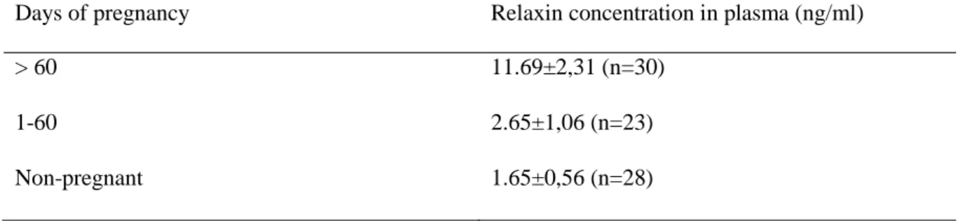
In one of the studies 36 female alpacas from six farms in Germany were involved (Volkery et al., 2012). The age of the animals varied from 3 to15 years. The first samples were taken from the females before mating, and thereafter 1-6 samples were taken from each mated female dur-ing pregnancy. Only animals with pregnancies confirmed by ultrasound were included. Blood samples were taken from the jugular vein with EDTA-blood collection tubes. The plasma was separated and frozen until analysis. The concentration of relaxin was measured with an enzyme immunoassay (EIA) described by Einspanier et al. (1999) using biotinylated porcine relaxin, polyclonal anti-relaxin serum from rabbit and secondary anti-rabbit IgG from goat. For detec-tion, a solution containing horseradish peroxidase-coupled streptavidin was used. The lowest concentration that the test could detect was 0.025 ng/ml (Einspanier et al., 1999). The conclu-sion of the study was that relaxin levels in plasma increased significantly after two months of pregnancy, as shown in Table 1 (Volkery et al., 2012).
Table 1. Relaxin concentration in alpacas according to Volkery et al. (2012)
Days of pregnancy Relaxin concentration in plasma (ng/ml)
> 60 11.69±2,31 (n=30)
1-60 2.65±1,06 (n=23)
Non-pregnant 1.65±0,56 (n=28)
1oncentrations in different body fluids of
The second study consisted of eleven alpacas from Peru (Bravo et al., 1996). Measurements were carried out using a radio immunoassay (RIA) developed for mares using antiserum from rabbits (Stewart, 1986). Blood samples were taken every day the first month and thereafter once a week until parturition (Bravo et al., 1996). Concentrations were basal (2.4ng/ml) the first two months. At three months they rose to >20ng/ml, declined to 4.5ng/ml at 5 months, and from 8 months until parturition rose to 25ng/ml.
Lateral flow test (LAT)
Lateral flow Test (LAT) is also designated as a test strip, immunochromatographic strip, im-munocapillary tests, lateral flow immunoassay (LFA) (Mark et al., 2010) or sol particle immu-noassay (Leuvering et al., 1980). It can either be a lateral flow immuimmu-noassay (LFIA) or a nucleic acid lateral flow assay (NALFA). The difference between them is that NALFA can detect ds-nuclein acid (amplicons) while LFIA only detects antigens such as antibodies, hormones and proteins (Koczula & Gallotta, 2016; Banerjee & Jaiswal, 2018). The first immune analysis in a
7
system driven by capillary forces was performed in 1978 (Glad & Grubb, 1978). Nowadays, the most well-known LAT pregnancy test is the home pregnancy test for women.
The LAT consists of a core and an outer cartridge. The cartridge contains a well where the test liquid is placed and a window where the test result is read. The core consists of multiple mate-rials providing essential biochemicals with enough capillary capacity to disperse the test sample over the whole surface of the core. The different materials in the core are antibodies, markers, test pad, conjugate pad, detection- and incubation pad, and absorption pad (Figure 1). The well on the outer cartridge where the sample is added leads to the test pad.
The marked antibodies that rehydrate and attach to the antigen, are placed on the conjugate pad. A buffer containing carbohydrates is used to preserve the conjugates. When carbohydrates are dry they surround the conjugates as a shield and prevent them from degrading. When the liquid from the sample reaches the conjugate pad the carbohydrates dissolve and enter the “stream” consisting of cross-linked silica dioxide (Banerjee & Jaiswal, 2018).
The marked antibodies in the conjugate pad attach to the antigen to be detected. The antibodies must have affinity and specificity, and be highly purified. There are two additional types of antigens attached to the membrane on the test: the one to catch the antigen is attached to the first marked antibody indicating a positive test result on the test line. The second type of addi-tional antigen catches the marked antigen and acts as a control line (Mark et al., 2010) . Mon-oclonal antibodies are mostly used as they are identical to the single B-cell from which they are derived (clone). In contrast, polyclonal antibodies are derived from multiple B-cells and are not identical (Tizard, 2013).
The antibodies are marked with a nanoparticle, normally with a size up to 800 nm. The most common material used is gold, but silver, carbon, selenium and coloured latex particles can also be used (Mark et al., 2010).
8
The detection and incubation pad, where the test and control lines with stationary antibodies are situated, is a membrane normally consisting of nitrocellulose with pore size between 0.05 and 12 µm. The length, material and the pore size determine the incubation time.
The task of the absorption pad is to draw the liquid through the membrane and to collect it when it has passed all the way through. It enables the use of larger test volumes which gives increased sensitivity. The absorption pad is normally made of cellulose.
There are two types of LAT - sandwich and competitive. Sandwich LAT is suitable for analysis of substances with more than two epitopes, one to attach to the marked antibody and the other to attach to the stationary antibody on the test strip. The competitive type is suitable for very small molecules or haptens. The immobilized analyte is conjugated with marked antibodies and is sprayed over the test line which gives a strong signal. When the substance to be analysed streams over the test it competes with the antibody for attachment, and the marked antibody detaches from the test line. Therefore, the signal decreases as the concentration of the analyzed substance increase (Banerjee & Jaiswal, 2018).
9 MATERIAL AND METHODS
Animal material
Ten pregnant and four non-pregnant female alpacas, aged between 3 and 9 years, were included in this study which was conducted at a farm in the United Kingdom. Eleven of the females were 89-90 days pregnant and one was 61 days pregnant. Of the non-pregnant females, three had not been mated this season, and the fourth had been mated but had been confirmed not pregnant by ultrasound examination. All animals were healthy at time of blood sampling.
Fast test
FASTest Relaxin is a semi-quantitative point-of-care test for relaxin that is produced and sold by Megacor (Hörbranz, Austria). It is a lateral flow test based on the immunochromatographic sandwich principle. The relaxin molecules in plasma attach to mobile monoclonal antibodies which are, in turn, attached to gold particles. When the complexes move over the nitrocellulose membrane the complex will attach to immobile monoclonal anti-relaxin antibodies from rabbits and a purple-pink line will appear (B) (see Figure 2.). A control line (C) is visible when the test has been performed correctly. If there are no, or insufficient, concentrations of complexes in the sample, only the control line will be visible.
The detection limit of the test is stated by the manufacturer to be 0.5 to 1ng/ml. A study conducted in 2004 by Schöne et al. however defined the cut off at 0.38ng/ml compared to RIA using biotinylated porcine relaxin.
Figure 2. Schematic view of FASTest Relaxin.
Method
Pregnancy was confirmed by Claire E. Whitehead with transabdominal ultrasound on the same day as the blood samples were taken. The blood used in this study was the remnants of blood samples collected for routine testing of selenium levels. The blood was taken from the jugular vein into heparinised tubes with a vacutainer. The samples were stored at room temperature until the test was performed.
Within two hours from sampling the blood was centrifuged and the plasma was separated. The plasma was transferred to test tubes using pipettes. The FASTest Relaxin test was carried out according to the manufacturer´s instructions. The test kits had been stored at room temperature. Plasma (80-100 µl) and two drops of buffer (80-100 µl) were put into the well (A). Reading of test results was performed after 5, 10, 15, 20, 25, 30 and 60 minutes at room temperature. The plasma from one pregnant alpaca was diluted 1:2, 1:4, 1:8 and 1:16 using PBS and heparin-NaCl. The dilutions were then used according to the instructions. The tests were read after 5, 10, 15, 20, 25, 30 and 60 min.
Plasma from each animal was frozen and transported on dry ice to the University of Leipzig three months later.
10 RESULTS
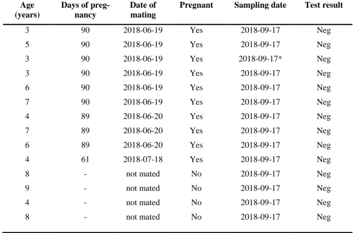
The results from the point-of-care test FASTest Relaxin on alpackas were all negative regard-less of age, days of pregnancy and confirmed pregnancy (see Table 2.).
The quantitative analysis, conducted at the University of Leipzig, showed that all pregnant fe-male alpackas had detectable and higher levels of relaxin than the non-pregnant ones. The ones that were 89-90 days pregnant had value between 2.84 to 9.63 ng/ml, with a mean value of 4.89 ng/ml. The 60 days pregnant female had a relaxin level of 1.86 ng/ml. The non-pregnant females had a relaxin concentration of 0.34 to 0.56 ng/ml with a mean value of 0.44 ng/ml (see Table 3).
Table 2. Results for FASTest Relaxin test
Age (years) Days of preg-nancy Date of mating
Pregnant Sampling date Test result
3 90 2018-06-19 Yes 2018-09-17 Neg 5 90 2018-06-19 Yes 2018-09-17 Neg 3 90 2018-06-19 Yes 2018-09-17* Neg 3 90 2018-06-19 Yes 2018-09-17 Neg 6 90 2018-06-19 Yes 2018-09-17 Neg 7 90 2018-06-19 Yes 2018-09-17 Neg 4 89 2018-06-20 Yes 2018-09-17 Neg 7 89 2018-06-20 Yes 2018-09-17 Neg 6 89 2018-06-20 Yes 2018-09-17 Neg 4 61 2018-07-18 Yes 2018-09-17 Neg
8 - not mated No 2018-09-17 Neg
9 - not mated No 2018-09-17 Neg
4 - not mated No 2018-09-17 Neg
8 - not mated No 2018-09-17 Neg
* dilution,1:2,1:4,1:8,1:16
Tabell 3. Quantitative analysis of relaxin
Days of preg-nancy
Number of animals
Relaxin concentration (ng/ml) Mean value Standard
deviat-ion Max. Min. 89-90 9 4.89 2.52 9.63 2.84 60 1 1.86 - - - 0* 4 0.44 0.09 0.56 0.34 *non-pregnant
11 DISCUSSION
The results from the quick test, FASTest Relaxin, were all negative, even though the quantita-tive analysis showed relaxin levels above the detection limit for the test and the enclosed in-structions for the test kit were followed exactly. The results from the quantitative assay also showed a significant difference between pregnant and non-pregnant animals. The reason for the negative results from the test kit is unknown; it could be due to a difference in the relaxin structure, substances interfering with the test, or another unknown factors. It could also be a combination of factors that together cause a negative result in the quick test.
The amino acid sequence, the secondary and the tertiary structure of relaxin has not been in-vestigated in alpacas and other camelids. As mentioned in the literature study, the prepro-re-laxin of dromedaries bears the greatest resemblance to pigs and horses. As the dromedary is the closest related species where this has been investigated, it is reasonable to presume that the preprorelaxin of alpacas is most similar to dromedaries. However, studies cited in the literature review showed that closely related species can have less in common than unrelated species. In order to be able to compare relaxin from dogs and cats with relaxin from alpacas, a study that shows the amino acid sequence and the structure of the molecule would be needed. Thus it could be possible to see which, if any, binding sites differ, and if it influences the attachment of the antibodies in the test. However, earlier studies showed that anti-relaxin antibodies from canine R6 can be used for RIA for dog, cat and alpaca relaxin. All species must therefore have a binding site which, if not identical, is sufficiently similar to each other so that the same anti-body can attach to them. In the case of relaxin from alpacas it could be that it attaches to the stationary anti-relaxin antibody from canine but not to the marked, movable antibodies. It has proved to be difficult to find information about how different materials in lateral flow tests influence the results, probably because this is regarded as proprietary information. There-fore, is very difficult to say if the material could have influenced the results obtained in this study.
The different structure of camelid IgG2 and IgG3 compared to IgG1 makes them interact with substances and materials in a different way. As the test is adapted for relaxin from dogs and cats, it is not adapted to these antibodies and could therefore react in unforeseen ways.
12 CONCLUSIONS
All results from FASTest Relaxin were negative even though the quantitative analysis showed detectable levels of relaxin in plasma. As stated in the introduction there is a demand for a reliable pregnancy test that can be used in early and late pregnancy in the field. This study has not been able to fill that gap. A method for reliable pregnancy diagnostics is necessary for better breeding performance and welfare. Since there are analyses for measuring relaxin in alpacas in laboratories it should be possible to develop a point-of-care test to be used in the field. Consid-ering the growing popularity and numbers of alpacas in Europe, there should be a market for such a test. Relaxin has the qualifications to be a suitable hormone for analysis since it indicates a normal pregnancy, is pregnancy specific and there are already point-of-care tests developed for other species.
13
POPULÄRVETENSKAPLIG SAMMANFATTNING
Sedan de första alpackorna importerades till Sverige på 1990-talet har deras antal och popula-ritet stadigt ökat, och 2013 beräknades det finnas runt 1500-2000 djur. De hålls främst för sin ull och som sällskapsdjur. Alpackor är ett kameldjur som härstammar ifrån bergskedjan An-derna i Sydamerika. Deras närmaste släktingar är de andra Sydamerikanska kameldjuren, lama, vicuña och guanaco. I Asien och Afrika återfinns de övriga kameldjuren nämligen dromedaren, Baktrianska kamelen och den vilda Baktrianska kamelen. För över 65 miljoner år sedan skiljdes de från en av de andra undergrupperna, idisslare (Ruminantia), i ordningen partåiga hovdjur (Artiodactyla). Vid åtskiljningen var de båda undergrupperna enkelmagade djur.
Alpackor får normalt bara en avkomma varje år, tvillingfödslar är mycket sällsynta. Den nor-mala dräktighetstiden är 342–350 dagar. Deras parning och förmåga att reproducera sig skiljer sig från de andra tamdjurens. Alpackahonan släpper inte ifrån sig mogna ägg med jämna mel-lanrum under parningssäsongen, utan äggen släpper från äggstockarna i samband med parning, när penis penetrerar vaginan. Nästan alltid så lossnar bara ett ägg från äggstockarna i taget. Honorna är brunstiga och villiga att para sig i upp till 36 dagar i sträck, därefter är hon inte mottaglig i upp till 48h.
Den vanligaste metoden för att se om honan är dräktig, och som djurägaren kan göra själv är att kolla på hennes beteende gentemot hanar. Om hon är dräktig ratar hon hanen efter ett par dagar och låter honom inte betäcka henne. Denna metoden är dock inte alltid särskilt säker. Det beror på att honan nyligen kan ha förlorat embryot och hennes hormoner fortfarande talar om att hon är dräktig, av samma anledning är det osäkert att ta prov på hormonet progesteron. Hon kan även ha någon annan sjukdom som påverkar hennes vilja att bli betäckt. Det är mycket svårt att se på en alpacka om den är dräktig, inte ens i sen dräktighet går det att se. Ett säkrare sätt att se om alpackan är dräktig är att veterinären använder ultraljud. Efter ca 3-5veckor kan veterinären se om honan är dräktig genom att sätta in ultraljudet i ändtarmen. Vid ungefär 60-80 dagars dräktighet kan ultraljudundersökningen ske genom huden på magen.
Anledningen till att det är viktigt att se om honan är dräktig är för att ca 50 % av de vuxna djuren inte får någon avkomma varje år. Det uppskattas att hälften av missfallen/aborterna sker under den första månaden som hon är dräktig, varför vet man inte. En anledning skulle kunna vara att den högra delen av livmodern inte kan bära på ett foster under hela dräktigheten och fostret därför dör.
Eftersom långt ifrån alla parade alpackor får en avkomma är det viktigt att veta vilka som är dräktiga. Dels för att öka antalet födda alpackor och veta när djuren kan paras igen, dels för att dräktiga djur behöver annan omvårdnad än icke-dräktiga.
Syftet med denna studie har därför varit att se om det går att använda ett snabbtest för att se om djuret är dräktigt som är gjort för hund och katt, FASTest Relaxin, skulle kunna användas även för alpackor. Testet upptäcker om det i blodet finns ett hormon som heter relaxin. Relaxin pro-duceras av äggstockarna, livmodern och moderkakan vid dräktighet. Relaxin stiger hos dräktiga alpackor från den andra till tredje dräktighetsmånaden, minskar vid fem månader och ökar se-dan från åtta månader fram till förlossningen. Testet fungerar i stora drag som ett graviditetstest
14
för människor fast istället för att sätta urin på stickan sätts en del av blodet, plasman, på. Om testet är positivt ses två linjer på testet, om det är negativt ses en linje.
Studien utfördes i Storbritannien. I studien ingick tolv stycken dräktiga alpackahonor och sex stycken som inte var dräktiga. Blodet som togs var det överblivna blodet i samband med att prover togs för att kolla halterna av ämnet selen. Samma dag centrifugerades blodet så att plas-man kunde utvinnas. Plasplas-man användes sedan enligt instruktionerna som följde med snabb-testet.
Alla resultaten från snabbtesterna var negativa trots att honorna samma dag konstaterades vara dräktiga med hjälp av ultraljud. Plasma skickades även till Tyskland för kvantitativ analys av koncentrationerna av relaxin och resultaten därifrån visar att dräktiga honor har en ökad kon-centration av relaxin i blodet jämfört med icke-dräktiga samt att dessa nivåer är på en nivå som snabbtestet ska klara av att detektera.
Varför snabbtestet inte fungerar går inte att säga. Det finns en del tänkbara orsaker som tas upp nedan. Det kan även vara en kombination av faktorer som gör att testet inte fungerar.
En anledning skulle kunna vara att kameldjurs antikroppar skiljer sig från övriga däggdjurs, där hund och katt ingår. Kameldjur har dels samma sorts antikroppar som andra däggdjur, dels antikroppar som ser annorlunda ut. De vanliga antikropparna består av två korta och två långa så kallade kedjor. Kameldjurens annorlunda antikroppar består av endast två långa kedjor. Detta gör att kameldjurens antikroppar fungerar lite annorlunda och kan binda in till andra ämnen och ställen som inte de vanliga kan och tvärtom. Det är därför möjligt att dessa antikroppar hindrar testet från att upptäcka relaxin.
Dessutom har aldrig alpackors relaxin studerats, så man vet inte exakt hur det ser ut. Det kan vara så att det skiljer sig så pass mycket från hundars och katters att det inte klarar av att binda in till båda ämnena i testet på samma sätt som deras relaxin gör.
Snabbtestet FASTest Relaxin kan alltså inte användas för att undersöka om en alpacka är dräk-tig. Själva hormonet relaxin har dock potential att kunna användas om det utvecklas ett lämpligt test.
15 REFERENCES
Adams, G. P. & Domínguez, M. (2007). Pregnancy diagnosis in llamas and alpacas. In: Youngquist, R. S. & Threlfall, W. R. (Eds) Current Therapy in Large Animal Theriogenology. 2. ed. Chapter 121, pp 889–895. Saint Louis: W.B. Saunders. ISBN 978-0-7216-9323-1.
Addiego, L., Tsutsui, T., Stewart, D. & Stabenfeldt, G. (1987). Determination of the source of immu-noreactive relaxin in the cat. Biology of Reproduction, 37(5), pp 1165–1169.
Alarcón, V., Sumar, J., Riera, G. S. & Foote, W. C. (1990). Comparison of three methods of preg-nancy diagnosis in alpacas and llamas. Theriogenology, 34(6), pp 1119–1127.
Alpaca Owners Association (2018). About alpacas. Available from http://www.alpacainfo.com/acad-emy/about-alpacas. [Accessed 2018-11-14].
Banerjee, R. & Jaiswal, A. (2018). Recent advances in nanoparticle-based lateral flow immunoassay as a point-of-care diagnostic tool for infectious agents and diseases. Analyst, 143(9), pp 1970– 1996.
Bathgate, R. a. D., Halls, M. L., van der Westhuizen, E. T., Callander, G. E., Kocan, M. & Summers, R. J. (2013). Relaxin family peptides and their receptors. Physiological Reviews, 93(1), pp 405– 480.
Bergfelt, D. R., Peter, A. T. & Beg, M. A. (2014). Relaxin: A hormonal aid to diagnose pregnancy sta-tus in wild mammalian species. Theriogenology, 82(9), pp 1187–1198.
Bravo, P. (1994). Reproductive endocrinology of llamas and alpacas. Veterinary Clinics of North
America-Food Animal Practice, 10(2), pp 265–279.
Bravo, P. W., Stewart, D. R., Lasley, B. L. & Fowler, M. E. (1996). Hormonal indicators of pregnancy in llamas and alpacas. Journal of the American Veterinary Medical Association, 208(12), pp 2027-30.
British Alpaca Society (2018). Alpacapedia. Available from http://www.bas-uk.com/alpacapedia/al-pacapedia. [Accessed 2018-11-13].
Brown, B. W. (2000). A review on reproduction in South American camelids. Animal Reproduction
Science, 58(3–4), pp 169–195.
Bryant, G. (1972). Detection of relaxin in porcine, ovine and human plasma by radioimmunoassay.
Endocrinology, 91(4), pp 1113-7.
Davison, P. (1987). A versatile procedure for the radioiodination of proteins and labeling reagents.
Bi-ochimica et Biophysica Acta, 926(2), pp 195–202.
van Dorsser, F. J. D., Swanson, W. F., Lasano, S. & Steinetz, B. G. (2006). Development, validation, and application of a urinary relaxin radioimmunoassay for the diagnosis and monitoring of preg-nancy in felids. Biology of Reproduction, 74(6), pp 1090–1095.
Einspanier, A., Nubbemeyer, R., Schlote, S., Schumacher, M., Ivell, R., Fuhrmann, K. & Marten, A. (1999). Relaxin in the marmoset monkey: secretion pattern in the ovarian cycle and early preg-nancy. Biology of Reproduction, 61(2), pp 512–520.
Fernandez-Baca, S., Hansel, W. & Novoa, C. (1970). Embryonic mortality in the alpaca. Biology of
Reproduction, 3(2), pp 243–251.
Fernandezbaca, S., Hansel, W., Saatman, R., Sumar, J. & Novoa, C. (1979). Differential luteolytic ef-fects of right and left uterine horns in the alpaca. Biology of Reproduction, 20(3), pp 586–595.
16
Fowler, M. E. (2011). Medicine and Surgery of Camelids [online]. Hoboken, United States: John Wiley & Sons, Incorporated. Available from: http://ebookcentral.proquest.com/lib/slub-ebooks/de-tail.action?docID=533951. [Accessed 2018-12-10].
Georges, D. & Schwabe, C. (1999). Porcine relaxin, a 500 million-year-old hormone? The tunicate Ciona intestinalis has porcine relaxin. Faseb Journal, 13(10), pp 1269–1275.
Glad, C. & Grubb, A. O. (1978). Immunocapillarymigration - A new method for immunochemical quantitation. Analytical Biochemistry, 85(1), pp 180–187.
Gutkowska, J., Stlouis, J. & Genest, J. (1985). Solid-phase radioimmunoassay for relaxin. Clinical and
Investigative Medicine, 8(2), pp 133–138.
Hombach-Klonisch, S., Abd-Elnaeim, M., Skidmore, J. A., Leiser, R., Fischer, B. & Klonisch, T. (2000). Ruminant relaxin in the pregnant one-humped camel (Camelus dromedarius). Biology of
Reproduction, 62(4), pp 839–846.
Jockenhovel, F., Peterson, M., Johnston, P. & Swerdloff, R. (1991). Directly iodinated rat relaxin as a tracer for use in radioimmunoassays. European Journal of Clinical Chemistry and Clinical
Bio-chemistry, 29(1), pp 71–75.
Knight, T., Ridland, M., Scott, I., Death, A. & Wyeth, T. (1995). Fetal mortality at different stages of gestation in alpacas (lama-pacos) and the associated changes in progesterone concentrations.
Ani-mal Reproduction Science, 40(1–2), pp 89–97.
Koczula, K. M. & Gallotta, A. (2016). Lateral flow assays. Essays in Biochemistry, 60(1), pp 111– 120.
Leuvering, J. H., Thal, P. J., van der Waart, M. & Schuurs, A. H. (1980). Sol particle immunoassay (SPIA). Journal of Immunoassay, 1(1), pp 77–91.
Mark, D., Haeberle, S., Roth, G., Stetten, F. von & Zengerle, R. (2010). Microfluidic lab-on-a-chip platforms: requirements, characteristics and applications. Chemical Society Reviews, 39(3), pp 1153–1182.
Novoa, C. (1970). Reproduction in Camelidae. Journal of Reproduction and Fertility, 22(1), pp 3-. Obyrne, E. & Steinetz, B. (1976). Radioimmunoassay (RIA) of relaxin in sera of various species using
an antiserum to porcine relaxin. Proceedings of the Society for Experimental Biology and
Medi-cine, 152(2), pp 272–276.
Parraguez, V. H., Cortez, S., Gazitua, F. J., Ferrando, G., MacNiven, V. & Raggi, L. A. (1997). Early pregnancy diagnosis in alpaca (Lama pacos) and llama (Lama glama) by ultrasound. Animal
Re-production Science, 47(1–2), pp 113–121.
Sanmartin, M., Copaira, M., Zuniga, J., Rodreguez, R., Bustinza, G. & Acosta, L. (1968). Aspects of reproduction in alpaca. Journal of Reproduction and Fertility, 16(3), pp 395-+.
Schwabe, C., Bullesbach, E., Heyn, H. & Yoshioka, M. (1989). Cetacean relaxin - isolation and se-quence of relaxins from Balaenoptera-Acutorostrata and Balaenoptera-Edeni. Journal of
Biologi-cal Chemistry, 264(2), pp 940–943.
Schwabe, C., Mcdonald, J. & Steinetz, B. (1977). Primary structure of B-chain of porcine relaxin.
Bio-chemical and Biophysical Research Communications, 75(2), pp 503–510.
Schöne, J., Einspanier, A., Kern, A. & Kern, A. (2004). Untersuchungen zur Eignung des FASTest® RELAXIN-Tests für den Trächtigkeitsnachweis beim Hund.
17
Sherwood, O., Chang, C., Bevier, G. & Dziuk, P. (1975). Radioimmunoassay of plasma relaxin levels throughout pregnancy and at parturition in pig. Endocrinology, 97(4), pp 834–837.
Sherwood, O. & Crnekovic, V. (1979). Development of a homologous radioimmunoassay for rat re-laxin. Endocrinology, 104(4), pp 893–897.
Sherwood, O. D. (2004). Relaxin’s physiological roles and other diverse actions. Endocrine Reviews, 25(2), pp 205–234.
Smith, C. L., Peter, A. T. & Pugh, D. G. (1994). Reproduction in llamas and alpacas: A review.
Theri-ogenology, 41(3), pp 573–592.
Steinetz, B. G., Bullesbach, E. E., Goldsmith, L. T., Schwabe, C. & Lust, G. (1996). Use of synthetic canine relaxin to develop a rapid homologous radioimmunoassay. Biology of Reproduction, 54(6), pp 1252–1260.
Steinetz, B., Goldsmith, L. & Lust, G. (1987). Plasma relaxin levels in pregnant and lactating dogs.
Biology of Reproduction, 37(3), pp 719–725.
Steven, D. H., Burton, G. J., Sumar, J. & Nathanielsz, P. W. (1980). Ultrastructural observations on the placenta of the alpaca (Lama pacos). Placenta, 1(1), pp 21–32.
Stewart, D. (1986). Development of a homologous equine relaxin radioimmunoassay. Endocrinology, 119(3), pp 1100–1104.
Stewart, D. & Stabenfeldt, G. (1985). Relaxin activity in the pregnant cat. Biology of Reproduction, 32(4), pp 848–854.
Sumar, J. (1988). Removal of the ovaries or ablation of the corpus-luteum and its effect. Acta
Veterin-aria Scandinavica, pp 133–141.
Svenska Alpackaföreningen (2013a). Vanliga frågor och svar. Available from: http://www.al-packaforeningen.se/vanliga-fragor-och-svar/. [Accessed 2018-11-14].
Svenska Alpackaföreningen (2013b). Svenska Alpackaföreningen s råd & rekommendationer. Avai-lable from: http://www.alpackaforeningen.se/rad-rekommendationer/. [Accessed 2018-12-11]. Tizard, R.I. (2013). Veterinary Immunology. 9. ed. St. Louis: Elsevier
Volkery, J., Gottschalk, J., Sobiraj, A., Wittek, T. & Einspanier, A. (2012). Progesterone, pregnane-diol-3-glucuronide, relaxin and oestrone sulphate concentrations in saliva, milk and urine of fe-male alpacas (Vicugna pacos) and their application in pregnancy diagnosis. The Veterinary
Rec-ord, 171(8), p 195.
Whitehead, C. E. (2017). Pregnancy diagnosis in camelids. Livestock, 22(6), pp 330–334.
Wilkinson, T. N., Speed, T. P., Tregear, G. W. & Bathgate, R. A. (2005). Evolution of the relaxin-like peptide family. BMC Evolutionary Biology, 5, p 14.