Ameloblastic fibrodentinoma and ameloblastic fibro-odontoma: an updated systematic review of cases reported in the literature
Bruno Ramos Chrcanovic, DDS, MSc, PhD student, Ricardo Santiago Gomez, DDS, MSc, PhD
PII: S0278-2391(16)31326-X
DOI: 10.1016/j.joms.2016.12.038 Reference: YJOMS 57600
To appear in: Journal of Oral and Maxillofacial Surgery
Received Date: 1 December 2016 Revised Date: 19 December 2016 Accepted Date: 20 December 2016
Please cite this article as: Chrcanovic BR, Gomez RS, Ameloblastic fibrodentinoma and ameloblastic fibro-odontoma: an updated systematic review of cases reported in the literature, Journal of Oral and
Maxillofacial Surgery (2017), doi: 10.1016/j.joms.2016.12.038.
This is a PDF file of an unedited manuscript that has been accepted for publication. As a service to our customers we are providing this early version of the manuscript. The manuscript will undergo copyediting, typesetting, and review of the resulting proof before it is published in its final form. Please note that during the production process errors may be discovered which could affect the content, and all legal disclaimers that apply to the journal pertain.
M
AN
US
CR
IP
T
AC
CE
PT
ED
Ameloblastic fibrodentinoma and ameloblastic fibro-odontoma: an updated systematic review of cases reported in the literature
Bruno Ramos Chrcanovic 1* Ricardo Santiago Gomez 2
1
DDS, MSc, PhD student; Department of Prosthodontics, Faculty of Odontology, Malmö University, Malmö, Sweden. bruno.chrcanovic@mah.se; brunochrcanovic@hotmail.com
2
DDS, MSc, PhD; Department of Oral Surgery and Pathology, School of Dentistry, Universidade Federal de Minas Gerais, Belo Horizonte, Brazil. rsgomez@ufmg.br
* Corresponding author:
Bruno Ramos Chrcanovic. Department of Prosthodontics, Faculty of Odontology, Malmö University, Carl Gustafs väg 34, SE-214 21, Malmö, Sweden. bruno.chrcanovic@mah.se; brunochrcanovic@hotmail.com Mobile: +46 725 541 545 Fax: +46 40 6658503
M
AN
US
CR
IP
T
AC
CE
PT
ED
Ameloblastic fibrodentinoma and ameloblastic fibro-odontoma: an updated systematic review of cases reported in the literature
ABSTRACT
Purpose: To integrate the available data published on ameloblastic fibrodentinoma (AFD) and ameloblastic fibro-odontoma (AFO) into a comprehensive analysis of its clinical/radiologic features. Methods: An electronic search was undertaken in August/2016. Eligibility criteria included publications reporting cases of AFD and/or AFO having enough clinical, radiological and histological information to confirm the diagnosis. Demographic data, lesion site and size, treatment approach, and recurrence were analyzed and compared between AFD and AFO.
Results: 55 publications reporting 64 AFDs (60 central, 4 peripheral) and 137 publications reporting 215 AFOs (211 central, 3 peripheral, 1 unknown) were included. The difference in recurrence rate (when the information about recurrence was provided) was not statistically significant. The mean age of the patients affected by AFD was not statistically significantly different from those affected by AFO.
Conclusions: AFD and AFO presented several similarities: higher prevalence in males and in mandibles, similar patients’ mean age, rate of cortical bone perforation and of the lesions’ association with displaced/unerupted teeth and tooth root resorption, mean lesion size, and recurrence rate. The lesions differed with regard to the presence of radiopacities and locularity. Taken together, our data do not support the concept of progressive maturation of these tumoral conditions.
KEYWORDS
Ameloblastic fibrodentinoma; ameloblastic fibro-odontoma; odontogenic tumors; clinical features; recurrence rate
M
AN
US
CR
IP
T
AC
CE
PT
ED
INTRODUCTIONAmeloblastic fibroma (AF) consists of odontogenic ectomesenchyme resembling the dental papilla and epithelial strands and nests resembling dental lamina and enamel organ. No dental hard tissues are present. Rarely, tumors with the histomorphology of AF may form dysplastic dentin, and are called ameloblastic fibrodentinomas (AFD), or dentin plus enamel, and be classified as ameloblastic fibro-odontoma (AFO).1 There is some controversy in the literature raising doubts whether these lesions represent separate entities or are the same lesion in a continuum representing different stages of evolution.2-6
The true AFD and AFO, not the developmental odontoma, are considered to be rare lesions. There are limited details in the literature regarding their clinical and radiologic features. The epidemiological study of such rare lesions is of great importance because it provides information that can improve the diagnostic accuracy and will allow pathologists and surgeons to make informed decisions and refine the treatment plan to optimize the clinical outcome. The aim of the present study was to integrate the available data published in the literature on AFD and AFO into an updated comprehensive comparative analysis of their clinical and radiologic features. Moreover, to report the frequency of recurrence of these lesions.
MATERIALS AND METHODS
This study followed the PRISMA Statement,7 which is an evidence-based minimum set of items for reporting in systematic reviews. PRISMA focuses on ways in which authors can ensure a transparent and complete reporting of this type of research. A review protocol does not exist.
M
AN
US
CR
IP
T
AC
CE
PT
ED
An electronic search without time or language restrictions was undertaken in August 2016 in the following databases: PubMed/Medline, Web of Science, and ScienceDirect. The following terms were used in the search strategies:
(“ameloblastic fibrodentinoma”) OR (“ameloblastic fibro-dentinoma”) OR (“ameloblastic fibro-odontoma”) OR (“ameloblastic fibro-odontome”) OR (“ameloblastic fibroodontoma”) OR (“mixed odontogenic tumor”) OR (“mixed odontogenic tumour”) OR (“ameloblastic fibroma”)
Moreover, Google Scholar was also checked. A manual search of related journals, including
Acta Odontologica Scandinavica, Acta Oto-Laryngologica, Annals of Otology Rhinology and Laryngology, British Journal of Oral and Maxillofacial Surgery, Cancer, Head & Neck, Head and Neck Pathology, International Journal of Oral and Maxillofacial Surgery, Japanese Journal of Oral and Maxillofacial Surgery, Journal of Dental Research, Journal of Craniofacial Surgery, Journal of Cranio-Maxillofacial Surgery, Journal of Japanese Society of Oral Oncology, Journal of the Japanese Stomatological Society, Journal of Laryngology and Otology, Journal of Maxillofacial and Oral Surgery, Journal of Nihon University School of Dentistry, Journal of Oral and Maxillofacial Surgery, Journal of Oral Pathology and Medicine, Journal of the Stomatological Society, Laryngoscope, Oral Diseases, Oral Oncology, Oral Surgery Oral Medicine Oral Pathology Oral Radiology, Otolaryngology -- Head and Neck Surgery, and Quintessence International, was performed. The reference list of the
identified studies and the relevant reviews on the subject were also scanned for possible additional studies. Publications with lesions identified by other authors as being AFD or AFO, even not having the terms “ameloblastic fibrodentinoma“ or “ameloblastic fibroodontoma” in the title of the article, were also re-evaluated by an author of the present study.
M
AN
US
CR
IP
T
AC
CE
PT
ED
Eligibility criteria included publications reporting cases of AFDs or AFOs. The studies needed to have enough clinical, radiological and histological information to confirm the diagnosis of AFD or AFO. Randomized and controlled clinical trials, cohort studies, case-control studies, cross-sectional studies, case series, and case reports were included. Exclusion criteria were review papers and immunohistochemical, histomorphometric, radiological, genetic expression, histopathological, cytological, cell proliferation/apoptosis, and in vitro studies, unless any of these publication categories had reported any cases with enough clinical, radiological and histological information. Hybrid tumors containing parts of AFD or AFO were not were not considered for this study, as they may behave differently from non-hybrid AFD and AFO tumors.
Study selection
The titles and abstracts of all publications identified through the electronic searches were read independently by the authors. For studies appearing to meet the inclusion criteria, or for which there were insufficient data in the title and abstract to make a clear decision, the full report was obtained. Disagreements were solved by discussion between the authors. The clinical and radiological aspects, as well as the histological description of the lesions were thoroughly assessed in order to confirm the diagnosis of AFD or AFO. All included cases of AFD had formation of dentinoid material, mentioned in the text and/or visible at the photomicrographs, besides the presence of ameloblastic fibroma-like areas. All published cases of AFO showing only dentine or dentinoid material deposition, but not enamel, were reclassified as AFD.
Data extraction
The authors independently extracted data using specially designed data extraction forms. Any disagreements were solved by discussion. For each of the identified studies included, the following data were then extracted on a standard form, when available: year of publication, number of patients, patient’s sex, age and race, follow-up period, duration of the lesion previously to
M
AN
US
CR
IP
T
AC
CE
PT
ED
treatment, lesion location (maxilla/mandible), anterior/posterior location (three categories: [a] anterior: lesions in the incisors/canine region; [b] premolar region; [c] posterior: lesions in the molars/retromolar region), recurrence, recurrence period, lesion size, presence of erosion of the subjacent cortical bone (for peripheral lesions), perforation of cortical bone, locularity radiological appearance (unilocular/multilocular), presence of radiopacities visible in the radiological exams, association of the lesion with a tooth (the tooth can either be erupted with the entire root(s) encompassed by the lesion or unerupted encompassing the entire tooth), and tooth displacement and tooth root resorption due to lesion’s growth. The lesion size was determined according to the largest diameter reported in the publications. Contact with authors for possible missing data was performed.
Analyses
The mean, standard deviation (SD), and percentages were presented as descriptive statistics. Kolmogorov–Smirnov test was performed to evaluate the normal distribution of the variables, and Levene’s test evaluated homoscedasticity. The performed tests for two independent groups were Student’s t-test or Mann-Whitney test, depending on the normality. Pearson’s chi-squared or Fisher’s exact tests were used for categorical variables, depending on the expected count of events in a 2x2 contingency table. The degree of statistical significance was considered p<0.05. All data were statistically analyzed using the Statistical Package for the Social Sciences (SPSS) version 23 software (SPSS Inc., Chicago, IL, USA).
RESULTS
Literature search
The study selection process is summarized in Figure 1. The search strategy in the databases resulted in 3610 papers. Search in Google Scholar resulted in 11 eligible papers not found in the
M
AN
US
CR
IP
T
AC
CE
PT
ED
three main databases. A number of 1158 articles were cited in more than one database (duplicates). The reviewers independently screened the abstracts for those articles related to the focus question. Of the resulted 2463 studies, 2218 were excluded for not being related to the topic. Additional hand-searching of journals and of the reference lists of selected studies yielded 22 additional papers. The full-text reports of the remaining 267 articles led to the exclusion of 75 publications because they did not meet the inclusion criteria (see Supplemental Appendix). The excluded studies did not report enough clinical, radiological and histological information to confirm a definite diagnosis of AFD or AFO. Thus, a total of 192 publications were included in the review. Examples of histopathological pictures of AFD and AFO are shown in Figure 2.
Description of the Studies and Analyses
Ameloblastic fibrodentinomas (AFD)
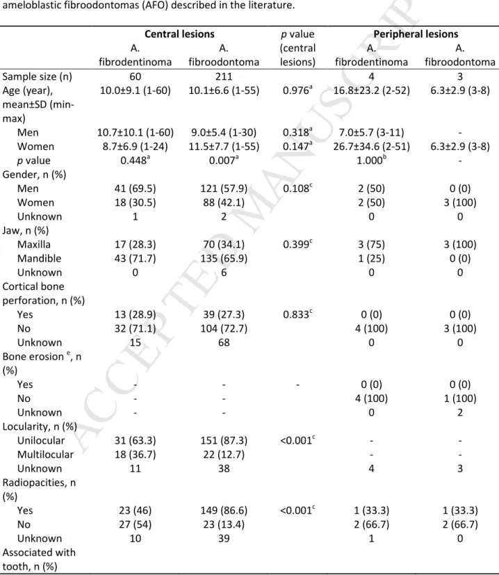
Fifty-five publications8-62 reporting AFDs were included in the present review, with 64 AFDs, 60 central and 4 peripheral lesions. All included cases of AFD showed formation of dentinoid material, mentioned in the text or visible in the photomicrographs, besides the presence of ameloblastic fibroma-like areas. Table 1 presents demographic and clinical features of all 64 AFDs. AFDs were more prevalent in men than in women, at a nearly 2:1 proportion. The mean age of the patients was higher for the peripheral lesions in relation to the central ones. Figure 3 shows the distribution of all lesions (n=64) according to age, with a higher prevalence in the first two decades of life. The age of the patients with peripheral lesions (n=4) was 2, 3, 11, and 51. The few peripheral lesions were located at/between teeth 11-21 buccal side (n=2), 25-26 buccal side (n=1), and 42-43 lingual side (n=1). The central lesions were more prevalent in the mandible in relation to the maxilla, and at the posterior region in relation to the anterior region (Figure 4). About 30% of the central lesions showed signs of cortical bone perforation and about 60% of the lesions had a radiological unilocular appearance. Radiopacities were observed in 46% of the AFDs, and approximately 80% of the lesions were associated with a tooth. Nearly 20% of the lesions caused root resorption of the
M
AN
US
CR
IP
T
AC
CE
PT
ED
adjacent teeth, most of them deciduous. Treatment of the lesions was known in 58 cases, of which 56 consisted of conservative surgery (6 excisions, 50 enucleations) and 2 cases were treated by segmental resection. The time of follow-up was informed for 37 central lesions, with a mean±SD of 50.4±54.0 months (min-max, 2-258). The race of the patient was reported in 49 cases. Eighteen cases (36.7%) were diagnosed in Caucasians, 11 in Indians, 10 in Asians, 5 in Turks, 2 in blacks, 2 in Hispanics, and 1 in a Persian.
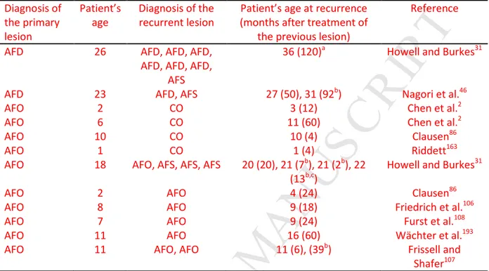
Table 2 shows the recurrence rate according to treatment and radiological locularity. There was information about recurrence for 42 lesions (40 lesions, if the information about the kind of treatment performed was also provided), of which only 2 recurred (4.8%), both central AFDs. One of the cases46 was treated by enucleation and recurred twice, with the first recurrence after 50 months, and the second recurrence as ameloblastic fibrosarcoma 92 months after the first one. The second case31 had several recurrences within a period of 10 years, until it recurred as an ameloblastic fibrosarcoma. Concerning locularity, the information about recurrence and locularity was provided for 37 lesions. Only one (5.9%) multilocular lesion recurred.
Ameloblastic fibro-odontoma (AFO)
Some studies reporting series of odontogenic studies and including AFOs were found, but their cases were not included here due to lack of enough clinical, radiological and histological information to confirm the diagnosis. These include, for example, Jing et al.63 with 4 cases and Luo and Li64 with 12 cases.
One hundred and thirty-seven publications2,29,31,32,48,61,65-195 reporting AFOs were included in the present review, reporting 215 AFOs, 211 central and 3 peripheral lesions, and one lesion for which it was not possible to identify whether it was central or peripheral, even though there was enough information to identify it as an AFO. All included cases of AFO showed formation of enamel with or without dentinoid material, mentioned in the text or visible in the photomicrographs, besides the presence of ameloblastic fibroma-like areas. Table 1 presents demographic and clinical features
M
AN
US
CR
IP
T
AC
CE
PT
ED
of the 214 AFOs known to be central or peripheral. AFOs were more prevalent in men than in women, at a 1.3:1 proportion. The mean age of the patients was higher for the central lesions in comparison to the peripheral ones. Figure 5 shows the distribution of the lesions (n=202; all cases for which the patients’ age were reported) according to age, with a high prevalence in the first two decades of life. For five lesions were diagnosed in patients in the age range between 20 to 24 years, three lesions were diagnosed in patients between 25 to 29 years old, and four lesions in patients who were 30 years-old or older. The age of the patients with peripheral lesions (n=3) was 3, 8, and 8. The few peripheral lesions were located at/between teeth 11 buccal side (n=1), 11-12 palatal side (n=1), and 13-14 palatal side (n=1). The central lesions were more prevalent in the mandible in comparison to the maxilla, and in the posterior region in comparison to the anterior region (Figure 4). About 27% of the central lesions showed signs of cortical bone perforation and 87% of the lesions had a radiological unilocular appearance. Radiopacities were observed in 86% of the AFOs, and approximately 80% of the lesions were associated with a tooth. Twenty percent of the lesions caused root resorption of adjacent teeth, most of them deciduous. Treatment of the lesions was known in 185 cases, of which 175 consisted of conservative surgery (3 excisions, 172 enucleations) and 10 cases were treated by resection (6 marginal, 4 with continuity). The time of follow-up was informed for 108 central lesions, with a mean±SD of 31.6±33.6 months (min-max, 1-168). The race of the patient was reported in 129 cases. Fifty-five cases (42.6%) were diagnosed in Caucasians, 47 in Asians, 11 in Indians, 6 in blacks, 5 in Hispanics, 2 in Turks, and 2 in Persians, and 1 in other. Table 2 shows the recurrence rate according to treatment and radiological locularity. There was information about recurrence for 146 lesions, with 10 recurrences (6.8%), all central AFOs. Four of these recurrences were diagnosed as complex odontomas,2,86,163 one of them as ameloblastic fibrosarcoma after multiple recurrences,31 and the other five recurrences as AFOs. Concerning locularity, 119 lesions had information about recurrence and locularity, showing a higher recurrence rate for unilocular lesions in comparison to the multilocular ones (p=0.658; Fisher’s exact test).
M
AN
US
CR
IP
T
AC
CE
PT
ED
AFD vs. AFOOnly two factors that were statistically significantly different between the two groups of central lesions. The first one was locularity, with AFDs being more frequently multilocular than AFOs (p<0.001). The second factor was the presence of visible radiopacities in the radiological exam, with a higher frequency for AFOs (p<0.001) (Table 1). The difference in recurrence rate (AFD 2/42, 4.8%; AFO 10/146, 6.8%) was not statistically significant (p=0.473; Fisher’s exact test).
Table 3 shows details about the cases of recurrence: diagnosis and age of the patients for primary and recurrent lesions.
DISCUSSION
The present study performed an updated comparative analysis of the clinical, radiologic features, and recurrence rate between AFDs and AFOs. The great majority of the cases were reported in isolated case reports or small case series. Peripheral lesions were extremely rare, as only 4 and 3 cases of AFD and AFO, respectively, were identified in the literature. Of the 42 cases of AFD described in the literature with information about recurrence, only 2 recurrences were reported, both diagnosed as ameloblastic fibrosarcoma after multiple recurrences. This suggests that the recurrence of AFDs is unusual. AFOs seem to have a similar recurrence rate to AFDs. Only one case of recurrent AFOs (10% of the recurrences) came back as ameloblastic fibrosarcoma after multiple recurrences.31 Forty percent of cases of AFOs recurred as complex odontomas.2,86,163 It seems odd that AFOs (a neoplasm) would recur as what is considered a hamartomatous process (complex odontoma), and this raises the possibility of the primary lesions in these particular cases actually being developing odontomas. On the other hand, the finding that six of the recurrent cases (one diagnosed as ameloblastic fibrosarcoma and five AFO) showed no sign of maturation is suggestive that at least some AFOs are not tooth malformations.
The comparisons of several parameters between AFD and AFO were performed taking into consideration the central lesions only, since the peripheral counterparts were so infrequently
M
AN
US
CR
IP
T
AC
CE
PT
ED
reported. AFD and AFO presented several similarities, such as a higher prevalence in males and in mandibles, similar mean age of the patients, rate of cortical bone perforation, rate of association with displaced/unerupted teeth and tooth root resorption, as well as the mean lesion size. The prevalence of AFDs could suggest a bimodal age distribution, even though with a far lower prevalence in the 6th and 7th decades of life than in the 1st and 2nd decades (Figure 3). A possible bimodal age distribution is less clear for AFOs (Figure 5), even having far more cases reported in the literature than AFDs.
AFD and AFO differed with regard to the presence of radiopacities visible in the radiological exams and to the radiographic locularity. It is understandable that AFOs would show a higher prevalence of visible radiopacities in the radiological exams in comparison to AFDs, as enamel is more radiopaque than dentin and dentinoid material. When it comes to locularity, despite the similarities of the mean size of the lesions in both groups, multilocularity was associated with AFD. We do not have any plausible hypothesis to explain such finding, but we have to consider that a statistically significant data may not be biologically relevant.196 Further studies are necessary to clarify the interpretation of these data.
It is important to mention that the WHO1 ranked the AFDs as an AF. There are doubts whether the two lesions are similar conditions, but it is of our opinion that AFDs should be considered as a distinct lesion from AFs until new evidence proves otherwise. The discussion about this issue probably began with Cahn and Blum,197 who postulated that AF represented the least histologically differentiated lesion that evolves from a moderately differentiated form to AFO and ultimately to odontoma. Over the years this concept has not become widely accepted and was put to the test. Eversole et al.3 and Slootweg5 argued that, if the AF is the least differentiated tumor and can develop progressively into an AFO and a complex odontoma, then this sequence should be reflected by AFs being found in a younger age group than odontomas, with tumors of intermediate differentiation occurring between the two ages. Yet, all lesions should show the same distribution according to site and the sex predilection should be the same.5 However, Eversole et al.’s3 data
M
AN
US
CR
IP
T
AC
CE
PT
ED
indicated that all these tumors occurred within the same age group. Moreover, they found completely different male-to-female ratios for AFs and complex odontomas. These ratios would be expected to be similar if AF routinely developed into odontomas. Slootweg5 observed in his review that a comparison of the data on age reveals that the more differentiated AFO occurs at a lower mean age than the histologically primitive AF. Therefore, since it cannot be accepted that a more differentiated lesion should occur at a lower mean age than the lesion from which it develops, the possibility of the AF developing into an AFO and then into an odontoma could be discarded.5 Considering the relationship between the AFO and the odontomas, Slootweg5 noted that his review’s data on age, site, and sex lead to believe that the AFO is an immature complex odontoma. He observed that the distribution according to location is approximately the same for both lesions and the complex odontoma occurs at a larger average age. However, although some “immature odontoma” may be included in every AFO review of cases, the neoplastic nature of this tumor is accepted in the literature.2
The discussion continued with Gardner,4 who stated that odontomas, being hamartomas, develop during the period of normal odontogenesis and, therefore, any apparent AF found after that time is unlikely to represent the early stage of a developing odontoma. From the results of the present review, it is interesting to note that twelve cases of AFO and eight cases of AFD affected patients of 20 years of age or older, which is beyond the age of an expected tooth formation. Later, Takeda198 suggested that an AFD may develop into an AFO. Philipsen et al.6 stated that even though there may histologically be a spectrum from the AF to the AFO with the AFD in an intermediate position, this does not necessarily suggest that the AF will differentiate over time into an odontoma. Residual or recurrent cases of AF have shown no evidence of further differentiation and maturation into a more developed odontogenic tumor.6 Although the present study observed that 40% of the recurrent cases of AFO were classified as complex odontomas, the other recurrent cases did not show such maturation process. Moreover, the mean age of the patients in AFD was similar to AFO, which do not support the concept of progressive maturation of these tumoral conditions. All these
M
AN
US
CR
IP
T
AC
CE
PT
ED
data support the hypothesis that some AFD and AFO tumors are true neoplastic conditions. We also believe that they should be considered separately from odontoma, unless some contrary evidence is demonstrated.
We agree with the concept of Takeda198 regarding the differential diagnosis of AFD and immature dentinoma. Microscopically, the epithelial component of 'immature dentinoma' is formed of strands and small islands of odontogenic epithelium without enamel organ-like structures, and the fibrous element varies from cellular to mature collagenous tissue, but the primitive dental papilla-like appearance is not found. The ‘immature dentinoma’ resembles odontogenic fibroma. On the other hand, the epithelial element in the AFD shows an enamel organ-like structure and the ectomesenchymal component resembles dental papilla. Except for the lack of enamel and the presence of dentinoid structures, AFD shows microscopic similarities to AFO and AF, respectively.
The results of the present study have to be interpreted with caution because of its limitations. First, all included studies were retrospective reports, which inherently results in flaws, manifested by the gaps in information and incomplete records. Second, many of the cases have a short follow-up, which could have led to an underestimation of the actual recurrence rate, because a longer follow-up period can lead to an increase in the recurrence rate. However, it is hard to define what it would be considered a short follow-up period to evaluate the recurrence of AFDs and AFOs. Third, the great majority of the cases described were published as isolated case reports or small case series.
CONCLUSIONS
AFD and AFO presented several similarities: higher prevalence in males and in mandibles, similar patients’ mean age, rate of cortical bone perforation and of the lesions’ association with displaced/unerupted teeth and tooth root resorption, mean lesion size, and recurrence rate. The lesions differed with regard to the presence of radiopacities and locularity. Taken together, our data do not support the concept of progressive maturation of these tumoral conditions.
M
AN
US
CR
IP
T
AC
CE
PT
ED
ACKNOWLEDGEMENTSThe authors would like to thank Dr. Paulo Goberlânio de Barros Silva for providing us his article, and Dr. Wagner Henriques Castro for providing us missing information on his publication.
REFERENCES
1. WHO: World Health Organization classification of tumours. Pathology and genetics of
head and neck tumours. Lyon, IARC Press, 2005
2. Chen Y, Li TJ, Gao Y, Yu SF: Ameloblastic fibroma and related lesions: a
clinicopathologic study with reference to their nature and interrelationship. J Oral Pathol Med 34:588, 2005
3. Eversole LR, Tomich CE, Cherrick HM: Histogenesis of odontogenic tumors. Oral Surg
Oral Med Oral Pathol 32:569, 1971
4. Gardner DG: The mixed odontogenic tumors. Oral Surg Oral Med Oral Pathol 57:395,
1984
5. Slootweg PJ: An analysis of the interrelationship of the mixed odontogenic tumors--ameloblastic fibroma, tumors--ameloblastic fibro-odontoma, and the odontomas. Oral Surg Oral Med Oral Pathol 51:266, 1981
6. Philipsen HP, Reichart PA, Praetorius F: Mixed odontogenic tumours and odontomas.
Considerations on interrelationship. Review of the literature and presentation of 134 new cases of odontomas. Oral Oncol 33:86, 1997
7. Moher D, Liberati A, Tetzlaff J, et al: Preferred Reporting Items for Systematic Reviews and Meta-Analyses: The PRISMA Statement. Ann Intern Med 151:264, 2009
8. Agarwal P, Kothari N, Girdhar M: A Case Report of Peripheral –Type Ameloblastic Fibrodentinoma and a Review. Br J Medic Med Res 8:891, 2015
M
AN
US
CR
IP
T
AC
CE
PT
ED
9. Ahmed M, Sadat SMA, Rita SN: Ameloblastic Fibro-Dentinoma of Mandible: A Case Report. J Bangladesh Coll Phys Surg 24:119, 2006
10. Akal UK, Gunhan O, Guler M: Ameloblastic fibrodentinoma. Report of two cases. Int J
Oral Maxillofac Surg 26:455, 1997
11. Anker AH, Radden BG: Dentinoma of the mandible. Oral Surg Oral Med Oral Pathol 67:731, 1989
12. Arai G, Yamada H, Nakaoka K, et al: A case of ameloblastic fibrodentinoma developing
in the mandible. Jap J Oral Maxillofac Surg 60:204, 2014
13. Azaz B, Ulmansky M, Lewin-Epstein J: Dentinoma. Report of a case. Oral Surg Oral Med
Oral Pathol 24:659, 1967
14. Baker WR, Swift JQ: Ameloblastic fibro-odontoma of the anterior maxilla. Report of a
case. Oral Surg Oral Med Oral Pathol 76:294, 1993
15. Bhargava M, Sood S, Rathore P: Ameloblastic Fibrodentinoma: Report of a Case in an
Infant. J Clin Diagn Res 10:Zd06, 2016
16. Bologna-Molina R, Salazar-Rodriguez S, Bedoya-Borella AM, et al: A histopathological
and immunohistochemical analysis of ameloblastic fibrodentinoma. Case Rep Pathol 2013:604560, 2013
17. Brandt SK, Mason MH, Barkley R: Ameloblastic fibrodentinoma: report of case. ASDC J
Dent Child 55:372, 1988
18. Cassidy JP, Crocker DJ, Grau WH: Ameloblastic fibrodentinoma. J Oral Maxillofac Surg
45:734, 1987
19. Chaudhry AP, Stickel FR, Gorlin RJ, Vickers RA: An unusual odontogenic tumor. Report
of a case. Oral Surg Oral Med Oral Pathol 15:86, 1962
20. Chen HS, Wang WC, Lin YJ, et al: Gingival ameloblastic fibro-dentinoma—Report of a
M
AN
US
CR
IP
T
AC
CE
PT
ED
21. Costa FW, Silva PG, Soares EC, et al: Surgical approach in a large ameloblastic fibrodentinoma. J Craniofac Surg 26:950, 2015
22. Fernández RL, Rodríguez JT, Molina RB: Fibrodentinoma ameloblástico en un paciente
pediátrico. Presentación de un caso. Rev Odontol Mex 13:43, 2009
23. Field HJ, Ackerman AA: Calcifying fibro-adamantoblastoma. Am J Orthod Oral Surg 28:B543, 1942
24. Giraddi GB, Garg V: Aggressive atypical ameloblastic fibrodentinoma: Report of a case.
Contemp Clin Dent 3:97, 2012
25. Godjesk JE, Dolinsky HB, Schneider LC, Doyle JL: Ameloblastic fibro-dentinoma in the
gingiva: report of a case. J Oral Med 35:59, 1980
26. Gorlin RJ, Chaudhry AP, Pindborg JJ: Odontogenic tumors. Classification,
histopathology, and clinical behavior in man and domesticated animals. Cancer 14:73, 1961
27. Günhan O, Celasun B: Ameloblastic fibro-odontoma. Case report. Aust Dent J 34:29, 1989
28. Gyulai-Gaal S, Takacs D, Szabo G, Suba Z: Mixed odontogenic tumors in children and adolescents. J Craniofac Surg 18:1338, 2007
29. Hansen LS, Ficarra G: Mixed odontogenic tumors: an analysis of 23 new cases. Head Neck Surg 10:330, 1988
30. Hietanen J, Calonius PE, Collan Y, Poikkeus P: Histology and ultrastructure of an ameloblastic fibroma. A case report. Proc Finn Dent Soc 69:129, 1973
31. Howell RM, Burkes EJ, Jr.: Malignant transformation of ameloblastic fibro-odontoma to ameloblastic fibrosarcoma. Oral Surg Oral Med Oral Pathol 43:391, 1977
32. Husted E, Pindborg JJ: Odontogenic tumours; clinical and roentgenological aspects, treatment and pathology. Odontol Tidskr 61:275, 1953
M
AN
US
CR
IP
T
AC
CE
PT
ED
33. Ikeda H, Minamizato T, Fujita S, Asahina I: Ameloblastic fibrodentinoma with a congenitally missing second premolar tooth: a case report. Oral Surg Oral Med Oral Pathol Oral Radiol 117:e88, 2014
34. Jacob OA, Padmakumar SK, Aloka D, Sooraj S: Ameloblastic Fibrodentinoma: A 12 Years follow-up of a Rare Entity. IJSS Case Rep Rev 1:31, 2015
35. Joseph S, Priya L, Gopal D, et al: Ameloblastic fibrodentinoma presenting as a false gingival enlargement in the maxillary anterior region. Case Rep Dent 2015:812087, 2015
36. Karasu HA, Akman H, Uyanik LO, Sayan NB: Ameloblastic fibrodentinoma. A case report. N Y State Dent J 70:22, 2004
37. Kaushik A, Krishna BA, Reddy ALG, et al: Ameloblastic fibrodentinoma involving anterior maxilla: A rare case report. J Indian Soc Pedod Prev Dent 31:275, 2013
38. Khurana AS, Munjal M, Narad M: Ameloblastic fibro-odontoma of the maxilla. Indian J
Otolaryngol Head Neck Surg 54:149, 2002
39. Kitano H, Matsubara M, Yoshida M, Ueki T: Mandibular Ameloblastic Fibro-odontoma.
Asian J Oral Maxillofac Surg 19:163, 2007
40. Kokuryo S, Tominaga K, Habu M, et al: A case of ameloblastic fibrodentinoma in the anterior maxilla. Jap J Oral Maxillofac Surg 55:364, 2009
41. Lúcio PS, Cavalcante RB, Maia RN, et al: Aggressive ameloblastic fibro-odontoma assessment with CBCT and treatment. Eur Arch Paediatr Dent 14:179, 2013
42. Lukinmaa PL, Hietanen J, Laitinen JM, Malmstrom M: Mandibular dentinoma. J Oral Maxillofac Surg 45:60, 1987
43. Maclennan WD, McKendrick AJ: UNUSUAL ODONTOGENIC TUMOR:REPORT OF CASE. J
Oral Surg Anesth Hosp Dent Serv 21:429, 1963
44. Minamizato T, I T, Ikeda H, et al: Peripheral-type ameloblastic fibrodentinoma with features of so-called "immature dentinoma". Oral Surg Oral Med Oral Pathol Oral Radiol 117:e61, 2014
M
AN
US
CR
IP
T
AC
CE
PT
ED
45. Miranda JL, Silva UH, Aires MEMCB, et al: Ameloblastic fibrodentinoma: radiographic
and histopathologic findings. Rev Cons Reg Odontol Pernamb 4:67, 2001
46. Nagori SA, Jose A, Bhutia O, et al: Ameloblastic fibrosarcoma developing 8 years after
resection of ameloblastic fibro-dentinoma: A unique presentation. J Oral Maxillofac Surg Med Pathol 27:143, 2015
47. O’Brien FV: Ameloblastic Odontoma: a Case Report. Br Dent J 131:71, 1971
48. Reilly JS, Supance JS: Pathologic quiz case 1. Ameloblastic fibro-odontoma. Arch Otolaryngol 109:200, 1983
49. Ribeiro CM, Santos TT, de Castro SR, et al: Extensive Mandibular Ameloblastic Fibro-Odontoma. J Craniofac Surg 27:e563, 2016
50. Salehinejad J, Langaroodi AJ, Shahakbari R, Yazdani N: Ameloblastic fibrodentinoma: report of a rare case. J Contemp Dent Pract 14:548, 2013
51. Sangala NB, Munishekar M, Krishna A, Shylaja S: Ameloblastic fibrodentinoma of mandible. J NTR Univ Health Sci 3:66, 2014
52. Sankireddy S, Kaushik A, Krishna BA, et al: Ameloblastic fibrodentinoma involving anterior maxilla: a rare case report. J Indian Soc Pedod Prev Dent 31:275, 2013
53. Scheuer HA, Holtje WJ: [Interdisciplinary treatment of a 3 1/2-year-old child with extensive ameloblastic fibro-odontoma. Case report and follow-up documentation over 21 1/2 years]. Mund Kiefer Gesichtschir 5:367, 2001
54. Sousa SF, Gomez RS, Diniz MG, et al: Defects of the Carney complex gene (PRKAR1A) in
odontogenic tumors. Endocr Relat Cancer 22:399, 2015
55. Steinberg MJ, Herrera AF, Frontera Y: Mixed radiographic lesion in the anterior maxilla in a 6-year-old boy. J Oral Maxillofac Surg 59:317, 2001
56. Takeda Y, Sato H, Satoh M, et al: Pigmented ameloblastic fibrodentinoma: a novel melanin-pigmented intraosseous odontogenic lesion. Virchows Arch 437:454, 2000
M
AN
US
CR
IP
T
AC
CE
PT
ED
57. Takeda Y, Sato H, Satoh M, et al: Immunohistochemical expression of neural tissue markers (neuron-specific enolase, glial fibrillary acidic protein, S100 protein) in ameloblastic fibrodentinoma: a comparative study with ameloblastic fibroma. Pathol Int 50:610, 2000
58. Ulmansky M, Bodner L, Praetorius F, Lustmann J: Ameloblastic fibrodentinoma: report
on two new cases. J Oral Maxillofac Surg 52:980, 1994
59. Umashankara KV, Nagaveni NB, Manjunath S: Ameloblastic fibro-dentinoma: Report of
a rare tumor with literature review. J Cranio Max Dis 1:141, 2012
60. Unsal H, Eren H, Inceoglu B, Oncul AM: Mature form of ameloblastic fibrodentinoma: a
case report. J Craniofac Surg 25:e299, 2014
61. Wakuda T, Saiki T, Watanabe S, et al: A case of pigmented ameloblastic
fibrodentinoma and one of pigmented ameloblastic fibroodontoma. Jap J Oral Maxillofac Surg 44:87, 1998
62. van Wyk CW, van der Vyver PC: Ameloblastic fibroma with dentinoid
formation/immature dentinoma. A microscopic and ultrastructural study of the epithelial-connective tissue interface. J Oral Pathol 12:37, 1983
63. Jing W, Xuan M, Lin Y, et al: Odontogenic tumours: a retrospective study of 1642 cases
in a Chinese population. Int J Oral Maxillofac Surg 36:20, 2007
64. Luo HY, Li TJ: Odontogenic tumors: a study of 1309 cases in a Chinese population. Oral
Oncol 45:706, 2009
65. Oral Pathology Quiz #62. Case number 4. Ameloblastic fibro-odontoma. J N J Dent Assoc 80:23, 2009
66. Adebayo ET, Ajike SO, Adekeye EO: A review of 318 odontogenic tumors in Kaduna, Nigeria. J Oral Maxillofac Surg 63:811, 2005
67. Album MM, Neff JH, Myerson RC: Ameloblastic fibro-odontoma: report of case. ASDC J
M
AN
US
CR
IP
T
AC
CE
PT
ED
68. Alderson GL, McGuff HS, Jones AC, Bandy DP: Oral and maxillofacial pathology case of
the month. Ameloblastic fibro-odontoma. Tex Dent J 121:427, 2004
69. al-Sebaei MO, Gagari E: Ameloblastic fibro-odontoma. J Mass Dent Soc 50:52, 2001
70. Anneroth G, Modeer T, Twetman S: Ameloblastic fibro-odontoma in the maxillae. A case report. Int J Oral Surg 11:130, 1982
71. Atarbashi Moghadam S, Mokhtari S: Peripheral Developing Odontoma or Peripheral Ameloblastic Fibroodontoma: A Rare Challenging Case. Case Rep Dent 2016:9379017, 2016
72. Atwan S, Geist JR: Ameloblastic fibro-odontoma: case report and review of the literature. J Mich Dent Assoc 90:46, 2008
73. Banihashem Rad SA, Mortazavi H, Eshghpour M, et al: A Large Ameloblastic Fibro-odontoma of the Maxillary Sinus. Iran J Otorhinolaryngol 26:111, 2014
74. Barone R, Ficarra G, Cudia G: [Ameloblastic fibro-odontoma. A case report]. Minerva Stomatol 39:97, 1990
75. Bernhoft CH, Bang G, Gilhuus-Moe O: Ameloblastic fibro-odontoma. Int J Oral Surg 8:241, 1979
76. Boxberger NR, Brannon RB, Fowler CB: Ameloblastic fibro-odontoma: a
clinicopathologic study of 12 cases. J Clin Pediatr Dent 35:397, 2011
77. Bramley P, Marsland EA: An aggressive odontome of infancy. Br J Oral Surg 1:33, 1963
78. Brocheriou C, Auriol M, Bendjaballah F, Guilbert F: Ameloblastic odontoma. Arch Anat
Pathol 20:71, 1972
79. Buchner A, Kaffe I, Vered M: Clinical and radiological profile of ameloblastic fibro-odontoma: an update on an uncommon odontogenic tumor based on a critical analysis of 114 cases. Head Neck Pathol 7:54, 2013
80. Capitaine P, Deffez JP, Plante P, Gros F: Ameloblastic odontoma in a very young child.
M
AN
US
CR
IP
T
AC
CE
PT
ED
81. Castellón ML, Fariña R, Martínez B, et al: Fibro-odontoma ameloblástico de la mandíbula. Rev Esp Cirug Oral y Maxilofac 35:87, 2013
82. Cavalcante AS, Anbinder AL, Costa NC, et al: Ameloblastic fibro-odontoma: a case report. Med Oral Patol Oral Cir Bucal 14:e650, 2009
83. Chang H, Precious DS, Shimizu MS: Ameloblastic fibro-odontoma: a case report. J Can
Dent Assoc 68:243, 2002
84. Chang JY, Marcantoni H, Kessler HP: Oral and maxillofacial pathology case of the month. Ameloblastic fibro-odontoma. Tex Dent J 124:686, 2007
85. Choukas NC, Toto PD: Ameloblastic odontoma. Oral Surg Oral Med Oral Pathol 17:10,
1964
86. Clausen F: Les Odontomes Ameloblastiques. Rev Stomatol 60:590, 1959
87. Cran JA, Herd JR, Chau KK: An unusual odontogenic lesion. Case report. Aust Dent J 21:520, 1976
88. Curran JB, Owen D, Lanoway J: Ameloblastic fibro-odontoma-case report. J Can Dent
Assoc 46:314, 1980
89. Daley TD, Lovas GL: Ameloblastic fibro-odontoma: report of a case. J Can Dent Assoc
48:467, 1982
90. Damm DD: Pericoronal radiolucency with radiopacities of posterior mandible.
Ameloblastic fibro-odontoma. Gen Dent 56:585, 2008
91. De Riu G, Meloni SM, Contini M, Tullio A: Ameloblastic fibro-odontoma. Case report and review of the literature. J Craniomaxillofac Surg 38:141, 2010
92. de Souza Tolentino E, Centurion BS, Lima MC, et al: Ameloblastic fibro-odontoma: a diagnostic challenge. Int J Dent 2010, 2010
93. de Visscher JG, van der Wal KG, Sievering NP, et al: [Ameloblastic fibro-odontoma. Examination in reference to a case]. Ned Tijdschr Tandheelkd 88:102, 1981
M
AN
US
CR
IP
T
AC
CE
PT
ED
94. Dhanuthai K, Kongin K: Ameloblastic fibro-odontoma: a case report. J Clin Pediatr Dent
29:75, 2004
95. Dolanmaz D, Pampu AA, Kalayci A, et al: An unusual size of ameloblastic fibro-odontoma. Dentomaxillofac Radiol 37:179, 2008
96. Doscher JC, Kramer JM, Fantasia JE: A large calcifying lesion of the maxilla in a child. J Am Dent Assoc 142:1026, 2011
97. Dutta A: Ameloblastic odontoma. Oral Surg Oral Med Oral Pathol 29:827, 1970
98. Duvigneaud S, Tant L, Loeb I, et al: [Mandibular ameloblastic fibro-odontoma]. Rev Stomatol Chir Maxillofac 105:223, 2004
99. Eda S, Tokuue S, Kato K, et al: A melanotic ameloblastic fibro-odontoma. Bull Tokyo Dent Coll 18:119, 1977
100. Euler H: Vom Zahnsystem ausgehende Geschwülste des frühesten Kindesalters. Dtsch
zahnärztl Wschr 26:206, 1933
101. Favia GF, Di Alberti L, Scarano A, Piattelli A: Ameloblastic fibro-odontoma: report of two cases. Oral Oncol 33:444, 1997
102. Ferracini R, Frizzera G, Bazzocchi F, et al: Ameloblastic odontoma. Zwr 83:passim, 1974 103. Flaitz CM, Hicks J: Delayed tooth eruption associated with an ameloblastic fibro-odontoma. Pediatr Dent 23:253, 2001
104. Forsberg A, Lagergren C, Martensson G: Ameloblastic odontoma. Report of a case. Oral Surg Oral Med Oral Pathol 14:726, 1961
105. Franco A, Riscala S, Kahoudji M, Croue A: [Endoscopic removal of a mandibular ameloblastic fibro-odontoma]. Rev Stomatol Chir Maxillofac 110:359, 2009
106. Friedrich RE, Siegert J, Donath K, Jakel KT: Recurrent ameloblastic fibro-odontoma in a 10-year-old boy. J Oral Maxillofac Surg 59:1362, 2001
107. Frissell CT, Shafer WG: Ameloblastic odontoma; report of a case. Oral Surg Oral Med
M
AN
US
CR
IP
T
AC
CE
PT
ED
108. Furst I, Pharoah M, Phillips J: Recurrence of an ameloblastic fibro-odontoma in a 9-year-old boy. J Oral Maxillofac Surg 57:620, 1999
109. G S, Reddy YI, P S, Prakash AR: Ameloblastic Fibro-odontome (AFO) of the Mandible: A
Case Report. J Clin Diagn Res 8:260, 2014
110. Gantala R, Gotoor SG, Kumar RV, Munisekhar MS: Ameloblastic fibro-odontoma. BMJ
Case Rep 2015, 2015
111. Geller LJ, Fielding AF: Ameloblastic fibro-odontoma. Report of case. J Dist Columbia Dent Soc 65, 1978
112. Ghalaut P, Wadhawan V, Kapoor P: Ameloblastic Fibro-Odontome: A case report with
review of literature. Ind J Basic Appl Med Res 3:109, 2014
113. Glickman R, Salman L, Chaudhry AP: Ameloblastic fibro-odontoma: a case report. Ann
Dent 48:25, 1989
114. Grätz KW, Makek M, Sailer HF: The ameloblastic fibro-odontoma. Dtsch Zahnärztl Z 46:71, 1991
115. Gupta R, Astekar M, Chittlangia RK: Ameloblastic fibro-odontoma of mandible. J Dent
Sciencies Oral Rehabil 5:103, 2014
116. Hamner JE, 3rd, Pizer ME: Ameloblastic odontoma. Report of two cases. Am J Dis Child
115:332, 1968
117. Hanna RJ, Regezi JA, Hayward JR: Ameloblastic fibro-odontoma: report of case with light and electron microscopic observations. J Oral Surg 34:820, 1976
118. Hawkins PL, Sadeghi EM: Ameloblastic fibro-odontoma: report of case. J Oral
Maxillofac Surg 44:1014, 1986
119. Hegde V, Hemavathy S: A massive ameloblastic fibro-odontoma of the maxilla. Indian J
Dent Res 19:162, 2008
M
AN
US
CR
IP
T
AC
CE
PT
ED
121. Hu YP, Liu B, Su T, et al: A huge ameloblastic fibro-odontoma of the maxilla. Oral Oncol Extra 42:160, 2006
122. Hunter AK, Muller S, Kalathingal SM, et al: Evaluation of an ameloblastic fibro-odontoma with cone beam computed tomography. Tex Dent J 129:619, 2012
123. Hutt PH: Ameloblastic fibro-odontoma: report of a case with documented four-year follow-up. J Oral Maxillofac Surg 40:45, 1982
124. Ide F, Kusama K: Hyaline bodies in ameloblastic fibro-odontoma. Histopathology 40:204, 2002
125. Ishigaki Y, Satoh T, Uchikawa Y, et al: A case of ameloblastic fibro-odontoma in the mandible. Jap J Oral Maxillofac Surg 46:593, 2000
126. Ito H, Suzuki M, Kimura H, et al: A case of ameloblastic fibro-odontoma. Jap J Oral Maxillofac Surg 35:1548, 1989
127. Ito N, Okuda T, Yasuoka T, et al: Ameloblastic Fibro-odontoma. Report of a Case and
Review of the Literature. J Jap Stomatol Soc 44:108, 1995
128. Josephsen K, Larsson A, Fejerskov O: Ultrastructural features of the epithelial-mesenchymal interface in an ameloblastic fibro-odontoma. Scand J Dent Res 88:79, 1980
129. Kawanaka M, Fukutsuji M, Sakurai K, et al: A Case of Mandibular Ameloblastic
Fibro-odontoma. J Jap Stomatol Soc 1998:4, 1998
130. Kemper JW, Root RW: Adamanto-odontoma. Report of a case. Am J Orthod Oral Surg
30:709, 1944
131. Kirjavainen A, Tuovinen V, Sandor GK: Large ameloblastic fibro-odontoma in a
7-year-old girl with analysis of 108 cases. Ann Maxillofac Surg 6:15, 2016
132. Kitano M, Tsuda-Yamada S, Semba I, et al: Pigmented ameloblastic fibro-odontoma with melanophages. Oral Surg Oral Med Oral Pathol 77:271, 1994
133. Kuzuhara T, Kinoshita Y, Inoue S, et al: A Case of Ameloblastic Fibro-odontoma in the
M
AN
US
CR
IP
T
AC
CE
PT
ED
134. Lin YC, Hsu HM, Liu CS, Yuan K: A peripheral ameloblastic fibro-odontoma in a
3-year-old girl: case report, immunohistochemical analysis, and literature review. Case Rep Dent 2014:321671, 2014
135. Manor E, Kan E, Bodner L: Ameloblastic fibroodontoma of the mandible with normal
karyotype in a pediatric patient. Case Rep Dent 2012:969687, 2012
136. Martinez Martinez M, Romero CS, Pina AR, et al: Pigmented ameloblastic fibro-odontoma: clinical, histological, and immunohistochemical profile. Int J Surg Pathol 23:52, 2015
137. Meerkotter VA: The ameloblastic fibro-odontome--report of a case and the
histogenesis of an anomalous denticle. J Dent Assoc S Afr 29:389, 1974
138. Miller AS, Lopez CF, Pullon PA, Elzay RP: Ameloblastic fibro-odontoma. Report of seven cases. Oral Surg Oral Med Oral Pathol 41:354, 1976
139. Minamikawa T, Umeda M, Ishida Y, et al: A case of congenital ameloblastic fibro-odontoma of the mandible. Jap J Oral Maxillofac Surg 51:82, 2005
140. Miyauchi M, Takata T, Ogawa I, et al: Immunohistochemical observations on a possible
ameloblastic fibro-odontoma. J Oral Pathol Med 25:93, 1996
141. Mummolo S, Marchetti E, Di Martino S, et al: Ameloblastic fibro-odontoma: a case report. Ann Stomatol (Roma) 1:11, 2010
142. Nagamine K, Okitsu M, Shimada J, et al: A case of ameloblastic fibro-odontoma in the
anterior region of the upper jaw. Jap J Oral Maxillofac Surg 38:1463, 1992
143. Nakagawa K, Nishide M, Kato R, et al: Two Cases of Ameloblastic Fibro-odontoma of
the Mandible. J Jap Stomatol Soc 39:973, 1990
144. Nascimento JE, Araújo LD, Almeida LY, et al: Ameloblastic fibro-odontoma: a
conservative surgical approach. Med Oral Patol Oral Cir Bucal 14:e654, 2009
145. Negishi A, Yamada T, Iwaki H, et al: A case of an ameloblastic fibro-odontoma in the
mandibular incisal region of a one-year-old boy. Jap J Oral Maxillofac Surg 40:1187, 1994
M
AN
US
CR
IP
T
AC
CE
PT
ED
147. Niki H, Uchida H, Nakamura S, et al: A case of ameloblastic fibro-odontoma in the mandible and statistical observations in Japan. Jap J Oral Maxillofac Surg 47:630, 2001
148. Oghli AA, Scuto I, Ziegler C, et al: A large ameloblastic fibro-odontoma of the right mandible. Med Oral Patol Oral Cir Bucal 12:E34, 2007
149. Ohtawa A, Aratani Y, Miyauchi S, et al: A case of ameloblastic fibro-odontoma. Jap J Oral Maxillofac Surg 40:293, 1994
150. Okura M, Nakahara H, Matsuya T: Treatment of ameloblastic fibro-odontoma without
removal of the associated impacted permanent tooth: report of cases. J Oral Maxillofac Surg 50:1094, 1992
151. Ozer E, Pabuccuoglu U, Gunbay U, et al: Ameloblastic fibro-odontoma of the maxilla:
case report. J Clin Pediatr Dent 21:329, 1997
152. Pantoja E, Wendth AJ, Luther PM: Ameloblastic odontoma. Clinicopathologic study. N
Y State J Med 76:224, 1976
153. Park WH, Lim WB, Kim JS, et al: Ameloblastic Fibro-Odontoma in Maxilla and Its Literature Review. Korean J Oral Maxillofac Pathol 36:347, 2012
154. Pflüger H: Über die vom Zahnbildenden Gewebe Ausgehenden Geschwülste
Adamantinom und Odontom. Dtsch Zahn Mund Kieferheilkd 25:97, 1956
155. Piette EM, Tideman H, Wu PC: Massive maxillary ameloblastic fibro-odontoma: case report with surgical management. J Oral Maxillofac Surg 48:526, 1990
156. Pontes FSC, Pontes HAR, Nogueira JES, et al: Ameloblastic fibro-odontoma: Case report
with maintenance of the involved teeth. Int J Pediatr Otorhinolaryngol Extra 3:48, 2008
157. Pontes HA, Pontes FS, Lameira AG, et al: Report of four cases of ameloblastic fibro-odontoma in mandible and discussion of the literature about the treatment. J Craniomaxillofac Surg 40:e59, 2012
158. Rao NR: Ameloblastic fibro odontoma: Report of two cases. J Indian Dent Assoc 70:90, 1999
M
AN
US
CR
IP
T
AC
CE
PT
ED
159. Reibel J, Gronbaek AB, Poulsen S: Peripheral ameloblastic fibro-odontoma or
peripheral developing complex odontoma: report of a case. Int J Paediatr Dent 21:468, 2011
160. Reich RH, Reichart PA, Ostertag H: Ameloblastic fibro-odontome. Report of a case, with ultrastructural study. J Maxillofac Surg 12:230, 1984
161. Reichart PA, Philipsen HP, Gelderblom HR, Stratmann U: Ameloblastic
fibro-odontoma––report of two cases with ultrastructural study of tumour dental hard structures. Oral Oncol Extra 40:8, 2004
162. Reis SR, de Freitas CE, do Espirito Santo AR: Management of ameloblastic fibro-odontoma in a 6-year-old girl preserving the associated impacted permanent tooth. J Oral Sci 49:331, 2007
163. Riddett SA: A composite odontome at a very early age. Brit Dent J 77:129, 1944
164. Sangu Y, Ando T, Kamiyama I, et al: Ameloblastic fibro-odontoma: Report of a case. Jap J Oral Maxillofac Surg 29:1648, 1983
165. Sardana D, Rattan V, Gauba K, et al: Surgical excision of large ameloblastic fibro-odontoma followed by alloplastic bone graft—2 year follow-up: A case report. J Oral Maxillofac Surg Med Pathol 27:41, 2015
166. Sassi LM, Stramandinoli RT, Dissenha JL, et al: Large ameloblastic fibro-odontoma in a 3 year-old child: case report. RSBO 8:114, 2011
167. Schloessmann H: Zur Pathologie der Odontome. Beitr Pathol Anat Allgem Pathol 44:311, 1908
168. Sekine J, Kitamura A, Ueno K, et al: Cell kinetics in mandibular ameloblastic
fibro-odontoma evaluated by bromodeoxyuridine and proliferating cell nuclear antigen
immunohistochemistry: case report. Br J Oral Maxillofac Surg 34:450, 1996
169. Shimomura E, Iwabuchi H, Uchiyama K, Tanaka Y: A case of ameloblastic fibro-odontoma in a mandibular molar A review of the literature on differences from developing odontoma. Jap J Oral Maxillofac Surg 49:218, 2003
M
AN
US
CR
IP
T
AC
CE
PT
ED
170. Shiratsuchi Y, Higuchi Y, Hara H, et al: A case of ameloblastic fibro-odontoma. J Jap Stomatol Soc 33:269, 1984
171. Silva GCC, Jham BC, Silva EC, et al: Ameloblastic fibro-odontoma. Oral Oncol Extra 42:217, 2006
172. Sivapathasundharam B, Manikandhan R, Sivakumar G, George T: Ameloblastic fibro odontoma. Indian J Dent Res 16:19, 2005
173. Slootweg PJ: Epithelio-mesenchymal morphology in ameloblastic fibro-odontoma: a light and electron microscopic study. J Oral Pathol 9:29, 1980
174. Soares RC, Godoy GP, Neto JC, et al: Ameloblastic fibro-odontoma: Report of a case presenting an unusual clinical course. Int J Pediatr Otorhinolaryngol Extra 1:200, 2006
175. Stokke T: Inductive Effect in Odontogenic Tumor. Nor Tannlaegeforen Tid 62:187, 1952
176. Suba Z, Szabo G, Szemenyei K, et al: [Clinical and pathological features of mixed odontogenic tumors (ameloblastic fibroma and ameloblastic fibro-odontoma)]. Fogorv Sz 81:111, 1988
177. Suenaga H, Teshima T, Marumo M, Maeda H: Ameloblastic fibro-odontoma: Report of
a case. Jap J Oral Maxillofac Surg 22:702, 1976
178. Surej Kumar LK, Manuel S, Khalam SA, et al: Ameloblastic fibro-odontoma. Int J Surg Case Rep 5:1142, 2014
179. Tachikawa N, Sugiyama Y, Asahina I, et al: A case report of ameloblastic fibro-odontoma. Jap J Oral Maxillofac Surg 38:1188, 1992
180. Takeda Y, Suzuki A, Kuroda M, et al: Pigmented ameloblastic fibro-odontoma:
detection of melanin pigment in enamel. Bull Tokyo Dent Coll 29:119, 1988
181. Terabe K, Takamori H, Saito T, et al: Two cases of Ameloblastic Fibro-odontoma. Jap J
Oral Maxillofac Surg 31:1209, 1985
182. Thoma KH, Goldman HM: Odontogenic Tumors: A Classification Based on Observations
M
AN
US
CR
IP
T
AC
CE
PT
ED
183. Tsuji K, Yoshida H, Watanabe S, et al: A case of ameloblastic fibro-odontoma accompanied with a number of impacted teeth. J Oral Maxillofac Surg Med Pathol 27:366, 2015 184. Tsuji T, Wen S, Suetsugu M, et al: A case of ameloblastic fibro-odontoma developing in the maxillary sinus. Jap J Oral Maxillofac Surg 40:1000, 1994
185. Tsuruki S, Kousuke K, Ichinokawa Y, Tsuruki T: A case of ameloblastic fibro-odontoma
occupying the left maxillosinus which was enucleated by the Le Fort I osteotomy. J Oral Maxillofac Surg Med Pathol 27:380, 2015
186. Uchiyama Y, Murakami S, Kishino M, Furukawa S: Ameloblastic fibro-odontoma arising
in the mandible: three case reports. Oral Radiol 25:71, 2009
187. Wainwright WW: Complex Odontoma. Report of a Case. Am J Orthod Oral Surg
31:447, 1945
188. Warnock GR, Pankey G, Foss R: Well-circumscribed mixed-density lesion coronal to an
unerupted permanent tooth. J Am Dent Assoc 119:311, 1989
189. Wassmund M: Hochdifferenziertes Odontom. Dtsch Zahnärztl Z 23:839, 1948
190. Wewel J, Narayana N: Ameloblastic fibro-odontoma of the anterior mandible in a
22-month-old boy. Indian J Dent Res 21:618, 2010
191. Vianna MR: Complex odontoma: report of case. J Oral Surg 24:450, 1966
192. Volkweis MR, Mottin RW, Rados PV, Wagner JCB: Fibro-Odontoma Amelobástico em
Terço Médio da Face. RGO 54:77, 2006
193. Wächter R, Remagen W, Stoll P: [Is it possible to differentiate between
odontoameloblastoma and fibro-odontoma? Critical position on basis of 18 cases in DOSAK list]. Dtsch Zahnarztl Z 46:74, 1991
194. Yagishita H, Taya Y, Kanri Y, et al: The secretion of amelogenins is associated with the induction of enamel and dentinoid in an ameloblastic fibro-odontoma. J Oral Pathol Med 30:499, 2001
M
AN
US
CR
IP
T
AC
CE
PT
ED
195. Zouhary KJ, Said-Al-Naief N, Waite PD: Ameloblastic fibro-odontoma: expansile mixed
radiolucent lesion in the posterior maxilla: a case report. Oral Surg Oral Med Oral Pathol Oral Radiol Endod 106:e15, 2008
196. Gomez RS, de Abreu MH, Abreu LG, Brennan PA: It is about time to stop misusing the
P-value? J Oral Pathol Med, 2016
197. Cahn LR, Blum T: Ameloblastic odontoma: case report critically analyzed. J Oral Surg 10:169, 1952
198. Takeda Y: So-called "immature dentinoma": a case presentation and histological comparison with ameloblastic fibrodentinoma. J Oral Pathol Med 23:92, 1994
M
AN
US
CR
IP
T
AC
CE
PT
ED
TABLESTable 1. Demographic and clinical features of ameloblastic fibrodentinomas (AFD) and ameloblastic fibroodontomas (AFO) described in the literature.
Central lesions p value Peripheral lesions
A. fibrodentinoma A. fibroodontoma (central lesions) A. fibrodentinoma A. fibroodontoma Sample size (n) 60 211 4 3 Age (year), mean±SD (min-max) 10.0±9.1 (1-60) 10.1±6.6 (1-55) 0.976a 16.8±23.2 (2-52) 6.3±2.9 (3-8) Men 10.7±10.1 (1-60) 9.0±5.4 (1-30) 0.318a 7.0±5.7 (3-11) - Women 8.7±6.9 (1-24) 11.5±7.7 (1-55) 0.147a 26.7±34.6 (2-51) 6.3±2.9 (3-8) p value 0.448a 0.007a 1.000b - Gender, n (%) Men 41 (69.5) 121 (57.9) 0.108c 2 (50) 0 (0) Women 18 (30.5) 88 (42.1) 2 (50) 3 (100) Unknown 1 2 0 0 Jaw, n (%) Maxilla 17 (28.3) 70 (34.1) 0.399c 3 (75) 3 (100) Mandible 43 (71.7) 135 (65.9) 1 (25) 0 (0) Unknown 0 6 0 0 Cortical bone perforation, n (%) Yes 13 (28.9) 39 (27.3) 0.833c 0 (0) 0 (0) No 32 (71.1) 104 (72.7) 4 (100) 3 (100) Unknown 15 68 0 0 Bone erosion e, n (%) Yes - - - 0 (0) 0 (0) No - - 4 (100) 1 (100) Unknown - - 0 2 Locularity, n (%) Unilocular 31 (63.3) 151 (87.3) <0.001c - - Multilocular 18 (36.7) 22 (12.7) - - Unknown 11 38 4 3 Radiopacities, n (%) Yes 23 (46) 149 (86.6) <0.001c 1 (33.3) 1 (33.3) No 27 (54) 23 (13.4) 2 (66.7) 2 (66.7) Unknown 10 39 1 0 Associated with tooth, n (%)
M
AN
US
CR
IP
T
AC
CE
PT
ED
Yes f 46 (82.1) 124 (79.5) 0.669c 0 (0) 0 (0) No 10 (17.9) 32 (20.5) 4 (100) 3 (100) Unknown 4 55 0 0 Tooth displacement/un erupted, n (%) Yes 42 (84) 166 (89.2) 0.308c 1 (25) 0 (0) No 8 (16) 20 (10.8) 3 (75) 3 (100) Unknown 10 25 0 0 Tooth root resorption, n (%) Yes 10 (19.6) 29 (20.1) 0.935c 0 (0) 0 (0) No 41 (80.4) 115 (79.9) 4 (100) 3 (100) Unknown 9 67 0 0 Treatment, n (%) Excision/curet tage 2 (3.7) 0 (0) 4 (100) 3 (100) Enucleation 50 (92.6) 172 (94.5) 0 (0) 0 (0) Marginal resection 0 (0) 6 (3.3) - 0 (0) 0 (0) Segmental resection g 2 (3.7) 4 (2.2) 0 (0) 0 (0) Unknown 6 29 0 0 Recurrence, n (%) Yes 2 (5) 10 (7) 1.000d 0 (0) 0 (0) No 38 (95) 133 (93) 2 (100) 3 (100) Unknown 20 68 2 0 Follow-up time (months), mean±SD (min-max) 50.4±54.0 (2-258; n=37) 31.6±33.6 (1-168; n=108) 0.053a 42.0±25.5 (24-60; n=2) 9.0±5.2 (3-12; n=3) Lesion size (cm), mean±SD (min-max) 3.6±2.3 (0.6-10.0; n=29) 3.6±2.2 (0.5-14.0; n=106) 0.973a 0.8±0.5 (0.4-1.6; n=4) 1.1±0.4 (0.8-1.5; n=3) For one AFO lesion, there was no information whether it was central or peripheralSD – standard deviation a Student’s t-test b Mann-Whitney test c
Pearson’s chi-squared test
d
Fisher’s exact test
e
Applied to peripheral lesions only
f
The tooth(teeth) can either be erupted with the entire root(s) encompassed by the lesion or unerupted encompassing the entire tooth
g
M
AN
US
CR
IP
T
AC
CE
PT
ED
Table 2. Recurrence rate according to treatment and radiological locularity – for the lesions with available information about both treatment and recurrence, or both locularity and recurrence.
Ameloblastic fibrodentinoma Ameloblastic fibroodontoma
Recurrence/total (% recurrence)
Central Peripheral Central Peripheral
Treatment Excision-curettage 1/2 (50) 0/2 (0) - 0/3 (0) Enucleation 1/36 (2.8) - 10/135 (7.4) - Marginal resection - - 0/6 (0) - Segmental resection a 0/2 (0) - 0/2 (0) - Total 2/40 (5) 0/2 (0) 10/143 (7) 0/3 (0) Locularity Unilocular 0/20 (0) - 7/101 (6.9) - Multilocular 1/17 (5.9) - 1/18 (5.6) - Total 1/37 (2.7) - 8/119 (6.7) - a
M
AN
US
CR
IP
T
AC
CE
PT
ED
Table 3. Cases of recurrence: age of the patients for primary and recurrent lesions.
Diagnosis of the primary lesion Patient’s age Diagnosis of the recurrent lesion
Patient’s age at recurrence (months after treatment of
the previous lesion)
Reference
AFD 26 AFD, AFD, AFD, AFD, AFD, AFD,
AFS
36 (120)a Howell and Burkes31
AFD 23 AFD, AFS 27 (50), 31 (92b) Nagori et al.46 AFO 2 CO 3 (12) Chen et al.2 AFO 6 CO 11 (60) Chen et al.2
AFO 10 CO 10 (4) Clausen86
AFO 1 CO 1 (4) Riddett163
AFO 18 AFO, AFS, AFS, AFS 20 (20), 21 (7b), 21 (2b), 22 (13b,c)
Howell and Burkes31
AFO 2 AFO 4 (24) Clausen86
AFO 8 AFO 9 (18) Friedrich et al.106 AFO 7 AFO 9 (24) Furst et al.108 AFO 11 AFO 16 (60) Wächter et al.193 AFO 11 AFO, AFO 11 (6), (39b) Frissell and
Shafer107
AFD - Ameloblastic fibrodentinoma, AFO - Ameloblastic fibroodontoma, AFS - Ameloblastic fibrosarcoma, CO – complex odontoma
a
During 10 years, six attempts of removal of the lesion by curettage were made, and the time period between these treatments was not informed. A new recurrence, which occurred 10 years after the primary lesion, was treated by resection of the mandible, with a diagnosis of ameloblastic fibrosarcoma
b
Time after previous treatment (also in months)
c
M
AN
US
CR
IP
T
AC
CE
PT
ED
FIGURE LEGENDSFigure 1. Study screening process.
Figure 2. Histopathological features of an ameloblastic fibrodentinoma (A and B) showing ameloblastic fibroma-like areas with deposition of dentin. Ameloblastic fibroodontoma with enamel formation (C and D).
Figure 3. Distribution of ameloblastic fibrodentinomas (n=64) according to age.
Figure 4. Distribution of the known precise location of central ameloblastic fibrodentinomas (AFD; n=51) and central ameloblastic fibroodontomas (AFO; n=177): AFD/AFO. Peripheral lesions are not included here (see text for the location of the peripheral lesions). Cases involving multiple regions (or an entire quadrant) are indicated between arrows. Numbers at the top and bottom of the lines indicate cases involving both adjoining regions: anterior/premolar, premolar/molar. One asterisk (*) indicates the number of lesions from the mandibular body that reached regions posterior to the 3rd mandibular molar (mandibular angle and/or ramus and/or coronoid process and/or condyle). For the rest of the AFDs, the location was ‘posterior’ for 1 lesion in the maxilla and 5 in the mandible, ‘right mandibular body’ for 1 lesion, ‘left mandibular body’ for 1 lesion, and ‘left maxillary sinus’ for 1 lesion. For the rest of the AFOs, the location was ‘anterior’ for 1 lesion in the maxilla and 5 in the mandible, ‘posterior’ for 9 lesions in the maxilla and 12 in the mandible, ‘maxilla’ for 1 lesion, ‘left maxillary sinus’ for 1 lesion, and no information was provided for 6 lesions.