Analysis
of
the
isotype
specificity
of
three
platypus
immunoglobulin
Fc
receptors
Srinivas
Akula
Degree project inmolecular biotechnology, 2012
Examensarbete imolekylär bioteknik 45 hp tillmasterexamen, 2012
Biology Education Centre and Molecular Immunology, Department ofCell and Molecular Biology, Uppsala University
Supervisor: Lars Hellman
Abstract
The host’s defense against diseases, called immunity, acts either via innate or adaptive defense mechanisms. Immunoglobulins (Ig’s) are important players in adaptive immunity. They have evolved both structurally and functionally during vertebrate evolution. The Fc region of Igs can interact with specific receptors on the surface of various immune cells; crosslinking of these Fc receptors can trigger a wide array of immune reactions. To trigger such reactions, higher mammals have five different classes of Igs (IgM, IgG, IgA, IgE and IgD) while amphibians, reptiles and birds have four (IgM, IgD, IgA and IgY). Our recent studies have revealed that the early mammals (Platypus) have eight Ig isotypes (IgM, IgD IgO, IgG1, IgG2, IgA1, IgA2 and IgE) and at least four Fc receptors: FcRA, FcRB, FcRC and FcRD. In this study we investigated the specificity of three of these platypus Fc receptors to get a better picture of their isotype specificity.
Analysis of the isotype specificity of three platypus
immunoglobulin Fc receptors
Popular Science Summary
SRINIVAS AKULA
The host defense mechanism against disease causing substances like microbes and macromolecules is called immunity. A complex set of cells and molecules involved in host immunity are collectively called the immune system. The immune system is broadly divided into early non-specific responses to microbes, innate immunity. Later responses to disease are termed adaptive immunity. In adaptive immunity, antibodies which are known as immunoglobulins play a vital role. Immunoglobulins are Y shaped proteins made of four polypeptide chains: two identical heavy chains and two identical light chains. Digestion with trypsin cleaves immunoglobulins into two fragments: a fragment for antigen binding (Fab) and fragment crystalline (Fc). The complexity of immunoglobulins has been gradually increased during vertebrate evolution.
Immune cells like mast cells, basophils, eosinophils, monocytes, macrophages, NK cells and dendritic cells have membrane receptors that interact with the Fc region of immunoglobulins. These receptors are called Fc receptors and they have three structural parts: a ligand binding extracellular part, consisting of several domains, a transmembrane region and a cytoplasmic tail. The cytoplasmic tail consists of ITAM (Immuno tyrosine activation motif) and ITIM (Immuno tyrosine inhibition motif) that are involved in signal transduction. Interaction of the Fc receptors to immunoglobulins regulate various immune reactions such as antibody dependent cytotoxicity, mast cell degranulation and phagocytosis. The aim of this project was to study the specificity of three platypus Fc receptors FcRA, FcRB, and FcRC towards different isotypes of the platypus immunoglobulins IgG1, IgG2I, gA1, IgA2 and IgE.
To study the isotype specificity of three platypus Fc receptors, recombinant clones of Fc receptors were constructed and transfected into HEK 293 cells. Expressed proteins from the HEK 293 c ells were purified by affinity chromatography and analyzed by SDS-PAGE. Unfortunately this was unsuccessful in our study. The possible reasons could be low efficiency of transfection, the sheer complexity of the mammalian expression system or low expression levels of Fc receptors. If we can express the desired protein in HEK 293 cells, we can study the interaction of these proteins with immunoglobulins using ELISA.
Degree project in molecular biotechnology, 2012
Examensarbete i molekylär bioteknik 45 hp till masterexamen, 2012
Biology Education Centre and Molecular Imunology, Department of Cell and Molecular Biology, Uppsala University
Contents
Abbreviations ... 6
Immune system ... 7
Antibodies (Immunoglobulins) ... 8
Fc receptors ... 9
Materials and Methods ... 12
Isolation of DNA constructs ... 12
Cloning of DNA constructs from pUC 57 vector into PCEP-Pu2 vector ... 12
Restriction digestion and ligation ... 13
Transformation and screening... 13
Transfection into HEK 293 EBNA cells ... 13
Protein purification ... 14
ELISA ... 14
Results ... 15
ELISA ... 15
Screening of clones ... 15
Transfection of Plasmid DNA into HEK 293 cells ... 17
Discussion ... 18
Conclusions ... 19
References ... 20
6
Abbreviations
FcγR Fc gamma receptors FcαR Fc alpha receptor FcεR Fc epsilon receptor FcαμR Fc alpha mue receptor FcδR Fc delta receptor
FcRL (1-6) Fc receptor like molecules Fab Fragment antigen binding
Fc Fragment crystalline
Ig Immunoglobulin
IGSF Immunoglobulin superfamily
ITAM Immunoreceptor tyrosine based activation motif ITIM Immunoreceptor tyrosine based inhibiting motif NK Natural killer
FcR Fc receptor
EC Extracellular
DMEM Dulbecco’s modified Eagle medium ELISA Enzyme linked immunosorbent assay FBS Fetal bovine serum
LB Luria – Bertani broth
Ni-NTA Nickel –nitotriacetic acid PBS Phosphate buffered saline
7
Introduction
Immune system
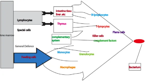
Invasion of foreign microbes and macromolecules into a host is the cause of many diseases1. A complex set of cells and molecules that collectively form the immune
system fight against these intruders (Fig.1). Cells such as B-lymphocytes, T-lymphocytes, neutrophils, eosinophils, mast cells, basophils, macrophages, dendritic cells and molecules such as complement proteins and cytokines play an important role in host immunity2.
The early response to microbes is called innate immunity while the late or adaptive response to an infection is called adaptive immunity. Innate immunity is non-adaptive and acts very quickly after the entry of the microbe, but it has no memory and acts with the same magnitude and kinetics the second time the body meets this microbe. Phagocytes like macrophages and neutrophils and complement molecules are involved in this early innate response. Adaptive immunity acts through B and T lymphocytes. B-lymphocytes produce and release antibodies to a specific antigen. During differentiation B cells change in function. Fully mature antigen activated B cells are called plasma cells and they are the primary producers of secreted antibodies against an antigen. Memory cells are another form of the B cell are specific to the antigen encountered during the primary immune response. T cells are found as several functionally different subpopulations. T killer cells are specialized to kill cells infected with intracellular parasites and T helper cells help T killer cells and B cells to become mature immunocompetent cells. [1,2]
Figure 1. The immune system consists of various immune cells and molecules: the picture adapted from reference 2.
8
Antibodies (Immunoglobulins)
Antibodies also known, as immunoglobulins are Y shaped proteins, made of four polypeptide chains: two identical heavy chains and two identical light chains that are connected by disulphide bonds. Each chain has two regions namely a variable region and a constant region. Digestion with trypsin cleaves immunoglobulins into two fragments: a fragment for antigen binding (Fab) and fragment crystalline3 (Fc) (Fig 2).
Mammals consist of at least five major Ig classes (IgM, IgG, IgA, IgE and IgD)4. These
were present more than 220 million years ago, when monotremes (egg laying mammals) separated from the other extant mammalian lineages. Amphibians, reptiles and birds have three classes of Igs5: IgM, IgA and IgY but not IgG and IgE. IgY has similar effector
functions as IgG and IgE indicating that IgY, by a gene duplication generated the ancestors of IgG and IgE during early mammalian evolution 6 (Fig.3). During mammalian evolution, monotremes (egg laying mammals) are the first branch to express all the five Ig classes7. Only three extant species of monotremes exist today, the platypus and two
species of ant–eaters, the echidnas 5.
Figure 2. The Y shaped antibody structure consists of the antigen binding region (Fab) and the constant (Fc) region; the figure is adapted from reference 3.
9
Figure3. Chicken IgY duplication and form mammals IgG and IgE: picture adapted from reference 8.
Fc receptors
Immune cells like mast cells, basophils, eosinophils, monocytes, macrophages, NK and dendritic cells have membrane receptors that interact with the Fc region of immunoglobulins9. These Fc region binding receptors on immune cells, the Fc receptors, have three structural parts: ligand binding extracellular part, transmembrane region and signal transducing cytoplasmic tail. The extra cellular part contains several domains and the number of these domains varies from receptor to receptor. The extracellular part resembles the domains in immunoglobulins and therefore they are members of the immunoglobulin super family (IgSF). Each immunoglobulin has a, specific Fc receptor (Fig.4): FcγR for IgG, FcεR for IgE, FcμR for IgM, FcδR for IgD and Fcαr for IgA10,11.
Fc receptors have been divided into three types. The classical Fc receptors (FcγR, FcεR, FcμR, FcδR, and Fcαr) that sit on the surface and bind the various immunoglobulin isotypes, the Fc receptors like molecules (FCRL1-FCRL6) and the intracellular receptor like proteins FCRLA and FCRLB. The function of Fc receptors like molecules and intracellular receptor like proteins is still obscure. [12,11,13]
FcγR is classified into FcγRI, FcγRII, and FcγRIII. Subtypes of the FcγRI (IA, 1B and 1C) are encoded by three genes and similarly subtypes of FcγRII (IIA, IIB and IIC) are encoded by three genes. Two gens code for FcγRIII (IIIA and IIIB). Except for FcγRIB and FcγRIC all other receptors have been found to have a specific function. FcγRI bind to antibodies with high affinity while other bind with low affinity10. FcαR is distantly
related to the other receptors14 and binds with a medium affinity (Table 1). The high
affinity receptor for IgE FcεRI is expressed on Langerhans cells, basophils and mast cells. A second receptor for IgE, the low affinity receptor or FcεRII (CD23 lectin family) is expressed on B cells eosinophils and monocytes. This receptor is not at all related to the other receptors but belong to a lectin family of proteins. The FcμR is expressed on B cells and bind to IgM. FcαμR bind both IgA and IgM. FcδR, the receptors for IgD are not well characterized and there are doubts if it at all exists. PIGR is a receptor for IgA (PIgA) and IgM (PIgM) at mucosal surfaces. FcRn are neonatal receptor of IgG expressed on epithelial and endothelial cells11. This latter receptor is also not related o the
10
classical Fc Receptors but is instead closely related to MHC class I molecules.
The Fc receptors are triggered by cross-linking of the receptor which lead to the phosphorylation of the cytoplasmic immuno tyrosine activator motif (ITAM). ITAM consists of two YXXL amino acid boxes that are separated by a seven amino acids. Src and Syk kinases mediated phosphorylation of the tyrosine in the ITAM motif and make it active. Some receptors (FcγRIIB) have immuno tyrosine inhibitory motifs (ITIM) in their cytoplasmic region that inhibits the immune response. SHIP-1 and SHIP kinases activate ITIM by tyrosine phosphorylation (Fig .5). Binding of Fc receptors to antibodies or immune complexes result in wide array of immuno-regulatory functions such as antibody dependent cytotoxicity, mast cell degranulation and phagocytosis. Their binding also regulates lymphocyte proliferation and antibody secretion15.
The aim of my project is to study the specificity of three platypus Fc receptors FcRA, FcRB, and FcRC towards different antibody isotypes of the platypus IgG1, IgG2, IgA1, IgA2 and IgE.
Figure 4. Human Fc receptors which are represent in d iffernrt colours in r eceptors contains variuos number of extra cellular d omains, transmembrane region and singal transduce cytoplasmic tail which conist of acivating ITAM(blue box) or inhibiting ITIM(red box) motifs adapted from reference 11.
11
Figure 5. Fc receptor signaling pathways, The signaling is mediated by Src family kinases, SHIP, receptors have ITAM or ITIM in cytoplasmic region, and the extra cellar part bind to antigen-antibody immune complexes. Adapted from reference 16.
Table 1 Characteristics of human Fc receptors retrieved and adapted from reference 11.
FcγRI FcγRII FcγRIII FcεRI FcεRII FcαRI FcαμR FcμR FcδR
Poly-Ig FcR n CD G en om ic lo ca tio n CD241 q21.1 CD321q 23-24 CD161q 23 1q23 CD231 9q CD891 9q13. 4 1q32.3 1q31-41 19q 13 M ole cu la r Wei gh t K D a 72 40 50-80 45-65 25-45 100 55- 70 58-60 46+14 G en es Is of or m s 3 3 2 1 1 1 1 1 2 Lig an d
IgG IgG IgG IgE
CR2, IgE, CR3 IgA, SIgA IgM and IgA polymer IgM IgD IgM and IgA polym er IgG A ffi ni ty L m ol - 108 -109 <10 7 Near 3*107 109 -1010 10 6 5*107 IgM 3*109 IgA 3*108 PH dep end ent
12
Materials and Methods
Isolation of DNA constructs
The DNA sequences to be used to produce recombinant mouse immunoglobulins IgG2a, IgE, mouse receptors FcγREC, FcεREC genes, platypus receptors FCRA, FCRB, FCRC genes and chicken immunoglobulin IgY gene were designed based on G enbank sequences and ordered as ready inserts from GenScript Corporation inserted in the plasmid pUC57. These plasmid DNA constructs having ampicillin selectivity were transformed into the DK1 bacterial cells and cultured overnight at 37oC in LB medium
containing ampicillin (1mg/ml). The respective plasmids isolated by using E.Z.N.A plasmid mini prep kit (Omega Bio-tek, Doraville U.S.A) according to the manufacturer’s instructions.
Cloning of DNA constructs from pUC 57 vector into PCEP-Pu2 vector
Isolated plasmids containing the respective DNA constructs were used to excise the insert fragment, then that was purified and ligated into the PCEP-Pu2 vector (Fig.6). PCEP-Pu2 vector has a BM 40 signal peptide (BM-40 SP), a His-myc-tag and restriction sites for XhoI, EcoRI, and BamHI.
Figure 6. S tructure of pCEP-Pu vector consists of restriction sites and signal peptide: adapted from reference 17.
13
Restriction digestion and ligation
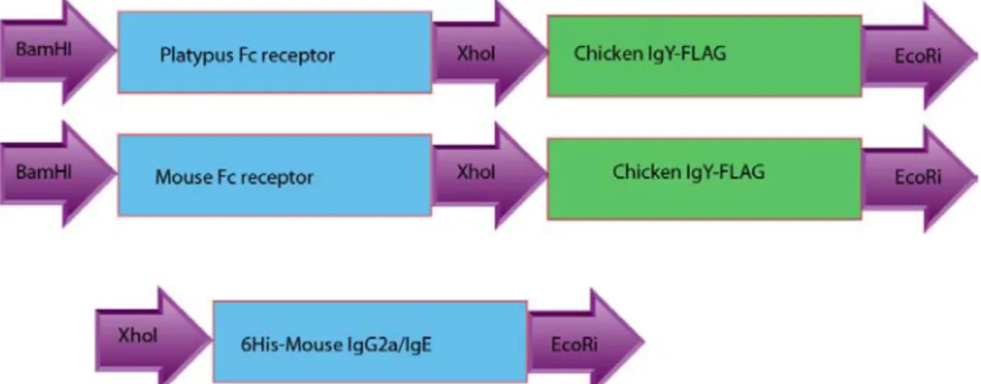
The clones containing immunoglobulin inserts (IgG2a, IgE and pUC57-IgY) were digested with XhoI and EcoRI, and clones containing the receptor sequences (pUC57- FcγREC, pUC57- FcεREC, pUC57- FCRA, pUC57- FCRB, and pUC57-FCRC) (Fig.7) were digested with BamHI and XhoI (Fermentas). The digested samples were analyzed on 1% agarose gels. The isolated DNA fragments were ligated with pCEP-pu2 vector using fast ligation kit (Fermentas) according to the protocol given in the kit.
Figure 7: Recombinant DNA constructs of platypus and mouse Fc receptors. The restriction sites used to excise the fragments are depicted in the figure.
Transformation and screening
Following ligation the vector constructs were transformed into E.coli DK1 competent cells by adding 5µl of DNA into 100µl of competent cells. This mixture was kept on ice for 30 mins and transferred to 42oC for 90 seconds and immediately transferred to when
it was keep ice for 2 m ins. After cooling, the mixtures were added with LB medium, plated on LB/ampicillin plates and incubated overnight at 37oC.
Colonies from the respective plates were collected and inoculated into LB medium containing ampicillin and grown overnight at 37oC. Plasmids from the respective cultures
were isolated and digested with the specific restriction enzymes used as above. The cleaved fragments were analyzed on 1% agarose gels to make sure that the proper fragments were inserted into the vector.
Transfection into HEK 293 EBNA cells
Human embryonic kidney cells (HEK) 293 E BNA cells were cultured in DMEM (Dulbecco’s Modified Eagle Medium) (Invitrogen) in T25 culture flasks by incubating at 37oC, 5%CO2. The cells were transferred into T75 flasks when the confluency reaches to
70% - 80%. Initially, clones of mouse IgG2a, IgE, FcγREC and FcεREC were transfected into HEK 293 c ells. The transfection mixture was prepared by adding 15ug of sterile DNA, 40μl of transfection reagent Lipofectamine 2000U (Invitrogen), 710μl of serum free DMEM (with 50ug/ml gentamycin). This mixture was vortexes vigorously for 2 min and kept at room temperature for 45 min. 6ml of serum free DMEM (with 50μg/ml
14
gentamycin) was added with 800μl transfection mixture and then added to HEK293 cells to express the transfected plasmids. After overnight incubation at 37oC, 5% CO2 in a
humidified incubator, 10% FBS was added to the cells and kept for incubation at 37oC,
5% CO2. Then the selection medium (DMEM with gentamycin (50μg/ml), puromycin
(0.5μg/ml) and heparin (5μg/ml)) was added to the cells and kept for overnight incubation at 37oC, 5% CO2. Dead cells were removed and new selection medium was
added to the cells, when the cells reaches to confluency, medium were collected the recombinant protein was purified by affinity chromatography.
Protein purification
Using Ni-NTA agarose beads (Qiagen) were purified expressed proteins of mouse Immunoglobulins IgG2a and IgE; using anti –FLAG beads (SIGMA) were purified expressed proteins of mouse receptors FcγREC and FcεREC. Recombinant proteins were purified from conditioned HEK293 cell medium, 140μl of Ni-NTA slurry and anti-FLAG beads were added to 100 ml of conditioned and filtered medium and rotated for 45 min at 4oC. Samples were centrifuged at 5000 rpm for 1 min at 4oC. Beads were collected and washed with washing solution (PBS tween 0.005%+10mM imidazole+1mMNaCl) two times. Then the solution containing beads were transferred into 2 m l columns and the solution passed through the column and the beads were washed three times with 1 ml, 2 ml and 2 ml of washing solution. Then six fractions were collected after adding elution solution (PBS tween 0.005%+100mM imidazole). These fractions were analyzed by SDS-PAGE.
ELISA
Binding to beads (my one streptavidin T1) (SIGMA), ten µl of horse serum (GIBCO) was added to 100 µl of receptor beads and incubated for 30 min at room temperature. Then platypus immunoglobulins (IgG1, IgG2, IgA1, IgA2 and IgE) (Preciously cloned and purified) were added to the above receptors and incubated at 4oC overnight. Secondary
antibody (Monoclonal anti-poly histidine alkaline phosphate) (SIGMA) was added to each sample at a 1:2000 dilution (antibody +Horse serum) and kept shaking at room temperature for one hour. Then washed with PBS/Tween20 solutions kept at 4oC for one
15
Results
ELISA
Conditioned HEK 293 medium from the platypus FcRA, FcRB, FcRC and mouse FcγRI and FcεRI transfected cultures were stored in cold room (several months) until they were purified and analyzed using SDS-PAGE (Fig.8).
Figure 8. Recombinant proteins on SDS-PAGE: M marker, platypus Fc receptors A, Platypus Fc receptors B and, platypus Fc receptors C., Mouse Fc gamma receptor (mFcγRI) and mouse Fc epsilon Receptor (mFcεRI).
Using ELISA, FCRA and FCRB proteins were tested against purified protein of the platypus immunoglobulin isotypes IgG1, IgG2, IgA1, IgA2 and IgE. Colour change was observed with FCRB but no color was seen with FCRA. This could be due to a problem in the construction of the clones, so clones were again analyzed by restriction enzyme digestion and nucleotide sequencing, see next section.
Screening of clones
To study the inserts of the clones, all clones (mouse IgG2a, IgE, FcγREC, FcεREC and platypus FCRA, FCRB and FCRC) were digested with restriction enzymes and analyzed on agarose gel (Fig 9 A). The sizes of the inserts are summarized in table 2.
Table 2. The sizes of Recombinant DNA Constructs ordered from Gene Script Name of species DNA constructs Size in bp
Mouse IgG2a 766 Mouse IgE 1000 Mouse FcγREC 789 Mouse FcεREC 534 Platypus FcRAEC 822 Platypus FcRBEC 822 Platypus FcRCEC 544 Chicken IgY 1038
16 (A)
(B) (C)
(D)
Figure 9: (A) Restriction analysis. Excised fragments were analyzed by agarose gel electrophoresis (1% agarose). The constructs analyzed were the platypus Fc receptors PFcRA, PFcRB, PFcRC and mouse FcγRI, FcεRI digestion with EcoRI, BamHI and EcoRI, BamHI and XhoI, mouse immunoglobulins were digested with EcoRI and XhoI. (B) Analysis by gel electrophoresis on 1% agarose gels, pCEP-Pu2 vector and IgE insert bands shown in yellow color, (C) Gel electrophoresis of restriction digests of IgE after EcoRI and XhoI digestion on a 1% agarose gel. (D) Restriction analysis by gel electrophoresis (1% agarose) of recombinant DNS constructs of platypus Fc receptors PFcRA, PFcRB, PFcRC and mouse FcγRI, The FcεRI clone was digested with EcoRI, BamHI and EcoRI, BamHI and XhoI, mouse immunoglobulins were digested with EcoRI and XhoI. Marker is 1000bp DNA ladder.
17
Of all the seven clones, one clone (IgE) did not give correct bands after restrictive digestion, which could be the result of a mix-up with other clones (Fig.9A). To construct a new IgE clone, plasmid DNA from the original GenScript clone was isolated and digested with restriction enzymes to isolate the IgE fragment (Fig.9 B). The isolated IgE fragment was cloned into with pCEP-Pu2 vector (Fig.9 C) and all the clones were screened and confirmed as above (Fig. 9 D).
Transfection of Plasmid DNA into HEK 293 cells
Initially, the plasmids DNA for four of the clones (IgG2a, IgE, FcγR1 and FcεR1) were sterilized by ethanol precipitation and transfected into HEK 293 cells. Condition medium containing the proteins was collected from HEK 293 cells and purified by affinity chromatography. Protein fractions were eluted during the purification and analyzed by SDS-PAGE (Fig.10). For unknown reasons we did not recover any protein and the transfection is being repeated. However, at the time of writing of the report no results from this second transfection is yet available.
Figure10. Six fractions of purified IgG2a and FcγREC were eluted and resolved by SDS-PAGE electrophoresis.
18
Discussion
To study the isotype specificity of three platypus Fc receptors, the coding regions for mouse IgG2a, IgE, FcγREC, FcεREC and platypus FcRAEC, FcRBEC, FcRCEC were inserted into the mammalian expression vector pCEP-Pu2 and transfected into HEK 293 cells (mouse clone for positive control). Initially we purified the platypus receptors from the medium containing the receptor proteins and the interaction with platypus immunoglobulins were studied by ELISA. Both FcRA and FcRB receptors have the affinity for immunoglobulins, but a color change was observed only with FcRB, but not with FcRA. The possible reason for this could be a problem in construction of clones or the clones could be mixed with other clones. In order to solve the problem, all the clones were screened by restriction analysis. Of the seven clones, six clones seems to be correct based on their size of the digested fragments whereas one clone IgE did not give correct band sizes (Fig.9A). So IgE was re-cloned into pCEP-Pu2 vector and all the clones were screened again by restriction digestion analysis (Fig. 9 B, C, D). Plasmids DNA from the respective clones were transfected into the HEK 293 EBNA cells. Initially mouse IgE, IgG2a, FcγREC and FcεREC were transfected in to the HEK 293 EBNA cells. Only with IgG2a and FcγREC clones the transfection was successful, due to low transfection efficiency of transfection reagent Lipofectamine (0.01-1%). By affinity chromatography the expressed proteins from the HEK 293 cells were analyzed by SDS-PAGE (Fig10). However no ba nds were seen indicating that our desired proteins were not expressed. This could be due to the complex mammalian expression system or the expression levels of the Fc receptors are very low. To overcome this issue, we need to do further studies on expression system of Fc receptors. If we can able to express the desired proteins in HEK 293 cells, we can study the interaction of these proteins with immunoglobulins using ELISA. These results will hopefully give us a better picture of the isotype specificity of these platypus Fc receptors and a cl ear picture of when IgE and IgG specificity was obtained by these receptors during mammalian evolution.
19
Conclusions
• Transfection of the recombinant constructs for the Fc receptors and the immunoglobulins into human embryonic kidney cells HEK 293 EBNA cells were performed.
• Proteins were purified using anti-FLAG columns for receptors and His tag columns for the immunoglobulins to identify the expressed proteins on S DS PAGE gels. The possible reason for very low expression of proteins in HEK 293 cells could be the Fc receptors have very low expression in mammalian cells or transfection efficiency of lipofectamine is very low.
• For future studies we have to check the expression system again. These results will hopefully give us a better picture of the isotype specificity of these Platypus Fc receptors.
20
References
1. Abbas, A. K., Lichtman, A. H. & Pillai, S. Cellular and Molecular Immunology, Updated
Edition. (Elsevier Health Sciences: 2009).
2. Kindt, T. J., Osborne, B. A. & Goldsby, R. A. Kuby Immunology, Sixth Edition. (W. H. Freeman & Company: 2006).
3. Check, E. Immunology: Pimp my antibody. Nature 446, 964–966 (2007).
4. Zhao, Y., Cui, Huiting, Whittington, Camilla M, Wei Zhiguo, Zhang Ziding, Yu Li, Ren Liming, Hu Xiaoxiang, Zhang yaping, Hellman Lars, Belov Katherine, Li Ning and Hammarström Lennart. Ornithorhynchus anatinus (Platypus) Links the Evolution of Immunoglobulin Genes in Eutherian Mammals and Nonmammalian Tetrapods. The
Journal of Immunology 183, 3285 –3293 (2009).
5. Nikolaidis, N., Klein, J. & Nei, M. Origin and evolution of the Ig-like domains present in mammalian leukocyte receptors: insights from chicken, frog, and fish
homologues. Immunogenetics 57, 151–157 (2005).
6. Taylor, A. I., Sutton, B. J. & Calvert, R. A. Mutations in an avian IgY-Fc fragment reveal the locations of monocyte Fc receptor binding sites. Dev. Comp. Immunol. 34, 97–101 (2010).
7. Vernersson, M., Aveskogh, M., Munday, B. & Hellman, L. Evidence for an early appearance of modern post-switch immunoglobulin isotypes in mammalian evolution (II); cloning of IgE, IgG1 and IgG2 from a monotreme, the duck-billed platypus, Ornithorhynchus anatinus. Eur. J. Immunol. 32, 2145–2155 (2002).
8. Taylor, A. I., Gould, H. J., Sutton, B. J. & Calvert, R. A. Avian IgY Binds to a Monocyte Receptor with IgG-like Kinetics Despite an IgE-like Structure. J. Biol. Chem. 283, 16384–16390 (2008).
9. Daëron, M. Fc receptor biology. Annual Review of Immunology 15, 203–234 (1997). 10. Ravetch, J. V. & Kinet, J. P. Fc Receptors. Annual Review of Immunology 9, 457–492
(1991).
11. Boross, P., Poel, K., Winkel, J. G. & Leusen, J. H. Fc Receptors. Encyclopedia of life
sciences. (2008).
12. Davis, R. S. Fc receptor-like molecules. Annul Review of Immunology 25, 525–560 (2007).
21
13. Fayngerts, S. A., Najakshin, A. M. & Taranin, A. V. Species-specific evolution of the FcR family in endothermic vertebrates. Immunogenetics 59, 493–506 (2007).
14. Van Egmond, Marjolein, Damen, Cora A, Van Spriel, Annemiek B, Vidarsson, Gestur, Van Garderen, Ever, Van de Winkel, Jan. G.J. IgA and the IgA Fc receptor. Trends in
Immunology 22, 205–211 (2001).
15. Amigorena, S. & Bonnerot, C. Fc receptor signaling and trafficking: a connection for antigen processing. Immunology Review 172, 279–284 (1999).
16. Gao H, Neff, T. & Ward, P. A. Regulation of Lung Inflammation in the Model of Igg Immune-Complex Injury. Annual Review of Pathology: Mechanisms of Disease 1, 215–242 (2006).
17. Warmerdam, P. A., Nabben, N. M., van de Graaf, S. A., van de Winkel, J. G. & Capel, P. J. The human low affinity immunoglobulin G Fc receptor IIC gene is a result of an unequal crossover event. Journal of Biological Chemistry 268, 7346 –7349 (1993).