DISSERTATION
3-CHLORO-P-TOLUIDINE HYDROCHLORIDE METABOLISM AND DETECTION OF EXPOSURE IN BIRDS
Submitted by David A. Goldade
Department of Environmental and Radiological Health Sciences
In partial fulfillment of the requirements For the Degree of Doctor of Philosophy
Colorado State University Fort Collins, Colorado
Summer 2017
Doctoral Committee:
Advisor: Marie Legare
Co-Advisor: William Hannemann Gregory Dooley
Copyright by David A. Goldade 2017 All Rights Reserved
ii ABSTRACT
3-CHLORO-P-TOLUIDINE HYDROCHLORIDE METABOLISM AND DETECTION OF EXPOSURE IN BIRDS
The avicide 3-chloro-4-methylanaline hydrochloride (chloro-p-toluidine hydrochloride, CPTH, DRC-1339) is used to control pest bird species that damage agricultural crops. While it is theorized that CPTH is a nephrotoxin, no definitive assessment of the mode of action has been performed. Additionally, the metabolic pathway of CPTH in birds has yet to be elucidated.
Radioactively labeled [14C]-3-chloro-4-methylaniline hydrochloride (250 µg per bird) was delivered to 21 red-winged blackbirds (Agelaius phoeniceus) and 21 dark-eyed juncos (Junco
hyemalis) via oral gavage, and the distribution and excretion of radioactivity were determined at 15
and 30 minutes and 1, 4, 8, 12, and 24 hours (n = 3 per time point). Direct measurement of radioactivity as well as measurement following combustion was accomplished using a liquid scintillation counter. Elimination from most tissues followed a two-compartment model, with very rapid elimination occurring between time 0 and 4 hours and a much slower elimination phase occurring after that. The average half-life of elimination for the initial phase in most tissues examined was 0.16 hours for juncos and 0.62 hours for blackbirds. The average for the slower second phase of elimination was 3.4 hours for juncos and 5.4 hours for blackbirds. The radioactivity in blackbird kidney tissues did not change significantly for the duration of the test, pointing toward the kidney as a possible site of action for this important agricultural chemical.
To further explore the mechanistic toxicology of CPTH, sub-cellular preparations were made from the liver and kidney of various avian species. In-vitro metabolism experiments were performed
iii
using these preparations and the resulting metabolites were identified and quantified. Two metabolites were identified: 3-chloro-4-methylacetanilide (CAT) and N-[3-chloro-4-(hydroxymethyl)phenyl]acetamide (OH-CAT).
A comparison of two methods was made for the analysis of CPTH and its metabolites. Due primarily to the solubility and volatility of the three compounds, CPTH and CAT performed well on gas chromatography tandem mass spectrometry (GC/MS/MS) and adequately on liquid chromatography tandem mass spectrometry (LC/MS/MS). Conversely, OH-CAT performed optimally on LC/MS/MS. LC/MS/MS was chosen as the technique for analysis of exposure data. Both methods generated residue values that demonstrated a high degree of variability between individuals. Despite the variability issues, the data showed that the primary chemical species present in the tissues of exposed birds was OH-CAT, and that the concentration of observed residue was related to the dose administered.
In an effort to identify the target for tissue binding of CPTH or its metabolites in the kidney of exposed red-winged blackbirds, protein samples were extracted and digested with trypsin. Several chemical compounds were found to be significantly different between treated and control groups (α=0.05) and were subjected to tandem mass spectrometry to identify their chemical structure. Results from this analysis did not yield any identification of specific protein binding. Limitations of sensitivity and lack of sample enrichment likely led to this outcome.
iv
ACKNOWLEDGEMENTS
I gratefully acknowledge Bruce Kimball for his assistance with statistical analysis of the data and for help in reviewing this document. Thanks go to Wendy O’Brien for insightful review as well. Thank you to the Animal Care and Chemistry Lab Units at the National Wildlife Research Center for their never-ending help in caring for the birds and providing analytical support. In particular, I would like to extend thanks to Benjamin Abbo, Steven Blamer, and Kirsten Sawtelle. Without their efforts, this work would not have been possible.
v DEDICATION
This work would not have been possible without the support of my loving wife, Mary. Her steadfast belief in me and my ability to do this has been incredible. I love her with all my heart and could not have done it without her.
vi TABLE OF CONTENTS ABSTRACT ... ii ACKNOWLEDGEMENTS ... iv DEDICATION ... v TABLE OF CONTENTS ... vi CHAPTER 1 – INTRODUCTION ... 1 1.1 Hypotheses ... 5
CHAPTER 2 – PHASE 1 – RADIOISOTOPE EXPERIMENTS ... 8
2.1 Introduction ... 8
2.1.1 Hypothesis ... 9
2.2 Materials and Methods ... 10
2.2.1 Animal Dosing ... 10
2.2.2 Sample Collection ... 11
2.2.3 Collection of Expired CO2 ... 12
2.2.4 Tissue Collection ... 12
2.2.5 Liquid Scintillation Counting Analysis ... 13
2.2.6 Data Collection ... 15
2.3 Data Analysis ... 15
2.4 Results and Discussion ... 20
2.4.1 Summary ... 30
CHAPTER 3 – PHASE 2 – IN-VITRO EXPERIMENTS ... 31
3.1 Introduction ... 31
3.1.1 Hypothesis ... 32
3.2 Materials and Methods ... 32
3.2.1 Sub-cellular Fraction Preparation ... 33
3.2.2 In-vitro Experiments ... 34
3.2.3 Liquid Chromatographic Analysis ... 35
3.3 Results and Discussion ... 37
3.3.1 Summary ... 47
CHAPTER 4 – PHASE 3 – IN-VIVO EXPERIMENTS ... 48
4.1 Introduction ... 48
4.1.1 Hypothesis ... 49
4.2 Materials and Methods ... 49
4.2.1 Trapping of birds ... 49
4.2.2 Purification of CPTH ... 50
4.2.3 Synthesis of Surrogate Compound ... 51
4.2.4 Administration of Dose ... 51
4.2.5 Tissue Collection ... 52
4.2.6 Gas Chromatography-Mass Spectrometry Analysis ... 52
4.2.7 Liquid Chromatography-Mass Spectrometry Analysis ... 53
4.3 Method Validation Samples ... 56
4.4 Results and Discussion ... 57
vii
4.4.2 Gas Chromatographic Results ... 60
4.4.3 Liquid Chromatographic Results ... 65
4.4.4 Comparison of LC and GC Results ... 79
4.4.5 Extrapolation to Field Data ... 80
4.4.6 Summary ... 81
CHAPTER 5 – PHASE 4 – PROTEOMICS AND METABOLOMICS EXPERIMENTS ... 82
5.1 Introduction ... 82
5.1.1 Hypothesis ... 83
5.2 Materials and Methods ... 84
5.2.1 Protein Extraction ... 84
5.2.2 Trypsin Digestion ... 84
5.2.3 Liquid Chromatography/Mass Spectrometry ... 85
5.2.4 Metabolomics Experiments ... 85
5.2.5 Data Handling ... 86
5.3 Results and Discussion ... 87
5.3.1 Proteomics Results ... 87 5.3.2 Metabolomics Results ... 89 5.3.3 Metabolite Identification ... 91 5.3.4 Summary ... 96 CHAPTER 6 – CONCLUSIONS ... 98 6.1 Summary of Findings ... 98 6.2 Tissue Binding ... 98 6.3 Cast Formation ... 100 6.4 Future Work ... 100 REFERENCES ... 102
1
CHAPTER 1 – INTRODUCTION
Agricultural crops suffer damage and losses from many sources. Among sources of damage are those due to birds feeding on the crops. The extent of this damage can vary dramatically from location to location. In some cases, the damage from these birds can be so extensive as to result in loss of an entire crop. Such effects can be devastating to the livelihoods of individual farmers (Kleingartner, 2003). Some tools that are available to control or minimize damage to crops include hazing, fumigants, shooting, habitat modification, and use of lethal agents.
During the post-World War II era, the United States experienced a rapid expansion in the chemical industry. New chemicals and products were being tested for any number of uses. Along with this increase in industrial chemical production came trials seeking new pesticides. Thousands of chemicals were screened for their bioactivity as pesticides (Eisemann et al., 2003). The results of this screening process were many new rodenticides and herbicides. Among those compounds identified for potential development was 3-chloro-p-toluidine hydrochloride (3-chloro-4-methylaniline hydrochloride; CPTH, DRC-1339), which was developed as an avicide. CPT (the non-hydrochloride free-base analog of CPTH) was a by-product of dye manufacture and of no commercial significance. It was found to have toxic effects at low exposure levels in blackbirds, starlings, rock doves, and corvids, all of which are species known or suspected to cause significant damage to agricultural crops as well as posing a risk to human health and safety.
CPTH is used for the control of pest bird species that damage agricultural crops, present hazards to aircraft, or have the potential to threaten human health or safety. Extensive research has been performed to determine its toxicity to many target and non-target bird species. It is a chemical agent that is a potent avicide to susceptible species and is capable of producing lethality following
2
ingestion of as little as one grain of rice containing 2% CPTH (Schafer, 1984). Integrated pest management (IPM) is a strategy for controlling pest species that incorporates multiple approaches to provide long term control of pests. IPM combines responsible pesticide use, habitat modification, use of pest-resistant crop strains, and changes to cultivation practices. It provides a holistic approach rather than focusing on one technique. As part of an IPM plan, use of CPTH has the potential to reduce crop damage caused by pest bird species.
Various products and approaches exist to minimize damage to crops from pest birds. An excellent review of the various approaches can be found in Linz et al. (2011). Hazing of the pest birds has been used since the first scarecrows were erected. Various scare devices such as air cannons have been used with limited effectiveness. The birds quickly become acclimated to the use of such devices and go back to feeding on crops. Chemical repellents containing a variety of compounds have been tested. Current products containing either methyl anthranilate or anthraquinone are the most popular. These products can be quite effective, but do not reduce populations merely pushing the over-populated birds to feed at another location. Habitat modification likewise discourages pest birds from feeding in certain regions, but does not reduce populations. Lethal control chemicals have also been tested over the years. Current products include Avitrol, which contains 4-aminopyridine, and DRC-1339 Concentrate, which contains CPTH. While Avitrol is a lethal control agent, the compound produces visible signs of distress in birds that consume the treated bait which has the added benefit of frightening other birds from the site of application. This can be a benefit to controlling bird-caused crop damage, but can cause public relations issues due to the obvious distress of the birds. CPTH produces lethality with very little evidence of distress and no vocalizations.
3
CPTH was initially registered with the U.S. Environmental Protection Agency (EPA) as an avicide in 1967 (Schafer, 1984), and is currently registered with the EPA under five labels: Compound DRC-1339 Concentrate – Feedlots, Compound DRC-1339 Concentrate – Gulls, Compound DRC-1339 Concentrate – Pigeons, Compound DRC-1339 Concentrate – Livestock, Nest & Fodder Depredations, Compound DRC-1339 Concentrate – Staging Areas. Under these five labels, CPTH is registered for controlling 22 bird species: Brewer’s blackbird (Euphagus
cyanocephalus), red-winged blackbird (Agelaius phoeniceus), yellow-headed blackbird
(Xanthocephalus xanthocephalus), tri-colored blackbird (Agelaius tricolor), common grackle (Quiscalus quiscula), boat-tailed grackle (Quiscalus major), great-tailed grackle (Quiscalus
mexicanus), brown-headed cowbird (Molothrus ater), European starling (Sturnus vulgaris), common
raven (Corvus corax), Chihuahuan raven (Corvus cryptoleucus), American crow (Corvus
brachyrhynchos), fish crow (Corvus ossifragus), black-billed magpie (Pica hudsonia), rock pigeon
(Columba livia), eurasian collared dove (Streptopelia decaocto), herring gull (Larus argentatus), great black-backed gull (Larus marinus), ring-billed gull (Larus delawarensis), laughing gull (Larus
atricilla), western gull (Larus occidentalis), and California gull (Larus californicus). It is registered
for use in feedlots, at nesting areas, airports, dumps, landfills, rooftops, rangeland, pasture land, stubble fields, harvested dormant hay fields, open grassy or bare-ground noncrop areas, roads, roadsides, rooftops, industrial and commercial structures, and secured parking areas.
CPTH is a restricted use pesticide, meaning that it can only be used by licensed pesticide applicators under very tightly controlled conditions. Typically, these restrictions include observation of the proposed baiting site by the applicators to verify that there are no threatened or endangered species present. Use of CPTH involves a pre-baiting event in which a non-toxic form of the bait is placed at the intended bait site to verify that the target birds will accept the bait and that non-target
4
species are absent from the test area. It can be applied at between 0.2 and 2.0% (w/w) on brown rice, rolled barley, whole or cracked corn, whole or rolled milo, rolled whole corn, high nutrition animal feed, whole raisins, cull French fries or waste potatoes, dry dog or cat food, distiller’s grain, bread slices, stale pastries, croutons or cubed bread, unpopped popcorn, dried peas, dried lentils, eggs, and meat cubes.
Detection of CPTH exposure in both target and non-target birds has been very problematic. Very early efforts to detect exposure were based on necropsies of the carcasses with examination to attempt to detect physiological indicators of exposure such as the accumulation of uric acid deposits (Johnston et al., 1999). Such deposits were thought to be indicative of CPTH exposure (DeCino et al., 1966). More recent advances have seen the use of deuterated standards and gas chromatography with mass spectrometry for the detection of CPTH residues at low levels in the tissues of exposed birds (Stahl et al., 2002). This newer technique has improved performance over older techniques with the added benefit of very good analytical sensitivity. The reported limit of detection was 12 ng/g for breast muscle and 25 ng/g for GI tract (Stahl et al., 2002). Despite these improvements, difficulties with noisy baselines and low extraction efficiencies led to very few verified exposures which were also prone to false positives.
CPTH underwent a reregistration eligibility process in 1995. In essence, the EPA evaluated the registration data and the respective registered uses and determined whether the product should remain available or be withdrawn as a bird control agent. In part, the EPA’s decision read “The Agency has determined that Starlicide products, labeled and used as specified in this Reregistration Eligibility Decision, will not pose unreasonable risks or adverse effects to humans or the environment. Therefore, the Agency concludes that products containing Starlicide for all uses are eligible for reregistration.” (EPA, 1995) Starlicide™ is the registered trademark for one formulated
5
product originally produced by Ralston-Purina (St. Louis, MO). It is also sold under the trade name DRC-1339 which was the designation given to it by the U.S. Fish and Wildlife Service’s Denver Research Center when it was tested in the 1960s.
Despite the EPA’s decision to reauthorize its use and the restrictions put upon its use, CPTH remains a very controversial chemical. Concerns among members of the public and environmental activists surround its continued use. Secondary hazards (in which birds that feed on the CPTH are then depredated by predators) and non-target hazards (in which birds other than the target pest birds consume the bait directly) are often cited as reasons to discontinue the use of CPTH as a bird control agent. Further investigation into the potential for secondary hazard, an understanding of the mechanism of toxicity, and the development of improved methods for the detection of CPTH exposure in non-target animals was needed.
1.1 Hypotheses
The present research efforts were undertaken in four phases. In Phase 1 we determined the distribution and excretion of CPTH when delivered to two species of birds via an oral gavage. The hypothesis for this phase was that CPTH will be rapidly excreted from most organs. Specifically, the questions we attempted to answer were:
• What is the excretion profile for CPTH and how does it differ between two different species of birds?
• Is there any evidence of strong tissue binding in either species?
In Phase 2 we determined what metabolites are formed by CPTH in-vitro. We also determined if an adduct could be formed in sub-cellular preparations from avian kidney. Our hypothesis for this
6
aim was that a reactive metabolite could be formed in-vitro and that it could attach to a target compound. Specifically, the questions we attempted to answer were:
• Can a reactive metabolite be formed in-vitro?
• Can glutathione or another suitable molecule be used as a substrate for this metabolite? • Can we use mass spectrometry to identify the metabolites?
In Phase 3 we used the information generated in Phase 2 to look for metabolites of CPTH in the tissues of birds exposed to CPTH in-vivo. The specific questions related to this hypothesis were:
• What are the residue levels of CPTH and its metabolites in birds that were given a field-relevant dose of CPTH?
• Can the metabolites from Phase 2 be used as a diagnostic test for field exposures to CPTH? • How does a newly developed liquid chromatography technique compare to a previously used
gas chromatography technique?
In Phase 4 we used proteomics and metabolomics to look for two things: any protein adduct formed by CPTH or one of its metabolites, and previously unknown metabolites that were not detected in Phase 2. Our working hypothesis for this aim was that CPTH formed a reactive metabolite in the kidney of exposed birds that formed a covalent bond with the structural components of the kidney. We also believed that mass spectrometry could be used to detect this adduct or metabolite. The questions we attempted to answer were:
7
• Can additional metabolites be identified in the kidney of birds following an in-vivo exposure?
8
CHAPTER 2 – PHASE 1 – RADIOISOTOPE EXPERIMENTS
2.1 Introduction1
Responsible use of any pesticide requires consideration of the potential effects to non-target species. Although specific baiting techniques and tools are employed to minimize potential exposures to secondary and non-target species (Pipas et al., 2003), inadvertent exposure is still possible. The potential risk for non-target species to be exposed should be investigated. The toxic effects of CPTH to birds have been known since the 1960s (Peoples and Henry, 1964; Schwab et al., 1964). Since that time, continued use has sparked extensive research of this chemical (Apostolou, 1969; DeCino et al., 1966; Mull, 1971; Westberg, 1969). A great deal is known about the acute toxicity of CPTH to various avian and mammalian species (Eisemann et al., 2003). Some investigations of the potential mode of action have been performed as well. These studies focused primarily on the pathological effects in exposed tissues and certain biochemical parameters such as blood pH (Apostolou, 1969; Mull, 1971; Mull et al., 1971). One thesis research project speculated which metabolites might be formed on the basis of likely metabolic pathways and then attempted to quantify these preselected metabolites in exposed bird species (Westberg, 1969). Packed-column gas-liquid chromatography was used to provide quantitation of the selected metabolites. However, no known attempt has been made to identify potential intermediate or unknown metabolites or to elucidate a mechanistic mode of action for this powerful toxicant.
1 The majority of the research presented in this chapter was originally published in Goldade, et al. (2004), but has been added to and modified for this dissertation. Additional data and figures related to the binding of radioactivity have been added. Figure numbers have been modified to reflect that they are specific to this chapter, e.g. Figure 1 is now Figure 2.1. The material has been approved for inclusion in this dissertation (copyright clearance: Appendix I). The original article may be accessed at: http://pubs.acs.org/doi/abs/10.1021/jf0493977#ChemWorx_10.1021__jf0493977.
9
CPTH is a fairly selective pesticide with respect to the amount of chemical required to produce toxic effects in various species. It is somewhat common for differences in toxic response to exist between taxonomical classes or even orders. It is less common for large differences to exist within taxonomic families; CPTH possesses such a feature. An extensive review of the literature concerning CPTH toxicity has been performed (Eisemann et al., 2003). The data reviewed clearly demonstrate the differences in toxic response between varied avian families. Those whose acute oral LD50 values (LD50 is defined as the concentration of a chemical which is likely to cause mortality in
50% of exposed individuals) are >25 mg/kg could be referred to as resistant. Those possessing LD50
values of <25 mg/kg are characterized as sensitive to the toxic effects of CPTH. Understanding these differences could contribute to the more effective and responsible use of this important compound.
2.1.1 Hypothesis
Our hypothesis is that CPTH will be rapidly excreted from most organs. Specifically, the questions we are attempting to answer are:
• What is the excretion profile for CPTH and how does it differ between two different species of birds?
• What is the rate of elimination for CPTH and its metabolites in two species of birds? • Is there any evidence of strong tissue binding in either species?
10 2.2 Materials and Methods
2.2.1 Animal Dosing
As a first step toward understanding these differences in toxic response, an aqueous solution (water from in-house filter system) containing ∼9.25 µCi of [14C]-CPTH (51.32 mCi/mmol; 98.77%
radiochemical purity; Wizard Laboratories Inc., West Sacramento, CA) was administered orally to two species of birds: red-winged blackbirds (Agelaius phoeniceus), a CPTH sensitive species (LD50=1.8-3.2 mg/kg), and dark-eyed juncos (Junco hyemalis), a CPTH resistant species (LD50=162
mg/kg). The radioactive CPTH was isotopically diluted with non-radioactive CPTH (98.8% pure; Purina Mills) to produce the desired exposure level of ∼250 µg per animal. The birds were housed in individual glass metabolism cages for a period of time ranging from 15 minutes to 24 hours until they were euthanized by carbon dioxide (CO2).
Test animals were trapped from wild populations in Colorado and housed indoors with free access to a standard wild bird seed mixture and water prior to testing. Test animals were held in quarantine for no less than 14 days prior to initiation of the test. For each time point, three birds were selected at random and given a dose of 250 µg of [14C]-CPTH in 100 µL of deionized water via oral gavage. The dosing solution was measured using a 100-µL Hamilton syringe and carefully transferred to a 1 cm3 plastic syringe. Adequate headspace was left in the syringe to permit full delivery of the dose. The blackbirds had an average body weight of 70 g (SD=3.5 g), whereas the juncos averaged 18 g (SD=2.2 g). The resultant dose of CPTH for each species averaged 3.6 and 14 mg/kg for blackbirds and juncos, respectively. The birds were held in a supine position by an assistant while the dose was delivered using a 1 cm3 syringe equipped with a 20 gauge animal feeding tube (Popper and Sons Inc., New Hyde Park, NY), that was inserted through the mouth
11
gently until the tip was just above the proventricular opening of the gizzard. Following administration of the dose, each bird was individually housed in a glass metabolism cage with a 10 L volume (Kent Scientific, Litchfield, CT) designed for large rodents (>300 g). The cages were large enough to permit the birds to stand upright without restricting their movement. The metabolism cages were set up as a closed system such that all excreted radioactivity could be collected. Air lines were connected to the cages to permit delivery of oxygen (300 mL/min) and collection of expired air. A coarse wire mesh was used as the floor of the cage, which permitted the collection of fecal-urate (birds excrete both urine and feces through the cloaca, so the term fecal-urate will be used to refer to all waste products). The birds had free access to food and water for the duration of the test period. All procedures involving animals were carried out with the approval of the NWRC Animal Care and Use Committee (NWRC protocol QA-771).
2.2.2 Sample Collection
For each treatment group, [14C]-CPTH was administered to three birds of each species as described above. At the conclusion of each exposure time, the birds were removed from their cages and euthanized via exposure to CO2. All birds were euthanized humanely in accordance with
American Veterinary Medical Association standards and practices. Each carcass was removed from the euthanizing chamber, and a sample of whole blood was taken via cardiac puncture into a heparinized syringe. The carcass was then placed in a plastic bag and frozen (at -30 °C) until dissection could be performed. The food and water dishes were emptied into individual glass jars with Teflon lids and stored at 4 °C until analysis for 14C content could be performed. Expired air
12
analysis. The cage was then washed with 2 L of deionized water, and the wash water was analyzed for 14C content as per the procedures for drinking water below.
2.2.3 Collection of Expired CO2
Expired CO2 was collected by using two glass trapping vessels in series. Each contained 30 mL
of a basic scintillation trapping solution (Carbon-14 Cocktail; R. J. Harvey Instrument Corp., Hillsdale, NJ). Expired air was initially collected from the birds in the 24-hour exposure time period and found to contain no detectable amounts of 14C. Therefore, the remainder of the treatment groups were housed with a wire mesh lid on the metabolism cage rather than a glass lid and no expired air was collected for those treatment groups. The behavior of the birds indicated that they were calmer when housed in the wire mesh topped cages.
2.2.4 Tissue Collection
Each carcass was allowed to thaw slightly before proceeding with the necropsy procedure. An incision was made in the skin covering the abdomen, and the birds were skinned completely. A lateral incision was then made and a pair of scissors used to cut the breast bone on each side. The breast was removed and a portion of the muscle tissue removed from it with a scalpel. The heart, lungs, liver, gastrointestinal (GI) tract (from esophagus to cloaca with contents included), and kidneys were then individually removed. Next the brain was removed by cutting the skull laterally between the orbital sockets with a pair of scissors. A cut was then made from one eye to the other around the circumference of the anterior skull and the top of the skull removed. The brain was cut free from the spinal cord and removed. Last, a portion of the leg muscle tissue was removed using a scalpel. A pair of scissors was used to mince each tissue before it was placed into a separate weighed glass homogenization tube. Fecal-urate samples were allowed to thaw and placed in a
pre-13
weighed glass homogenization tube. Sample weights were recorded, and a measured volume of deionized water was added to each tube to produce approximately a 3:1 water-to-sample ratio. Each tissue was homogenized using a Teflon and glass Potter-Elvehjem homogenization tube and stored at -30 °C in individual glass vials until combustion analysis could be performed. Tissue and fecal-urate samples for individual birds were processed and stored separately and were not pooled. Feed samples were ground to a fine powder using a coffee grinder, weighed, and stored at -30 °C until analysis. Drinking water, cage wash water, and whole blood samples were directly analyzed as collected without further sample preparation steps.
2.2.5 Liquid Scintillation Counting Analysis
Depending on the nature of the sample matrix (liquid, solid, or homogenate), the samples were prepared for analysis and counted on the liquid scintillation counter (Table 2.1). Radioactivity was determined using a Packard Tri-Carb 1600TR liquid scintillation counter (LSC). Samples were counted in triplicate for 10 min (4-156 keV). Drinking water and cage wash water samples were analyzed for 14C content by pipetting 1 mL into a 20-mL scintillation vial containing 20 mL of
Scintiverse BD scintillation cocktail (Fisher Chemicals, Fair Lawn, NJ). The response of a background blank of deionized water was subtracted from each response and the total disintegrations per minute (DPM) determined for each sample.
Subsamples of each homogenate, feed, and whole blood sample were weighed in duplicate in porcelain combustion boats containing 0.5 g of mannitol (reagent grade; EM Science, Gibbstown, NJ). The samples were combusted in an R. J. Harvey model OX-600 biological oxidizer. Oxygen and nitrogen flows were 350 mL/min. The combustion and catalyst zone temperatures were held at 900 and 680 °C, respectively, and samples were combusted for 4 min. The CO2 produced was
14
Table 2.1: Sample Analysis Techniques Employed To Determine Total Radioactive Residue
Levels in Various Matrices Matrix
Direct Scintillation
Analysis
Liquid Sample Combustion
Analysis Homogenate
Expired Air yes no no N/A
Drinking Water yes yes no N/A
Cage Wash yes yes no N/A
Feed no no yes no
Fecal-Urate no no yes yes
Brain no no yes yes
Breast Muscle no no yes yes
Leg Muscle no no yes yes
GI Tract no no yes yes
Heart no no yes yes
Kidney no no yes yes
Liver no no yes yes
Lung no no yes yes
Whole Blood no no yes no
N/A = Samples of this type could not be analyzed using this method.
trapped in a 14C cocktail (R. J. Harvey). The cocktail was transferred to a glass vial and counted on
the LSC using the method described above.
To investigate potential tissue binding, samples were subjected to serial extraction with 20:1 (v/v) heptane:chloroform (Solvent A), 20% (w/v) trichloroacetic acid (Solvent B), 10% (w/v) trichloroacetic acid (Solvent C), methanol (Solvent D), 2:2:1 (v/v) ethanol:diethyl ether:chloroform (Solvent E), and acetone (Solvent F). Following extraction with acetone, the tissue pellet was placed in a round-bottom flask and refluxed with a solution of 1 N hydrochloric acid for one hour. Following the acid reflux step, the pellet was weighed and subjected to combustion analysis as detailed above.
15
2.2.6 Data Collection
For data obtained from direct measurement of aliquots of aqueous samples, the raw counts were corrected for counting efficiency by the LSC through the use of a standard of known activity and reported as disintegrations per minute (DPM). The DPM of a background sample of deionized water was subtracted from all results including control samples. The data were further corrected by subtracting the average DPM of control samples from the results obtained for test samples from the same matrix. This yielded a background-corrected value (BCV). The BCV was divided by sample mass or volume to give DPM per unit mass or volume for each sample.
Data obtained from combustion analyses were treated in a similar manner, with the following exceptions. The efficiency of the combustion apparatus was determined by combusting an aliquot of mannitol fortified with a known quantity of [14C]-CPTH at the beginning and end of each day’s analyses. Recovered radioactivity was determined by counting on the LSC, and the percent recovery of DPM was calculated for each fortified sample. The average recovery of the two fortified samples was used as a correction factor for that day’s analyses. All results obtained on that particular day were corrected for this efficiency value. A BCV was also obtained for each tissue type by subtracting the mean DPM value of the samples obtained from control animals from the observed DPM values for treated animals. Finally, a calculation was performed to relate the obtained result back to initial tissue weight instead of homogenate weight. This was accomplished through simple ratio calculation of unit mass of tissue per unit mass of homogenate.
2.3 Data Analysis
The data were analyzed on the basis of total radioactivity in each tissue type or “compartment”. This value represents an estimate of the total DPM based on the average response for duplicate
16
analyses of each subsample. This value incorporates any dilutions of the sample resulting from sample preparation. In most cases, the entire organ was processed as a tissue homogenate in water. Subsamples of breast muscle, leg muscle, and blood were analyzed and the resulting values used to estimate the total DPM for the entire tissue. In the case of blood samples, it was assumed that blood volume was ∼10% of the body weight of the bird (Phillips, 2002). For breast muscle and leg muscle, values of 26.9 and 8.9% of body weight were chosen for blackbird breast and leg; values of 22.7 and 10.5% were chosen for junco breast and leg, respectively (Hartman, 1961). These values were used to calculate the percentage of administered dose found in each tissue compartment at each time point (Table 2.2).
The data were also evaluated on the basis of concentration of CPTH and metabolites in parts per billion of CPTH equivalents. CPTH equivalents are defined as un-metabolized CPTH and all metabolites of CPTH containing 14C. During statistical analysis of the data, it was determined that a log transformation of the data produced a better linear regression result. This determination was based on inspection of the residuals and an evaluation of the R2 values. Using non-transformed data, plots of the residuals demonstrated that the variability was not uniform, with the data points closer to time 0 having much larger residuals than those at later time points (data not shown). Residual plots of the transformed data produced much more uniform variability. In addition, using log-transformed data improved the R2 values significantly. The data demonstrated a two-phase
elimination profile, with a very rapid early elimination phase and a much slower secondary elimination phase. A nonlinear two-compartment model could have been used to describe the data very well. However, calculation of the half-life of elimination was a prime goal of the research. Therefore, a linear model using log of concentration as the response versus time was employed, and the data were evaluated from 0 to 4 hours and from 4 to 24 hours as two separate linear regressions.
17
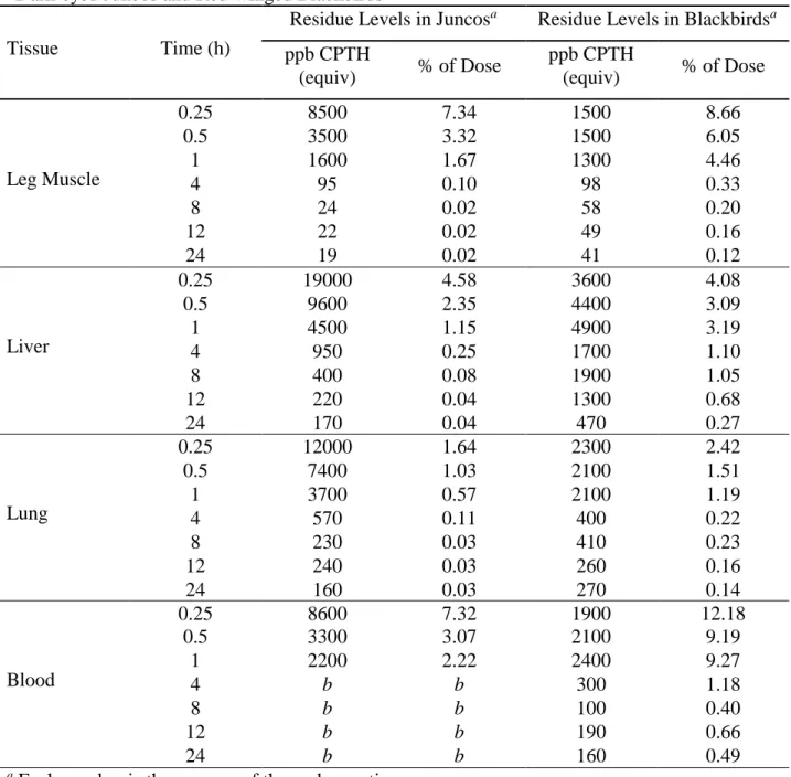
Table 2.2: Mean Concentration (CPTH Equivalents) and Percent of Recovered Dose Values for
Dark-eyed Juncos and Red-winged Blackbirds
Tissue Time (h)
Residue Levels in Juncosa Residue Levels in Blackbirdsa
ppb CPTH (equiv) % of Dose ppb CPTH (equiv) % of Dose Brain 0.25 7500 1.51 1900 1.94 0.5 2600 0.56 1400 0.91 1 730 0.17 1300 0.75 4 39 <0.01 55 0.03 8 7.2 <0.01 20 0.01 12 9.1 <0.01 23 0.01 24 11 <0.01 15 <0.01 Breast muscle 0.25 8000 15.05 2100 35.42 0.5 3200 6.72 1900 22.58 1 960 2.25 1700 18.13 4 83 0.17 100 1.05 8 7.8 0.02 32 0.34 12 8.4 0.01 39 0.39 24 7.3 0.01 26 0.22 GI Tract 0.25 58000 46.99 6300 18.24 0.5 53000 45.08 8600 19.93 1 47000 39.54 11000 22.48 4 8800 7.17 4500 7.98 8 1200 0.84 2200 3.91 12 2100 1.19 1500 2.05 24 360 0.24 640 1.01 Heart 0.25 11000 1.20 2000 1.48 0.5 4600 0.53 1900 1.07 1 2000 0.24 1600 0.74 4 230 0.03 160 0.08 8 64 <0.01 86 0.04 12 79 <0.01 100 0.04 24 48 <0.01 70 0.03 Kidney 0.25 35000 3.03 6900 2.41 0.5 20000 1.35 22000 5.40 1 17000 1.31 16000 3.59 4 13000 1.00 12000 2.59 8 1400 0.09 1100 2.45 12 5100 0.31 18000 4.13 24 490 0.04 11000 2.20
18
Table 2.2: Mean Concentration (CPTH Equivalents) and Percent of Recovered Dose Values for
Dark-eyed Juncos and Red-winged Blackbirds
Tissue Time (h)
Residue Levels in Juncosa Residue Levels in Blackbirdsa
ppb CPTH (equiv) % of Dose ppb CPTH (equiv) % of Dose Leg Muscle 0.25 8500 7.34 1500 8.66 0.5 3500 3.32 1500 6.05 1 1600 1.67 1300 4.46 4 95 0.10 98 0.33 8 24 0.02 58 0.20 12 22 0.02 49 0.16 24 19 0.02 41 0.12 Liver 0.25 19000 4.58 3600 4.08 0.5 9600 2.35 4400 3.09 1 4500 1.15 4900 3.19 4 950 0.25 1700 1.10 8 400 0.08 1900 1.05 12 220 0.04 1300 0.68 24 170 0.04 470 0.27 Lung 0.25 12000 1.64 2300 2.42 0.5 7400 1.03 2100 1.51 1 3700 0.57 2100 1.19 4 570 0.11 400 0.22 8 230 0.03 410 0.23 12 240 0.03 260 0.16 24 160 0.03 270 0.14 Blood 0.25 8600 7.32 1900 12.18 0.5 3300 3.07 2100 9.19 1 2200 2.22 2400 9.27 4 b b 300 1.18 8 b b 100 0.40 12 b b 190 0.66 24 b b 160 0.49
a Each number is the average of three observations.
b Observed values were below values for control samples.
The results of this transformation were also used to calculate elimination constants (KEl) and
half-life of elimination (t½) values for both elimination phases in each tissue or bodily fluid as well as
19
the half-life of elimination was easily determined. Because a half-life is commonly defined as the amount of time for half of the remaining radioactivity to be excreted from the body, 100% elimination can never be mathematically achieved. A close approximation can be found using 7 times the value of t½. This accounts for elimination of 99.2% of the radioactivity. The values for
each statistic as well as the p value for the slope of the regression lines are given (Tables 2.3 and 2.4).
Table 2.3: Log DPM Linear Regression Results, KEl, and t½ Values for Time 15 min to 4 h (n = 12
per Species) Tissue R2 Slope p SE KEl t½ (h) 95% CI of t½ 99.2% Elimi-nation Dark-eyed Junco Brain 0.840 -3.0129 0.0003 0.4598 6.939 0.10 0.08, 0.15 0.70 Breast Muscle 0.830 -2.7576 0.0004 0.4360 6.351 0.11 0.08, 0.16 0.77 GI Tract a -0.3237 0.4549 0.4092 a a a a Heart 0.804 -2.1478 0.0007 0.3698 4.946 0.14 0.1, 0.22 0.98 Kidney a -0.9618 0.1650 0.6204 a a a a Leg Muscle 0.646 -2.4066 0.0055 0.6096 5.542 0.13 0.08, 0.27 0.91 Liver 0.794 -1.9284 0.0008 0.3413 4.441 0.16 0.11, 0.25 1.12 Lung 0.661 -1.4220 0.0047 0.3489 3.275 0.21 0.14, 0.44 1.47 Blood 0.547 -2.3436 0.0137 0.7176 5.397 0.13 0.08, 0.36 0.91 Carcass 0.501 -1.0753 0.0199 0.3581 2.476 0.28 0.16, 0.92 1.96 Red-winged Blackbird Brain 0.949 -1.0049 <0.0001 0.0777 2.314 0.30 0.26, 0.36 2.10 Breast Muscle 0.958 -0.8850 <0.0001 0.0614 2.038 0.34 0.3, 0.4 2.38 GI Tract 0.387 -0.2169 0.0323 0.0839 0.500 1.39 0.77, 7.27 9.73 Heart 0.929 -0.7642 <0.0001 0.0704 1.760 0.39 0.33, 0.49 2.73 Kidney a -0.1257 0.2182 0.0941 a a a a Leg Muscle 0.958 -0.8233 <0.0001 0.0570 1.896 0.37 0.32, 0.43 2.59 Liver 0.803 -0.3019 0.0003 0.0492 0.693 1.00 0.74, 1.51 7.00 Lung 0.962 -0.5173 <0.0001 0.0344 1.191 0.58 0.51, 0.68 4.06 Blood 0.927 -0.6039 <0.0001 0.0564 1.391 0.50 0.42, 0.62 3.50 Carcass 0.945 -0.4467 <0.0001 0.0360 1.029 0.67 0.58, 0.81 4.69
20
Table 2.4: Log DPM Linear Regression Results, KEl, and t½ Values for Time 4 to 24 h (n = 12 per
Species) Tissue R2 Slope p SE KEl t½ (h) 95% CI of t½ 99.2% Elimi-nation Dark-eyed Junco Brain a -0.0466 0.2073 0.0336 a a a a Breast Muscle 0.337 -0.0879 0.0592 0.0391 0.202 3.43 1.77, 49.12 24.01 GI Tract 0.678 -0.1587 0.0039 0.0376 0.365 1.90 1.27, 3.76 13.30 Heart 0.398 -0.0963 0.0406 0.0384 0.222 3.12 1.7, 18.98 21.84 Kidney 0.414 -0.1310 0.0365 0.0508 0.302 2.29 1.27, 12.17 16.03 Leg Muscle 0.327 -0.0681 0.0626 0.0308 0.157 4.41 2.27, 81.81 30.87 Liver 0.610 -0.0770 0.0079 0.0209 0.177 3.92 2.49, 9.08 27.44 Lung 0.614 0.0508 0.0076 0.0137 0.117 5.92 3.78, 13.6 41.44 Blood a a a a a a a a Carcass 0.698 -0.1425 0.0031 0.0323 0.328 2.11 1.43, 4.01 14.77 Red-winged Blackbird Brain 0.430 -0.0539 0.0122 0.0177 0.124 5.59 3.31, 17.8 39.13 Breast Muscle 0.342 -0.0772 0.0269 0.0298 0.178 3.89 2.16, 20.27 27.23 GI Tract 0.559 -0.0868 0.0031 0.0224 0.200 3.47 2.25, 7.55 24.29 Heart 0.234 -0.0469 0.0635 0.0225 0.108 6.42 3.22, 139.59 44.94 Kidney b -0.0049 0.8655 0.0280 b b b b Leg Muscle 0.363 -0.0424 0.0225 0.0157 0.098 7.07 3.99, 31.75 49.49 Liver 0.387 -0.0750 0.0182 0.0266 0.173 4.01 2.3, 15.59 28.07 Lung 0.257 -0.0456 0.0533 0.0208 0.105 6.60 3.38, 147.76 46.20 Blood b -0.0217 0.3724 0.0233 b b b b Carcass 0.463 -0.0500 0.0089 0.0154 0.115 6.03 3.66, 17.02 42.21
b Values less than background; data not used. b Slope not significantly different from zero; no values
can be calculated.
2.4 Results and Discussion
Red-winged blackbirds and dark-eyed juncos were chosen to represent species that exhibit varied sensitivities to CPTH. Red-winged blackbirds, a target species, are highly sensitive to CPTH, having an LD50 of 1.8-3.2 mg/kg (Schafer, 1972 and 1994), whereas dark-eyed juncos, a non-target species,
21
were considered in the design of this experiment: CPTH dose level, 14C activity in dose, route and method of exposure, and length of exposure time.
Selection of dose level, that was representative of a real-world exposure, was one parameter considered. The selection of an appropriate dose level was based upon ingestion of a single grain of 2% CPTH-treated rice bait. Previous research with CPTH indicated that a single treated rice grain would typically be sufficient to induce acute toxicity in sensitive species, and that by diluting treated rice 1:25 with untreated rice, the desired exposure could be achieved (Schafer, 1984). This is equivalent to a dose of ∼4 mg/kg for a 100-g bird. During study design, we assumed that the weight of the more sensitive blackbirds would be ∼60 g. Therefore, a final dose of ∼250 µg of CPTH was selected. The dose was uniform for all birds and was not adjusted for differences in body weight. Another key consideration was the desire to produce no mortality during the course of the study. The dose level chosen was below the published LD50 values for juncos and below a level expected to
produce mortality in blackbirds in <24 hours. No mortalities occurred during the study as a result of CPTH toxicity: therefore, the dose seemed to be appropriate for the intended purpose.
The level of 14C activity administered to each bird was based on the limit of detection (DL) for the LSC and biological oxidizer. The limit of detection for the biological oxidizer was ∼50 DPM. Using this value, a method limit of detection for the samples was found to be ∼2000 DPM for a 10-g tissue sample. If the bird retained as little as 1% of the administered dose in its body and a small portion of that dose was contained in the tissue being analyzed, the response for that sample should be ∼10-fold above the method DL.
The route and method of exposure were chosen to permit a reasonable assurance that the entire dose was ingested by each test animal. The predominant route of exposure in a field application is via ingestion; therefore, an oral gavage of CPTH in 100 µL of deionized water was used.
22
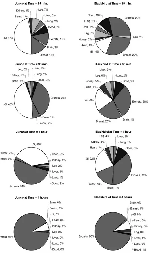
Sampling times were selected to provide a cross-section of exposure. Previously published data indicated that most of the CPTH would be excreted in <24 hours (Giri et al., 1976; Westberg, 1969). Information on rate of distribution would also be important; therefore, the sampling times were focused toward shorter time periods rather than being evenly spaced over the 24-hour test period. The times selected were 15 and 30 minutes and 1, 4, 8, 12, and 24 hours post dose. The results for the 15- and 30-minute and 1- and 4-hour time points are found in Figure 2.1. No further time points are shown as greater than 90% of the radioactivity was excreted by the 4-hour time point.
In most of the tissues and fluids collected from both bird species tested, a similar pattern was observed in plots of the radioactive residue (RR) as a function of time post dose. In the case of every tissue except blackbird kidney, greater than 80-90% of the administered dose was excreted from the body of the test animal by 4 hours after exposure. This supports the conclusion that CPTH is not retained in the carcass of exposed birds in significant quantities, with the exception of the RR in liver and kidney of exposed birds.
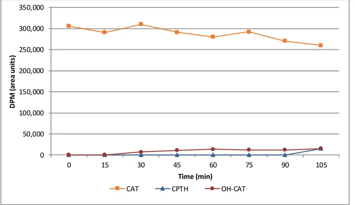
In the case of blackbird kidney (Figure 2.2), RR levels significantly higher than background levels were observed for the duration of the test. This level of radioactivity is approximately equivalent to a mean kidney concentration of 15 mg/kg of CPTH equivalents. A slight elevation of RR level was also observed in junco kidney (Figure 2.2) and in blackbird liver (Figure 2.3). However, the results were not as pronounced as those for blackbird kidney. Possible reasons for this difference in elimination relate to potential binding of CPTH metabolites to these sensitive tissues.
23
24
Figure 2.2: Logarithmic plot of elimination of radiolabeled CPTH and metabolites from kidney of red-winged blackbirds
and dark-eyed juncos following a single oral dose (n = 3 per time point; error bars indicate standard deviation).
Figure 2.3: Logarithmic plot of elimination of radiolabeled CPTH and metabolites from liver of red-winged blackbirds
25
The plots of RR in the blood versus time also demonstrate one very distinct feature. There is a virtual lack of any type of uptake curve for CPTH (Figure 2.4). This suggests that CPTH delivered in this manner is readily absorbed and rapidly perfused through the body. This is further supported by a comparison of oral LD50 to intraperitoneal (ip) LD50 values for CPTH in starlings (DeCino et al.,
1966). The value for an oral exposure has been reported at 3.8 mg/kg, whereas that for an ip injection was found to be 3.5 mg/kg. Examination of the half-life of elimination values (Tables 2.2 and 2.3) reveals that the radioactivity took longer to clear from blackbirds than from juncos as evidenced by the longer half- lives. This is further bolstered by the differences in whole body elimination rates between the two species (Figure 2.5). This could be partially due to the larger body mass of the blackbird as opposed to the junco. However, because the data were evaluated as concentrations and
Figure 2.4: Logarithmic plot of elimination of radiolabeled CPTH and metabolites from whole blood of red-winged
blackbirds and dark-eyed juncos following a single oral dose (n = 3 per time point; error bars indicate standard deviation).
26
adjusted for body mass, it is equally likely that any differences observed are due to differential metabolism between the two species. The R2 values for each tissue type in the first elimination phase demonstrated a reasonable fit to a linear model in most cases (Table 2.2). The R2 values for the second elimination phase demonstrated a far less desirable fit for a linear regression (Table 2.3). As the residue levels approached background levels, the variability of the results increased, resulting in lower R2 values overall. In the case of several tissue types, the slope of the regression line was found to not be significantly different from zero (p = 0.10). In the first elimination phase, junco GI tract and kidney and blackbird kidney did not meet the criteria for a linear regression. Therefore, no
Figure 2.5: Logarithmic plot of elimination of radiolabeled CPTH and metabolites from whole body of red-winged
blackbirds and dark-eyed juncos following a single oral dose (n = 3 per time point; error bars indicate standard deviation).
27
elimination rate constants or half-lives were calculated for these tissues. In the second elimination phase, rate constants could not be generated for junco brain and blood or for blackbird kidney.
Elimination of CPTH from the kidney of both bird species was slower than for most other tissue types. The difference in elimination was even more pronounced for blackbird than for junco (Figure 2.2). This further supports the hypothesis that CPTH is metabolized differently in junco than it is in blackbird. The slope of the line for junco kidney very closely mirrors that for liver tissue (Figures 2.2 and 2.3). Results of a two-tailed t test reveal that the slopes of the elimination curves, which are directly related to the half-life, for junco kidney and junco liver are not statistically different (p = 0.3239). Conversely, the half-life of elimination for blackbird kidney could not be calculated because it has no significant slope.
Previous research has hypothesized that the mode of action for CPTH toxicity involves damage to the kidneys (Giri et al., 1976), more specifically, damage to proximal tubular cells of the kidney (Apostolou, 1969; DeCino et al., 1966; Mull et al., 1972). Additionally, observations of increased blood uric acid levels (Apostolou, 1969) have been made. The appearance of uric acid deposits in the abdominal cavities of exposed birds is also used as a method of determining exposure in certain cases (Cummings et al., 2003; DeCino et al., 1966; Johnston et al., 1999).
Renal damage of this type can be indicative of a highly reactive chemical that may be able to covalently bind to tissues. Such a highly reactive chemical species could cause the extensive tissue damage observed in the previously mentioned studies, resulting in a failure of normal kidney functions and the subsequent uric acid increases. The fact that the RR in the kidney of the more sensitive blackbird species did not change significantly over time in comparison to the that of the less sensitive junco points to the kidney as a possible site of action. This observation is consistent with the hypothesis that covalent binding is occurring in the kidney of blackbirds to a greater degree than
28
in the kidney of exposed juncos, especially when compared to the rapidly declining RR in the other compartments of the blackbird.
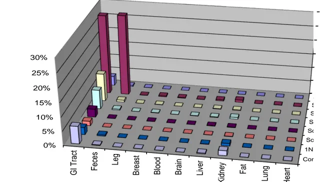
To further investigate potential tissue binding, samples collected at the 15 minute exposure time were subjected to serial extraction with various solvents as well as an acid reflux. The purpose of these extractions was to remove as much radioactivity from the tissues as possible prior to combustion of the remaining pellet. A very robust measure of extractable versus non-extractable
S S S S So So 1N Com 0% 5% 10% 15% 20% 25% 30% H ea rt Lu ng Fa t K idn ey Li ver B ra in B loo d B re as t Leg F ec es G I T rac t P er cen t o f R eco ver ed D o se
Figure 2.6: Bound and extractable recovery of radioactivity from junco tissues and fluids 15 minutes after receiving a
single, oral dose of 14C CPTH.
radioactive residue was determined as a result of this experiment. In the less sensitive junco species, tightly bound residues were found in both the kidney and the GI tract (Figure 2.6). A similar pattern was observed in the more sensitive blackbird species, with bound residue also being observed in the
29
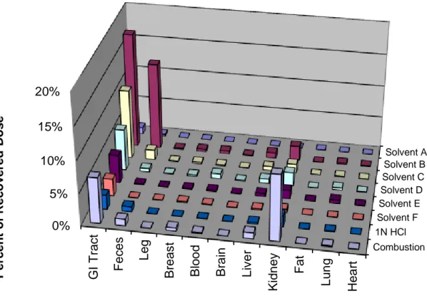
liver (Figure 2.7). In both the junco and blackbird GI tract, the amount of bound radioactivity, expressed as a percentage of the administered dose, was around 6-7%. In the junco, the bound radioactivity in the kidney was only around 2% while it was 10% in the blackbird.
Solvent A Solvent B Solvent C Solvent D Solvent E Solvent F 1N HCl Combustion 0% 5% 10% 15% 20% H e a rt Lu ng Fa t K idn ey Li v er B ra in B loo d B re a s t L eg F ec es G I T rac t P er cen t o f R eco v er ed D o se
Figure 2.7: Bound and extractable recovery of radioactivity from blackbird tissues and fluids 15 minutes after receiving
a single, oral dose of 14C CPTH.
Although it is apparent that CPTH or more likely one of its metabolites is strongly retained by the kidney of red-winged blackbirds, the bulk of the parent and metabolites are rapidly excreted from the bodies of both sensitive and non-sensitive species. Factoring in observations from previous studies
30
that time to death is typically >24 hours (DeCino et al., 1966), it seems fairly clear that carcasses of exposed birds found in the field are unlikely to contain significant residues of CPTH or its metabolites. Furthermore, it is likely that any metabolites present might be tightly bound to tissues and thus biologically unavailable to any predators that might consume them. These findings are significant with respect to estimating potential secondary exposure of wildlife that may consume CPTH-containing pest bird carcasses which could cause mortality in non-target birds. The results presented further suggest that future research aimed at elucidating the bio-activation pathway by which CPTH is retained by renal tissues of susceptible bird species would greatly increase our understanding of the mode of action of CPTH and possibly lead to the development of more effective and ecologically friendly avicides.
2.4.1 Summary
Our central hypothesis for Phase 1 was that CPTH would be rapidly excreted from most organs. This has been proven with the results listed above. We have addressed the specific questions asked by the hypothesis as well:
• The excretion profile for CPTH exhibited a 2 phase behavior with a rapid phase lasting approximately 4 hours and a slow phase lasting 24 hours or more in most tissues.
• The excretion profile was the same for most tissues tested in both a sensitive and less sensitive species of bird.
• The relative residue levels were higher in the blackbird (a more sensitive species) than in the junco (a less sensitive species).
• The rate of elimination was established for all tissues tested (Tables 2.3 and 2.4).
31
CHAPTER 3 – PHASE 2 – IN-VITRO EXPERIMENTS
3.1 Introduction
3-Chloro-p-toluidine hydrochloride (CPTH) has been used successfully to control pest bird species, but a deeper understanding of its mode of action is required. This chemical appears to pose minimal hazard to mammalian species (LD50s greater than 1000 mg/kg) while being extremely toxic
to the target species (LD50 for starlings <10 mg/kg). In addition, CPTH appears to be less toxic to
non-target bird species such as hawks and songbirds (LD50s between 100 and 500 mg/kg). However,
concerns surround use of this chemical on a wide-spread basis. Very little is known about the metabolism and fate of this chemical after it is consumed by a bird. A more in-depth understanding of this facet of CPTH is required to assuage concerns with its continued use.
Toxic substances can cause damage to exposed tissues in many ways. The chemical can cause damage directly or it can require biotransformation to convert it to its toxic form. In some cases multiple biotransformation steps might be required to cause toxicity. The site of toxic action can be general and spread throughout the body or can be quite specific and localized to a target organ. As a primary mechanism for filtering and removal of toxicants or waste products, the kidney is a potential target of damage for many toxic substances.
What information is known of CPTH comes from pathological examination of exposed birds. CPTH was found to cause damage to the proximal convoluted tubules in sensitive avian species. Experiments with [14C]-CPTH demonstrated that significantly more radiolabeled material bound tightly to the kidneys of sensitive species than a less sensitive species. These observations, along with an observed elevation in the level of uric acid in the birds, lead us to classify this compound as a nephrotoxin. The hypothetical mode of action involves bio-activation resulting in damage to the
32
kidney and interruption of normal excretion processes leading to death by uric acid poisoning. Experiments to investigate the mechanistic metabolism and provide insight into the mode of action of CPTH were designed and conducted.
3.1.1 Hypothesis
Our hypothesis for this aim is that a reactive metabolite can be formed in-vitro and that it can attach to a target compound. Specifically, the questions we will attempt to answer are:
• Can we use mass spectrometry to identify the metabolites of CPTH? • Can a reactive metabolite be formed in-vitro?
• Can glutathione or another suitable molecule be used as a target compound for this metabolite?
3.2 Materials and Methods
In order to investigate the mechanistic toxicology of CPTH, comparisons of metabolism between various species were performed. Several species of birds (mallard duck (Anas platyrhynchos), boat-tailed grackle (Quiscalus major), and red-winged blackbird (Agelaius phoeniceus)) were chosen for inclusion in this research. Blackbirds and grackles are classified as highly sensitive species with respect to CPTH intoxication (LD50 = 1 to 8.0 mg/kg), while mallards are more resistant (LD50 = 100
mg/kg) (Eisemann et al., 2003). Birds selected for inclusion in this research were free of any exposures to veterinary drugs or toxicants and were donated from other research projects conducted at the USDA’s National Wildlife Research Center (NWRC). All birds were euthanized humanely in accordance with American Veterinary Medical Association standards and practices under the supervision of the Attending Veterinarian at the NWRC. All procedures involving animals were carried out with the approval of the Animal Care and Use Committee.
33
All chemicals and solvents were obtained from either Fisher Scientific (Pittsburgh, PA) or Sigma Aldrich (St. Louis, MO). All chemicals were reagent grade.
3.2.1 Sub-cellular Fraction Preparation
The liver and kidneys from multiple individuals were immediately removed from euthanized birds, pooled, weighed, homogenized in an ice-cold homogenization buffer, and subjected to differential centrifugation. The buffer solution consisted of 250 mM sucrose, 50 mM 4-(2-hydroxyethyl)-1-piperazineethanesulfonic acid (HEPES), 25 mM potassium chloride, 5 mM magnesium chloride, and 0.1 mM ethylenediaminetetraacetic acid (EDTA). The homogenization buffer was prepared in ultra-pure water and was adjusted to pH 7.4 with 1 M potassium hydroxide. Tissues were minced with scissors in 2 volumes (v/w) of homogenization buffer. The tissues were then homogenized with three passes of a Potter-Elvehjem homogenizer that was kept on ice. The homogenized tissues were placed in centrifuge tubes and subjected to differential centrifugation. The S-9 fraction was defined as the supernatant fraction obtained from an organ homogenate by centrifuging at 9000 × g for 20 minutes in a suitable medium; this fraction contained both cytosol and microsomes. The S-9 fraction was collected as a sub-sample of the supernatant following centrifugation at 9,000 × g for 10 minutes at 4 °C. This fraction contains both cytosol and microsomes and is somewhat more representative of the metabolic activity of the intact organ. The remaining supernatant was transferred to a clean centrifuge tube and placed in the centrifuge at 15,000 × g for 20 minutes at 4 °C. The supernatant was removed and again transferred to a clean centrifuge tube that was centrifuged at 105,000 × g for 60 minutes at 4 °C. This produced the cytosol fraction which was collected as the supernatant of this centrifugation step. The pellet from this centrifugation step is the microsomes. These were gently rinsed three times with
34
homogenization buffer before being resuspended in homogenization buffer with 3 passes of a Potter-Elvehjem homogenizer that was kept on ice. Following transfer to a clean centrifuge tube, the sample was centrifuged at 105,000 × g for a second time. The supernatant was removed and discarded. The microsomes were resuspended in 1 volume (v/w) of homogenization buffer with 3 passes of a Potter-Elvehjem homogenizer that was kept on ice.
After collection, 1 mL fractions of S-9, cytosol, and microsomes were flash frozen in liquid nitrogen. The tubes were stored in a freezer at -80 ºC until used. In later experiments, the results were normalized for protein content of the fraction being tested. Protein content was determined using a standard Bradford assay kit (Sigma Aldrich, St. Louis, MO).
3.2.2 In-vitro Experiments
In-vitro enzyme experiments were conducted in plastic 1.5 mL snap-cap tubes. A 100 µL aliquot
of enzyme source (either microsomes, cytosol, or S-9) and 100 µL of a cofactor solution consisting of 16.5 mM nicotinamide adenine dinucleotide phosphate (NADP), 17 mM glucose-6-phosphate, and 5 mM glucose-6-phosphate dehydrogenase were added to a tube containing enough phosphate buffer (100 mM; pH 7.2) to produce 1 mL of solution. In some experiments, between 20 and 100 µL of acetyl coenzyme A solution was added to the reaction tubes. This solution was prepared by dissolving 2 mg of acetyl coenzyme A in 10 mL of phosphate buffer. All solutions were stored in a refrigerator at 4 °C until used. Depending on the experiment being conducted, a quantity of CPTH or one of its metabolites, that were prepared in phosphate buffer, were added and the tubes were placed in a shaking water bath at 37 ºC for a fixed period of time. The reaction times varied between 15 minutes and 2 hours depending on the study design of the particular experiment. At the end of the reaction, a 500 µL aliquot of 1% acetic acid in methanol was added to each tube. The tubes were
35
stoppered, vortexed, and placed on ice for 10 minutes. The tubes were then centrifuged at 9,000 × g for 5 minutes and the supernatant removed for analysis via high performance liquid chromatography (HPLC).
3.2.3 Liquid Chromatographic Analysis
All samples were injected into an Agilent 1200 series liquid chromatograph equipped with an auto injector, quaternary pump, temperature-controlled column compartment and diode array detector (Table 3.1). Separation was achieved using a 2.1 x 50 mm Phenomenex Kinetex XB-C18 column (100 Å; 2.6 µm).
Table 3.1: Liquid Chromatograph/Ultra Violet Conditions
Mobile Phase A: 20 mM acetic acid
Mobile Phase B: 20 mM acetic acid in 9:1 methanol:water Gradient:
Time (min) % A % B Flow Rate (mL/min)
0.0 80 20 0.400 1.0 80 20 0.400 1.1 60 40 0.400 6.0 20 80 0.400 6.1 0 100 0.800 Injection Volume: 5 µL Column Temperature: 60 °C
Run Time: 8 minutes
Post Time: 4.5 minutes
Detector: UV @ 241 nm
Metabolites were collected and purified prior to identification. A fraction collector was attached to the HPLC system and multiple injections were made of in-vitro experiments conducted using red-winged blackbird hepatic S-9 with a reaction time of 60 minutes. The fractions collected were pooled and purified using a Phenomenex Strata-X solid phase extraction column (SPE). Briefly, the fractions were placed in a heated water bath at approximately 40 °C under a stream of nitrogen to
36
remove methanol from the fraction. The aqueous portion of the fraction was loaded onto a 200 mg SPE cartridge which had been conditioned with methanol and water. After loading the fraction onto the SPE, it was washed with a solution of 30% methanol in water. The metabolites were eluted in methanol, evaporated to dryness under a stream of nitrogen, and reconstituted via the addition of 500 µL of a diluent consisting of 0.1% formic acid in 80:20 water:methanol. Identification of metabolites was performed using an Agilent 1200 series liquid chromatograph coupled to an Agilent 6410 mass spectrometer (LC/MS/MS). The HPLC was equipped with an auto injector, binary pump, and temperature-controlled column compartment. Separation was achieved using the same column as previously described.
Table 3.2: Liquid Chromatograph/Mass Spectrometer Conditions
Mobile Phase A: 0.1% formic acid
Mobile Phase B: 0.1% formic acid in methanol Gradient:
Time (min) % A % B Flow Rate (mL/min)
0.0 80 20 0.350 1.0 80 20 0.350 6.0 20 80 0.350 6.5 0 100 0.350 Injection Volume: 10 µL Column Temperature: 40 °C
Run Time: 8 minutes
Post Time: 4 minutes
Scan Range: 50 to 1000 m/z
Fragmentor Voltage: 65 V
Polarity: Positive
Gas Temperature: 350 °C
Gas Flow: Nitrogen @ 11 L/minute
Nebulizer Pressure: 35 psi Capillary Voltage: 4000 V
37 3.3 Results and Discussion
Combining CPTH with hepatic S-9, microsomes, or cytosol did not produce any detectable metabolism in any species tested until a source of an acetyl donor group such as acetyl coenzyme A was added. Upon addition of acetyl coenzyme A, CPTH was rapidly metabolized to two metabolites that were initially designated as Metabolite A and Metabolite B (Figure 3.1). Metabolite A was formed very rapidly upon addition of acetyl coenzyme A and had a longer retention time in a reversed-phase liquid chromatography system. We can conclude from its relative retention that was less polar than the parent molecule (CPTH). A second metabolite (Metabolite B) was observed following formation of Metabolite A. The retention time of Metabolite B was much shorter than that of either CPTH or Metabolite A, suggesting it was more polar and more water soluble than CPTH. No other significant metabolites were observed in chromatograms from in-vitro experiments. This includes experiments with Phase II metabolic systems (sulfates and glucuronides) or with bovine serum albumin or glutathione as targets for reactive metabolites (data not shown).
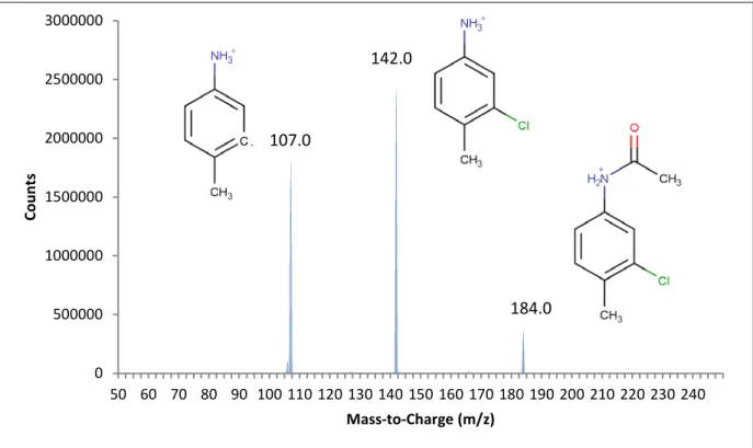
Pooled fractions of Metabolite A produced from hepatic microsomes were injected into the LC/MS/MS using the parameters outlined in Table 3.2 and produced the fragments shown in Figure 3.2. A product ion scan was conducted on the largest ion (184 m/z) producing the fragments shown in Figure 3.3. Based on the interpretation of the mass spectra, Metabolite A was assigned a structure and identified as 3-chloro-4-methylacetanilide (CAT). The formation of this metabolite appeared to be a non-enzymatic process as it did not require the presence of cellular material to occur. The lack of metabolism without the presence of an acetyl source indicated that the acetylation of CPTH was a necessary first step in the metabolic pathway.
38 -50 450 950 1450 1950 2450 0 1 2 3 4 5 6 Re sp on se (m AU )
Retention Time (minutes)
Metabolite B
CPTH
Metabolite A
Figure 3.1: Chromatogram of red-winged blackbird hepatic S-9 sample injected using the conditions found in Table 3.1.
0 1000000 2000000 3000000 4000000 5000000 6000000 7000000 50 60 70 80 90 100 110 120 130 140 150 160 170 180 190 200 210 220 230 240 250 Co un ts Mass-to-Charge (m/z) 184.0
39 0 500000 1000000 1500000 2000000 2500000 3000000 50 60 70 80 90 100 110 120 130 140 150 160 170 180 190 200 210 220 230 240 Co un ts Mass-to-Charge (m/z) 107.0 142.0 184.0
Figure 3.3: LC/MS/MS Spectra of the 184.0 m/z peak of Metabolite A injected using the conditions found in Table 3.2.
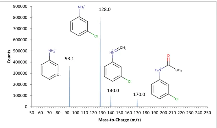
Pooled fractions of Metabolite B produced from hepatic microsomes were injected into the LC/MS/MS and produced the fragments shown in Figure 3.4. A product ion scan was conducted on the largest ion (200 m/z) producing the fragments shown in Figure 3.5. Based on the interpretation of the mass spectra, Metabolite B was assigned a structure and identified as N-[3-chloro-4-(hydroxymethyl)phenyl]acetamide (OH- CAT). The hydroxylation reaction was likely a cytochrome P-450 mediated action since the presence of microsomes was necessary for it to occur. Mass spectral analysis of this product indicated that the site of hydroxylation was likely on the benzyl methyl. Hydroxylation of this site did not occur without prior acetylation of the amine group. The acetyl group was necessary to initiate and/or stabilize this enzymatic process.
40 0 1000000 2000000 3000000 4000000 5000000 6000000 50 60 70 80 90 100 110 120 130 140 150 160 170 180 190 200 210 220 230 240 250 Co un ts Mass-to-Charge (m/z) 200.0
Figure 3.4: LC/MS Spectra of Metabolite B injected using the conditions found in Table 3.2.
0 100000 200000 300000 400000 500000 600000 700000 800000 900000 50 60 70 80 90 100 110 120 130 140 150 160 170 180 190 200 210 220 230 240 250 Co un ts Mass-to-Charge (m/z) 140.0 170.0 128.0 93.1