ARTICLE
Inhibition of the prostaglandin D
2–GPR44/DP2 axis improves humanislet survival and function
Shadab Abadpour1,2&Björn Tyrberg3&Simen W. Schive1&Charlotte Wennberg Huldt3&Peter Gennemark3,4&
Erik Ryberg3&Tina Rydén-Bergsten3&David M. Smith3,5&Olle Korsgren6&Stanko Skrtic3,7&Hanne Scholz1,2&
Maria Sörhede Winzell3
Received: 13 September 2019 / Accepted: 28 February 2020 / Published online: 29 April 2020
Abstract
Aims/hypothesis Inflammatory signals and increased prostaglandin synthesis play a role during the development of diabetes. The prostaglandin D2(PGD2) receptor, GPR44/DP2, is highly expressed in human islets and activation of the pathway results in impaired insulin secretion. The role of GPR44 activation on islet function and survival rate during chronic hyperglycaemic conditions is not known. In this study, we investigate GPR44 inhibition by using a selective GPR44 antagonist (AZ8154) in human islets both in vitro and in vivo in diabetic mice transplanted with human islets.
Methods Human islets were exposed to PGD2or proinflammatory cytokines in vitro to investigate the effect of GPR44 inhibition on islet survival rate. In addition, the molecular mechanisms of GPR44 inhibition were investigated in human islets exposed to high concentrations of glucose (HG) and to IL-1β. For the in vivo part of the study, human islets were transplanted under the kidney capsule of immunodeficient diabetic mice and treated with 6, 60 or 100 mg/kg per day of a GPR44 antagonist starting from the transplantation day until day 4 (short-term study) or day 17 (long-term study) post transplantation. IVGTT was performed on mice at day 10 and day 15 post transplantation. After termination of the study, metabolic variables, circulating human proinflammatory cytokines, and hepatocyte growth factor (HGF) were analysed in the grafted human islets.
Results PGD2or proinflammatory cytokines induced apoptosis in human islets whereas GPR44 inhibition reversed this effect. GPR44 inhibition antagonised the reduction in glucose-stimulated insulin secretion induced by HG and IL-1β in human islets. This was accompanied by activation of the Akt–glycogen synthase kinase 3β signalling pathway together with phosphorylation and inactivation of forkhead box O-1and upregulation of pancreatic and duodenal homeobox-1 and HGF. Administration of the GPR44 antagonist for up to 17 days to diabetic mice transplanted with a marginal number of human islets resulted in reduced fasting blood glucose and lower glucose excursions during IVGTT. Improved glucose regulation was supported by increased human C-peptide levels compared with the vehicle group at day 4 and throughout the treatment period. GPR44 inhibition reduced
Maria Sörhede Winzell and Hanne Scholz contributed equally to this study.
Electronic supplementary material The online version of this article
(https://doi.org/10.1007/s00125-020-05138-z) contains peer-reviewed but
unedited supplementary material, which is available to authorised users. * Hanne Scholz
hanne.scholz@medisin.uio.no * Maria Sörhede Winzell
maria.sorhede-winzell@astrazeneca.com
1
Department of Transplant Medicine and Institute for Surgical Research, Oslo University Hospital, Sognsvannsveien 20, 0027 Oslo, Norway
2
Hybrid Technology Hub, Centre of Excellence, Institute of Basic Medical Sciences, University of Oslo, Oslo, Norway
3
Research and Early Development, Cardiovascular, Renal and Metabolism, BioPharmaceuticals R&D, AstraZeneca, Peppredsleden 1, 431 83 Mölndal, Gothenburg, Sweden
4 Department of Biomedical Engineering, University of Linköping,
Linköping, Sweden
5
Hit Discovery, Discovery Sciences, BioPharmaceuticals R&D, AstraZeneca, Cambridge, UK
6
Department of Immunology, Genetics and Pathology, Science for Life Laboratory, University of Uppsala, Uppsala, Sweden
7 Institute of Medicine at Sahlgrenska Academy, University of
Gothenburg, Gothenburg, Sweden
plasma levels of TNF-α and growth-regulated oncogene-α/chemokine (C-X-C motif) ligand 1 and increased the levels of HGF in human islets.
Conclusions/interpretation Inhibition of GPR44 in human islets has the potential to improve islet function and survival rate under inflammatory and hyperglycaemic stress. This may have implications for better survival rate of islets following transplantation.
Keywords DP2 . GPR44 . Human islets . Islet apoptosis . Islet survival rate . Prostaglandin D2
Abbreviations
CXCL1 Chemokine (C-X-C motif) ligand 1 EIA Enzyme immunoassay
FOXO1 Forkhead box O-1
GRO-α Growth-regulated oncogene-α GSK3β Glycogen synthase kinase 3β HG High concentration of glucose HGF Hepatocyte growth factor HPMC Hydroxypropylmethylcellulose
MafA V-Maf musculoaponeurotic fibrosarcoma onco-gene homolog A
PDX-1 Pancreatic and duodenal homeobox-1 PGD2 Prostaglandin D2
PGE2 Prostaglandin E2
PI3K Phosphoinositide 3-kinases PIP Phosphatidylinositol
Introduction
GPR44 (also known as PTGDR2, DP2 or CRTh2) is a trans-membrane G-protein coupled receptor for prostaglandin D2 (PGD2), shown to be highly expressed in human beta cells through proteomics screening analysis [1]. Furthermore, it has been identified as a useful imaging biomarker for visualising of human beta cell mass [2]. The physiological role of GPR44 in human islet function and survival in the diabetic milieu is however unknown.
One hallmark of both type 1 and type 2 diabetes is the loss of functional beta cell mass resulting in an insufficient release of insulin and development of hyperglycaemia [3]. Prolonged hyperglycaemia has an adverse effect on pancreatic beta cell
function and mass due, at least partly, to the activation of proinflammatory responses leading to beta cells apoptosis [4]. Proinflammatory prostaglandins are lipid molecules derived from arachidonic acid in cell-membrane phospho-lipids and their synthesis is triggered by inflammatory signals [5]. IL-1β, a proinflammatory cytokine known to induce beta cell injury and loss, has been found to increase prostaglandin production in islets through activation of NF-κB [6].
PGD2signals through two G-protein coupled receptors, GPR44 and DP1 [7], the former being highly expressed in human islets [1]. Although, there is not much known about the role of PGD2and the regulatory mechanism of PGD2– GPR44 in human islets, it has been demonstrated that PGD2 is synthesised and secreted most likely by the pancreatic stel-late cells in human islets [8] and that elevated levels of glucose and IL-1β increase PGD2synthesis in vitro in rat and human islets in a similar fashion as previously described for prosta-glandin E2(PGE2) [8,9]. Furthermore, PGD2signalling in immune cells via GPR44 couples to inhibitory G-proteins resulting in reduction of intracellular cAMP [10]. This suggests that the activation of GPR44 in beta cells could also result in reduction of cAMP and inhibition of glucose-stimulated insulin secretion. The same observation has been previously reported with PGE2[5,8]. Significant reduction of GPR44 protein expression has been noted in insulin-negative human islets in pancreatic sections from individuals with long-standing type 1 diabetes [1]. In contrast, the expression of this receptor was upregulated in islets from individuals with type 2 diabetes compared with healthy individuals [11]. Taken together, these observations suggest that the activation of GPR44 may have a negative impact on islet function in an inflammatory and hyperglycaemic state and that inhibition of GPR44 may improve islet function and survival rate.
In the present study, we used the specific GPR44 antagonist AZ8154 to investigate the role of GPR44 in the function and survival rate of human islets exposed to a type 1 diabetes-like milieu both in vitro (mimicked by islet exposure to high concentration of glucose [HG] and to IL-1β) and in vivo (using human islets transplanted to a diabetic immunodefi-cient mouse model). We also explored the intracellular responses associated with GPR44 inhibition in isolated human islets in vitro.
Methods
Animal studies All experiments and methods using human islets were approved by and performed in accordance with the guidelines and regulations made by the regional commit-tee for medical and health research ethics central in Norway (2011/782). The animal experiments were approved by the Norwegian National Animal Research Authority (FOTS ID 7005) and were performed in accordance with the European
Directive 2010/63/EU and The Guide for the Care and Use of Laboratory Animals, 8th edition (NRC 2011, National Academic Press) or by the Local Ethics Review Committee for Animal Experiments (Gothenburg, Sweden). The AstraZeneca Gothenburg animal unit is accredited by the Association for the Assessment and Accreditation of Laboratory Animal Care (AAALAC).
Compounds and in vitro pharmacology The GPR44 antago-nist AZ8154 is an analogue to the previously published GPR44 a n t a g o n i s t A Z D 1 9 8 1 , 4 ( a c e t y l a m i n o ) 3 [ ( 4 -chlorophenyl)thio]-2-methyl-1H-indole-1-acetic acid [12], a clinical drug candidate intended for the treatment of asthma [13,14]. Both compounds were synthesised by AstraZeneca and came from two neighbouring chemical series. Potencies of AZ8154 and AZD1981 on binding to human and mouse GPR44 and DP1 receptors (electronic supplementary material [ESM]Methods, ESM Table1) and inhibition of GPR44 recep-tor on EndoC-βH1 cells (human beta cell line, Univercell Biosolutions, Toulouse, France [mycoplasma negative]) using insulin secretion and cAMP measurement assays (ESM Table 2, ESM Fig. 1) are described in detail in the ESM Methods. Selectivity analysis of AZD1981 and AZ8154 on the PGE2receptor, EP3 shows that neither of the AZ8154 and AZD1981 antagonists bound to EP3 in human primary adipo-cytes and therefore did not have an inhibitory effect on EP3 (ESMMethodsand ESMResults/EP3 selectivity for AZD1981 and AZ8154, ESM Fig. 2). AZ8154 was chosen over AZD1981 for this study due to its improved pharmacokinetic profile in mice (ESMMethods, ESM Figs.3,4).
Human islet culture and experimental condition Human islets from non-diabetic donors were obtained from Prodo laborato-ry (Aliso Viejo, CA, USA). Islets were dispersed and exposed to a stable PGD2analogue, 15(R)-15-methyl-PGD2(Cayman Chemicals, Ann Arbor, MI, USA), at the concentrations 1, 10 or 100 nmol/l, or to a mixture of the cytokines IL-1β (10 ng/ml), IFN-γ (50 ng/ml) and TNF-α (50 ng/ml) (R&D systems, Minneapolis, MN, USA) with or without addition of AZ8154 (1, 3 or 10μmol/l), to evaluate the effect of the ligand PGD2and the GPR44 antagonist on islet apoptosis.
Human islets were obtained from the JDRF award 31-2008-416 (ECIT Islet for Basic Research program) and isolat-ed as previously describisolat-ed [15] from male/female 7/3 non-diabetic brain-dead donors with mean age 55 years (35– 69 years) and mean BMI 24 kg/m2(23–42 kg/m2) after appro-priate informed consent from relatives for multi-organ dona-tion and for use in research. A human islet checklist is provid-ed in theESM. All experiments and methods using human islets were approved by and performed in accordance with the guidelines and regulations made by regional committee for medical and health research ethics central in Norway (2011/782). An islet purity of >50% was used in this study,
judged by digital imaging analysis [16] or dithizone staining. Equally sized islets were manually hand-picked and distribut-ed blindly among experimental groups. Islets were culturdistribut-ed at 37°C (5% CO2) for 48 h in petri dishes (Sterilin, Newport, UK) with CMRL 1066 medium supplemented with 2% human AB serum (Milan ANALYTICA, Rheinfelden, Switzerland), 1% penicillin/streptomycin, 10 mmol/l HEPES (Life Technologies, Carlsbad, CA, USA) without (untreated islets) or with a combination of HG (20 mmol/l glucose) and IL-1β (10 ng/ml) (R&D systems, Minnneapolis, MN, USA) with or without AZ8154 (10μmol/l).
Cell death and apoptosis analysis Programmed cell death and the level of caspase activity was analysed in human islets by using the Cell Death Detection ELISAPLUS kit (Roche Diagnostics, Mannheim, Germany) and Caspase-Glo 3/7 assay (Promega Biotech, Madison, WI, USA). See ESM Methodsfor details.
Glucose-stimulated insulin secretion assay Human islets were incubated in 1.67 mmol/l glucose for 45 min at 37°C followed by 45 min incubation with 20 mmol/l glucose. Insulin secre-tion was analysed in the supernatant fracsecre-tion using human insulin enzyme immunoassay (EIA) (Mercodia, Uppsala, Sweden). See ESMMethodsfor details.
Islet transplantation and rationale for dose selection of the GPR44 antagonist Diabetes was introduced in 8- to 10-week-old male NMRI-nude HsdCpb:NMRI-Foxn1numice (Harlan, Indianapolis, IN, USA) by one intravenously administered dose of Alloxan monohydrate 75 mg/kg (Sigma Aldrich, Oslo, Norway) 3 days prior to islet transplantation. Mice with non-fasted blood glucose ≥20 mmol/l for two consecutive days were selected as diabetic recipients. In all experiments, 500 hand-picked human islets were transplanted under the left kidney capsules as previously described [17]. Transplantation was performed blinded as the surgeon did not know the design of the experiments. Mice were randomly selected into groups and treated with the GPR44 antagonist AZ8154 or vehicle (0.5% hydroxypropyl methyl cellulose (wt/vol.) [HPMC]) 1 day prior to islet transplantation. Mouse body weights were measured every other day until termination of the studies. Mice were divided into three cohorts. The first cohort consisted of eight diabetic mice transplanted with human islets from one donor. These mice were administered orally with 100 mg/kg per day of AZ8154 up to day 17 post transplanta-tion. As control, eight mice received vehicle treatment. The dose of AZ8154 was chosen to achieve full inhibition of GPR44 over 24 h (more details can be found in the ESM Methods/Pharmacokinetic analysis of AZ8154 and AZD1981, ESM Results/Pharmacokinetic evaluation of AZ8154 for determination of dose in the human islet transplantation studies and ESM Fig.4a,b). The second cohort
consisted of 24 mice transplanted with human islets. These mice were divided into three groups and a dose–response study was performed with two doses of AZ8154 (6 mg/kg per day [n = 8] and 60 mg/kg per day [n = 8], or vehicle [HPMC, n = 8]) for 17 days. The low dose of AZ8154 was set to achieve exposure in the range of the in vitro IC50value, while the high dose was chosen to achieve maximal inhibition over 24 h (more details can be found in the ESMMethods/ Pharmacokinetic analysis of AZ8154 and AZD1981, ESM Results/Pharmacokinetic evaluation of AZ8154 for determination of dose in the human islet transplantation studies and ESM Fig.4a,c). The third cohort of mice was used to investigate the effect of GPR44 inhibition in vivo on the early phase post transplantation. Twenty-eight mice transplanted with human islets from two donors were treated with 60 mg/kg per day of AZ8154 (n = 14) or vehicle (n = 14). The experiment was terminated 4 days post transplantation to explore signalling pathways that were activated following GPR44 inhibition. Islet grafts were collected and immediately snap-frozen in liquid nitrogen and stored at −70°C until further gene and protein expression analysis. No animals were excluded from the studies.
Glucose measurements and IVGTT Human islet response to glucose was investigated by performing an IVGTT on day 10, 4 h after administration of the GPR44 antagonist to the mice, and on day 15 without the morning administration of the GPR44 antagonist. See ESMMethodsfor details.
Biochemical measurements In vivo human-specific C-peptide, proinsulin, insulin, TNF-α and growth-regulated oncogene-α (GRO-α) were measured in plasma samples. The protein level of hepatocyte growth factor (HGF), phosphorylated Akt and glycogen synthase kinase 3β (GSK3β) were measured in the human islet lysate. See ESMMethodsfor details.
Immunofluorescent staining At termination of the in vivo studies, the entire grafts were harvested and fixed in 10% formalin, embedded in paraffin and sectioned for immunoflu-orescent staining of insulin. Slides were scanned and images were taken by the slide scanner Axioscan Z1 (Carl Ziess, Oberkochen, Germany) operated by the ZEN lite blue soft-ware. See ESMMethodsfor details.
Real-time qPCR Frozen islet grafts were homogenised, follow-ed by total RNA isolation. TaqMan primers and probes were used for mRNA analysis of human MAFA, PDX-1, TNF-α (also known as TNF) and GRO-α (also known as CXCL1). Results were normalised to the housekeeping gene human RPL30. See ESMMethodsfor details.
Western blot analysis Equal amounts of total protein (20μg) were analysed for phosphorylated forkhead box O-1 (FOXO1),
total FOXO1, pancreatic and duodenal homeobox-1 (PDX-1) and GAPDH. See ESMMethodsfor details.
Statistical analysis Data are presented as means ± SD. Differences among the three groups were evaluated by one-way ANOVA with Bonferroni corrections. A Mann–Whitney U test was performed for difference analysis between two groups. Significance was set at p < 0.05. Data were analysed using GraphPad Prism software, version 6.0 (La Jolla, CA, USA).
Results
GPR44 inhibition using the antagonist AZ8154 reverses PGD2-induced apoptosis and protects human islet function under type 1 diabetes-like milieu Both GPR44 antagonists, AZ8154 and AZD1981, were equally potent in blocking PGD2
receptors on human and mouse HEK293 cells overexpressing PGD2 receptors, GPR44 and DP1 (ESM Results, ESM Table 1). Both antagonists were also potent in inhibition of cellular responses mediated by GPR44 receptor in human EndoC-βH1 cells (ESM Results, ESM Table 2, ESM Fig. 1a,d). The stable PGD2 analogue 15(R)-15-methyl-PGD2 potently induced apoptosis in dispersed human islets, with maximal caspase 3/7 activity detected at 1 nmol/l PGD2 (Fig. 1a). Addition of the specific GPR44 antagonist AZ8154 (10 μmol/l) significantly reduced the caspase 3/7 activity back to basal levels at all three tested concentrations (1, 10, 100 nmol/l) of 15(R)-15-methyl-PGD2(Fig. 1a). Furthermore, treatment with the GPR44 antagonist signifi-cantly reduced caspase 3/7 activity induced by proinflamma-tory cytokine mix (IL-1β, TNF-α and INF-γ) but did not normalised the activity to basal levels (Fig.1b). We demon-strated increased cell death in human islets treated with HG + IL-1β for 48 h (Fig. 1c), and the GPR44 antagonist
Fig. 1 Inhibition of GPR44 results in reduced apoptosis and improved function in human islets. (a) Caspase 3/7 activity in human islets treated with 15(R)-15-methyl-PGD2(1, 10 and 100 nmol/l) with or without
AZ8154 (10μmol/l) for 24 h, n = 3, with each incubation condition run in duplicate. (b) Caspase 3/7 activity in human islets treated with a proin-flammatory cytokine mix (10 ng/ml IL-1β, 50 ng/ml TNF-α and 50 ng/ml INF-γ) with or without AZ8154 (1, 3 and 10 μmol/l) for 24 h, n = 3–9, with each incubation condition run in duplicate. (c) Apoptosis evaluated in isolated human islets treated with either AZ8154 (10μmol/l) or HG (20 mmol/l) + IL-1β (10 ng/ml) with or without AZ8154 (10 μmol/l) for
48 h, n = 7 individual donors. (d, e) Insulin secretion in response to basal (1.67 mmol/l) and stimulated (20 mmol/l) levels of glucose measured by EIA (d) and calculated as stimulation index (e) for human islets treated with either AZ8154 (10μmol/l) or HG (20 mmol/l) + IL-1β (10 ng/ml) with or without AZ8154 (10μmol/l) for 48 h, n = 6 independent donors. In all analyses, data are presented as means ± SD and analysed by one-way ANOVA with Bonferroni corrections. *p < 0.05, **p < 0.01 and ***p < 0.001; Abs., absorbance; PCM, proinflammatory cytokine mix (IL1-β, TNF-α and INF-γ); RLU, relative light units
significantly reduced the level of cell death induced by HG + IL-1β (Fig.1c).
HG + IL-1β reduced insulin secretion in response to HG in human islets by increasing the basal insulin secretion and consequently reduced the stimulation index compared with the untreated islets (Fig.1d,e). The adverse effect of HG + IL-1β on insulin secretion was reversed by GPR44 antagonist treatment (Fig.1d,e).
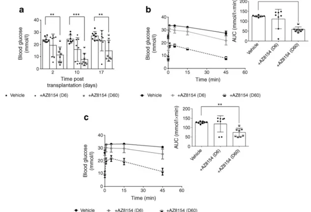
GPR44 inhibition in vivo improves glucose tolerance in diabetic mice transplanted with a marginal dose of human islets Diabetic mice bearing human islet grafts showed signif-icant improvement in fasting blood glucose measured on day 2 and day 17 post transplantation following treatment with GPR44 antagonist (60 mg/kg per day) vs vehicle (Fig.2a). In response to an intravenous glucose challenge performed on day 10, the mice treated with 60 mg/kg per day of the GPR44 antagonist AZ8154 showed significantly improved glucose tolerance compared with the vehicle group as shown by AUC quantitative analysis of the glucose excursion curves (Fig.2b). To exclude acute effects of the GPR44 antagonist on improved glucose response in islets, an IVGTT was
performed on day 15 post transplantation after an 18 h wash-out period of AZ8154. The blood glucose AUC was signifi-cantly improved in mice treated with 60 mg/kg per day of the GPR44 antagonist AZ8154 compared with vehicle (Fig.2c). The low dose of AZ8154 (6 mg/kg per day) affected neither the fasting blood glucose nor the glucose tolerance (Fig.2a– c). Body weight measurement every other day throughout the study showed no significant difference among the groups (data not shown).
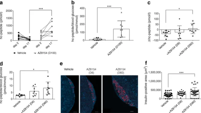
In vivo GPR44 inhibition preserves transplanted human islet grafts Circulating human C-peptide measured on day 2 and 17 post transplantation in diabetic mice was signifi-cantly improved in mice treated with 100 mg/kg per day AZ8154 vs vehicle (Fig. 3a). Furthermore, the ratio of human C-peptide over fasting blood glucose was increased in mice treated with 100 mg/kg per day AZ8154 vs vehicle (Fig.3b).
We also observed a significant increase in human C-peptide levels in mice treated with 60 mg/kg per day of AZ8154 vs vehicle (Fig.3c), calculated as the difference (Δ value) between day 2 and day 17 post transplantation. In
Fig. 2 GPR44 inhibition enhances glucose tolerance of human islets transplanted in diabetic mice. (a) Fasting blood glucose was measured on day 2, 10 and 17 in mice transplanted with human islets and treated with 6 or 60 mg/kg per day of AZ8154 or vehicle. (b, c) Blood glucose levels during IVGTT and the corresponding AUCs represent the differ-ence in the level of glucose in mice transplanted with human islets and treated with AZ8154 or vehicle on day 10, 4 h after administration of
AZ8154 (b) and on day 15, 18 h after administration of AZ8154 (c). Data are presented as means ± SD. The differences in AUC corresponding to the glucose levels during IVGTT were analysed with one-way ANOVA with Bonferroni corrections. **p < 0.01 and ***p < 0.001. For all analy-sis n = 8 mice/experimental group. D6, 6 mg/kg per day; D60, 60 mg/kg per day
addition, the ratio of human C-peptide over fasting blood glucose was increased in mice treated with 60 mg/kg per day AZ8154 vs vehicle on day 17 post transplantation (Fig. 3d). The transplanted human islet grafts analysed for insulin at termination of the study showed a significant increase in insulin-positive area in mice treated with 60 mg/kg per day AZ8154 vs vehicle (Fig.3e,f). The lower dose of AZ8154 (6 mg/kg per day) had no effect on preservation of the islet graft function or insulin area. The plasma concentration of AZ8154 was measured at the end of the experiment (ESM Figs.3,4a–c). For the low dose (6 mg/kg per day), the concen-tration of AZ8154 only reached sufficient exposure levels for a few hours, which was not sufficient to achieve any treatment effects. It is suggested that full inhibition of GPR44 is needed to protect the islet function.
In vivo GPR44 inhibition protects human islets from inflam-matory responses Levels of human C-peptide measured 4 days post transplantation were increased in mice treated with 60 mg/kg per day of AZ8154 vs vehicle (Fig.4a). In addition, the ratio of proinsulin to insulin was lower in mice treated with
AZ8154 vs vehicle (Fig. 4b). The mRNA expression of TNF-α but not GRO-α was significantly decreased in mice treated with AZ8154 vs vehicle group (data not shown). However, the circulating levels of the proinflammatory cyto-kines GRO-α and TNF-α were reduced in mice treated with AZ8154 vs vehicle (Fig.4c). An increase in the survival factor HGF was observed in the grafted islets in the AZ8154-treated mice (Fig. 4d). Furthermore, we also found a significant increase (50%) in the mRNA level of the beta cell-specific transcription factor MAFA (p < 0.05) (Fig.4f) but not PDX-1 (Fig.4e) in islet grafts following treatment with AZ8154 vs vehicle.
GPR44 inhibition mediates upregulation of PDX-1 via phos-phorylation of Akt, GSK3β and FOXO1 in human islets In vitro exposure of human islets to HG + IL-1β resulted in a twofold reduction in phosphorylation of Akt (Fig.5a) and GSK3β (Fig. 5b) compared with exposure to vehicle. These reductions were reversed by AZ8154, which restored the phosphorylation of Akt and GSK3β to levels similar to those observed in vehicle-treated islets (Fig.
Fig. 3 GPR44 inhibition improves human islet function and preserves beta cell mass post transplantation in diabetic mice. (a) Human C-peptide levels measured on day 2 and day 17 in mice transplanted with human islets and treated with 100 mg/kg per day AZ8154 or vehicle, n = 8 mice/each experimental group. (b) The ratio of human C-peptide over fasting blood glucose analysed on day 17 in mice transplanted with human islet grafts and treated with 100 mg/kg per day AZ8154 or vehicle, n = 8 mice/each experimental group. (c) Assessment of difference (Δ value) in the level of plasma human C-peptide measured on day 2 and day 17 in mice transplanted with human islets and treated with 60 mg/kg per day AZ8154 or vehicle, n = 8 mice/each experimental group. (d) The
ratio of human C-peptide over fasting blood glucose analysed on day 17 in mice transplanted with human islet grafts and treated with 60 mg/kg per day AZ8154 or vehicle, n = 8 mice/each experimental group. (e, f) Representative images showing immunofluorescent staining of insulin (red) together with DAPI nuclear staining (blue) on day 17 (e) and quan-tification of the area of insulin-positive cells within transplanted islet grafts (f) treated with AZ8154 or vehicle. Magnification ×10; scale bar, 200μm. In all analyses, data are presented as means ± SD and analysed with one-way ANOVA with Bonferroni corrections. *p < 0.05 and ***p < 0.001. D6, 6 mg/kg per day; D60, dose 60 mg/kg per day; D100, 100 mg/kg per day; hc-peptide, human C-peptide
5a,b). In addition, HG + IL-1β reduced secretion of HGF by human islets and this effect was reversed by AZ8154 (Fig.5c). Western blot analysis of human islets treated with HG + IL-1β revealed a twofold reduction in phosphoryla-tion of FOXO1 (Fig. 5d,e) compared with the vehicle, whereas inhibition of GPR44 reversed the effect of HG + IL-1β (Fig.5d,e). PDX-1 was slightly but not significantly reduced by HG + IL-1β, while AZ8154 increased the PDX-1 level in the islets exposed to this inflammatory environment (Fig. 5d,g). The protein level of total FOXO1 was unaffected regardless of the treatment (Fig. 5d,f). These data suggest that GPR44 inhibition protected human islet function at least partly through activation of the Akt–GSK3β pathway, phosphorylation of FOXO1 and upregulation of PDX-1 (Fig.6).
Discussion
Inhibition of GPR44 in human islets has the following effects: (1) reduced PGD2- and proinflammatory cytokine-induced apoptosis; (2) preserved islet grafts; and (3) improved glucose regulation in vivo in human islets exposed to a type 1 diabetes-like milieu. The mechanism is potentially related to reduction in inflammatory responses as plasma levels of TNF-α and GRO-α were decreased, while the cell survival
marker HGF was increased in human islets exposed to a type 1 diabetes-like milieu in vitro and in vivo. The inhibition of GPR44 in human islets is involved in restoration of Akt, GSK3β and FOXO1 phosphorylation and increase in the expression of the transcription factors PDX-1 and V-Maf musculoaponeurotic fibrosarcoma oncogene homolog A (MafA). Our findings suggest an important role for the PGD2–GPR44 pathway during stress-induced responses in islets exposed to a hyperglycaemic and proinflammatory envi-ronment, representing a potential target to improve islet survival rate and function.
Hyperglycaemia and systemic inflammation, which are the main hallmarks of diabetes, induce production of prostaglan-din molecules through upregulation of the COX2 enzyme [18]. Among the prostaglandin family, the PGE2 pathway has been extensively studied in pancreatic islets [5, 19]. PGE2reduces glucose-stimulated insulin secretion in beta cell lines and in rodent and human islets [20,21]. Moreover, intra-venous infusion of PGE2to rats decreased insulin secretion in response to glucose and, by using a transgenic mouse model overexpressing COX2 and PGE2synthase-1, reduced insulin level and hyperglycaemia were demonstrated [22,23]. PGE2 signals through four types of EP receptor (EP1–4); EP3 is the only G-protein coupled PGE2receptor that couples to inhibi-tory G-proteins and reduces insulin secretion in response to glucose through reduced cAMP levels [21]. Antagonising the
Fig. 4 GPR44 inhibition improves islet function and ameliorates secre-tion of proinflammatory cytokines in early phase post transplantasecre-tion. (a– c) Human C-peptide (a), ratio of human proinsulin over insulin (b) and plasma levels of the human proinflammatory cytokines TNF-α and GRO-α (c) analysed on day 4 post transplantation in mice transplanted with human islets and treated with 60 mg/kg per day AZ8154 or vehicle. (d–f) Human HGF protein level (d) and mRNA expression of PDX-1 (e) and
MAFA (f), normalised to RPL30, in transplanted human islet lysates obtained from mice treated with AZ8154 60 mg/kg per day or vehicle on day 4 post transplantation. For all analyses n = 14 mice/experimental group; data are presented as mean ± SD and analysed by Mann–Whitney U test. *p < 0.05 and **p < 0.01. D60, 60 mg/kg per day; hGRO-α, human GRO-α; hc-peptide, human C-peptide; hTNF-α, human TNF-α
EP3 receptor improves insulin secretion in response to glucose in MIN6 beta cells and in rat islets [24]. EP3 inhibition also enhances insulin secretion in human islets isolated from donors with type 2 diabetes [21].
Elevated levels of PGD2impair glucose-stimulated insulin secretion and reduce cAMP levels [7,8]. Our data show that PGD2increased apoptosis in human islets to a similar degree as proinflammatory cytokines. This effect was blocked by administration of the GPR44 antagonist AZ8154, which is an analogue to the clinical GPR44 antagonist drug candidate AZD1981 [12]. AZD1981 has been shown to reverse the effect of PGD2on suppressing insulin secretion in human islets [8]. Administration of the antagonist AZ8154 to hyperglycaemic mice transplanted with a marginal number of human islets improved glucose response, increased plasma levels of human C-peptide and increased the insulin-positive area of the transplanted islets, which could suggest improve-ment in human islet grafts [25]. These effects were accompa-nied by an increased ratio of human C-peptide to fasting blood glucose, which has been shown to correlate with increase in beta cell area [26]. A recent clinical study explored the acute effect of GPR44 inhibition in individuals with poorly controlled type 2 diabetes [8]. The outcome was negative in terms of the administration of the GPR44 antagonist
AZD1981 having no acute effect on insulin secretion. It is possible that these individuals had already developed signifi-cant islet dysfunction and therefore did not respond to acute administration of the drug, or an elevated PGD2tone is not present in the islet vicinity. Moreover, considering the specific expression of the GPR44 receptor on beta cells, the presence of too few functional beta cells could be another reason for the absence of an effect of GPR44 inhibition in individuals with type 2 diabetes [1,8].
In the islet transplantation process, it is well known that proin-flammatory cytokines are involved in pancreatic islet injury and islet graft destruction post transplantation [27]. In particular, elevated levels of proinflammatory cytokines IL-1β, TNF-α and INF-γ observed after islet transplantation can mediate islet injury at the early phase post transplantation [28]. IL-1β produc-tion is triggered by itself and by high concentraproduc-tion of glucose within islets [29]. Furthermore, production of the proinflammato-ry cytokine GRO-α has been found to increase 6 h after human islet exposure to whole blood using an ex vivo loop blood model mimicking the graft loss post transplantation [30]. GPR44 is high-ly expressed in human islets [1,8,11] and we found that the inhibition of GPR44 led to reduced plasma levels of human GRO-α and TNF-α in the post islet transplantation phase. This could support the positive impact of GPR44 inhibition on
Fig. 5 GPR44 inhibition increases protein levels of PDX-1 via the Akt– GSK3β–FOXO1 signalling pathway in isolated human islets. (a–c) Phosphorylation of Akt (a; n = 8 independent donors) and GSK3β (b; n = 8 independent donors), and secretion of HGF (c; n = 6 independent donors) in human islets treated with either AZ8154 (10μmol/l) alone or HG (20 mmol/l) + IL-1β (10 ng/ml) with or without AZ8154 (10 μmol/l) for 48 h. (d) Western blot analysis of phosphorylated FOXO1, total FOXO1 and PDX-1 in human islets treated with either AZ8154
(10μmol/l) alone or HG (20 mmol/l) + IL-1β (10 ng/ml) with or without AZ8154 (10μmol/l) for 48 h. (e–g) Quantification of blots shown in (d): p-FOXO1 (e; n = 7 independent donors), total FOXO1 (f; n = 5 indepen-dent donors) and PDX-1 (g; n = 7 indepenindepen-dent donors). Band densities were normalised to GAPDH and presented as fold of changes over control (vehicle-treated) islets. For all analyses, data were analysed with one-way ANOVA with Bonferroni corrections and presented as mean ± SD. *p < 0.05 and **p < 0.01. hHGF, human HGF
reducing the adverse effects of the inflammatory responses that are often responsible for major loss of functional beta cell mass after islet transplantation [31].
Increased secretion of proinsulin could occur due to the pres-ence of a stress micro-environment such as prolonged exposure to hyperglycaemia and elevated demand for insulin secretion [32, 33]. Increased ratio of proinsulin to insulin has been reported in individuals with new onset of type 1 diabetes and in allo-islet transplanted patients [34,35]. It is not clear from our study the extent to which the reduced ratio of proinsulin to insulin in GPR44-treated mice contributes to the preserved functionality of transplanted islets, although the reduction in this ratio accompa-nied by elevated level of C-peptide and mRNA expressions of PDX-1 and MAFA together with a reduction in secretion of proin-flammatory cytokines could suggest an overall improvement of the human islet grafts.
PGE2has been found to both decrease DNA synthesis and to reduce islet survival rate in rat islets [36]. This was suggested to be mediated through PGE2coupling to the EP3 receptor, leading to dephosphorylation of FOXO1 either via
activation of c-Jun N-terminal kinase (JNK1) or inhibition of the Akt survival signalling pathway [37,38]. Subsequently there is an increase in translocation of FOXO1 to the nucleus where it could participate in nuclear exclusion of critical beta cell transcription factors, PDX-1 and MafA [37,39]. MafA and PDX-1 have been previously suggested to be key activa-tors of insulin synthesis and master regulaactiva-tors of genes involved in maintaining beta cell function [40]. We observed a reduction in protein expression of PDX-1 in human islets exposed to a diabetic milieu in vitro, and also found a decrease in phosphorylation of Akt and its downstream target GSK3β. Importantly, the effect of the stressors was reversed by inhibi-tion of GPR44, shown by upregulainhibi-tion of PDX-1 protein expression, increased phosphorylation of FOXO1 and activa-tion of the Akt–GSK3β signalling pathway. Furthermore, we demonstrated upregulation of HGF in islet grafts in the early phase post transplantation and in human islets exposed to a type 1 diabetes-like milieu and treated with GPR44 antagonist in vitro. HGF has been shown to be involved in regeneration and restoration of damaged tissue including pancreatic beta
Fig. 6 Schematic overview of the PGD2–GPR44 axis in human islets
exposed to hyperglycaemia and proinflammatory cytokines. HG and presence of proinflammatory cytokines increases the level of PGD2in
the vicinity of islets [8,9]. PGD2signals through GPR44 and induces
apoptosis in human islets via increase in caspase 3/7 activity (green arrow). Inhibition of the PGD2–GPR44 axis improves human islet
viabil-ity and function via increased activation of the Akt–GSK3β signalling
pathway. Inhibition of GPR44 during stress responses restores phosphor-ylation of FOXO1 and upregulates the expression of beta cell transcrip-tion factors, PDX-1 and MafA (grey arrows). It has been suggested that unphosphorylated FOXO1 translocates to the nucleus where it binds to PDX-1 gene and downregulates its transcription [37,39]. Therefore, increased phosphorylation of FOXO1 could, at least partly, be involved in the upregulation of key beta cell transcription factors
cells [41]. Proinflammatory cytokines have been reported to modulate secretion of HGF in human islets in vitro [42]. Adenoviral delivery of HGF to islets from non-human primates demonstrated a marked improvement in transplanted islet function and mass suggesting a possible role for HGF in the survival of islets exposed to diabetogenic agents [43]. HGF is also known for improving insulin secretion, mainly through activation of the Akt signalling pathway [44]. Therefore, HGF could at least partly be involved in activation of the Akt signalling pathway and improvement in islet func-tion. Despite similar adverse effects of PGD2and PGE2on human islets, and similar signalling pathways, the positive effects we observed with AZ8154 was only related to GPR44 since the compound had no activity on the PGE2 receptor EP3.
This study supports a role for the PGD2–GPR44 path-way in human islet dysfunction and suggests that the inhi-bition of GPR44 could improve islet survival rate and function during severe hyperglycaemia. Inhibition of GPR44 may have the potential to represent a rescue strat-egy for islets exposed to an acute inflammatory and hyperglycaemic environment and may preserve human islet grafts after transplantation.
Acknowledgements Open Access funding provided by University of Oslo (incl Oslo University Hospital). The authors thank M. Høyem from Oslo University Hospital for technical assistance with the in vivo animal experiments. Human pancreatic islets were obtained from The Nordic Network for Clinical Islet Transplantation supported by the Swedish national strategic research initiative EXODIAB and JDRF. The authors are grateful to all members at the human islet isolation facilities at University of Uppsala and Oslo University Hospital.
Parts of this study were presented in abstract form at the 16th International Congress Pancreas and Islet Transplant Association, Oxford, UK, 8th EPITA Symposium & 37th AIDPIT Workshop, Innsbruck, Austria, 2018 and at the 52nd EASD meeting, Munich, Germany, 2016.
Data availability The datasets generated during and/or analysed during the current study and presented in the main article and assupplementary
materialsare available from the corresponding authors upon a reasonable
request. The chemical structure of AZ8154 compound, which was gener-ated by Astra Zeneca and used in the current study, is not publicly avail-able due to the fact that the compound has not been patented yet but is available from the corresponding author upon reasonable request. Funding The study was supported by grants from the South Eastern Norway Regional Health Authority (Reference no. 2012031) and partly supported by the Research Council of Norway through the Center of Excellence funding scheme, project number 262613. AstraZeneca funded parts of the study.
Authors’ relationships and activities BT, CW-H, PG, ER, TR-B, DMS, SS and MS-W are employed by AstraZeneca. AstraZeneca was not involved in the design of the study; the collection, analysis, and interpre-tation of data; writing the report; and did not impose any restrictions regarding the publication of the report. The remaining authors declare that there are no relationships or activities that might bias, or be perceived to bias, their work.
Contribution statement SA, DMS, OK, SS, HS and MS-W contributed to the conception and design of the study. SA, BT, SWS, CW-H, PG, ER, TR-B, HS and MS-W contributed to acquisition of data, analysis and interpretation of data. OK and HS performed human islet isolation. SA, MS-W and HS contributed to drafting the article and are the guarantors of this work, and as such have full access to all data in the study and take responsibility for the integrity of the data and the accuracy of the data analysis. SA, BT, SWS, DMS, PG, CW-H, ER, TR-B, OK, SS, HS and MS-W contributed to revising the article critically for important intellec-tual content. All authors have approved the final version of the article for publication.
Open Access This article is licensed under a Creative Commons Attribution 4.0 International License, which permits use, sharing, adap-tation, distribution and reproduction in any medium or format, as long as you give appropriate credit to the original author(s) and the source, provide a link to the Creative Commons licence, and indicate if changes were made. The images or other third party material in this article are included in the article's Creative Commons licence, unless indicated otherwise in a credit line to the material. If material is not included in the article's Creative Commons licence and your intended use is not permitted by statutory regulation or exceeds the permitted use, you will need to obtain permission directly from the copyright holder. To view a copy of this licence, visithttp://creativecommons.org/licenses/by/4.0/.
References
1. Lindskog C, Korsgren O, Ponten F, Eriksson JW, Johansson L, Danielsson A (2012) Novel pancreatic beta cell-specific proteins: antibody-based proteomics for identification of new biomarker candidates. J Proteome 75(9):2611–2620.https://doi.org/10.1016/ j.jprot.2012.03.008
2. Eriksson O, Johnstrom P, Cselenyi Z et al (2018) In vivo visualiza-tion ofβ-cells by targeting of GPR44. Diabetes 67(2):182–192. https://doi.org/10.2337/db17-0764
3. Donath MY, Storling J, Maedler K, Mandrup-Poulsen T (2003) Inflammatory mediators and isletβ-cell failure: a link between type 1 and type 2 diabetes. J Mol Med 81(8):455–470.https://doi.org/10. 1007/s00109-003-0450-y
4. Federici M, Hribal M, Perego L et al (2001) High glucose causes apoptosis in cultured human pancreatic islets of Langerhans: a potential role for regulation of specific Bcl family genes toward an apoptotic cell death program. Diabetes 50(6):1290–1301. https://doi.org/10.2337/diabetes.50.6.1290
5. Carboneau BA, Breyer RM, Gannon M (2017) Regulation of pancreaticβ-cell function and mass dynamics by prostaglandin signaling. J Cell Commun Signal 11(2):105–116.https://doi.org/ 10.1007/s12079-017-0377-7
6. Parazzoli S, Harmon JS, Vallerie SN, Zhang T, Zhou HR, Robertson RP (2012) Cyclooxygenase-2, not microsomal prosta-glandin E synthase-1, is the mechanism for interleukin-1β-induced prostaglandin E-2 production and inhibition of insulin secretion in pancreatic islets. J Biol Chem 287(38):32246–32253.https://doi. org/10.1074/jbc.M112.364612
7. Hata AN, Breyer RM (2004) Pharmacology and signaling of pros-taglandin receptors: multiple roles in inflammation and immune modulation. Pharmacol Ther 103(2):147–166.https://doi.org/10. 1016/j.pharmthera.2004.06.003
8. Skrtic S, Tyrberg B, Broberg M et al (2018) Exploring the insulin secretory properties of the PGD2-GPR44/DP2 axis in vitro and in a randomized phase-1 trial of type 2 diabetes patients. PLoS One 13(12):e0208998.https://doi.org/10.1371/journal.pone.0208998
9. Evans MH, Pace CS, Clements RS (1983) Endogenous prostaglan-din synthesis and glucose-induced insulin-secretion from the adult-rat pancreatic-islet. Diabetes 32(6):509–515.https://doi.org/10. 2337/diabetes.32.6.509
10. Hata AN, Zent R, Breyer MD, Breyer RM (2003) Expression and molecular pharmacology of the mouse CRTH2 receptor. J Pharmacol Exp Ther 306(2):463–470.https://doi.org/10.1124/jpet. 103.050955
11. Hellstrom-Lindahl E, Danielsson A, Ponten F et al (2016) GPR44 is a pancreatic protein restricted to the human beta cell. Acta Diabetol 53(3):413–421.https://doi.org/10.1007/s00592-015-0811-3 12. Luker T, Bonnert R, Brough S et al (2011) Substituted
indole-1-acetic acids as potent and selective CRTh2 antagonists-discovery of AZD1981. Bioorg Med Chem Lett 21(21):6288–6292.https://doi. org/10.1016/j.bmcl.2011.08.124
13. Kuna P, Bjermer L, Tornling G (2016) Two phase II randomized trials on the CRTh2 antagonist AZD1981 in adults with asthma. Drug Des Devel Ther 10:2759–2770.https://doi.org/10.2147/ DDDT.S105142
14. Schmidt JA, Bell FM, Akam E et al (2013) Biochemical and phar-macological characterization of AZD1981, an orally available selective DP2 antagonist in clinical development for asthma. Br J Pharmacol 168(7):1626–1638.https://doi.org/10.1111/bph.12053 15. Friberg AS, Stahle M, Brandhorst H, Korsgren O, Brandhorst D
(2008) Human islet separation utilizing a closed automated purifi-cation system. Cell Transplant 17(12):1305–1313.https://doi.org/ 10.3727/096368908787648100
16. Friberg AS, Brandhorst H, Buchwald P et al (2011) Quantification of the islet product: presentation of a standardized current good manufacturing practices compliant system with minimal variability. Transplantation 91(6):677–683. https://doi.org/10.1097/TP. 0b013e31820ae48e
17. Sahraoui A, Winzell MS, Gorman T et al (2015) The effects of exendin-4 treatment on graft failure: an animal study using a novel re-vascularized minimal human islet transplant model. PLoS One 10(3):e0121204.https://doi.org/10.1371/journal.pone.0121204 18. Sorli CH, Zhang HJ, Armstrong MB, Rajotte RV, Maclouf J,
Robertson RP (1998) Basal expression of cyclooxygenase-2 and nuclear factor-interleukin 6 are dominant and coordinately regulat-ed by interleukin 1 in the pancreatic islet. Proc Natl Acad Sci U S A 95(4):1788–1793.https://doi.org/10.1073/pnas.95.4.1788 19. Robertson RP (1983) Hypothesis. PGE, carbohydrate homeostasis,
and insulin secretion. A suggested resolution of the controversy. Diabetes 32(3):231–234.https://doi.org/10.2337/diab.32.3.231 20. Robertson RP, Tsai P, Little SA, Zhang HJ, Walseth TF (1987)
Receptor-mediated adenylate cyclase-coupled mechanism for PGE2 inhibition of insulin secretion in HIT cells. Diabetes 36(9): 1047–1053.https://doi.org/10.2337/diab.36.9.1047
21. Kimple ME, Keller MP, Rabaglia MR et al (2013) Prostaglandin E2 receptor, EP3, is induced in diabetic islets and negatively regulates glucose- and hormone-stimulated insulin secretion. Diabetes 62(6): 1904–1912.https://doi.org/10.2337/db12-0769
22. Sacca L, Perez G, Rengo F, Pascucci I, Condorelli M (1975) Reduction of circulating insulin levels during the infusion of differ-ent prostaglandins in the rat. Acta Endocrinol 79(2):266–274. https://doi.org/10.1530/acta.0.0790266
23. Oshima H, Taketo MM, Oshima M (2006) Destruction of pancre-aticβ-cells by transgenic induction of prostaglandin E2 in the islets. J Biol Chem 281(39):29330–29336.https://doi.org/10.1074/jbc. M602424200
24. Shridas P, Zahoor L, Forrest KJ, Layne JD, Webb NR (2014) Group X secretory phospholipase A2 regulates insulin secretion through a cyclooxygenase-2-dependent mechanism. J Biol Chem 289(40): 27410–27417.https://doi.org/10.1074/jbc.M114.591735
25. Caiazzo R, Gmyr V, Kremer B et al (2008) Quantitative in vivo islet potency assay in normoglycemic nude mice correlates with primary
graft function after clinical transplantation. Transplantation 86(2): 360–363.https://doi.org/10.1097/TP.0b013e31817ef846
26. Meier JJ, Menge BA, Breuer TG et al (2009) Functional assessment of pancreaticβ-cell area in humans. Diabetes 58(7):1595–1603. https://doi.org/10.2337/db08-1611
27. Barshes NR, Wyllie S, Goss JA (2005) Inflammation-mediated dysfunction and apoptosis in pancreatic islet transplantation: impli-cations for intrahepatic grafts. J Leukoc Biol 77(5):587–597. https://doi.org/10.1189/jlb.1104649
28. Bottino R, Fernandez LA, Ricordi C et al (1998) Transplantation of allogeneic islets of Langerhans in the rat liver: effects of macro-phage depletion on graft survival and microenvironment activation. Diabetes 47(3):316–323.https://doi.org/10.2337/diabetes.47.3.316 29. Boni-Schnetzler M, Thorne J, Parnaud G et al (2008) Increased interleukin (IL)-1β messenger ribonucleic acid expression in β-cells of individuals with type 2 diabetes and regulation of IL-1β in human islets by glucose and autostimulation. J Clin Endocrinol Metab 93(10):4065–4074.https://doi.org/10.1210/jc.2008-0396 30. Hardstedt M, Lindblom S, Karlsson-Parra A, Nilsson B, Korsgren
O (2016) Characterization of innate immunity in an extended whole blood model of human islet allotransplantation. Cell Transplant 25(3):503–515.https://doi.org/10.3727/096368915X688461 31. Monti P, Vignali D, Piemonti L (2015) Monitoring inflammation,
humoral and cell-mediated immunity in pancreas and islet trans-plants. Curr Diabetes Rev 11(3):135–143.https://doi.org/10.2174/ 1573399811666150317125820
32. Abadpour S, Gopel SO, Schive SW, Korsgren O, Foss A, Scholz H (2017) Glial cell-line derived neurotrophic factor protects human islets from nutrient deprivation and endoplasmic reticulum stress induced apoptosis. Sci Rep 7(1):1575.https://doi.org/10.1038/ s41598-017-01805-1
33. Fu Z, Gilbert ER, Liu D (2013) Regulation of insulin synthesis and secretion and pancreatic Beta-cell dysfunction in diabetes. Curr Diabetes Rev 9(1):25–53
34. Rodriguez-Calvo T, Zapardiel-Gonzalo J, Amirian N et al (2017) Increase in pancreatic proinsulin and preservation ofβ-cell mass in autoantibody-positive donors prior to type 1 diabetes onset. Diabetes 66(5):1334–1345.https://doi.org/10.2337/db16-1343 35. Klimek AM, Soukhatcheva G, Thompson DM et al (2009)
Impaired proinsulin processing is a characteristic of transplanted islets. Am J Transplant 9(9):2119–2125.https://doi.org/10.1111/j. 1600-6143.2009.02740.x
36. Sjoholm A (1996) Prostaglandins inhibit pancreaticβ-cell replica-tion and long-term insulin secrereplica-tion by pertussis toxin-insensitive mechanisms but do not mediate the actions of interleukin-1β. Biochim Biophys Acta 1313(2):106–110.https://doi.org/10.1016/ 0167-4889(96)00058-4
37. Meng ZX, Lv JH, Luo Y et al (2009) Forkhead box O1/pancreatic and duodenal homeobox 1 intracellular translocation is regulated by c-Jun N-terminal kinase and involved in prostaglandin E-2-induced pancreaticβ-cell dysfunction. Endocrinology 150(12):5284–5293. https://doi.org/10.1210/en.2009-0671
38. Meng ZX, Sun JX, Ling JJ et al (2006) Prostaglandin E2 regulates Foxo activity via the Akt pathway: implications for pancreatic islet beta cell dysfunction. Diabetologia 49(12):2959–2968.https://doi. org/10.1007/s00125-006-0447-5
39. Kitamura T, Nakae J, Kitamura Y et al (2002) The forkhead tran-scription factor Foxo1 links insulin signaling to Pdx1 regulation of pancreaticβ cell growth. J Clin Invest 110(12):1839–1847.https:// doi.org/10.1172/JCI16857
40. Arcidiacono B, Iiritano S, Chiefari E et al (2015) Cooperation between HMGA1, PDX-1, and MafA is essential for glucose-induced insulin transcription in pancreatic beta cells. Front Endocrinol 5:ARTN 237. https://doi.org/10.3389/fendo.2014. 00237
41. Mellado-Gil JM, Cobo-Vuilleumier N, Gauthier BR (2012) Islet β-cell mass preservation and regeneration in diabetes mellitus: four factors with potential therapeutic interest. J Transplant 2012: 230870.https://doi.org/10.1155/2012/230870
42. Mellado-Gil J, Rosa TC, Demirci C et al (2011) Disruption of hepatocyte growth factor/c-Met signaling enhances pancreatic β-cell death and accelerates the onset of diabetes. Diabetes 60(2):525–
536.https://doi.org/10.2337/db09-1305
43. Fiaschi-Taesch NM, Berman DM, Sicari BM et al (2008) Hepatocyte growth factor enhances engraftment and function of
nonhuman primate islets. Diabetes 57(10):2745–2754.https://doi. org/10.2337/db08-1085
44. Fiaschi-Taesch N, Stewart AF, Garcia-Ocana A (2007) Improving islet transplantation by gene delivery of hepatocyte growth factor (HGF) and its downstream target, protein kinase B (PKB)/Akt. Cell Biochem Biophys 48(2–3):191–199. https://doi.org/10.1007/ s12013-07-0024-7
Publisher’s note Springer Nature remains neutral with regard to jurisdic-tional claims in published maps and institujurisdic-tional affiliations.