This is the published version of a paper published in Sports Biomechanics.
Citation for the original published paper (version of record): Aasa, U., Bengtsson, V., Berglund, L., Öhberg, F. (2019)
Variability of lumbar spinal alignment among power- and weightlifters during the deadlift and barbell back squat
Sports Biomechanics
https://doi.org/10.1080/14763141.2019.1675751
Access to the published version may require subscription. N.B. When citing this work, cite the original published paper.
Permanent link to this version:
Full Terms & Conditions of access and use can be found at
https://www.tandfonline.com/action/journalInformation?journalCode=rspb20
ISSN: 1476-3141 (Print) 1752-6116 (Online) Journal homepage: https://www.tandfonline.com/loi/rspb20
Variability of lumbar spinal alignment among
power- and weightlifters during the deadlift and
barbell back squat
Ulrika Aasa, Victor Bengtsson, Lars Berglund & Fredrik Öhberg
To cite this article: Ulrika Aasa, Victor Bengtsson, Lars Berglund & Fredrik Öhberg (2019):
Variability of lumbar spinal alignment among power- and weightlifters during the deadlift and barbell back squat, Sports Biomechanics, DOI: 10.1080/14763141.2019.1675751
To link to this article: https://doi.org/10.1080/14763141.2019.1675751
© 2019 The Author(s). Published by Informa UK Limited, trading as Taylor & Francis Group.
Published online: 13 Nov 2019.
Submit your article to this journal
Article views: 457
View related articles
Variability of lumbar spinal alignment among power- and
weightlifters during the deadlift and barbell back squat
Ulrika Aasaa, Victor Bengtssona, Lars Berglundband Fredrik Öhbergc
aDepartment of Community Medicine and Rehabilitation, Physiotherapy, Umeå University, Umeå, Sweden; bDepartment of Community Medicine and Rehabilitation, Sports Medicine, Umeå University, Umeå, Sweden; cDepartment of Radiation Sciences, Umeå University, Umeå, Sweden
ABSTRACT
The aims of the study were to evaluate the relative and absolute variability of upper (T11-L2) and lower (L2-S2) lumbar spinal alignment in power- and weightlifters during the deadlift and back squat exer-cises, and to compare this alignment between the two lifting groups. Twenty-four competitive powerlifters (n = 14) and weightlifters (n = 10) performed three repetitions of the deadlift and the back squat exer-cises using a load equivalent to 70% of their respective one-repetition maximum. The main outcome measures were the three-dimensional lumbar spinal alignment for start position, minimum and maximum angle of their spinal alignment, and range of motion measured using inertial measurement units. Relative intra-trial reliability was calculated using the two-way random model intraclass correlation coefficient (ICC) and absolute reliability with minimal detectable change (MDC). The ICC ranged between 0.69 and 0.99 and the MDC between 1°-8° for the deadlift. Correspondingfigures for the squat were 0.78–0.99 and 1°-6°. In all participants during both exercises, spinal adjustments were made in both thoracolumbar and lumbopelvic areas in all three dimensions. In conclusion, when performing three repetitions of the deadlift and the squat, lumbar spinal alignment of the lifters did not change much between repetitions and did not differ significantly between power- and weightlifters.
ARTICLE HISTORY
Received 18 April 2019 Accepted 26 September 2019
KEYWORDS
Inertial sensors; powerlifting; weightlifting; lifting technique
Introduction
Deviations from a neutral spinal alignment during performance of the barbell back squat, henceforth referred to as the squat, and deadlift strength training exercises could be a risk for future low back pain (Sjoberg, Aasa, Rosengren, & Berglund,2018). Since the interest of strength sports is growing worldwide, and injuries in the low back are common in powerlifters (Stromback, Aasa, Gilenstam, & Berglund, 2018) and are becoming more common in weightlifters (Burekhovich et al.,2018), physiotherapists often treat patients whose pain is associated with either performance of the squat or the deadlift. In the process of rehabilitation, the physiotherapists analyse the movement patterns of lifters during performance of the pain-provoking exercises to examine whether side bending, rotating orflexing of the lumbar spine during the lift is associated with the lifters’ pain experience. If an increased relativeflexibility (Sahrmann, Azevedo, & Dillen, 2017) in
CONTACTUlrika Aasa ulrika.aasa@umu.se
© 2019 The Author(s). Published by Informa UK Limited, trading as Taylor & Francis Group.
This is an Open Access article distributed under the terms of the Creative Commons Attribution License (http://creativecommons.org/ licenses/by/4.0/), which permits unrestricted use, distribution, and reproduction in any medium, provided the original work is properly cited.
either of these directions is found, the functional impairments causing it are targeted in the rehabilitation. Excessive movements in these planes are also considered contra-indicated for a number of other reasons, not least the correlation between spinal align-ment and degenerative changes. For example, it has been suggested that axial twisting in
combination with repetitive flexion-extension motion might predispose joint diseases
such as vertebral stress fractures (Leone, Cianfoni, Cerase, Magarelli, & Bonomo,2011) as well as bulging of the lumbar discs and herniation (Marshall & McGill,2010).
When evaluating movement patterns for the squat and deadlift, it is important to remember the inherent variability evident within the human movement system (Hamill, Palmer, & Van Emmerik,2012). In fact, the movement system has the ability to sponta-neously reorganise movement coordinative strategies in a variety of ways to adapt to external and internal factors (functional variability) (Davids, Glazier, Araujo, & Bartlett,
2003). For example, the ability of the lifters to always initiate the deadlift without rounding their lower back could be affected by internal factors such as fatigue (Santamaria & Webster, 2010). Repeatability is the opposite to variability. Repeatability practices were introduced by Bland and Altman (1999). Relative intra-trial repeatability could be used to evaluate the consistency of the spinal alignment within a training session when performing repetitive repetitions of the squat or deadlift, and hence gain a better understanding of whether lifters perform the exercises in a similar manner across repetitions. To our knowledge, only one previous study has investigated the relative reliability of repeated measures of spinal alignment during squat performance (McKean, Dunn, & Burkett,2010). In that study, calculation of the intrarater test–retest relative reliability was reported with good (>0.7) to excellent (>0.9) intraclass correlation coefficients (ICC) indicating low intra-individual variability of the lumbar and pelvic movements between repetitions when the squat was performed with 50% of body weight. Further, in addition to the relative reliability, the absolute reliability can be used to calculate the minimal detectable change (MDC) in order to reveal whether measured variations are genuine changes or not (Bland & Altman,1996). No previous study has examined the absolute intra-trial reliability or MDC of the spinal alignment during deadlift or squat performance.
A growing body of scientific literature has investigated the utility of inertial measure-ment units (IMUs) for monitoring of spinal alignmeasure-ment during resistance training (Gleadhill, Lee, & James, 2016) and to evaluate exercise technique (Taylor, Almeida,
Hodgins, & Kanade, 2012). IMUs are portable tri-axial motion sensors (containing
gyroscope, accelerometer and magnetometer) that are attached on the skin and can be wirelessly connected to a computer. They have been shown to be as effective as marker-based systems at measuring joint angles in sport (Bonnet, Mazza, Fraisse, & Cappozzo,
2013) and work-related (Stenlund et al.,2014) tasks, and have been shown to distinguish between acceptable and aberrant squat techniques with excellent accuracy and to identify exact technique deviations (O’Reilly, Whelan, Ward, Delahunt, & Caulfield,2017).
The deadlift and the squat are essential exercises among both power- and weightlifters (Wretenberg, Feng, & Arborelius, 1996). However, since they are also two of three competitive lifts in the sport of powerlifting (Keogh, Hume, & Pearson, 2006; Siewe et al.,2011; Swinton, Lloyd, Agouris, & Stewart,2009), but not in weightlifting, power-lifting and weightpower-lifting athletes might use different power-lifting styles and thereby move differently in their lumbar spines. Today, it is unknown whether they show different spinal alignment during the lifts.
The present study aimed to 1) evaluate the relative and absolute variability of the spinal alignment during both the squat and deadlift exercises, and 2) determine whether power- and weightlifters show different spinal alignment in their lumbar spine.
We hypothesised that 1) the movement patterns of the lifters would not change between three repetitions when performing the lifts at approximately 70% 1RM and that 2) compe-titive power- and weightlifters would show similar spinal alignment during the lifts.
Methods Study design
This cross-sectional study measured the tree-dimensional movements in the upper (thoracolumbar, T11-L2) and lower (lumbopelvic, L2-S2) lumbar spine with IMUs when performing the squat and deadlift exercises. Data collection was performed in a gym. Spinal alignment was measured during the performance of one set of three repetitions of 70% of self-estimated one-repetition maximum (1RM) squats and deadlifts.
Participants
Fourteen powerlifters (men = 10, women = 4) and 10 weightlifters (men = 4, women = 6) were recruited from local clubs in Umeå, Sweden (for characteristics, seeTable 1). Inclusion criteria were power- and weightlifters≥150 cm in height with at least two years of lifting experience, without injuries that could affect performance, and with the intent of compet-ing. To ensure that eligibility criteria were met, all participants completed a questionnaire. They also signed an informed consent form prior to participation. At the day of data collection, one of the women powerlifters chose not to perform the squat. Written informed consent was obtained from all lifters prior to participation and the study was approved by the Regional Ethical Review Board of Umeå, Sweden (Dnr 2014-285-3M).
Procedures
First, the participants completed a self-administered warm-up with the intention to be prepared for heavy deadlifts. The warm-up typically consisted of sub-maximal deadlifts with increasing loads. Thereafter, three calibrated IMUs were affixed to their backs using double-sided tape and self adherent wraps at the following anatomical landmarks:
Processus spinosus Th11 and L2, and Sacrum (S2) (Figure 1). The placements of the
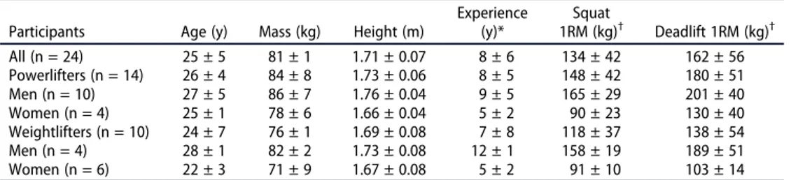
Table 1.Participant characteristics (mean±SD).
Participants Age (y) Mass (kg) Height (m)
Experience (y)* Squat 1RM (kg)† Deadlift 1RM (kg)† All (n = 24) 25 ± 5 81 ± 1 1.71 ± 0.07 8 ± 6 134 ± 42 162 ± 56 Powerlifters (n = 14) 26 ± 4 84 ± 8 1.73 ± 0.06 8 ± 5 148 ± 42 180 ± 51 Men (n = 10) 27 ± 5 86 ± 7 1.76 ± 0.04 9 ± 5 165 ± 29 201 ± 40 Women (n = 4) 25 ± 1 78 ± 6 1.66 ± 0.04 5 ± 2 90 ± 23 130 ± 40 Weightlifters (n = 10) 24 ± 7 76 ± 1 1.69 ± 0.08 7 ± 8 118 ± 37 138 ± 54 Men (n = 4) 28 ± 1 82 ± 2 1.73 ± 0.08 12 ± 1 158 ± 19 189 ± 51 Women (n = 6) 22 ± 3 71 ± 9 1.67 ± 0.08 5 ± 2 91 ± 10 103 ± 14 *Strength training experience;†Self-estimated one-repetition maximum (1RM)
units were palpated with the lifters standing erect by the same experienced person. Before commencing the respective data collections of the deadlift and squat exercises, the lifters further performed one set of the relevant exercise to ensure that the IMUs did not hinder performance.
For data collection, the lifters performed one set of three repetitions at 70% of his/her self-estimated 1RM for each exercise. The lifters stood holding the barbell, which was
placed on thefloor in front of them, with straight arms and an optional grip and with
Figure 1.Three calibrated IMUs were affixed at the level of the Processus spinosus at Th11 and L2, and at Sacrum (S2). Thisfile/figure is licensed under the Creative Commons Attribution-Share Alike 4.0 International license.
flexed knees and hips (Start position deadlift). In accordance with the rules of the International Powerlifting Federation (2019), the barbell was then lifted by extension of the knees and hips until the lifter was standing erect with their shoulders back (Stop position). Once the lifter was motionless in the Stop position a down signal was given by the test leader and the lifter then lowered the barbell to the ground before releasing their grip. The lifters were instructed to stand erect momentarily before beginning with the next repetition. Since only conventional style deadlifts were allowed, so that the measure-ments would be uniform, all participants used a conventional deadlift style where the barbell is held with the arms laterally to the legs. For the squat, participants were standing erect (Start position) with the barbell on their shoulders and instructed to descend by flexing at the hip, knee and ankle joints until the crease of the hip was lower than the top of the knee in accordance with the rules of the International Powerlifting Federation (2019). From the bottom position, the participants ascended to the start position by extending the same joints. The lifters were instructed to stay in this position until a start signal was given by the test leader to begin the next repetition. Notably, apart from the practical instructions, no more instructions were given. The basic premise of this form of evaluation is to measure the individual movement and joint loading patterns of each participant (Nielsen et al., 2017). No equipment (knee wraps, belts) other than wrist wraps was allowed. The lifters were allowed to use chalk and, if preferred, they could use alternated hand grip for the deadlift.
Instruments and measurements
We wanted ourfindings to be representative for the real-life environments (Dingenen &
Gokeler, 2017), hereby acknowledging the importance of the environmental and task
constraints within the dynamic system theory (Davids et al., 2003; Holt, Wagenaar, & Saltzman,2010). We used a portable movement analysis system in a gym to assess spinal alignment in the thoracolumbar and lumbopelvic areas during the lifts. The system
included three tri-axial IMUs (MoLabTMPOSE, AnyMo AB, Umeå, Sweden) that were
wirelessly connected using WiFi to a computer equipped with the software MoLabTM
measure (AnyMo AB, Umeå, Sweden). Each sensor was 60 × 45 x 10 mm (length x width x height), included a three-dimensional gyroscope, accelerometer and magnetometer and weighed 14 g. The sampling frequency was 128 Hz with a 16-bit resolution and an anti-aliasing low passfilter set at 64 Hz. The full-scale range was ±1000°/s for the gyroscopes, ±8 g for the accelerometers and ±4800 µT for the magnetometers. The measurement
precision and accuracy of the MoLabTMPOSE system for measurements in the spine has
been validated against a gold standard optical system (Ertzgaard, Ohberg, Gerdle, & Grip,
2016; Öhberg, Lundström, & Grip,2013). Outcome measures were based on the IMUs
detection of three-dimensional spinal alignment and real-time orientation (Öhberg et al.,
2013). The IMUs sent information regarding their orientation relative to each other. We recorded both the thoracolumbar (measured as the angle between the IMU on processus spinosus Th11 and the IMU on processus spinosus L2) and lumbopelvic (measured as the angle between the IMU on processus spinosus L2 and the IMU on processus spinosus S2) angles since the human body functions as an integrated series of highly interacting multiple segments across multiple planes within a kinetic chain.
Five measures for the deadlift and four measures for the squat were selected to quantify the spinal alignment in the thoracolumbar and lumbopelvic areas, respectively: (1) Start position, (2) Stop position (deadlift only) (3) Min angle (the minimum angle in degrees [°] during each exercise), (4) Max angle (the maximum angle in degrees during
each exercise) and (5) range of motion (ROM) (difference in degrees between the min
and max angles during each exercise).
Data handling and statistical analysis
Orientation data (i.e., segment angles) from the IMUs were processed in MoLabTM
analysis (AnyMo AB, Umeå, Sweden). The Euler sequence used for the segment angles were X (rotations in the sagittal plane), Y (rotations in the frontal plane), and Z (rotations in the transverse plane). A more detailed description of the used algorithms can be found in Öhberg et al. (2013).
Statistical analyses were performed using the Statistical Package for the Social Sciences (SPSS) version 23 (IBM Corp., Armonk, NY, USA). Intrarater test-retest relative reliability and absolute reliability were used as outcome variables. Intrarater test-retest relative reliability was estimated by ICC and ICC values <0.75 represented poor to moderate reliability, 0.75–0.90 good reliability, and 0.91–1 adequate reliability for clinical measure-ment (Portney & Watkins,2014). The ICC was calculated with a 95% confidence interval
(CI) and a two-way random model was used since tester and participants were considered random effects. The absolute reliability was calculated as described by Bland and Altman (1999) using all three measurements. Accordingly, a one-way analysis of variance was carried out to obtain the group residual mean square. The square root of the within-group residual mean square is the within-subject standard deviation (sw). The minimal
detectable change (MDC) is based on swand is calculated using the equation√2 * 1.96 * sw.
This provides information about the MDC with a 95% CI.
A factorial-repeated measures analysis of variance (ANOVA) was conducted to
compare the influence of the independent variable (Group 1 = powerlifters and Group
2 = weightlifters) and the effects of the depended variables. For the deadlift, segment angle‘SegAng’ at four different time points (Point 1 = Angle in Start position, Point 2 = Angle in Stop position, Point 3 = Minimum angle at any time point, Point 4 = Maximum angle at any time point) and total ROM for the sagittal, horizontal and frontal planes, respectively, were chosen as dependent variables. For the squat, segment angel at three different time points (Point 1 = Angle in Start position, Point 2 = Minimum angle at any time point, Point 3 = Maximum angle at any time point) and total ROM for the sagittal, horizontal and frontal planes, respectively, were chosen as dependent variables. The analyses were performed for both the upper and lower lumbar spine areas. For each time point, we used the mean values for the three repetitions. Sphericity was calculated using Mauchly’s test of Sphericity. If sphericity was not assumed, a correction was made using the Greenhouse-Geisser estimation. If significant within-participants effects were found, post-hoc pairwise comparisons were calculated. Effect sizes of within-participants effects were calculated with partial eta squared.
Significance level was set at 0.05 and Bonferroni corrections were performed for multi-ple comparisons.
Results
The relative intra-trial reliability analysis for the deadlift (Table 2) showed adequate ICC values for 16 of the 24 measures and good ICC values for seven measures. For the squat (Table 3), the correspondingfigures were 18 of 24 and six of 24, respectively.Table 4
shows the values of MDC for the measures included in the description of the spinal alignment. For the deadlift, the smallest amount of difference in individual scores that represents a true change for ROM was 8° in the upper and 4° in the lower lumbar spinal areas. For the squat, the correspondingfigures were 4° in the upper and 5° in the lower lumbar spine.
The three-dimension angles of the upper and lower lumbar spine during the deadlift for the Start position, Min angle and Max angle, and ROM are presented inTables 5and6, respectively. The three-dimension angles of the upper and lower lumbar spine during the squat for the Start position, Min and Max angle, and ROM are presented inTables 7and8, respectively. Regarding the independent variable Group, there were no statistically sig-nificant differences between powerlifters and weightlifters in segment angles in Start position, Stop position, Minimum angle or Maximum angle, or in ROM, neither for the deadlift nor for the squat. Regarding the dependent variables, for the deadlift there was a significant main effect for segmental angles in the upper lumbar spine [sagittal plane (F (1.1, 24.1) = 44.8, p < 0.001), frontal plane (F(1.4, 29.7) = 19.6, p < 0.001), horizontal plane (F(1.7, 37.4) = 40.1, p < 0.001] and in the lower lumbar spine [sagittal plane (F(1.1, 23.9) = 229.8, p < 0.001), frontal plane (F(1.7, 37.4) = 17.6, p = <0.001), horizontal plane (F(1.5, 33.8) = 26.3, p < 0.001)]. For the squat there was a significant main effect for segmental angle in the upper [sagittal plane (F(2, 42) = 83.4, p = <0.001), frontal plane (F(1.4, 30.1) = 103.9, p = <0.001), horizontal plane (F(2, 42) = 67.4, p = <0.001)] and in the lower lumbar spine [sagittal plane (F(1.3, 27.5) = 245.4, p = <0.001), frontal plane (F(1.5, 30.9) = 82.1, p = <0.001), horizontal plane (F(2, 42) = 101.5, p = <0.001)]. Effect sizes (Partial Eta Squared) were small or very small (<0.02) for all comparisons (Table 5–8).
Discussion and implication
In accordance with our hypothesis that spinal alignment of the lifters would not change between three repetitions when performing the lifts at approximately 70% 1RM, the relative intra-trial reliability analysis of the 24 measures for the deadlift and the 24 for the squat showed that the majority of measures had adequate ICC levels. According to Portney and Watkins (2014), all ICC values for Start position, min and max angle values, could be considered having adequate reliability for clinical measurement. For total ROM, the variation between repetitions was larger, but could still be considered as good reliability (Portney & Watkins, 2014). Thefinding that ICC levels were good/adequate is positive, since excess variability between consecutive repetitions within a consistent environment may indicate less optimal coordination between different components of the dynamic system theory, resulting in less efficient movement (Harbourne & Stergiou,
2009). For example, it has been shown that women athletes who undergo anterior
cruciate ligament reconstruction and return to full sport participation have an increased coordinative variability during a side-stepping task compared to non-injured controls (Pollard, Stearns, Hayes, & Heiderscheit, 2015). An increased coordinative variability
Table 2. Deadlift: The ICC values for the variables start position, minimum [min] angle, maximum [max] angle and range of motion [ROM] that were chosen to quantify the movement patterns in the upper (thoracolumbar) and lower (lumbopelvic) lumbar areas in all lifters (n = 24) and separately for the powerl ifters (n = 14) and weightlifters (n = 10). Thoracolumbar region Lumbopelvic region Sagittal plane Frontal plane Horizontal plane Sagittal plane Frontal plane Horizontal plane Start position All lifters 0.98 (0.97 –0.99) 0.97 (0.95 –0.99) 0.89 (0.81 –0.95) 0.97 (0.94 –0.99) 0.99 (0.97 –0.99) 0.94 (0.89 –0.97) Powerlifters 1.00 (0.99 –1.00) 0.97 (0.93 –0.99) 0.93 (0.85 –0.98) 0.98 (0.95 –0.99) 0.99 (0.97 –1.00) 0.94 (0.87 –0.98) Weightlifters 0.97 (0.91 –0.99) 0.98 (0.94 –0.99) 0.84 (0.61 –0.95) 0.95 (0.85 –0.99) 0.98 (0.89 –1.00) 0.95 ()0.86 –0.99 Min angle All 0.99 (0.97 –0.99) 0.97 (0.94 –0.98) 0.72 (0.54 –0.86) 0.98 (0.96 –0.99) 0.98 (0.96 –0.99) 0.94 (0.89 –0.97) Powerlifters 0.99 (0.99 –1.00) 0.97 (0.92 –0.99) 0.62 (0.32 –0.84) 0.98 (0.96 –0.99) 0.97 (0.93 –0.99) 0.95 (0.89 –0.98) Weightlifters 0.96 (0.90 –0.99) 0.97 (0.91 –0.99) 0.80 (0.54 –0.94) 0.97 (0.92 –0.99) 0.99 (0.97 –1.00) 0.93 (0.81 –0.98) Max angle All 0.95 (0.90 –0.98) 0.92 (0.84 –0.96) 0.88 (0.78 –0.94) 0.97 (0.94 –0.99) 0.99 (0.97 –0.99) 0.88 (0.78 –0.94) Powerlifters 0.93 (0.84 –0.98) 0.90 (0.78 –0.96) 0.89 (0.76 –0.96) 0.97 (0.93 –0.99) 0.99 (0.97 –1.00) 0.84 (0.66 –0.94) Weightlifters 0.97 (0.92 –0.99) 0.96 (0.86 –0.99) 0.87 (0.67 –0.96) 0.95 (0.88 –0.99) 0.98 (0.94 –1.00) 0.91 (0.77 –0.98) ROM All 0.87 (0.77 –0.94) 0.84 (0.72 –0.92) 0.69 (0.50 –0.84) 0.95 (0.90 –0.98) 0.92 (0.85 –0.96) 0.83 (0.69 –0.91) Powerlifters 0.82 (0.63 –0.93) 0.62 (0.33 –0.84) 0.66 (0.39 –0.87) 0.96 (0.90 –0.99) 0.93 (0.84 –0.97) 0.81 (0.62 –0.93) Weightlifters 0.93 (0.81 –0.98) 0.96 (0.89 –0.99) 0.77 (0.49 –0.93) 0.93 (0.82 –0.98) 0.91 (0.76 –0.97) 0.85 (0.64 –0.96)
Table 3. Squat: The ICC values for the variables start position, minimum [min] angle, maximum [max] angle and range of motion [ROM] that were chosen to quantify the movement patterns in the upper (thoracolumbar) and lower (lumbopelvic) lumbar areas in all lifters (n = 23) and separately for the powerl ifters (n = 13) and weightlifters (n = 10). Thoracolumbar region Lumbopelvic region Sagittal plane Frontal plane Horizontal plane Sagittal plane Frontal plane Horizontal plane Start position All 0.96 (0.91 –0.98) 0.96 (0.92 –0.98) 0.91 (0.83 –0.96) 0.96 (0.92 –0.98) 0.99 (0.98 –0.99) 0.80 (0.63 –0.91) Powerlifters 0.96 (0.89 –0.99) 0.98 (0.94 –0.99) 0.94 (0.87 –0.98) 0.96 (0.91 –0.99) 0.99 (0.98 –1.00) 0.81 (0.60 –0.93) Weightlifters 0.96 (0.88 –0.99) 0.91 (0.78 –0.98) 0.77 (0.49 –0.93) 0.90 (0.73 –0.97) 0.98 (0.93 –0.99) 0.78 (0.47 –0.93) Min angle All 0.99 (0.99 –1.00) 0.98 (0.96 –0.99) 0.95 (0.89 –0.98) 0.99 (0.99 –1.00) 0.98 (0.97 –0.99) 0.95 (0.90 –0.98) Powerlifters 0.99 (0.99 –1.00) 0.99 (0.96 –1.00) 0.97 (0.93 –0.99) 1.00 (0.99 –1.00) 0.98 (0.96 –0.99) 0.96 (0.91 –0.99) Weightlifters 0.99 (0.98 –1.00) 0.96 (0.90 –0.99) 0.86 (0.66 –0.96) 0.99 (0.97 –1.00) 0.98 (0.96 –1.00) 0.86 (0.65 –0.96) Max angle All 0.98 (0.95 –0.99) 0.98 (0.95 –0.99) 0.93 (0.86 –0.97) 0.96 (0.92 –0.98) 0.99 (0.99 –1.00) 0.91 (0.83 –0.96) Powerlifters 0.98 (0.94 –0.99) 0.99 (0.96 –1.00) 0.96 (0.90 –0.99) 0.97 (0.93 –0.99) 0.99 (0.98 –1.00) 0.91 (0.79 –0.97) Weightlifters 0.98 (0.94 –0.99) 0.95 (0.85 –0.99) 0.82 (0.58 –0.95) 0.92 (0.78 –0.98) 0.99 (0.97 –1.00) 0.93 (0.82 –0.98) ROM All 0.82 (0.68 –0.91) 0.93 (0.88 –0.97) 0.86 (0.74 –0.93) 0.86 (0.75 –0.93) 0.78 (0.61 –0.89) 0.83 (0.70 –0.92) Powerlifters 0.78 (0.54 –0.92) 0.95 (0.87 –0.98) 0.95 (0.88 –0.98) 0.93 (0.84 –0.98) 0.81 (0.61 –0.32) 0.88 (0.74 –0.96) Weightlifters 0.88 (0.70 –0.97) 0.92 (0.79 –0.98) 0.63 (0.29 –0.88) 0.82 (0.59 –0.95) 0.73 (0.40 –0.91) 0.77 (0.48 –0.93)
during high-load movements such as a maximal attempt in the squat/deadlift might increase the risk of injury since only consistency in movement pattern between attempts means loading the same tissues that have been strengthened during training.
Notably in our study, despite low variability in movement patterns between repeti-tions as indicated by the high ICC values, the absolute reliability demonstrated that it is hard to detect changes smaller than 4–8° in movements in the sagittal plane for the
deadlift and 4–5° for the squat regarding ROM as shown by the minimal detectable
change. This means that if a lifter is asked to move less in the lower lumbar spine during the deadlift (total range om motion was on average 22°), the change has to be greater than 4° to be a valid change that is not due to chance. Likewise, if a lifter is asked to move less Table 4.The minimum detectable change for the variables start position, minimum [min] angle, maximum [max] angle and range of motion [ROM] in degrees [°] that were chosen to quantify spinal alignment in the upper (thoracolumbar) and lower (lumbopelvic) lumbar spinal areas in all lifters.
Thoracolumbar region Lumbopelvic region
Sagittal plane Frontal plane Horizontal plane Sagittal plane Frontal plane Horizontal plane Deadlift (N = 24) Start position 3 2 2 .7 2 2 Min angle 2 2 3 3 1 2 Max angle 8 2 2 5 1 2 ROM 8 3 2 4 1 2 Squat (N = 23) Start position 6 2 2 5 2 3 Min angle 2 2 2 2 2 2 Max angle 4 1 2 4 1 2 ROM 4 1 2 5 2 2
Table 5.The three-dimensional angles (SegAng) in degrees [°] of the upper lumbar spine (thoraco-lumbar region) during the deadlift for the Start position, Stop position, Minimum (Min) angle, Maximum (Max) angle and range of motion (ROM) as well as results of the two-way factorial-repeated measures ANOVA (within-participants effect) in all lifters (n = 24) and separately for the powerlifters (n = 14) and weightlifters (n = 10).
Within-participants effect SegAng*group Within-participants effect SegAng SegAng Start [°] SegAng Stop [°] Min SegAng [°] Max SegAng [°] ROM [°] P Partial Eta Squared P Partial Eta Squared All Sagittal plane† 4.6 ± 7.5 13.0 ± 11.5# 2.2 ± 6.6# 14.0 ± 11.8# 11.8 ± 7.3 0.636 0.012 <0.001 0.671 Frontal plane −1.8 ± 4.8 −0.4 ± 2.4 −3.3 ± 4.3# 1.0 ± 2.7# 4.3 ± 2.7 0.171 0.081 <0.001 0.471 Horizontal plane 0.3 ± 2.2 −0.8 ± 2.0 −1.9 ± 2.0# 1.5 ± 2.0# 3.4 ± 1.4 0.458 0.033 <0.001 0.646 Powerlifters Sagittal plane† 3.5 ± 7.8 12.7 ± 10.9 1.9 ± 7.5 13.4 ± 10.9 11.5 ± 6.5 Frontal plane −2.9 ± 4.6 −0.3 ± 2.8 −3.8 ± 4.2 0.5 ± 3.0 4.3 ± 1.8 Horizontal plane 0.2 ± 2.3 −1.0 ± 1.8 −2.4 ± 1.7 1.2 ± 2.0 3.6 ± 1.5 Weightlifters Sagittal plane† 6.1 ± 7.1 13.3 ± 12.9 2.8 ± 5.5 14.9 ± 13.4 12.1 ± 8.7 Frontal plane −0.2 ± 4.8 −0.5 ± 1.8 −2.6 ± 4.5 1.6 ± 2.3 4.2 ± 3.6 Horizontal plane 0.3 ± 2.1 −0.4 ± 2.4 −1.3 ± 2.3 2.0 ± 2.0 3.2 ± 1.3
†A positive sagittal plane angle indicated a lordotic spinal alignment and negative sagittal plane angle indicated
a kyphotic spinal alignment
Table 6.The three-dimensional angles (SegAng) in degrees [°] of the lower lumbar spine (lumbopelvic region) during the deadlift for the Start position, Stop position, Minimum (Min) angle, Maximum (Max) angle and range of motion (ROM) as well as results of the two-way factorial-repeated measures ANOVA (within-participants effect) in all lifters (n = 24) and separately for the powerlifters (n = 14) and weightlifters (n = 10). Within-participants effect SegAng*group Within-participants effect SegAng SegAng Start [°] SegAng Stop [°] Min SegAng [°] Max SegAng [°] ROM [°] P Partial Eta Squared P Partial Eta Squared All Sagittal plane† −3.7 ± 7.4 16.2 ± 9.3# −4.7 ± 7.5# 17.0 ± 9.2# 21.7 ± 6.4 0.787 0.004 <0.001 0.913 Frontal plane 2.2 ± 4.4 1.9 ± 3.2 0.6 ± 3.4# 3.4 ± 3.7# 2.8 ± 1.7 0.449 0.034 <0.001 0.444 Horizontal plane −0.2 ± 2.6 0.2 ± 1.9 −1.4 ± 2.2# 1.4 ± 2.0# 2.8 ± 1.3 0.812 0.006 <0.001 0.545 Powerlifters Sagittal plane† −6.1 ± 6.8 13.8 ± 10.1 −6.7 ± 6.6 14.6 ± 9.7 21.3 ± 7.1 Frontal plane 2.0 ± 4.8 1.3 ± 3.1 0.2 ± 3.4 3.2 ± 4.2 3.1 ± 1.9 Horizontal plane −0.2 ± 2.6 0.1 ± 1.6 −1.5 ± 2.2 1.3 ± 1.6 2.8 ± 1.3 Weightlifters Sagittal plane† −0.3 ± 7.2 19.5 ± 7.1 −1.9 ± 8.0 20.4 ± 7.7 22.3 ± 5.6 Frontal plane 2.5 ± 3.8 2.7 ± 3.2 1.3 ± 3.5 3.7 ± 3.2 2.4 ± 1.2 Horizontal plane −0.3 ± 2.8 0.4 ± 2.3 −1.3 ± 2.3 1.5 ± 2.6 2.8 ± 1.5
†A positive sagittal plane angle indicated a lordotic spinal alignment and negative sagittal plane angle indicated
a kyphotic spinal alignment.
#
Significant difference to Start position after adjustment for multiple comparisons using the Bonferroni correction.
Table 7.The three-dimensional angles (SegAng) in degrees [°] of the upper lumbar spine (thoraco-lumbar region) during the squat for the Start position, Minimum (Min) angle, Maximum (Max) angle and range of motion (ROM) as well as results of the two-way factorial-repeated measures ANOVA (within-participants effect) in all lifters (n = 24) and separately for the powerlifters (n = 13) and weightlifters (n = 10). Within-participants effect SegAng*group Within-participants effect SegAng Start SegAng [°] Min angle SegAng [°] Max SegAng [°] ROM [°] P Partial Eta Squared P Partial Eta Squared All Sagittal plane† 7.8 ± 9.8 4.0 ± 7.7# 13.7 ± 9.5# 9.7 ± 3.3 0.593 0.025 <0.001 0.799 Frontal plane 0.3 ± 3.1 −1.9 ± 4.1# 1.7 ± 3.3# 3.6 ± 2.0 0.939 0.001 <0.001 0.634 Horizontal plane −0.3 ± 2.9 −2.2 ± 2.7# 1.5 ± 2.3# 3.7 ± 1.5 0.630 0.022 <0.001 0.762 Powerlifters Sagittal plane† 6.6 ± 11.5 3.2 ± 8.8 12.2 ± 10.5 9.0 ± 3.2 Frontal plane −0.1 ± 3.4 −2.4 ± 4.9 1.3 ± 3.8 3.7 ± 2.2 Horizontal plane −0.8 ± 3.4 −2.5 ± 3.1 1.3 ± 2.7 3.8 ± 1.7 Weightlifters Sagittal plane† 9.5 ± 7.0 5.1 ± 6.3 15.6 ± 7.8 10.6 ± 3.1 Frontal plane 0.8 ± 2.5 −1.3 ± 2.9 2.3 ± 2.5 3.6 ± 1.8 Horizontal plane 0.2 ± 1.9 −1.8 ± 1.9 1.7 ± 1.7 3.5 ± 1.2
†A positive sagittal plane angle indicated a lordotic spinal alignment and negative sagittal plane angle indicated
a kyphotic spinal alignment.
#
in the lower lumbar spine during the squat (total range of motion was on average 18°), the change has to be greater than 5° to be a true value.
Beforehand, we hypothesised that although competitive power- and weightlifters use different lifting styles (powerlifters generally perform the low-bar back-squat that typi-cally require them to increase their upper body forward lean whereas weightlifters preferably perform a high-bar squat and strive to keep their upper body upright),
would show similar spinal alignment. This was confirmed in the analyses. It was
shown that in all participants during both exercises, spinal adjustments were made in
both thoracolumbar and lumbopelvic areas in all three dimensions. Thisfinding should
be contrasted to the findings of Sjöberg et al. (2018) where an expert panel of eight powerlifting experts (researchers, coaches, lifters) identified side bending and twisting of the lower back during the deadlift and squat exercises as important risk factors for the development of injuries. Further risk factors included aflexed lower back during initia-tion of the deadlift and aflexed lower spine in the bottom position of the squat. This is also in line with instructions in gym settings, and when the deadlift exercise is included in rehabilitation (Aasa, Berglund, Michaelson, & Aasa,2015; Michaelson, Holmberg, Aasa, & Aasa,2016), where it is commonly indicated that the spine should be kept in‘its neutral spinal alignment’ during the lifts. A neutral spinal alignment is, however, not necessarily a singular, static position, but more of a zone or‘a region of intervertebral motion around the neutral posture’ as described by Panjabi (1992a, 1992b). In this zone, the load is equally distributed on the tissues, whereas in the outer range of a motion the load will be unequally distributed between the loaded tissues, which might eventually lead to micro-trauma in areas that are excessively loaded (McGill,2001; Sahrmann et al.,2017). There Table 8.The three-dimensional angles (SegAng) in degrees [°] of the lower lumbar spine (lumbopelvic region) during the squat for the Start position, Minimum (Min) angle, Maximum (Max) angle and range of motion (ROM) as well as results of the two-way factorial-repeated measures ANOVA (within-participants effect) in all lifters (n = 24) and separately for the powerlifters (n = 13) and weightlifters (n = 10). Within-participants effect SegAng*group Within-participants effect SegAng Start SegAng [°] Min angle SegAng [°] Max SegAng [°] ROM [°] P Partial Eta Squared P Partial Eta Squared All Sagittal plane† 12.2 ± 8.9 −4.5 ± 7.9# 13.6 ± 8.4# 18.1 ± 4.9 0.336 0.047 <0.001 0.921 Frontal plane 0.2 ± 4.7 −1.8 ± 4.6# 1.3 ± 4.7# 3.1 ± 1.2 0.759 0.009 <0.001 0.796 Horizontal plane −0.8 ± 2.4 −2.6 ± 2.4# 0.8 ± 2.1# 3.5 ± 1.3 0.315 0.054 <0.001 0.829 Powerlifters Sagittal plane† 7.3 ± 6.8 −8.2 ± 6.0 9.1 ± 6.2 17.4 ± 4.0 Frontal plane 0.4 ± 5.7 −1.5 ± 5.1 1.5 ± 5.5 3.0 ± 1.3 Horizontal plane −1.0 ± 2.9 −2.6 ± 3.0 0.6 ± 2.3 3.2 ± 1.3 Weightlifters Sagittal plane† 18.5 ± 7.2 0.3 ± 7.5 19.5 ± 7.2 19.2 ± 5.8 Frontal plane −0.0 ± 3.2 −2.2 ± 4.0 1.1 ± 3.7 3.3 ± 1.1 Horizontal plane −0.5 ± 1.7 −2.8 ± 1.3 1.1 ± 1.6 3.8 ± 1.3
†A positive sagittal plane angle indicated a lordotic spinal alignment and negative sagittal plane angle indicated
a kyphotic spinal alignment.
#Signi
seem, however, not to be described in scientific literature what angles of movements in the sagittal, frontal and horizontal planes that could be considered harmful.
In the present study, the highest ROM during both the deadlift (22°) and the squat (18°) was found forflexion-extension movements in the lower lumbar spine, but spinal adjustments were made in all three dimensions also in the upper lumbar spine. The mean ROM of flexion-extension motions indicates that most powerlifters and the weightlifters keep their spines within their neutral zones. The reason why we conclude so is that it has been described that the lumbar lordosis of the unloaded lumbar spine of a standing person is about 25° and the
normal ROM for flexion from this point is about 50° (Ng, Richardson, Kippers, &
Parnianpour,2002) (or in other words from upright standing the spine canflex about 80° (Dvorak, Vajda, Grob, & Panjabi,1995)). The participants’ starting position as measured with
the IMUs was at about 8° in the upper and 12° in the lower lumbar spine, and their total ROM was about 8° and 18° for the upper and lower lumbar spine, respectively. For the deadlift, the mean total ROM was 22° and the stop position 16°. The standard deviationfigures indicate, however, that some of the lifters might be close to the outer range offlexion in the start position.
Notably, according to the dynamic system theory (Davids et al., 2003), the ability to maintain the spine in its neutral zone depends on both intrinsic and extrinsic factors and may therefore vary between individuals. Examples of intrinsic factors are the individual lifters’ pattern of relativeflexibility, for example, between the lumbar spine and hip joints (Sahrmann et al., 2017), neuromuscular activation patterns, muscle strength and tissue tolerance. Regarding relativeflexibility, the body is considered as a linked system of interdependent segments achieving the desired movement in an efficient manner (Karandikar & Vargas,
2011) and each segment in this system influences the motions of its adjacent segments
(Sahrmann et al.,2017). Therefore, if a lifter is relatively stiffer in the hips than in the lumbar
spine, the lumbar spine will more readily move intoflexion compared to another lifter with less stiff hips or more stiff lumbar spine (Sahrmann et al.,2017). It is important though to differentiate whether rounding of the back is due to stiffness in the hips or whether the muscles surrounding the lower back are relatively too weak (and are therefore not able to maintain neutral spinal alignment). Also, it must be noted that there are successful deadlifters that seem to use back-dominant lifting techniques with visually rounded backs to shorten the external moment arm (Cholewicki & McGill,1992). This shorter external moment arm allows the lifters to handle heavier weights. The importance of moving away from the zone of neutral posture may vary between individuals due to many factors (Davids et al.,2003). Regarding elite lifters who lift with visually rounded backs and have no low back injuries, there might also be a source of selection bias (‘healthy worker effect’ (Shah,2009)), i.e., only lifters whose tissues
withstand heavy flexion loading are still competing at this level. They probably have
a tremendous tissue tolerance since they have been practising for a long time. Namely, the human movement system has a great ability to adapt to tissue loading to maintain tissue homoeostasis and function (Hodges & Smeets,2015); the more the tissues are loaded the stronger they become. Sometimes it is therefore argued that it might not be important to maintain the lumbar spine in its neutral zone since ‘repetitive flexion-extension loading’ strengthens the tissues (Lehman,2018). However, all tissues have a breaking point (Marras, Davis, Ferguson, Lucas, & Gupta,2001) (although tissue breakdown might not always be associated with low back pain (Brinjikji et al.,2015)), but the issue is whether all tissues can adapt and be strengthened indefinitely or if there is a maximum, or a maximum ROM, at
which tissues cannot be further strengthened. To this date, no study has investigated the ultimate potential for tissue strengthening during power and weightlifting.
Limitations
When calculated for powerlifters and weightlifters separately, the ICC values decreased for both groups. This could be explained by the smaller sample sizes when the groups were analysed separately.
We wanted to investigate the variability between repetitions. However, the exact number of repetitions needed to have an appropriate outcome measure is not straight-forward and dependent on the activity, the subject and the variable under investigation (Preatoni et al.,2013). We chose three repetitions to ensure that the total number of lifts would stay within the lifters capacity and to avoid introducing fatigue which would have undoubtedly changed the kinematics during the set (Hooper et al.,2014).
Conclusions
Despite the general consensus that a neutral spinal alignment should be maintained during execution of the deadlift and squat exercises, our study found that when experienced powerlifters and weightlifters perform three repetitions of each exercise at approximately 70% 1RM, they adjust their lumbar spinal alignments in all three planes. However, the three-dimensional spinal alignment adjustments show low variability and do not seem to
reach outer ranges of lumbar spinal flexion or extension for the squat. Lumbarspinal
alignment of the lifters did not differ significantly between power- and weightlifters.
Acknowledgments
The authors wish to thank Jimmy Falk for his help with the data collection.
Disclosure statement
Ulrika Aasa, Victor Bengtsson and Lars Berglund report no conflict of interest. Fredrik Öhberg is currently involved in the startup company AnyMo AB which is manufacturing the system used in this study.
Funding
This work was supported by The Swedish Research Council for Sport Science (P2017-0148) and Umeå School of Sports Sciences, Umeå University (SE) (Dnr 5.2-32-2016).
References
Aasa, B., Berglund, L., Michaelson, P., & Aasa, U. (2015). Individualized low-load motor control exercises and education versus a high-load lifting exercise and education to improve activity, pain intensity, and physical performance in patients with low back pain: A randomized con-trolled trial. Journal of Orthopaedic & Sports Physical Therapy, 45, 77–85, B71–74. doi:10.2519/ jospt.2015.5021
Bland, J. M., & Altman, D. G. (1996). Measurement error. British Medical Journal, 313, 744. doi:10.1136/bmj.313.7059.744
Bland, J. M., & Altman, D. G. (1999). Measuring agreement in method comparison studies. Statistical Methods in Medical Research, 8, 135–160. doi:10.1177/096228029900800204
Bonnet, V., Mazza, C., Fraisse, P., & Cappozzo, A. (2013). Real-time estimate of body kinematics during a planar squat task using a single inertial measurement unit. IEEE Transactions on Biomedical Engineering, 60, 1920–1926. doi:10.1109/TBME.2013.2245131
Brinjikji, W., Luetmer, P. H., Comstock, B., Bresnahan, B. W., Chen, L. E., Deyo, R. A., & Jarvik, J. G. (2015). Systematic literature review of imaging features of spinal degeneration in asymptomatic populations. Americal Journal of Neuroradiology, 36, 811–816. doi:10.3174/ajnr.A4173
Burekhovich, S. A., Newman, J. M., Shah, N. V., Onuoha, K. O., Le, C., Persaud, C. S., & Zikria, B. A. (2018). Epidemiology and trends of weightlifting-related sprains and strains that presented to emergency departments in the United States. Journal of Long-term Effects of Medical Implants, 28, 111–117. doi:10.1615/JLongTermEffMedImplants.2018026168
Cholewicki, J., & McGill, S. M. (1992). Lumbar posterior ligament involvement during extremely heavy lifts estimated from fluoroscopic measurements. Journal of Biomechanics, 25, 17–28. doi:10.1016/0021-9290(92)90242-s
Davids, K., Glazier, P., Araujo, D., & Bartlett, R. (2003). Movement systems as dynamical systems: The functional role of variability and its implications for sports medicine. Sports Medicine, 33, 245–260. doi:10.2165/00007256-200333040-00001
Dingenen, B., & Gokeler, A. (2017). Optimization of the return-to-sport paradigm after anterior cruciate ligament reconstruction: A critical step back to move forward. Sports Medicine, 47, 1487–1500. doi:10.1007/s40279-017-0674-6
Dvorak, J., Vajda, E. G., Grob, D., & Panjabi, M. M. (1995). Normal motion of the lumbar spine as related to age and gender. European Spine Journal, 4, 18–23. doi:10.1007/bf00298413
Ertzgaard, P., Ohberg, F., Gerdle, B., & Grip, H. (2016). A new way of assessing arm function in activity using kinematic exposure variation analysis and portable inertial sensors–A validity study. Manual Therapy, 21, 241–249. doi:10.1016/j.math.2015.09.004
Gleadhill, S., Lee, J. B., & James, D. (2016). The development and validation of using inertial sensors to monitor postural change in resistance exercise. Journal of Biomechanics, 49, 1259–1263. doi:10.1016/j.jbiomech.2016.03.012
Hamill, J., Palmer, C., & Van Emmerik, R. E. (2012). Coordinative variability and overuse injury. Sports Medicine, Arthroscopy, Rehabilitation, Therapy & Technology, 4, 45. doi: 10.1186/1758-2555-4-45
Harbourne, R. T., & Stergiou, N. (2009). Movement variability and the use of nonlinear tools: Principles to guide physical therapist practice. Physical Therapy Journal, 89, 267–282. doi:10.2522/ptj.20080130
Hodges, P. W., & Smeets, R. J. (2015). Interaction between pain, movement, and physical activity: Short-term benefits, long-term consequences, and targets for treatment. The Clinical Journal of Pain, 31, 97–107. doi:10.1097/AJP.0000000000000098
Holt, K. G., Wagenaar, R. O., & Saltzman, E. (2010). A dynamic systems/constraints approach to rehabilitation. Revista Brasileira De Fisioterapia, 14, 446–463. doi: 10.1590/S1413-35552010000600002
Hooper, D. R., Szivak, T. K., Comstock, B. A., Dunn-Lewis, C., Apicella, J. M., Kelly, N. A., & Kraemer, W. J. (2014). Effects of fatigue from resistance training on barbell back squat biomechanics. Journal of Strength & Conditioning Research, 28, 1127–1134. doi:10.1097/ JSC.0000000000000237
Internation Federation of Powerlifting. (2019). Disciplines of powerlifting. Technical rules book [Internet].http://www.powerlitfting-ipf.com.
Karandikar, N., & Vargas, O. O. (2011). Kinetic chains: A review of the concept and its clinical applications. PM&R, 3, 739–745. doi:10.1016/j.pmrj.2011.02.021
Keogh, J., Hume, P. A., & Pearson, S. (2006). Retrospective injury epidemiology of one hundred one competitive Oceania power lifters: The effects of age, body mass, competitive standard, and gender. Journal of Strength & Conditioning Research, 20, 672–681. doi:10.1519/R-18325.1
Lehman, G. J. (2018). The role and value of symptom-modification approaches in musculoskeletal practice. Journal of Orthopaedic & Sports Physical Therapy, 48, 430–435. doi:10.2519/ jospt.2018.0608
Leone, A., Cianfoni, A., Cerase, A., Magarelli, N., & Bonomo, L. (2011). Lumbar spondylolysis: A review. Skeletal Radiology, 40, 683–700. doi:10.1007/s00256-010-0942-0
Marras, W. S., Davis, K. G., Ferguson, S. A., Lucas, B. R., & Gupta, P. (2001). Spine loading characteristics of patients with low back pain compared with asymptomatic individuals. Spine, 26, 2566–2574. doi:10.1097/00007632-200112010-00009
Marshall, L. W., & McGill, S. M. (2010). The role of axial torque in disc herniation. Clinical Biomechanics, 25, 6–9. doi:10.1016/j.clinbiomech.2009.09.003
McGill, S. M. (2001). Low back stability: From formal description to issues for performance and rehabilitation. Exercise and Sport Sciences Reviews, 29, 26–31. doi: 10.1097/00003677-200101000-00006
McKean, R. M., Dunn, K. P., & Burkett, B. J. (2010). The lumbar and sacrum movement pattern during the back squat exercise. Journal of Strength & Conditioning Research, 24, 2731–2741. doi:10.1519/JSC.0b013e3181e2e166
Michaelson, P., Holmberg, D., Aasa, B., & Aasa, U. (2016). High load lifting exercise and low load motor control exercises as interventions for patients with mechanical low back pain: A randomized controlled trial with 24-month follow-up. Journal of Rehabilitation Medicine, 48, 456–463. doi:10.2340/16501977-2091
Ng, J., Richardson, C., Kippers, V., & Parnianpour, M. (2002). Comparison of lumbar range of movement and lumbar lordosis in back pain patients and matched controls. Journal of Rehabilitation Medicine, 34, 109–113. doi:10.1080/165019702753714129
Nielsen, R. O., Bertelsen, M. L., Moller, M., Hulme, A., Windt, J., Verhagen, E., & Parner, E. T. (2017). Training load and structure-specific load: Applications for sport injury causality and data analyses. British Journal of Sports Medicine. doi:10.1136/bjsports-2017-097838
O’Reilly, M. A., Whelan, D. F., Ward, T. E., Delahunt, E., & Caulfield, B. M. (2017). Technology in S&C: Assessing bodyweight squat technique with wearable Sensors. Journal of Strength & Conditioning Research, 31. doi:10.1519/JSC.0000000000001957
Öhberg, F., Lundström, R., & Grip, H. (2013). Comparative analysis of different adaptive filters for
tracking lower segments of a human body using inertial motion sensors. Measurement Science and Technology, 24, 085703. doi:10.1088/0957-0233/24/8/085703
Panjabi, M. M. (1992a). The stabilizing system of the spine. Part I. Function, dysfunction, adaptation, and enhancement. Journal of Spinal Disorders, 5, 383–389. doi: 10.1097/00002517-199212000-00001
Panjabi, M. M. (1992b). The stabilizing system of the spine. Part II. Neutral zone and instability hypothesis. Journal of Spinal Disorders, 5, 390–396. doi:10.1097/00002517-199212000-00002
Pollard, C. D., Stearns, K. M., Hayes, A. T., & Heiderscheit, B. C. (2015). Altered lower extremity movement variability in female soccer players during side-step cutting after anterior cruciate ligament reconstruction. Americal Journal of Sports Medicine, 43, 460–465. doi:10.1177/ 0363546514560153
Portney, L., & Watkins, M. (2014). Foundations of clinical research: Applications to practice (pp. 594–595). Harlow: Pearson Education.
Preatoni, E., Hamill, J., Harrison, A. J., Hayes, K., Van Emmerik, R. E., Wilson, C., & Rodano, R. (2013). Movement variability and skills monitoring in sports. Sports Biomechanics, 12, 69–92. doi:10.1080/14763141.2012.738700
Sahrmann, S., Azevedo, D. C., & Dillen, L. V. (2017). Diagnosis and treatment of movement system impairment syndromes. Brazilian Journal of Physical Therapy, 21, 391–399. doi:10.1016/ j.bjpt.2017.08.001
Santamaria, L. J., & Webster, K. E. (2010). The effect of fatigue on lower-limb biomechanics during
single-limb landings: A systematic review. Journal of Orthopaedic & Sports Physical Therapy, 40, 464–473. doi:10.2519/jospt.2010.3295
Shah, D. (2009). Healthy worker effect phenomenon. Indian Journal of Occupational & Environmental Medicine, 13, 77–79. doi:10.4103/0019-5278.55123
Siewe, J., Rudat, J., Rollinghoff, M., Schlegel, U. J., Eysel, P., & Michael, J. W. (2011). Injuries and overuse syndromes in powerlifting. International Journal of Sports Medicine, 32, 703–711. doi:10.1055/s-0031-1277207
Sjoberg, H., Aasa, U., Rosengren, M., & Berglund, L. (2018). Content validity index and reliability of a new protocol for evaluation of lifting technique in the powerlifting squat and deadlift. Journal of Strength & Conditioning Research. doi:10.1519/JSC.0000000000002791
Stenlund, T., Öhberg, F., Lundström, R., Lindroos, O., Häger, C., & Rehn, B. (2014). Inter- and intra-tester reliability when measuring seated spinal postures with inertial sensors. International Journal of Industrial Ergonomics, 44, 732–738. doi:10.1016/j.ergon.2014.06.002
Stromback, E., Aasa, U., Gilenstam, K., & Berglund, L. (2018). Prevalence and consequences of injuries in powerlifting: A cross-sectional study. Orthopaedic Journal of Sports Medicine, 6, 2325967118771016. doi:10.1177/2325967118771016
Swinton, P. A., Lloyd, R., Agouris, I., & Stewart, A. (2009). Contemporary training practices in elite British powerlifters: Survey results from an international competition. Journal of Strength & Conditioning Research, 23, 380–384. doi:10.1519/JSC.0b013e31819424bd
Taylor, P. E., Almeida, G. J., Hodgins, J. K., & Kanade, T. (2012, August 28–September 1).
Multi-label classification for the analysis of human motion quality. In 34th Annual International Conference of the IEEE EMBS (pp. 2214–2218). San Diego, CA. doi:10.1109/ EMBC.2012.6346402
Wretenberg, P., Feng, Y., & Arborelius, U. P. (1996). High- and low-bar squatting techniques during weight-training. Medicine & Science in Sports & Exercise, 28, 218–224. doi:10.1097/ 00005768-199602000-00010
![Table 5. The three-dimensional angles (SegAng) in degrees [°] of the upper lumbar spine (thoraco- (thoraco-lumbar region) during the deadlift for the Start position, Stop position, Minimum (Min) angle, Maximum (Max) angle and range of motion (ROM) as well](https://thumb-eu.123doks.com/thumbv2/5dokorg/4252761.93916/12.739.88.651.129.305/dimensional-segang-degrees-deadlift-position-position-minimum-maximum.webp)
![Table 7. The three-dimensional angles (SegAng) in degrees [°] of the upper lumbar spine (thoraco- (thoraco-lumbar region) during the squat for the Start position, Minimum (Min) angle, Maximum (Max) angle and range of motion (ROM) as well as results of the](https://thumb-eu.123doks.com/thumbv2/5dokorg/4252761.93916/13.739.95.650.593.849/dimensional-segang-degrees-thoraco-position-minimum-maximum-results.webp)
