MEDIATED BY CONFORMATIONAL DYNAMICS IN THE CYCLOPHILIN FAMILY OF PEPTIDYL-PROLYL ISOMERASES
by
MICHAEL JOSEPH HOLLIDAY B.A., Dartmouth College, 2005
M.S., Thayer School of Engineering, Dartmouth College, 2007
A thesis submitted to the Faculty of the Graduate School of the University of Colorado in partial fulfillment
of the requirements for the degree of Doctor of Philosophy
Molecular Biology Program 2015
ii
This thesis for the Doctor of Philosophy degree by Michael Joseph Holliday
has been approved for the Molecular Biology Program
by
Robert A. Sclafani, Chair Kirk C. Hansen David N. Jones Jeffrey S. Kieft
Rui Zhao
Elan Z. Eisenmesser, Advisor
Holliday, Michael Joseph (Ph.D., Molecular Biology)
Allosteric Communication Networks and Enzymatic Regulation as Mediated by Conformational Dynamics in the Cyclophilin Family of Peptidyl-Prolyl Isomerases Thesis directed by Associate Professor Elan Z. Eisenmesser
ABSTRACT
Proteins exhibit conformational flexibility over a wide range of timescales, ranging from picoseconds to hours. These motions are not simply random fluctuations, but are intimately involved in a range of critical protein functions including substrate binding and release, enzymatic catalysis, and allosteric communication. Critical to our increasing understanding of these dynamics and their relation to function have been nuclear magnetic resonance (NMR) and molecular dynamics (MD) based studies, which are able to inform on motions over a wide timescale at atomic resolution. This manuscript outlines a number of related studies in which NMR and MD, along with additional biophysical approaches, have been utilized to study conformational dynamics in the cyclophilin family of peptidyl-prolyl isomerases. We have determined the NMR solution structure of the first characterized thermophilic cyclophilin (GeoCyp) and found that it catalyzes isomerization at a remarkably high rate at low temperatures and utilizes a conserved catalytic mechanism as compared to human Cyclophilin A (CypA). We additionally identified a novel mechanism by which GeoCyp increases substrate affinity via a dynamic ‘clamp’ over the substrate binding pocket. Using lineshape analysis, we have determined the rate constants defining the catalytic cycles of multiple human cyclophilins, including CypA, Cyclophilin B, and Cyclophilin C, as well as GeoCyp. This study revealed a conserved isomerization rate among the human cyclophilins with variable substrate affinities mediated by differing on- and off-rates. Structural and dynamic comparison suggests that variable substrate affinity is primarily mediated by dynamic differences among the human cyclophilins. In CypA, we identified, via
iv
a combination of NMR dynamic experiments and analysis of MD ensembles, distinct networks of allosteric communication mediated among non-coherently dynamic residues. Through generation of active site distal mutations, we have altered the conformational sampling in the CypA active site independent of changes to the ground state of the substrate interface. These dynamic changes alter CypA binding affinity and catalytic turnover towards multiple substrates, likely via shifting the inherent conformational selection mechanism of substrate engagement, indicating a direct link between conformational fluctuations in CypA and its enzymatic function.
The form and content of this abstract are approved. I recommend its publication. Approved: Elan Z. Eisenmesser
vi
ACKNOWLEDGEMENTS
I would like to thank my parents, Mark and Mary Holliday, for their constant support throughout my life and for instilling the love of learning in me from as early as I can remember. I would like to thank my brother Andy and sister Caroline. I would like to thank Jess, my best friend and lifelong partner, for her endless support and love. I thank Lily, for helping me to keep it all in perspective.
I would like to thank my advisor, Elan Eisenmesser for his guidance over the years, and particularly for his constant excitement and enthusiasm for scientific exploration. I would like to thank previous mentors, Simon Shepherd, for helping to shape the way that I think about scientific questions, and Carrie McCurdy, for giving me the chance to dive full-on into biological research. I would like to thank current and former members of the Eisenmesser Lab for creating an enjoyable and engaging lab environment, and particularly, Aga Kendrick, Jasmina Redzic, and Thomas Chi for great friendships throughout the years. I would like to thank my committee for their suggestions and guidance. I would like to thank the students, faculty, and administrators of the Department of Biochemistry and Molecular Genetics, The Molecular Biology Program, and the Program in Structural Biology and Biochemistry, for providing such a valuable and gratifying research and training environment.
TABLE OF CONTENTS CHAPTER
I. LITERATURE REVIEW AND THE CURRENT STATE OF THE FIELD ... 1
The cyclophilins ... 1
Cyclophilin biology ... 1
The role of cyclophilins in pathology ... 5
Methods to monitor protein dynamics ... 7
Biomolecular NMR spectroscopy ... 8
NMR methodologies to monitor protein dynamics ... 9
The role of conformational exchange in regulating protein function ... 14
The role of dynamics in binding ... 14
The role of dynamics in enzymatic catalysis ... 15
The role of dynamics in mediating allosteric interactions ... 17
Dynamic relationships between protein segments ... 19
Studies of protein dynamics among homologues ... 23
Studies of dynamics in human Cyclophilin A ... 24
Experimental studies of dynamics in human Cyclophilin A ... 24
Computational studies of dynamics in Cyclophilin A ... 26
II. A STRUCTURAL, FUNCTIONAL, AND DYNAMIC COMPARISON OF THE CYCLOPHILIN FROM THE THERMOPHILIC BACTERIUM GEOBACILLUS KAUSTOPHILUS WITH HUMAN CYCLOPHILIN A ... 33
Introduction ... 33
Results ... 34
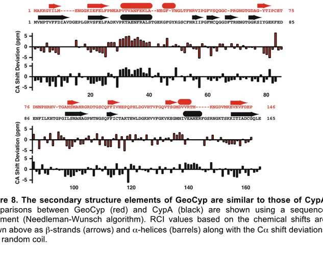
Chemical shift determination and secondary structure prediction of GeoCyp ... 34
Solution structure of GeoCyp and comparison to CypA ... 36
GeoCyp is thermostabilized as compared to CypA ... 38
Differential conformational sampling between GeoCyp and CypA when bound to a peptide substrate ... 38
viii
A gatekeeper residue that regulates substrate binding ... 39
An active site electric field is conserved between CypA and GeoCyp ... 41
GeoCyp and CypA exhibit comparable temperature dependent activities ... 42
Dynamics are similar over multiple timescales between CypA and GeoCyp, with variability in temperature dependence and magnitude ... 43
GeoCyp weakly self associates in the millisecond timescale ... 45
Summary ... 45
III. DETERMINATION OF THE FULL CATALYTIC CYCLE AMONG MULTIPLE CYCLOPHILIN FAMILY MEMBERS ... 63
Introduction ... 63
Results ... 64
Functional characterization of multiple members of the cyclophilin family ... 64
Structural and dynamic comparison of multiple human cyclophilins ... 66
Elucidation of the complete catalytic cycle for multiple cyclophilins utilizing lineshape analysis ... 68
Summary ... 74
IV. LIMITATIONS ON THE APPLICATION OF CPMG-RD IN REVERSIBLE CATALYTIC SYSTEMS ... 82
Introduction ... 82
Results ... 83
Modeling CPMG-RD in reversible systems ... 83
Experimental measurement of non-catalyzed enzyme-perspective cis↔trans interconversion ... 87
Segmental dynamics in CypA in the bound form ... 88
Summary ... 88
V. MAPPING NETWORKS OF NON-COHERENT DYNAMIC ALLOSTERIC COUPLING IN CYCLOPHILIN A ... 97
Introduction ... 97
Identification of a distal residue allosterically connected to active site
dynamics ... 99
Identifying putative communication pathways in chemical shift guided MD ensembles. ... 101
Identification of allosteric pathways via CPMG-RD measurement on multiple single site mutations ... 104
Integration of the influence of multiple active site distal mutations on CypA dynamics ... 105
Active site distal residues influence substrate binding and catalysis ... 108
Lineshape analysis suggests that altered conformational sampling regulates substrate binding ... 110
Summary ... 111
VI. MATERIALS AND METHODS ... 128
Protein expression and purification ... 128
GeoCyp Purification ... 128
CypA Purification ... 128
CypB Purification ... 128
CypC Purification ... 129
Protein Deuteration ... 129
Peptide purification ... 129
NMR assignments ... 130
GeoCyp assignments ... 130
CypC assignments ... 130
Peptide assignments ... 130
NMR solution structure determination ... 131
Circular dichroism monitored thermal denaturation ... 131
NMR titrations for measuring binding affinity ... 132
ZZ-exchange to measure isomerization rate ... 132
x
Molecular dynamics ensembles ... 134
Electric field calculations ... 134
Bacillaceae sequence alignment ... 135
Isothermal titration calorimetry ... 135
Lineshape analysis ... 135
CPMG-RD simulations ... 135
VII. FUTURE DIRECTIONS ... 137
Alternative regulation of CypA dynamics ... 137
Application to other protein systems ... 139
REFERENCES ... 142
LIST OF TABLES TABLE
1. Structural statistics for the GeoCyp RASREC Rosetta structures ... 49
2. GSFGPDLRAGD model peptide chemical shift assignments ... 50
3. Binding affinities and catalytic efficiencies for CypA, GeoCyp, and mutants ... 50
4. Apparent binding affinities and observed isomerization rates for multiple
cyclophilins ... 76
5. Microscopic rate constants determined for CypA using different values of
R2-Bound ... 76
6. Best-fit solutions of cyclophilin microscopic rate constants ... 76
7. Catalyzed and uncatalyzed exchange rates for multiple cyclophilins ... 90
8. Apparent dissociation constants and isomerization rates of cyclophilin mutants
towards the FGP peptide ... 114
9. Apparent dissociation constants as measured by ITC ... 114
10. Apparent dissociation constants and isomerization rates of cyclophilin
mutants towards the HAP2A peptide ... 114
11. Best-fit solutions of microscopic rate constants for CypA and mutants ... 115
xii
LIST OF FIGURES FIGURE
1. Peptidyl prolyl isomerization ... 29
2. Cyclophilin A structure bound to cyclosporine and model peptide ... 29
3. NMR chemical exchange regimes ... 30
4. CPMG-RD refocusing ... 31
5. CPMG-RD profiles ... 31
6. ZZ-exchange example ... 31
7. Classic models of allostery ... 32
8. The secondary structure elements of GeoCyp are similar to those of CypA ... 51
9. The structure of GeoCyp is similar to CypA, with some changes in substrate interface ... 52
10. GeoCyp is stable over a broad range of temperatures ... 53
11. CypA and GeoCyp binding interfaces ... 54
12. Peptide bound MD ensembles of CypA and GeoCyp ... 55
13. Homologous mutations in the context of mutation to the catalytic arginine ... 56
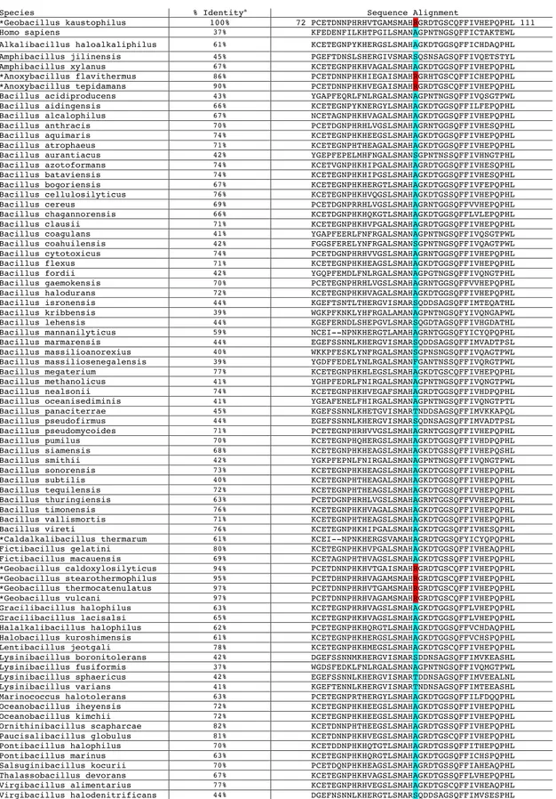
14. Sequence alignment of all annotated cyclophilins from Bacillaceae family members in the RefSeq database ... 57
15. GeoCyp exhibits a -Z electric field within the active site ... 58
16. Catalytic efficiency for CypA and GeoCyp from 0-45°C ... 58
17. For GeoCyp, Rex values, but not R20 or R1 values, exhibit a large concentration dependence ... 59
18. R1 relaxation rates for CypA and GeoCyp ... 60
19. R20 relaxation rates for CypA and GeoCyp ... 61
20. GeoCyp self-associates on the millisecond time scale ... 62
21. Functional characterization of multiple cyclophilins ... 77
22. Highly conserved structure and fast timescale dynamics among CypA, CypB, and CypC, and variable µs timescale exchange ... 78
24. Testing self association in CypB and CypC ... 79
25. Divergent µs-ms motions in CypA and CypC ... 80
26. Four state peptide-perspective minimal exchange model of cyclophilin catalysis ... 80
27. Extraction and fitting lineshape data ... 81
28. Minimal three state enzyme-perspective exchange model ... 91
29. Validation of stochastic CPMG-RD model. ... 91
30. CPMG-RD can report on uncatalyzed exchange in reversible enzymatic systems ... 92
31. Rex as a function of substrate concentration for uncatalyzed peptide interconversion ... 92
32. Cyclophilin peptide saturation ... 93
33. CPMG-RD simulations performed over a range of kex-catalyzed and KD-app values ... 94
34. Comparing catalytic turnover of the model peptide and its thioamide variant by CypA ... 95
35. Measured CPMG-RD data on 2H15N CypA when it is saturated with catalyzable and uncatalyzable substrates ... 96
36. Identification of an active site distal residue coupled to active site dynamics ... 116
37. Active site distal methyl CPMG data alone and with FGP peptide ... 116
38. Identification of putative allosteric pathways between the CypA active site and Val 29 in an MD ensemble ... 117
39. Conformational transitions reported on by Ser 99 and Phe 113 ... 119
40. A second putative allosteric pathway between Val 29 and the active site ... 120
41. Val 29 mutants are well folded and do not alter the global structure of CypA ... 121
42. Experimental identification of allosteric communication pathways in CypA ... 122
43. Additional allosteric coupling stemming from Val 29 ... 123
44. Identification of a second active site distal location and characterization of its communication pathway to the active site ... 124
45. Dynamic response to Val 6 mutants and double mutant ... 125
xiv
47. Peptide on-rates mirror CPMG-RD monitored shifts in dynamics ... 127
48. 15N-HSQC spectra of AAG, AlkA, DMG2, and EndoV ... 141
CHAPTER I
LITERATURE REVIEW AND THE CURRENT STATE OF THE FIELD The cyclophilins
Cyclophilin biology
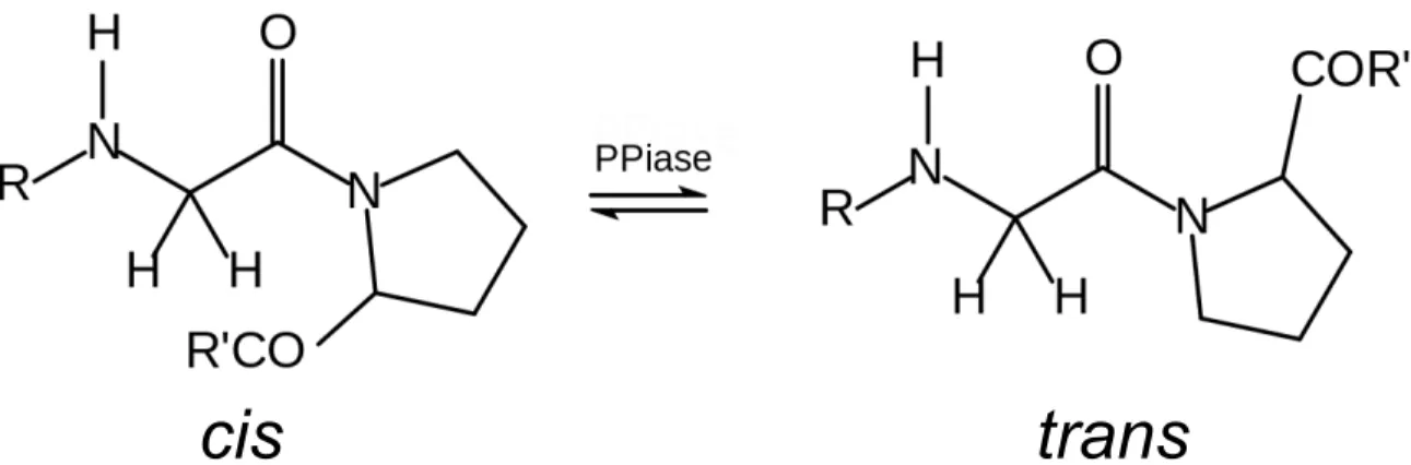
Cyclophilins are a near ubiquitously expressed protein family, found in all domains of life, and only known to be absent in a small number of obligately symbiotic bacteria and archaea.1,2 Among organisms with cyclophilins, they are highly abundant (0.1-0.3% of total protein in human tissues) and are often present as multiple isoforms (e.g., 17 isoforms exist in humans, 8 in S. cerevisiae, and 2 in E. coli).3-5 Most, although not all, cyclophilins catalyze the cis↔trans isomerization of peptidyl-prolyl bonds (see Figure 1), and are therefore also referred to as peptidyl-prolyl isomerases (PPIases). Cyclophilins are one of three major families of PPIases, along with the structurally unrelated FK506 binding proteins and the parvulins.6
The structure of cyclophilins is highly conserved and consists of a flattened eight-sheet anti-parallel β-barrel capped by two 2-3 turn α-helices and a short 3-10 helix adjacent to the active site, which lies parallel to the β-sheet along one side of the barrel (Figure 2). Amino acid sequence is fairly well conserved within the cyclophilin family, with a representative 34-36% homology between the two E. coli cyclophilins and human Cyclophilin A (CypA). Within the protein active site, certain residues are highly or absolutely conserved, including a catalytic arginine, which is absolutely conserved among known catalytically active cyclophilins.7 As some cyclophilins have evolved to fulfill niches that do not require isomerization activity, the catalytic arginine has not been absolutely conserved among these non-catalytic cyclophilins.8
2
The prototypical and first characterized cyclophilin, CypA, encoded by the PPIA gene, was initially identified independently by two groups for its peptidyl prolyl isomerase activity and as being the binding partner of the fungal cyclic undecapeptide and immunosuppressant, cyclosporine A (CsA).9,10 Soon thereafter, it was recognized that these two functions were mediated by the same protein.11 The immunosuppressant activity of CsA, however, has been shown to be independent of the PPIase activity of CypA; upon binding, the CypA-CsA interface binds to and blocks catalytic activity of the phosphatase Calcineurin, which dephosphorylates the nuclear factor for activation of T-cells (NF-AT), a critical step in T-cell activation.12,13 CsA has also been shown to bind to the majority of other human cyclophilins,5 although in mice the immunosuppressant activity of CsA has been shown to be specifically dependent on CypA.14
Cyclophilins are involved in a wide range of biological functions. Perhaps the most well understood role of the proteins is as a chaperone/foldase. While peptide bonds consisting of X-nonPro amino acids, where X is any residue and nonPro is any non-proline residue, overwhelmingly adopt a trans conformation (~99.9% of bonds in both unstructured peptides and in protein structures), X-Pro peptide bonds adopt the cis conformation with significant frequency (~10-30% in unstructured peptides and ~6.5% of bonds in protein structures).15 As a given protein structure will generally only contain a single isomer at each proline position and the un-catalyzed isomerization rate is on the order of 10s-100s of seconds (shown in Chapter 2 to be >>1 s for the model peptide utilized here), the cis↔trans interconversion of prolyl-peptide bonds represents the limiting step in the folding of many proteins, particularly when the correct isomer is required for multiple prolines.16,17 Cyclophilins, along with other peptidyl-prolyl isomerases, act to accelerate these interconversions by a factor of ~105, circumventing this bottleneck to protein folding.1,9,17,18 Cyclophilins have been shown to specifically promote proper folding of several human proteins, including transferrin,19 collagen,20 and the α7 neuronal nicotinic and type 3
serotonin homo-oligomeric receptors.21 Additionally, the conserved hydrophobic active site allows cyclophilins to act as chaperones by shielding partially folded intermediates from misfolding and preventing misfolded proteins from aggregating.18
More recently, multiple additional biological roles of CypA have been identified in humans, independent of its chaperone activity. CypA regulates signal transduction through the tyrosine kinase Itk, with a CypA mediated proline conformational switch inhibiting Itk activity and subsequent downstream signaling events.22 Additional signal transduction pathways are proposed to rely on conformational switching by CypA, including via interactions with Calcineurin23 and NF-κB.24 CypA has also been implicated in mediating intra-cellular trafficking of numerous protein targets, including apoptosis inducing factor,25 heterogeneous nuclear ribonucleoprotein A2,26 and zinc finger protein 1.27
While CypA is predominantly cytoplasmically localized, several other human cyclophilins are localized to specific cellular compartments with specific, although generally less well understood, functions. Human cyclophilins Cyclophilin B (CypB) and Cyclophilin C (CypC) both contain target sequences that localize them to the endoplasmic reticulum (ER), with CypB suppressing ER stress-related apoptosis.28,29 Cyclophilin D (PPIF) interacts with and regulates calcium sensitivity of the mitochondrial permeability transition pore.30 Cyclophilin H (PPIH) and PPI-like protein 1 (PPIL1) have both been shown to stably interact the spliceosome.31-33 Cyclophilin E regulates gene expression through interactions with the mixed lineage leukemia histone methyltransferase.34 The mechanisms by which cyclophilins carry out most of these functions remain poorly characterized, including whether PPIase activity is required.
In addition to the specific cellular localizations of the human cyclophilins, CypA, CypB, and CypC are secreted extracellularly,35-37 acting as cytokines, with a role in driving many cancerous and inflammatory conditions, including those described below.38 CypA’s major known extracellular binding partner is extracellular matrix metalloprotease inducer
4
(EMMPRIN, also known as CD147); blocking EMMPRIN leads to a potent reduction in cell migration in multiple contexts.39,40 The interaction of CypA with EMMPRIN, however, is very weak in vitro, with a dissociation constant of more than 1 mM, suggesting that other binding partners may play a role in mediating CypA extracellular activity.41 CypB has likewise been found to utilize CD147 as a receptor for its extracellular activity.42
Among the many biological roles identified for cyclophilins, their ability to provide tolerance to a range of stresses, including high salinity, oxidative stress, osmotic stress, infection, cold, and heat, has been identified across many species.43-46 Multiple studies have also exogenously expressed cyclophilins from one species into another, often conferring a range of tolerances in the recipient species.47-49 For example, a cyclophilin derived from a salt-tolerant strain of rice, when expressed in E. coli or S. cerevisiae, provides a survival advantage not only under high salt conditions, but also when exposed to osmotic, oxidative, or heat stress.47 The specific mechanism by which cyclophilins provide these various stress appears to be multifaceted. For example, some studies have pointed to targeted functions such as RNA binding activity or hyperpolarization of the mitochondrial membrane as mechanisms by which specific cyclophilins provide survival advantages.46 However, the breadth of protection provided has led most studies to hypothesize that, in general, tolerance is mediated through protein chaperone activity as cyclophilins act to maintain protein homeostasis and promote proper protein folding.6
Given the evolutionary conservation of the family and the range of biological functions of cyclophilins, knockouts of cyclophilins are surprisingly well tolerated in many organisms. In S. cerevisiae, simultaneous knockout of all 8 cyclophilins results in viable yeast, with only mild growth inhibition and slight temperature sensitivity.50 Overexpression of CypA from S. cerevisiae is, however, able to rescue knockout of the one otherwise essential PPIase, the parvulin Ess1, indicating some overlapping roles in the function of these two functionally similar yet structurally distinct proteins.51 Knockout of the most abundant
cyclophilin, CypA, in mice results in somewhat decreased viability, but with largely healthy adults with normal lifespans. PPIA knockout mice do exhibit an allergic disease phenotype, consistent with hyperactivity of Itk without CypA downregulation, and are resistant to immunosuppression by CsA.14,52 In D. melanogaster, knockout of the human CypB homologue, ninaA, prevents proper generation of two isoforms of the rhodopsin G-protein-coupled receptor in the photoreceptor cells, presumably through a chaperone or foldase activity, suggesting that, while cyclophilins can target substrates promiscuously, certain cyclophilins may have highly specialized roles.53 Interestingly, mutations to CypB in humans have been shown not to impact rhodopsin, but to lead to Osteogenesis Imperfecta, a heterogeneous bone disease caused by impaired collagen biosynthesis, indicating that the specific protein targets of cyclophilins are not necessary evolutionarily conserved.
The role of cyclophilins in pathology
In addition to the known biological roles of cyclophilins catalogued above, cyclophilins have been implicated in a wide range of pathological conditions, including roles in promoting viral infectivity, multiple inflammatory diseases, and the progression of multiple cancers.
Human immunodeficiency virus (HIV) specifically incorporates host CypA into its viral coat54,55 via binding to a loop region within p24 capsid protein including the critical proline 90.56 This incorporation has been shown to enhance both viral replication and infectivity via numerous proposed mechanisms,57 including stabilization of the capsid and transport to the nucleus58 as well as in coordinating proper uncoating of the capsid.59 CypA has also been implicated in masking or otherwise protecting the virus from host recognition by pathogen associated molecular pattern receptors.60 Interestingly, cloning of the p24 binding sequence into the simian immunodeficiency virus (SIV), which does not naturally incorporate CypA, does lead to viral incorporation of CypA; the incorporation, however, inhibits viral replication in SIV, indicating that other structural components of the p24 capsid protein, specifically a
6
presence or absence of a type II tight turn, dictate whether CypA enhances or inhibits replication.61 While no relevant polymorphisms within the CypA coding region have been identified in relation to HIV susceptibility or other clinical outcomes, one polymorphism in the regulatory region has been associated with increased susceptibility to HIV,62 suggesting that levels of CypA production may influence HIV infectivity in vivo. Additionally, serum levels of CypA are increased in HIV infected patients.63
Human hepatitis C virus (HCV) likewise utilizes cyclophilins to promote infectivity and replication. While the specific mechanisms are less well understood in HCV as compared to HIV, HCV is though to incorporate CypA via binding to numerous sites within the NS5A protein which promotes formation of a double membrane vesicle that can protect the virus during replication from host detection and restriction.57,64 CypB is likewise thought to play a role in HCV replication via positive regulation of the viral RNA polymerase.65 Numerous other viruses have been shown to utilize various human host cyclophilins in their life cycles, including hepatitis virus B,66 influenza,67 and human papilloma virus.68 Given the role of cyclophilins in promoting the life cycle of these numerous viruses, significant efforts have been made to develop non-immunosuppressant homologues of CsA, including several currently in clinical trials.69,70
CypA has been found to be up-regulated in a wide range of cancers, including lung,71 pancreatic,72 hepatocellular carcinoma,73 colorectal,74 and squamous cell carcinoma.75 In addition to potential application as a biomarker, in many of these cases, the up-regulation appears to be functionally relevant in the cancer development and/or metastasis.76-79 CypA effects cancer promotion through multiple complimentary inter- and extracellular mechanisms, including driving cell proliferation,76,77 blocking apoptosis,80 protection from reactive oxygen species,81 and promoting cell migration and invasion.82 Additionally, CypA has been shown to promote chemotherapeutic resistance via up-regulation of drug metabolism and transport proteins80,83 and has been found to be down-regulated by a
number of treatments.84-86 CypA levels are regulated by both p53 and HIF-1α,80,87 and CypA may engage in a positive feedback mechanism whereby it stabilizes p53.88 Other human cyclophilins are also up-regulated in multiple cancers, including CypB,89 CypD,90,91 and Cyp40,92 where they have been found to block apoptotic cell death, and CypC,37 where it has been found to increase cell migration and invasion.
Cyclophilins have also been implicated in the progression of a wide range of inflammatory diseases. These include cardiovascular disease, where CypA has been shown to promote vascular remodeling, vascular smooth muscle cell proliferation, inflammation, and the recruitment of immune cells.93,94 In rheumatoid arthritis, macrophages release high concentrations of extracellular CypA, resulting in an increased release of inflammatory cytokines and extracellular matrix reconstruction, potentially through interactions with EMMPRIN.95 Similarly, CypA has been associated with increased inflammation and immune cell recruitment in Alzheimer’s disease,96 sepsis,97 asthma,40 and peritonitis.98
Methods to monitor protein dynamics
Proteins exist as a conformationally diverse collection of structures, interconverting, locally and globally, over a large range of timescales, from picoseconds to hours.99-102 The dominant theories of protein binding posit that dynamic mobility is crucial for protein-ligand interactions to occur and, in studies of enzymes, alterations to dynamics have been correlated with changes to both substrate interaction and catalytic turnover.103-108 Recognition of neither the existence nor the importance of dynamics in proteins is new; indeed, since ‘lock-and-key’ models began facing scrutiny over half a century ago, descriptions of protein interactions have evolved toward our current understanding, a broad combination of conformational selection and induced fit models.107,109 Only recently, however, have advances in experimental techniques and instrumentation, particularly in biomolecular nuclear magnetic resonance (NMR), permitted direct observation and
8
quantitation of these dynamic motions over a range of biologically important timescales at atomic resolution.
Biomolecular NMR spectroscopy
NMR spectroscopy relies on the phenomenon by which atomic nuclei with non-zero spin exhibit both a magnetic moment and angular momentum. While the dominant natural hydrogen isotope (1H) does exhibit a non-zero spin, biomolecular NMR experiments often require isotopic labeling of additional atoms within a protein, generally 15N and/or 13C which have spin 1/2, as the most abundant isotope of carbon (12C) has is zero-spin and is therefore insensitive to magnetic manipulation and the most abundant isotope of nitrogen (14N) has a spin of 1, which introduces quadrupole relaxation that is prohibitively rapid for use in biomolecular NMR. Introducing non-zero spin nuclei into a strong static magnetic field B0 (14.1-21.1 T for experiments described herein) leads to a preferential alignment with the B0, providing a net alignment of the bulk magnetization (M) of a sample along the magnetic field (+z). This bulk magnetization can then be manipulated by application of a short radio frequency (RF) pulse, generating a weaker magnetic field perpendicular to B0 (i.e., in x or y), causing rotation of M into the x-y (transverse) plane, where the magnetization then processes about B0 with a characteristic Larmor frequency determined by B0 and the atom type. Additional small changes in the procession frequency of individual atoms are introduced by changes to the local magnetic environment due to their localization in the context of the rest of the protein. The NMR signal is recorded by measuring the current induced by the nuclear procession of all nuclei in a sample after cessation of the RF pulses, a signal known as the free induction decay (FID). To deconvolute the FID, a Fourier transform is applied to the FID to transform it to the frequency domain, yielding peaks with a distinct frequency, also known as chemical shift (ω), associated with each monitored atom in the protein. M can be transferred between different NMR active nuclei within the protein,
allowing collection of correlated chemical shift information among homo- or heteronuclei and yielding multidimensional spectra upon Fourier transformation.
The evolution of M in the absence of an applied RF pulse is described by the Bloch equations, written here in the rotating reference frame110:
Equation 1 �� �� = −�! −Ω 0 Ω −�! 0 0 0 −� ! � + �!�! 0 0 1
where Ω is the frequency offset (i.e., Ω=ω-ωrotating frame) and M0 is the initial magnetization aligned with B0. R1 (or alternatively, its inverse, T1) describes longitudinal or spin-lattice relaxation that reflects the stochastic return of magnetization to the equilibrium state, aligned with the background magnetic field. R2 (or alternatively its inverse T2) describes transverse or spin-spin relaxation and reflects the loss of coherence of magnetization in the transverse plane.
Three fundamental properties of a signal can be monitored by NMR spectroscopy. The chemical shift (ω) is reflective of the local magnetic environment of a given atom within the protein, and may provide information on local secondary structural elements of the protein. The signal intensity (I), given by the peak area, is reflective of the total number of atoms processing at a given frequency. The signal linewidth (λ), given by the peak width at half height which is related to transverse relaxation by R2=λπ (in Hz). By monitoring these signal properties in the context of various NMR experiments, dynamics over wide range of timescale can be monitored.
NMR methodologies to monitor protein dynamics
Fast motions (ps-ns) in proteins are generally described in terms of the rigidity of a given bond vector, which in turn is reflective of the structural constraint on that vector due to the local secondary structure within the protein. Fast timescale motions in proteins can be
10
monitored by measurement of the transverse (R2) and longitudinal (R1) relaxation rates described above, along with the heteronuclear-NOE (hnNOE), a measurement of through space magnetization transfer between two heteronuclei (i.e., transfer from a proton to an attached 15N-labeled nitrogen). R2 and R1 can be directly monitored by observing the decay of magnetization as a function of delay time while magnetization is stored in the transverse plane or longitudinal axis, respectively. hnNOEs are measured on 15N amides by comparing signal intensity with or without proton pre-saturation. R1, R2, and hnNOE each have distinct dependencies on the spectral density function J(ω), which describes the amplitudes of motion experienced at a given frequency. J(ω) is influenced by the global tumbling of a protein, which occurs on the ns timescale, as well as any localized bond motions, which generally occur on the ps timescale. R2 is additionally impacted by slower motions in the µs-ms timescale.
Interpretation of the relaxation measurements can take several forms. The most common is to use the Lipari-Szabo model-free approach,111 which assumes that internal motions are faster than and independent of the global tumbling of the protein, but otherwise assumes no structural information about the motions. The description of motions is defined by a global tumbling time (τm), a site-specific correlation time (τe), a site-specific squared order parameter (S2), and any influence from µs-ms exchange (Rex). Of these parameters, S2 is most informative of fast timescale motions, and is generally interpreted in terms of the degree of restricted motion of the within a cone where a value of 1 is completely rigid and a value of 0 is completely unrestrained. Alternatively, R1, R2, and hnNOE can be interpreted directly across the sequence of a protein as reflective of regional internal motions within the protein.
Descriptions of slower motions (µs-s) are generally described as an exchange between distinct chemical environments (chemical exchange), described by the population
occupancies of the states (PA and PB), the rate of exchange (kex = kAàB + kBàA), and the chemical shift difference between the states (Δω). Depending upon the relationship between kex and Δω, the exchange can fall into fast, intermediate, or slow exchange. In slow exchange, where kex >> Δω, distinct peaks are observable (assuming sufficiently high populations) for each chemical environment; in fast exchange, where kex >> Δω, a single peak is present at the population-averaged chemical shift position; intermediate exchange describes any case in between, kex ≈ Δω, wherein the peaks are linebroadened due to chemical exchange (see Figure 3 for representative spectra across the three regimes).
Slower timescale motions, from ~100-5000 s-1 can be quantitatively monitored by the Carr-Purcell-Meiboom-Gill relaxation dispersion experiment (CPMG-RD), also known as R2 relaxation dispersion.112,113 These motions are on the timescale of sidechain reorientation, loop motions, and other structural rearrangements. The experiment consists of a constant relaxation time (~20-80 ms) during which magnetization is allowed to decay via R2 relaxation. During this relaxation time, 180° refocusing pulses are applied in the transverse plane with varied frequency (νcpmg) from ~50-1000 s-1. Over that relaxation time, signal will decay according to the background R20 relaxation rate, with additional contribution from any chemical exchange on the µs-ms timescale (Rex).
Equation 2
�!= �!"+ �!"
The application of the refocusing pulses leads to the retention of additional signal as a function of the refocusing frequency (see Figure 4), with increasing signal retained at higher refocusing frequencies. The signature of R2as a function of refocusing frequency is described for generalized two state exchange by the Carver-Richards equations114:
12 Equation 3 �!= �!"+ 1 2 �!"− 1 2����ℎ !! � !cosh �! − �!cosh �! �± = 1 2 ±1 + � + 2Δ�! (�!+�!)!.! �±= 2� 2 ±� +(� !+�!)!.! !.! � = (�!�!"− �!�!")!− Δ�!+4�!�!�!"! � = 2Δ��!"(�!− �!)
where R2 is the observed R2relaxation, R20 is the exchange independent component of R2 relaxation, τ is the period of CPMG refocusing (τ = 1/νcpmg), kex is the exchange rate, defined as kex=kAB+kBA, where kAB and kBA are the forward and reverse rate constants, respectively, Δω is the chemical shift difference between the two states, and PA and PB are the populations of the two states. Observed R2 relaxation is given by:
Equation 4 �!=– 1 Τ��� �! �!
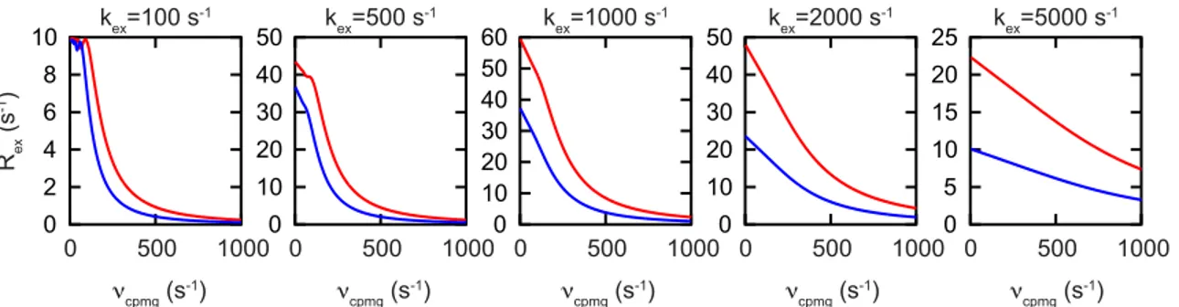
where Τ is the constant time relaxation period, Iτ is the peak intensity at a given refocusing time τ, and I0 is the peak intensity in the absence of the constant time relaxation period. Data are generally collected at two magnetic field strengths, which has been shown to be required to sufficiently constrain fits to the above equations.115 In theory, by fitting the data to the Carver-Richards equations, kinetic (kex), thermodynamic (PA) and structural (Δω) information about the exchange process can be extracted, although this is only possible in the intermediate exchange regime (See Figure 5 for representative CPMG-RD Rex profiles over a range of kex values).
CPMG-RD can be used to monitor exchange processes fully in the slow and fast exchange regimes, although less information can be determined about the exchange. Specifically, in the limit of slow exchange, R2 is described by116:
Equation 5
�!= �!"+ �!"− �!"!"# (!!") ∆!"
which contains information only on the ‘away’ rate (i.e., rate of exchange from AàB) and the chemical shift difference between the states. Alternatively, in the fast limit, R2 is described by117: Equation 6 �! = �!"+ �!�!∆�! �!" 1 − 2tanh (�!"�) 2�!"�
for which the populations and Δω are inherently coupled into a single parameter, precluding independent determination of either of them.
For much slower processes (~0.5-20 s-1), fully in the slow exchange regime, and for which both states can be observed, ZZ-exchange spectroscopy (also known as exchange spectroscopy or EXSY) can be used to measure rates of exchange between states. In ZZ-exchange spectroscopy, the magnetization of each probe is labeled with its chemical shift and stored alone the Z-axis, such that it is subject to R1, and not R2 relaxation. As R1 is much smaller than R2 for proteins, this allows for monitoring events occurring over timescales as long as multiple seconds. After a mixing time of varied length, the magnetization is transferred back to the transverse plane and recorded. Any atoms that have switched state during the mixing time will register as ‘cross-peaks’ as shown in Figure 6. The dependence of each of the four peaks is given by118:
14 Equation 7 �!! � = �!! 0 (− �!− �!! �!!!!+ �!− �!! �!!!!)/(�!− �!) �!! � = �!! 0 (− �!− �!! �!!!!+ �!− �!! �!!!!)/(�!− �!) �!"(�) = �!(0)(�!"�!!!!− �!"�!!!!)/(�!− �!) �!"(�) = �!(0)(�!"�!!!!− �!"�!!!!)/(�!− �!) � !,! = 1/2 (�!!+ �!! ± (�!!− �!!)!+4�!"�!" !.!] � = �!+ �!" −�!" −�!" �!+ �!"
where IAA and IBB are the peak intensities of peaks A and B, IAB and IBA are exchange peak intensities, T is the mixing time, R is the longitudinal relaxation rate (R1), kAB and kBA are the AàB and BàA exchange rates where kBA = (PA/PB)kAB and PA and PB are the populations at equilibrium. By recording spectra while varying T over ~0.1-2 seconds, the exchange rate, kex= kAB + kBA, and chemical species-specific R1 relaxation parameters can be determined.
The role of conformational exchange in regulating protein function The role of dynamics in binding
The first qualitative descriptions of protein-substrate interactions followed the “lock-and-key” model by which a given protein is pre-formed to interact precisely with its partner.119 This model has been largely supplanted by two competing, if not directly contradictory, models of protein-substrate interactions, induced fit and conformational selection. The induced fit model posits that given binding partner will interact weakly with a protein, and in doing so, introduce conformational changes in the protein such that it then binds more tightly.120 Alternatively, the conformational selection model holds that a given protein is inherently sampling an ensemble of structures, dubbed the conformational landscape, with varying degrees of affinity for a given substrate, and that the binding event to a high affinity sub-population then shifts the overall population toward that high-affinity conformation.109,121 The two descriptions are analogous to the traditional competing models
of allostery, the Koshland-Némethy-Filmer (KNF) and the Monod–Wyman–Changeux (MWC) models, respectively, discussed further below and illustrated in Figure 7.
A particularly compelling landmark set of experiments in favor of conformational selection utilized low temperature (<200 K) flash photolysis to inhibit transitions between conformational states in myoglobin and demonstrated a variable affinity for O2 among the various substates.122,123 Additionally, the advent of high-resolution NMR and atomic resolution molecular dynamics simulations has demonstrated the sampling of high energy, bound-like states in native proteins, supporting the notion of conformational selection.124-126
The two models of binding are not mutually exclusive, however, and instead represent extremes on a continuum of mechanisms by which binding may occur. A more generalized and inclusive model, termed the ‘extended conformational selection’ model, suggests that conformational heterogeneity does exist within most free proteins, consistent with the conformational selection model, but that during the binding event, further conformational adjustment often occurs in the protein and/or binding partner, consistent with an induced fit.107,127,128 Analytical methods have been developed to theoretically distinguish the extent to which conformational selection or induced fit act in a given system, however, experimental limitations often limit the applicability of these approaches.129,130
The role of dynamics in enzymatic catalysis
As described above, the role of inherent conformational fluctuations in substrate binding, via conformational selection, is well established. Likewise, numerous studies have established the role of inherent conformational fluctuations in the ligand-bound protein in regulating substrate release, as various substates of the bound complex also exhibit varied affinity for the substrate. For example, in dihydrofolate reductase (DHFR), a loop motion occurs in the ligand-bound form which confines dihydrofolate in the major form, trapping the substrate sufficiently long to permit catalysis, yet the loop transiently samples a low-population higher energy open form that permits substrate release.131 In this respect, the
16
role of inherent conformational fluctuations in regulating the complete catalytic cycle has been demonstrated.
Significant controversy, however, surrounds the idea that the inherent dynamics of a system influence the rate of catalysis subsequent to substrate binding. Broadly, enzymatic catalysis is thought to occur via an enzyme’s high affinity for the transition state of a reaction, stabilizing the transition state, and thereby lowering the activation energy required to transverse to product.132 The potential effect of conformational fluctuations in reaching the stabilized transition state can be dissected into two distinct impacts on catalysis: the conformational search of the enzyme-substrate system in reaching the optimal configuration to stabilize the transition state and a direct influence of dynamical motions on the chemical step of catalysis.
While the debate over the relevance of dynamics in catalysis has been waged by many parties, the controversy has been most notably argued between recent co-recipients of the Nobel Prize, Drs. Karplus and Warshel.133,134 Warshel has strongly argued against a role for conformational dynamics in influencing enzymatic catalysis, while Karplus has advocated its role. With regard to a direct influence on the chemical step, Warshel argues that the inherent friction present in a protein system prevents any ‘memory’ in the progression of the substrate over the activation energy barrier, such that any dynamic motions present prior to reaching the chemical step of catalysis are irrelevant in passing over the chemical barrier. He additionally argues that, in order to be considered as a dynamical influence on catalysis, motions must exhibit behavior deviating from a Boltzmann distribution, as otherwise any observed motions are simply a reflection of the energy landscape. Karplus largely agrees with the former point, indicating that energy dissipation is generally sufficiently fast as to prevent non-equilibrium behavior in a bound enzyme-substrate system. Oh the later point, however, Karplus argues that inherent conformational fluctuations in the system permit lowering of the quasithermodynamic free energy of
activation, providing evidence in the form of simulations in which a flexible enzyme proceeds through a lower activation barrier than a rigid one.135 The essence of the argument on the latter point may be largely semantic, as the motions of an enzyme necessarily follow Boltzmann behavior, and are dictated by (and reflective of) the energy landscape; nonetheless, understanding the particular motions present in an enzyme:substrate complex and the pathway by which the system reaches the activation barrier is important to understanding the catalytic mechanism of a given system.
The role of dynamics in mediating allosteric interactions
While Pauling initially introduced the idea of intramolecular protein regulation to control oxygen binding in hemoglobin in 1935,136 the term allostery was not applied to the phenomenon until 1961 by Monod and Jacob.137 Subsequent to the determination of the structure of the prototypical allosteric protein, hemoglobin, in 1960,138 the long-range nature of the allosteric information transfer became apparent, as well as that the phenomenon was applicable not only to hemoglobin, but also to many additional proteins. Two phenomenological models arose to explain the nature of allostery (depicted in Figure 7). Both models concern symmetric connected ‘subunits’ that transition between distinct ‘tense’ and ‘relaxed’ states. The first, the ‘sequential’ or KNF model, which is similar to the model
originally proposed by Pauling, posits that each subunit can individually transition from the tense state to the relaxed state upon ligand binding, and that the likelihood of ligand binding is influenced by the state of other subunits.120 The second, ‘symmetric’ or MWC model, assumes global transitions, such that all subunits adopt the tense or relaxed states simultaneously, with ligand affinity increased at each subunit in the relaxed state and the relaxed state stabilized by ligand binding.139 The KNF and MWC models essentially represent two-state versions of the induced fit and conformational selection models of binding discussed above. While the original formulations of these models concerned positive cooperativity in symmetric homooligomeric subunits, they have been expanded to
18
accommodate allosteric interactions within a single domain, and to incorporate negative cooperativity.140-142
Inherent in the MWC model is the concept that proteins are constantly sampling a higher energy state, even in the absence of ligand.140,142 This concept has been expanded upon to encompass not only two-state transitions, but also systems for which a broad ensemble of structures is being sampled. This expanded view of allostery, referred to as ‘dynamic allostery,’ the ‘population-shift model,’ or the ‘ensemble nature of allostery,’ center on the view that all proteins are dynamically transitioning to some extent between multiple states, and that any influence (e.g., binding event, post-translational modification, mutation) that preferentially stabilizes certain sub-populations within the ensemble will shift the population distribution with subsequent impact on protein function.143-145 In this regard, it has been suggested that, with perhaps the exception of fibrous proteins, all proteins are allosteric, with the predominant variability being in the degree to which known influences impact population sampling and the degree to which population shifts influence function.146 This model incorporates an important notion absent in the two-state model, namely that allosteric effects may be mediated absent significant changes to the average or ground state structure of a given protein. The homodimeric transcription factor CAP, which exhibits negative cooperativity between subunits for binding cyclic AMP (cAMP), presents an illustrative example of this effect. Binding of a single cAMP induces increased conformational flexibility in the other subunit without altering the ground-state structure, increasing the entropic penalty for a second cAMP binding, and thereby reducing affinity.147 Many other allosteric systems have been probed via CPMG-RD and networks of coherent or non-coherent residues have been identified that propagate dynamic allosteric information independent of ground state structural changes.148-151 Unlike the more rigid two-state model, this expanded, ensemble-perspective model also accommodates intrinsically disordered
proteins, which inherently sample an extremely broad conformational landscape that can nonetheless be allosterically modulated to influence subsequent binding events.152,153
Dynamic relationships between protein segments
As described above, NMR provides a powerful approach to quantitatively measure motion with residue specific probes in proteins, particularly motions in the µs-ms timescale near many biologically relevant events via CPMG-RD. While the particular dispersion curves are readily obtainable for many systems, the mechanistic interpretation of the data is often challenging. Of particular interest is the establishment of the relationship between various sites for which dispersion is detected. Broadly, three ‘dynamic relationships’ may exist between any two probe sites.101 Two sites may be directly reporting on the same physical transition, for which we would expect identical exchange rates (kex) and population occupancies, as well as identical reactions to changing conditions (e.g., temperature, buffer, mutation). Alternatively, two probe sites may report on entirely independent physical phenomena, for which we may see similar or different kex and population values, and similar or different responses to changing conditions (i.e., similar rates, populations, and responses to changing conditions are suggestive of reporting on the same physical phenomenon, but not definitive). Finally, two probe sites may report on distinct, yet coupled physical motions, for which the sites may report similar or different kex and population values, but will experience correlated responses to altered conditions.
Systems for which largely coherent motions dominate have been most extensively studied in terms of relating protein dynamics and function. While experimental and technical improvements have opened the application of relaxation dispersion experiments to a wider range of applications, extensive studies of the role of µs-ms conformational dynamics have revolved around a thorough understanding of a small number of model systems that are readily amenable to study by NMR spectroscopic methods.
20
Kay and co-workers have been the dominant force in developing novel relaxation dispersion methods, including new labeling schemes and pulse sequences to access an increasing number of probe sites.154-158 In particular, the lab has utilized these techniques to access structural information about low-population states that are otherwise invisible to other structural methods.159-162 The most notable success of this approach was the determination of an atomic resolution model of a transient folding intermediate of a small FF domain with a millisecond lifetime and a population of 2-3%.163 To achieve this, relaxation dispersion experiments were carried out on five backbone probe sites (Cα, CO, Hα, HN, and NH); the data were fit to a single two-site exchange model, from which the low-population chemical shifts could be determined for each of the probe sites experiencing exchange. Using previously established empirical relationships between chemical shift values and secondary structure, an atomic resolution model of the low-population state could be reconstructed. This success highlights the power of relaxation dispersion approach, but likewise exposes limitations in that achieving an atomic resolution model was dependent on the global two-state nature of the conformational change and its application to a small (71 residue) protein.
Global two-state exchange does, however, appear to play a role in the catalytic cycles of many biologically relevant and NMR accessible systems. For instance, Loria and co-workers identified concerted conformational motions in an active site loop in ribonuclease A representing transitions between an open and closed state of the substrate binding pocket.164 Notably, the transition between the open and closed sates exists in both the substrate-free and a substrate-mimic-bound form, with the substrate-mimic simply shifting the major state to the closed rather than open form, suggesting a role for the motion in substrate release. The rate constant of the coherent exchange remains similar in each case, and is likewise similar to the rate of substrate release, koff, and to kcat; combined, these data indicate that the transition to the open state is the limiting step in catalytic turnover in
ribonuclease A, such that the inherent conformational motions are directly responsible for regulation of the catalytic cycle.
Relaxation dispersion experiments have also been applied to systems with a surprisingly complex set of conformational rearrangements over a multi-step cycle. DHFR undergoes a five step catalytic cycle involving separate, sequential binding steps for the dihydrofolate substrate and NADPH co-factor, the catalytic step of proton transfer, and sequential release of the product and oxidized cofoactor.131,165 Wright and co-workers have analyzed the conformational fluctuations in DHFR for each of the five steps in the cycle by utilizing a non-catalyzable mimic, allowing data collection on each of the five ground-state structures in the cycle. For each state of the cycle, they discovered that the enzyme samples a high-energy, low-population minor sate; analysis of the chemical shift values of these minor states demonstrated that, in each case, the minor state sampled is similar to that of the next structure in the cycle. This study demonstrates the importance of conformational selection in both substrate binding and release, and also highlights the ability of ligand binding to impact both the ground state structure and the low-population minor state sampled. While the exchange rate and population occupancies of the minor states vary between ground state structures, in each case, the exchange parameters were sufficiently similar to fit to a global two-state exchange model. Wright and co-workers further identified a mutation in DHFR that abolishes detectable millisecond timescale dynamics within the active site independent of changes to the ground-state structure of the enzyme and observed a dramatic drop in kcat.108 These results suggest that dynamics permit the conformational search for the optimized transition-state necessary for efficient catalysis in addition to regulating substrate binding and release.
In addition to the globally coherent systems described above, a smaller number of studies have investigated systems with more localized conformational sampling. Kay and co-workers investigated µs-ms motions in the enzyme complex of NAD(P)H:flavin
22
oxidoreductase bound to flavin adenine dinucleotide and identified three distinct regions of localized motion.166 One of the observed groups of residues appears to result from a localized unfolding event of one α-helix and is seemingly unconnected to the other groups. The other two groups, however, include residues involved in binding the proton acceptor (FAD) and the proton donor (Tyr 35), respectively, and are therefore in close physical proximity. No attempt was made to ascertain the degree to which motions in the second two groups are linked (i.e., if any allosteric interactions exist), so these two groups may exhibit fully independent localized motions or coupled, yet district motions.
McDonald et al.150 investigated conformational motions in the bacterial signaling protein CheY, which is known to exhibit allosteric regulation in which phosphorylation switches the protein to a predominantly active conformation. Utilizing CPMG-RD, they discovered that this conformational switch does not proceed through a coherent two state conformational switch. Rather, it appears that multiple residues or small groups of residues undergo localized two-state switching, yet are nonetheless coupled in that alterations to dynamics induced by Mg2+ binding are propagated throughout the conformational flexible regions of the protein.
Kleckner et al.151 likewise investigated dynamic allostery in the trp-RNA binding attenuation protein via methyl 13C-CPMG-RD and identified a non-coherent network of coupled residues. The motions of these residues are quenched upon Trp binding and are proposed to coalesce, promoting subsequent binding to trp-RNA. This model represents a hybrid of localized MWC and KNF models in which individual segments each sample ‘active’ conformations, but the lack of coherence renders the entire binding site unlikely to simultaneously adopt a binding competent state. Trp binding, however, induces coherence among the segments, permitting the adoption of a fully binding competent site. A similar model is proposed by Xiao et al.167 for extracellular signal-regulated kinase 2 (ERK2). In the absence of phosphorylation, ERK2 exhibits largely non-coherent methyl dynamics; upon
phosphorylation, however, these motions appear to become largely coherent, such that a single global exchange phenomenon can explain the bulk of the observed dynamics.
These studies serve to highlight the diversity of mechanisms by which nature utilizes conformational flexibility in protein systems and the difficulty in formulating a coherent theory of the precise role of dynamics in protein systems.
Studies of protein dynamics among homologues
Given the appreciation for the role of conformational flexibility in regulating protein motions, many researchers have sought to determine the degree to which protein dynamics are conserved within a protein family, as structure tends to be.
Wolf-Watz et al.168 examined µs-ms dynamics in a mesophilic and thermophilic adenylate kinase (Adk) and identified a conserved regulatory mechanism by which a concerted lid opening is the rate-limiting step in catalytic turnover. This mechanism has been evolutionarily tuned and dramatically slows kinase activity in the thermophile at low temperatures, yet permits activity at high temperatures as the rate of lid opening increases. Further studies by the group have suggested that Adk provides a framework for continuous evolutionary tuning of loop dynamics to accommodate the specific needs of a given organism.169 Whittier et al.170 suggest a comparable mechanism among protein tyrosine phosphatases (PTPs), which exhibit a loop closing mechanism that has been evolutionarily tuned to accommodate the particular environment of its host.
Alternatively, Bhabha et al.171 found that, despite the highly conserved structure of DHFR between humans and E. coli, the inherent dynamics of the enzyme have been shifted dramatically in both nature and timescale, effecting adoption of a different catalytic cycle (i.e., ordering of folate and NADP(H) binding and release) in the two organisms. These changes are suggested to result from evolutionary adjustment to the levels of THF and NADP+ in human vs. E. coli cells, but point to a fully altered functional mechanism as opposed to the continuous tuning of motions observed in Adk and PTPs.
24
Studies of dynamics in human Cyclophilin A Experimental studies of dynamics in human Cyclophilin A
Along with systems described above, CypA has been an instrumental model system in understanding the role of inherent conformational fluctuations in regulating protein dynamics. The original studies originating from the Kern group (Eisenmesser et. al (2002)172 and Eisenmesser et. al (2005)106) examined conformational dynamics in CypA alone and in the presence of high concentrations of a short tetra-peptide Suc-Ala-Phe-Pro-Phe-4-NA via CPMG-RD. The studies assumed that at sufficiently high peptide concentrations, dynamics measured on near-saturated CypA would be reporting solely on cis↔trans isomerization, with no effect from substrate binding and release. As demonstrated in Chapter 4 of this manuscript, the assumptions made in utilizing this weakly binding peptide are incorrect, and reported dynamics in the presence of the weakly binding tetra-peptide cannot be representative of on-enzyme isomerization. However, these studies did identify significant µs-ms conformational flexibility in CypA both alone and in the presence of substrate, in both cases localized predominantly to the active site and surrounding residues. This finding suggests that these motions are somehow linked to protein function. In the free protein, these studies identified two distinct regions of coherent motions, while identifying a globally coherent exchange in the bound state (although one that cannot be indicative of isomerization, as noted). Further, comparison of CPMG-RD profiles in multiple active site mutants suggested a ‘common dynamic network’ in which populations of all mobile resides in the protein are shifted proportionally in response to perturbation.
More recently, Schlegel et al.173 employed deuterated CypA, which improves signal quality by reducing the background R20 relaxation rate,174 and data collection at multiple magnetic field strengths, which improves the accuracy of parameters derived from fitting the data,175 to measure µs-ms motions in free CypA over a range of temperatures from 0-25°C.
Because of the improvement in data collection, Rex was observed for nearly 50% of the residues in the protein, with nearly the entire active site exhibiting measureable exchange. Within the active site, they identified significant variability in the temperature dependency of different residues, indicating that the motions within CypA do not comprise two coherent regional motions as previously suggested. Rather, by individually fitting the residues within the active site to the Carver-Richards equations to extract exchange rates, they suggest that conformational fluctuations consist of a ‘dynamic continuum’ for which non-coherent rates of motion exist spanning nearly an order of magnitude across the active site. Furthermore, by generating single and double active site mutants of CypA, they demonstrate regional, not global, responses to mutational perturbation, indicating the existence of localized dynamic networks rather than the ‘common dynamic network’ previously suggested.
Expanding upon those studies utilizing the weakly binding tetra-peptide described above, Bosco et al.176 examined conformational dynamics of CypA in complex with the N-terminus of the HIV Capsid protein (CAN). As described in Chapter 4, CypA’s binding affinity for CAN is sufficiently tight that measurement of CPMG-RD on the complex can potentially monitor isomerization, although all measured motions in the complex are not a priori indicative of isomerization. In the context of the complex, both CypA and CAN exhibit similar rates of motion, and rates that differ significantly from those of either protein alone, supporting the notion that the measured dynamics are reporting on isomerization. They further analyze the impact of a number of active site mutants in terms of their ability to catalyze isomerization of the tetra-peptide and CAN. Interestingly, several of the mutations decrease isomerization of the peptide by ~1000 fold, while only minimally impacting CAN isomerization. This dichotomy is a result of the rate-limiting step for CypA’s interaction with either substrate. With respect to the peptide, on-enzyme isomerization is the rate limiting step for CypAWT and all mutants; however, with respect to CAN, substrate release from CypAWT is nearly two orders of magnitude slower than on-enzyme isomerization. Analysis