ORIGINAL ARTICLE
Second premolar agenesis is associated with
mandibular form: a geometric morphometric analysis
of mandibular cross-sections
Michael H Bertl
1, Kristina Bertl
2,3, Manuel Wagner
3, André Gahleitner
3,4, Andreas Stavropoulos
2,
Christian Ulm
3and Philipp Mitteroecker
5The aim of this study was to compare mandibular form (i.e., size and shape) between patients with agenesis of the lower second premolar (P2) and a control group with no agenesis. Three hypotheses were tested: (H1) agenesis causes a change in
mandibular morphology because of inadequate alveolar ridge development in the area of the missing tooth (mandibular plasticity); (H2) agenesis is caused by spatial limitations within the mandible (dental plasticity); and (H3) common genetic/ epigenetic factors cause agenesis and affect mandibular form (pleiotropy). A geometric morphometric analysis was applied to cross-sectional images of computed tomography (CT) scans of three matched groups (n = 50 each): (1) regularly erupted P2; (2) agenesis of P2 and the primary second molarin situ; and (3) agenesis of P2 and the primary second molar missing for 43 months. Cross-sections of the three areas of interest (first premolar, P2, first molar) were digitized with 23 landmarks and superimposed by a generalized Procrustes analysis. On average, the mandibular cross-sections were narrower and shorter in patients with P2 agenesis compared with that in the control group. Both agenesis groups featured a pronounced submandibular fossa. These differences extended at least one tooth beyond the agenesis-affected region. Taken together with the large interindividual variation that resulted in massively overlapping group distributions, thesefindings support genetic and/or epigenetic pleiotropy (H3) as the most likely origin of the observed covariation between mandibular form and odontogenesis. Clinically, reduced dimensions and greater variability of mandibular form, as well as a pronounced submandibular fossa, should be expected during the treatment planning of patients with P2 agenesis.
International Journal of Oral Science advance online publication, 18 November 2016; doi:10.1038/ijos.2016.41 Keywords: agenesis; cross-sections; geometric morphometrics; mandibular form
INTRODUCTION
Lower second premolars (P2) are the most common congenitally missing teeth—not considering third molars—with a prevalence of 3%.1–5 Although the primary second molars may be kept in some cases,6–7 more often, other treatment options are required for the replacement of the missing second premolar.8–10Treatment options include autotransplantation,11–12 comprehensive orthodontic treat-ment with either space closure13or implant site development14and/
or an implant-supported restoration.15–16 In such cases, there are specific spatial requirements for the size and cross-sectional shape of the mandibular site to ensure full bony coverage of the implant or transplanted tooth root surface.
The form of the mandible is determined by continuous bone growth and remodelling throughout ontogeny and adulthood, influ-enced both by genetic and epigenetic factors, such as muscle activity and biomechanical forces during mastication.17–20After the cessation
of craniofacial growth, the alveolar ridge remains heavily influenced by the presence and position of teeth; it undergoes significant remodelling after tooth loss.21Tooth agenesis is likely to affect mandibular shape, at least locally. Conversely, dental development, starting from the formation of the tooth bud, takes place within the growing mandible. Mandibular dimensions may therefore have the potential to affect tooth development, eventually leading to tooth agenesis if certain key dimensions in the mandible are below a “minimum threshold”. Indeed, it has been shown that agenesis patients differ from normal individuals in various aspects of facial morphology22 and that the
cross-sectional dimension of the mandible covary with other facial dimensions.23–24This association of cranial and mandibular morphol-ogy with dental agenesis may also result from variation in pleiotropic genes, that is, genes that are involved both in craniofacial development and odontogenesis. Numerous syndromes and genes related to tooth agenesis have been identified,25 many of which also influence other
1Division of Orthodontics, School of Dentistry, Medical University of Vienna, Vienna, Austria;2Department of Periodontology, Faculty of Odontology, University of Malmö, Malmö,
Sweden;3Division of Oral Surgery, School of Dentistry, Medical University of Vienna, Vienna, Austria;4Department of Diagnostic Radiology, Division of Osteoradiology, General
Hospital, Medical University of Vienna, Vienna, Austria and5Department of Theoretical Biology, University of Vienna, Vienna, Austria
Correspondence: Dr MH Bertl, Division of Orthodontics, School of Dentistry, Sensengasse 2a, Vienna 1090, Austria E-mail: michael.bertl@meduniwien.ac.at
craniodental traits, such as maxillary retrognathia22,26–27and palatally displaced or transposed canines.9,28
Therefore, three hypotheses can be formulated about the mechan-isms linking dental agenesis to mandibular morphology:
(H1) Tooth agenesis may affect mandibular skeletal development owing to the lack of tissue growth induction (i.e., mandibular plasticity). In this case, it would be expected that differences in mandibular morphology between agenesis and non-agenesis patients would be primarily located in the vicinity of the missing tooth and other regions would remain largely unaffected.
(H2) If a minimum space within the mandible is required for successful tooth development, limited mandibular dimensions may lead to agenesis simply because of spatial limitations for the emerging tooth bud (i.e., dental plasticity). In this case, it would be expected that global mandibular morphology in agenesis patients would be con-siderably different compared with that in non-agenesis patients. Additionally, a minimum spatial requirement for odontogenesis should produce well-separated distributions of mandibular form between those groups.
(H3) Genetic and/or epigenetic factors affect both the mandibular form and the probability of tooth agenesis (i.e., genetic and/or epigenetic pleiotropy). Similarly to the previous hypothesis, this would also be supported by an association of tooth agenesis and global mandibular morphology. However, because of the highly polygenic basis of mandibular form,19–20,29–30 the distributions of mandibular shape for agenesis and non-agenesis patients are likely to overlap massively.
We tested these hypotheses by comparing mandibular form (i.e., size and shape) between patients with agenesis of the lower P2 and a control group with no agenesis. To this end, we applied geometric morphometric methods to cross-sectional outlines of the mandibular body in the vicinity of P2. Geometric morphometrics, which is based on landmark coordinates instead of distance measure-ments, allows for the analysis and visualization complex shape differences and variance patterns.31–33
MATERIALS AND METHODS Data collection
Three groups of 50 patients each, 418 years of age, with regularly erupted mandibularfirst premolars and first molars, were formed by screening computed tomography (CT) records of the University Clinic
of Dentistry (Medical University of Vienna, Vienna, Austria) between 2004 and 2014: group 1 (“control”) consisted of patients with the lower P2 regularly erupted andin situ; group 2 (“agenesis”) consisting of patients with the agenesis of at least one lower P2 with the primary second molarin situ (if agenesis of the lower P2 was bilateral, the test side was chosen by coin toss); group 3 (“agenesis post-ex”) consisting of patients with agenesis of at least one lower P2, where the primary second molar had been extracted at least 3 months before the CT scan. Groups 1 and 3 were matched to group 2 regarding sex, age (±3 years) and mandibular side. Patients who had previously received augmenta-tion procedures in this area were excluded.
The study protocol was approved by the ethics committee of the Medical University of Vienna (EK-No. 2005/2012).
Dental CT scans and image preparation
Dental CT scans had been recorded with a standard dental CT investigation protocol34 with two different devices (Tomoscan
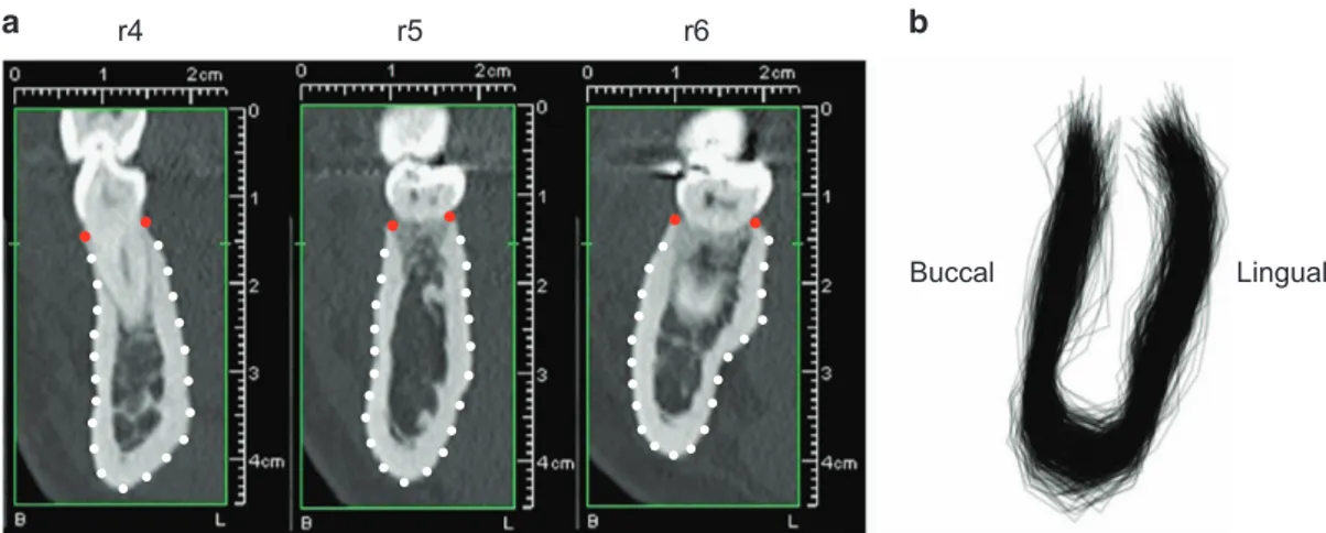
SR-6000 (Philips Medical Systems, Eindhoven, The Netherlands) with 1 mm slice thickness, 1 mm table feed, 120 kV, 75 mA, 2 s scan time, 100–120 mm field of view, high-resolution bone filter; Somatom Sensation 4 (Siemens, Forchheim, Germany) with 0.5 mm slice thickness, 1 mm table feed, 120 kV, 80 mA, 1 s scan time, 100–120 mm field of view, high-resolution bone filter). Axial slices were used to build an orthoradial multiplanar reconstruction, which was calculated perpendicular to a manually drawn central line of the mandibular arch and to the mandibular plane. At the side of the mandible included in the analysis, three regions of interest were identified: the area of the first premolar (r4); the area of P2, the second primary molar, or the gap between thefirst premolar and first molar (r5); the area of the first molar (r6) (Figure 1a). For each region, a cross-sectional image was selected at either the mesiodistal midpoint of the tooth crown or at the centre of the gap between thefirst premolar and first molar.
All images were then arranged with the buccal side to the left and the lingual side to the right and presented in a random order to a single investigator (MW). The examiner scaled the images and identified and digitally placed the following landmarks using tpsUtil (version 1.68)35and tpsDIG2 (version 2.22)36software solutions. Two landmarks were placed on the buccal and lingual alveolar crest, and 21 landmarks were placed approximately equidistantly along the buccal and lingual contour of the mandible (Figure 1a).
r4 r5 r6
Buccal Lingual
Figure 1 Landmark scheme for the mandibular cross-sections. (a) Cross-sectional computed tomography (CT) reconstructions at the three regions of interest:
first premolar (r4), second premolar (r5) and first molar (r6) with fixed landmarks (red) and semilandmarks (white). (b) Reconstructed mandibular outlines of
Morphometric analysis
The form of each mandibular cross-section was represented by the 23 landmarks, of which the two landmarks representing the buccal and lingual alveolar crest were treated asfixed anatomical landmarks and the remaining 21 as semilandmarks along the mandibular contour. After the initial, approximately equidistant placement, their exact positions were estimated by the sliding landmark algorithm to minimize the root summed squared distances (Procrustes distance) among all superimposed cross-sections and their sample average.37–38 The resulting 450 configurations of 23 landmarks were superimposed by a Generalized Procrustes Analysis33,39 (Figure 1b). This least
squares-based superimposition standardizes the landmark configura-tions for overall position, scale and orientation, yielding a set of shape coordinates for each cross-section. For the present study, not only the shape of the cross-sections but also their size was of interest. Thus, for certain analyses, the shape coordinates were rescaled by the config-uration’s centroid size to perform the analyses of the full form information (i.e., size and shape).40–41 Centroid size is a standard measure of the overall size of a landmark configuration; it equals the square root of the summed squared distances of the landmarks from their centroid (average landmark position).
Group-wise means and variances of the cross-sectional area (based on the polygon defined by the measured landmarks) and centroid size, as well as the total variance of cross-sectional shape were calculated. The group mean forms, together with extrapolated versions, were represented as reconstructed cross-sectional forms. The differences between the group mean forms were analysed by a principal component analysis (PCA) of the average rescaled shape coordinates. The statistical significance of group mean differences was estimated by permutation tests,42based on the Euclidean distance between-group
mean forms as the test statistic and 10 000 random permutations. To represent the individual variation around these means, the individual configurations were projected onto the two principal components (PCs) of the mean forms (between-group PCA).43
In addition, scores (orthogonal projections) for each individual along the mean difference vectors were computed. These scores represent the individual expression of the form features that differ most between the groups. Histograms of these scores allowed for the assessment of separation or overlap among groups.
RESULTS
Each group comprised 19 male and 31 female patients. In 24 of these patients, the left side of the mandible was measured, and in 26, the right side. The mean ages of the control, agenesis and agenesis post-ex groups were 22.9± 5.5, 22.8 ± 5.6 and 25.0 ± 8.1 years, respectively.
The cross-sectional area was significantly smaller in the two agenesis groups compared with that in the control group (Po0.005 for both r4 and r5;P = 0.02 for r6; Table 1). Similarly, centroid size, as a measure of overall size, was smaller in both agenesis groups compared with the
control group for the three regions of interest (Figure 2). In contrast, the variance of both the centroid size and shape of the mandibular cross-section was smallest in the control group (Figure 2). Differences in the variance of the cross-sectional area were not as pronounced as those for centroid size and shape. However, the corresponding coefficients of variation for cross-sectional area (standard deviation divided by the mean as a size correction) were clearly the smallest in the control group.
Apart from these size differences, the mandibular cross-sections also differed in shape between the agenesis groups and the control group. Although the average differences were subtle, they were consistent across both agenesis groups and across the three regions of interest (P = 0.004 for r4 and P = 0.02 for both r5 and r6). Figures 3 and 4 show the group mean forms along with extrapolations of their mutual differences. On average, the three regions of interest were narrower and shorter in the agenesis groups compared with those in the control group. Furthermore, in patients with dental agenesis the mandibular cross-sections featured a pronounced lingual alveolar plate and a distinct concavity (submandibular fossa) underneath the mylohyoid line. The extent of this concavity increased from the r4 to the r6 region.
The consistent pattern of form differences is also shown by the PCA in Figure 5. Whereas PC 1 mainly contrasts the three analysed dental regions, PC 2 similarly distinguishes the control group from the agenesis patients for all three regions of interest. Extraction of the primary molar at the r5 region in the post-ex group induced more pronounced shape differences compared with the other groups; thus, the corresponding mean form deviated from the other agenesis groups in the PC plot.
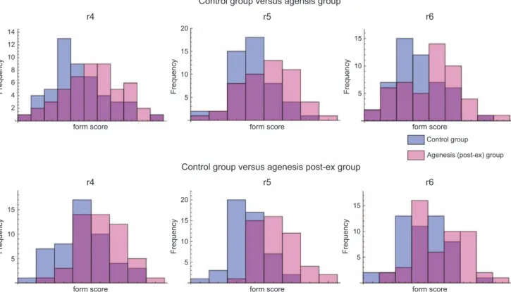
Despite the clear pattern of average form differences, individual variation around these means was substantial, leading to a massive overlap of group distributions. To display this group overlap, Figure 6 presents histograms of individual form scores that maximize the average difference between the control and agenesis groups. Despite this maximization of group differences, the form scores show strong overlap for all group comparisons. Similarly, the between-group PCA of all individuals in Supplementary Figure S1 shows a strong multi-variate overlap among the groups.
Table 1 Means and standard deviation of the mandibular cross-sectional areas (in cm2) at the three regions of interest (r4, r5, r6) for the control group and the two agenesis groups
Groups r4 r5 r6 Control 2.68 (0.47) 2.65 (0.42) 2.72 (0.37) Agenesis 2.41 (0.50) 2.37 (0.50) 2.52 (0.49) Agenesis post-ex 2.35 (0.44) 2.11 (0.45) 2.52 (0.46)
mean centroid size
centroid size variance
shape variance 0.000 0.002 0.004 0.006 0.008 r4 r5 r6 r4 r5 r6 r4 r5 r6 control agenesis agenesis post-ex 0.00 0.05 0.10 0.15 0.20 0.25 4.0 4.2 4.4 4.6 4.8 4.0
r4
r5
r6
Control Agenesis Agenesis × 5
Control × 5
Control Agenesis Agenesis × 5
Control × 5
Control Agenesis Agenesis × 5
Control × 5
Figure 3 Mean forms of the control group and the agenesis group for all three regions (r4, r5 and r6), together withfivefold extrapolations of these group
differences. For example, to compute to the upper right configuration—the extrapolated agenesis form—five times the average difference between agenesis
and control group was added to the average control form.
r4 r5 r6 Control Agenesis post-ex Agenesis post-ex Agenesis post-ex Agenesis post-ex x 5 Agenesis post-ex x 2 Agenesis post-ex x 5 Control x 5 Control Control x 2 Control Control x 5
Figure 4 Mean forms of the control group and the agenesis post-ex group for all three regions (r4, r5 and r6), together withfivefold extrapolations of these
DISCUSSION
The results of this study clearly showed that the cross-sectional mandibular form (shape and size) in patients with agenesis of the lower P2 differed significantly from that of patients without agenesis of P2. These differences were apparent not only in the region of P2 (r5) but also in the neighbouring regions of thefirst premolar (r4) and first molar (r6). Mandibular form differences extending (at least one tooth) beyond the agenesis area are not consistent with H1, the hypothesis of mandibular plasticity, which predicted that skeletal differences are
constrained to the close vicinity of the affected region (r5). Only the alveolar crest reacted locally to deciduous tooth loss (compare the pronounced shape differences at r5 between the control and agenesis post-ex groups in Figures 4 and 5). It has been shown previously that extraction of the primary molar in agenesis patients results in a 25% reduction of the original alveolar ridge width within 3 years and a 30% reduction within 6 years.44
Despite the average form differences, individual shape distributions greatly overlapped between both agenesis groups and the control
2 4 6 8 10 12 14 5 10 15 20 5 10 15 5 10 15 5 10 15 20 5 10 15 Control group versus agensis group
Control group
Frequency
Frequency Frequency Frequency Frequency Frequency
form score
form score form score form score form score form score
r4 r5 r6
Control group versus agenesis post-ex group
r4 r5 r6
Agenesis (post-ex) group
Figure 6 Histograms of individual scores for the form features that differ most between the control and agenesis groups (upper panel) and between the
control and agenesis post-ex group (lower panel). These scores are orthogonal projections of the rescaled shape coordinates on the mean difference vectors.
PC 2 PC 1 PC 1 PC 2 – + – + Agenesis r4 Agenesis r5 Agenesis r6 Control r4 Control r5 Control r6 Agensis post-ex r4 Agensis post-ex r5 Agensis post-ex r6
Figure 5 Principal component analysis (PCA) of mandibular form. (a) Scatterplot of thefirst two principal components (PCs) of the group mean forms. (b, c)
group. Thisfinding favours H3, genetic or epigenetic pleiotropy, over H2, the hypothesis of dental plasticity. If a consistent minimum of mandibular dimensions acts as a threshold for dental development, the mandibular form variations would be more clearly separated between agenesis and control groups. The strong overlap that we found could to some degree result from a threshold that is highly variable across individuals. It seems more likely, however, that genetic or epigenetic pleiotropic factors influence both mandibular form and odontogen-esis. Such pleiotropic factors would account for the association between tooth agenesis and average mandibular form, whereas the highly polygenic basis of both traits would explain the considerable individual variation and the group overlap of mandibular form distributions.
The genetic network underlying tooth agenesis has received increased attention in the recent literature. Multiple signalling path-ways important to tooth development as well as more than 150 syndromes and 80 genes related to tooth agenesis have been identified todate.25The homeobox geneMSX1, which controls proliferation and
differentiation in a variety of cell types, is one of the many candidate genes underlying agenesis. Several MSX1 mutations have been identified in tooth agenesis patients, and MSX1 is also a direct downstream target of WNT/β-catenin signalling during craniofacial development. Imbalances in these signalling interactions may account for failures in both craniofacial development and odontogenesis.25,45 A pleiotropic genetic basis of tooth agenesis is also supported by the frequent co-occurrence with other dental anomalies, such as palatally displaced or transposed canines.9,28
A polygenic basis of mandibular morphology with numerous pleiotropic effects has been documented extensively in mice.19–20,29,46 Cheverud et al.29 identified 26 chromosomal regions that affected
more than two mandibular traits, half of which were related to the tooth-baring alveolar region. Workman et al.20found that many of
these traits also relate to molar morphology. In humans, maxillary retrognathia has been associated with mild22,27and severe26forms of maxillary tooth agenesis. Similarly, mandibular retrognathia was prevalent in patients with mandibular tooth agenesis, suggesting an impeded sagittal development of the jaw associated with tooth agenesis.22 Vertical relations appear less affected.22,47 Similar to our results, these studies report standard deviations in the agenesis groups that markedly and consistently exceed those of individuals with normal dental development. Such a destabilization of development by genetic or environmental perturbations, leading to increased phenotypic variability, has been documented in various biological and biomedical contexts.48–53
The cross-sectional form of the human mandible has been studied in anatomical sections54as well as CT scans,24,55–56and was associated
with varying facial dimensions24,55 and dental inclinations.56 The
submandibular fossa was previously found to increase from the premolar to the molar region, ranging from 0 to 5 mm in depth,57
which is reflected by PC 1 in our study. The extent of this fossa has been described as highly variable.57While we found the
submandib-ular fossa to increase in depth from r4 to r6, the variance of both size and shape decreased from r4 to r6 in the control group (Figure 2). It has been previously shown that cross-sectional dimensions of the mandible are sexually dimorphic;24 indeed, we also found sexual
differences in cross-sectional size and shape that were consistent across regions of interest and agenesis groups. Therefore, these differences did not confound our results on agenesis despite the unequal number of males and females herein, which simply represents the higher prevalence of tooth agenesis in females.3,47
Ourfindings of reduced mandibular dimensions in agenesis patients have clinical implications for the restoration of congenitally missing P2 by endosseous replacement (tooth autotransplantation or implant installation) with specific minimum bone volume requirements.58
The shape differences between agenesis and non-agenesis patients— particularly the prevalence of a pronounced submandibular fossa in agenesis patients—may pose a risk for lingual perforation during tooth extraction or implantation. Early reports of such perforations due to extractions date back more than half a century,59–60 whereas more recently, similar complications have been reported for the placement of dental implants.61–63 A submandibular fossa depth 42 mm has been identified as a risk factor for lingual cortex perforation during implant placement,64 putting 10%–18% of patients at risk when
installing a 10-mm implant.57,65Penetration of the lingual plate in the
submandibular fossa endangers the Nevus mylohyoideus, the A. and V. mylohyoidea, the glandula submandibularis, and the M. mylohyoi-deus. Because the submandibular fossa depth is often difficult to estimate by clinical examination, from only the alveolar bone width, or by standard two-dimensional imaging,62,64 three-dimensional radio-graphic examination of the posterior mandible is indicated for the treatment planning of agenesis patients.
CONCLUSIONS
1. Cross-sectional mandibular size and shape differ significantly between patients with and without agenesis of the lower P2. 2. This effect extends beyond a localized morphological change,
suggesting pleiotropic genetic/epigenetic effects as mechanisms that link craniofacial growth and odontogenesis.
3. Clinically, a greater variability in mandibular form, a more pronounced lingual alveolar plate, and a distinct submandibular fossa underneath the mylohyoid line should be expected when treating patients with lower P2 agenesis.
ACKNOWLEDGEMENTS
We thank Helge Schöchtner for his support in sampling the CT scans. Further,
the authors declare no conflict of interest and no external funding was obtained
for the present study.
1 Bergström K. An orthopantomographic study of hypodontia, supernumeraries and other
anomalies in school children between the ages of 8–9 years. An epidemiological study.
Swed Dent J 1977; 1(4): 145–157.
2 Locht S. Panoramic radiographic examination of 704 Danish children aged 9-10 years.
Community Dent Oral Epidemiol 1980; 8(7): 375–380.
3 Polder BJ, Van’t Hof MA, Van der Linden FP et al. A meta-analysis of the prevalence of
dental agenesis of permanent teeth. Community Dent Oral Epidemiol 2004; 32(3):
217–226.
4 Rølling S, Poulsen S. Agenesis of permanent teeth in 8138 Danish schoolchildren: prevalence and intra-oral distribution according to gender. Int J Paediatr Dent 2009; 19(3): 172–175.
5 Rakhshan V. Meta-analysis of observational studies on the most commonly missing permanent dentition (excluding the third molars) in non-syndromic dental patients or randomly-selected subjects, and the factors affecting the observed rates. J Clin Pediatr
Dent 2015; 39(3): 199–207.
6 Sletten DW, Smith BM, Southard KA et al. Retained deciduous mandibular molars in adults: a radiographic study of long-term changes. Am J Orthod Dentofacial Orthop
2003; 124(6): 625–630.
7 Bergendal B. When should we extract deciduous teeth and place implants in young
individuals with tooth agenesis. J Oral Rehabil 2008; 35(Suppl 1): 55–63.
8 Albers DD. Ankylosis of teeth in the developing dentition. Quintessence Int 1986;
17(5): 303–308.
9 Bjerklin K, Kurol J, Valentin J. Ectopic eruption of maxillaryfirst permanent molars and
association with other tooth and developmental disturbances. Eur J Orthod 1992;
10 Winter GB, Gelbier MJ, Goodman JR. Severe infra-occlusion and failed eruption of deciduous molars associated with eruptive and developmental disturbances in the permanent dentition: a report of 28 selected cases. Br J Orthod 1997; 24(2):
149–157.
11 Josefsson E, Brattström V, Tegsjö U et al. Treatment of lower second premolar agenesis by autotransplantation: four-year evaluation of eighty patients. Acta Odontol Scand.
1999; 57(2): 111–115.
12 Bokelund M, Andreasen JO, Christensen SS et al. Autotransplantation of maxillary second premolars to mandibular recipient sites where the primary second molars were impacted, predisposes for complications. Acta Odontol Scand 2013; 71(6):
1464–1468.
13 Fines CD, Rebellato J, Saiar M. Congenitally missing mandibular second premolar: treatment outcome with orthodontic space closure. Am J Orthod Dentofacial Orthop
2003; 123(6): 676–682.
14 Eliášová P, Marek I, Kamínek M. Implant site development in the distal region of the
mandible: bone formation and its stability over time. Am J Orthod Dentofacial Orthop 2014; 145(3): 333–340.
15 Sabri R. Management of congenitally missing second premolars with orthodontics and single-tooth implants. Am J Orthod Dentofacial Orthop 2004; 125(5): 634– 642.
16 Borzabadi-Farahani A. Orthodontic considerations in restorative management of hypodontia patients with endosseous implants. J Oral Implantol 2012; 38(6):
779–791.
17 Linder-Aronson S. The relation between nasorespiratory function and dentofacial
morphology. Am J Orthod 1983; 83(5): 443–444.
18 Yamada T, Tanne K, Miyamoto K et al. Influences of nasal respiratory obstruction on
craniofacial growth in young Macaca fuscata monkeys. Am J Orthod Dentofacial Orthop 1997; 111(1): 38–43.
19 Klingenberg CP, Leamy LJ, Routman EJ et al. Genetic architecture of mandible shape in mice: effects of quantitative trait loci analyzed by geometric morphometrics. Genetics
2001; 157(2): 785–802.
20 Workman MS, Leamy LJ, Routman EJ et al. Analysis of quantitative trait locus effects on the size and shape of mandibular molars in mice. Genetics 2002; 160(4):
1573–1586.
21 Schropp L, Wenzel A, Kostopoulos L et al. Bone healing and soft tissue contour changes following single-tooth extraction: a clinical and radiographic 12-month prospective
study. Int J Periodont Restor Dent 2003; 23(4): 313–323.
22 Kreczi A, Proff P, Reicheneder C et al. Effects of hypodontia on craniofacial structures
and mandibular growth pattern. Head Face Med 2011; 7(1): 138–140.
23 Kohakura S, Kasai K, Ohno I et al. Relationship between maxillofacial morphology and morphological characteristics of vertical sections of the mandible obtained by CT
scanning. J Nihon Univ Sch Dent 1997; 39(2): 71–77.
24 Swasty D, Lee J, Huang JC et al. Cross-sectional human mandibular morphology as assessed in vivo by cone-beam computed tomography in patients with different vertical
facial dimensions. Am J Orthod Dentofacial Orthop 2011; 139(4 Suppl): e377–e389.
25 Yin W, Bian Z. The Gene Network Underlying Hypodontia. J Dent Res 2015; 94(7):
878–885.
26 Ogaard B, Krogstad O. Craniofacial structure and soft tissue profile in patients with
severe hypodontia. Am J Orthod Dentofacial Orthop 1995; 108(5): 472–477.
27 Wisth PJ, Thunold K, Böe OE. The craniofacial morphology of individuals with
hypodontia. Acta Odontol Scand 1974; 32(4): 281–290.
28 Bartolo A, Calleja N, McDonald F et al. Dental anomalies infirst-degree relatives of
transposed canine probands. Int J Oral Sci 2015; 7(3): 169–173.
29 Cheverud JM, Routman EJ, Irschick DJ. Pleiotropic effects of individual gene loci on mandibular morphology. Evolution 1997; 51(6): 2006–2016.
30 Claes P, Liberton DK, Daniels K et al. Modeling 3D facial shape from DNA. PLoS Genet 2014; 10(3): e1004224.
31 Bookstein FL. Morphometric tools for landmark data: geometry and biology. New York: Cambridge University Press, 1991.
32 Slice DE. Modern morphometrics in physical anthropology. New York: Kluwer Press, 2005.
33 Mitteroecker P, Gunz P. Advances in geometric morphometrics. Evol Biol 2009; 36(2):
235–247.
34 Gahleitner A, Watzek G, Imhof H. Dental CT: imaging technique, anatomy, and
pathologic conditions of the jaws. Eur Radiol 2003; 13(2): 366–376.
35 Rohlf FJ. tpsUtil, file utility program. Stony Brook: Department of Ecology and
Evolution, State University of New York, 2004.
36 Rohlf FJ. tpsDig. Stony Brook: Department of Ecology and Evolution, State University of New York, 2006.
37 Bookstein FL. Landmark methods for forms without landmarks: morphometrics of group differences in outline shape. Med Image Anal 1997; 1(3): 225–243.
38 Gunz P, Mitteroecker P. Semilandmarks: a method for quantifying curves and surfaces.
Hystrix 2013; 24(1): 103–109.
39 Rohlf FJ, Slice D. Extensions of the Procrustes method for the optimal superimposition
of landmarks. Syst Biol 1990; 39(1): 40–59.
40 Mitteroecker P, Gunz P, Bernhard M et al. Comparison of cranial ontogenetic
trajectories among great apes and humans. J Hum Evol 2004; 46(6): 679–698.
41 Mitteroecker P, Gunz P, Windhager S et al. A brief review of shape, form, and allometry in geometric morphometrics, with applications to human facial morphology. Hystrix 2013; 24(1): 59–66.
42 Good P. Permutation tests: a practical guide to resampling methods for testing hypotheses. New York: Springer, 2000.
43 Mitteroecker P, Bookstein F. Linear discrimination, ordination, and the visualization of
selection gradients in modern morphometrics. Evol Biol 2011; 38(1): 100–114.
44 Ostler MS, Kokich VG. Alveolar ridge changes in patients congenitally missing
mandibular second premolars. J Prosthet Dent 1994; 71(2): 144–149.
45 Medio M, Yeh E, Popelut A et al. Wnt/β-catenin signaling and Msx1 promote outgrowth
of the maxillary prominences. Front Physiol 2012; 3: 375.
46 Leamy LJ, Routman EJ, Cheverud JM. A search for quantitative trait loci affecting asymmetry of mandibular characters in mice. Evolution 1997; 51(3):
957–969.
47 Bauer N, Heckmann K, Sand A et al. Craniofacial growth patterns in patients with
congenitally missing permanent teeth. J Orofac Orthop 2009; 70(2): 139–151.
48 Bookstein FL, Streissguth AP, Sampson PD et al. Corpus callosum shape
and neuropsychological deficits in adult males with heavy fetal alcohol exposure.
Neuroimage 2002; 15(1): 233–251.
49 Hallgrímsson B, Brown JJ, Ford-Hutchinson AF et al. The brachymorph mouse and the developmental-genetic basis for canalization and morphological integration. Evol Dev
2006; 8(1): 61–73.
50 Hermisson J, Wagner GP. The population genetic theory of hidden variation and genetic robustness. Genetics 2004; 168(4): 2271–2284.
51 Mitteroecker P. The developmental basis of variational modularity: insights from quantitative genetics, morphometrics, and developmental biology. Evol Biol 2009; 36(4): 377–385.
52 Ramler D, Mitteroecker P, Shama LN et al. Nonlinear effects of temperature on body form and developmental canalization in the threespine stickleback. J Evol Biol 2014;
27(3): 497–507.
53 Thornhill R, Møller AP. Developmental stability, disease and medicine. Biol Rev Camb
Philos Soc 1997; 72(4): 497–548.
54 Schubert W, Kobienia BJ, Pollock RA. Cross-sectional area of the mandible. J Oral
Maxillofac Surg 1997; 55(7): 689–692.
55 Tsunori M, Mashita M, Kasai K. Relationship between facial types and tooth and bone characteristics of the mandible obtained by CT scanning. Angle Orthod 1998; 68(6):
557–562.
56 Yu Q, Pan XG, Ji GP et al. The association between lower incisal inclination and
morphology of the supporting alveolar bone–a cone-beam CT study. Int J Oral Sci 2009;
1(4): 217–223.
57 de Souza LA, de Souza Picorelli Assis NM, Ribeiro RA et al. Assessment of mandibular posterior regional landmarks using cone-beam computed tomography in dental implant
surgery. Ann Anat 2016; 205: 53–59.
58 Bertl K. Alveolar bone remodelling after extraction of the deciduous tooth in patients with agenesis of the lower second premolar. A radiographic analysis. Clin Oral Implants Res 2015; 26(S12): 45.
59 Edwards RW. Foreign bodies in the submandibular fossa. Oral Surg Oral Med Oral Pathol 1949; 2(9): 1118–1121.
60 Stacy GC, Orth D. Perforation of the lingual alveolar plate in association with mandibular third molars. Report of a case. Oral Surg Oral Med Oral Pathol 1964; 17:
586–591.
61 Angelopoulos C, Thomas SL, Thomas S et al. Comparison between digital panoramic
radiography and cone-beam computed tomography for the identification of the
mandibular canal as part of presurgical dental implant assessment. J Oral Maxillofac
Surg 2008; 66(10): 2130–2135.
62 Chan HL, Brooks SL, Fu JH et al. Cross-sectional analysis of the mandibular lingual concavity using cone beam computed tomography. Clin Oral Implants Res 2011; 22(2):
201–206.
63 Chan HL, Benavides E, Yeh CY et al. Risk assessment of lingual plate perforation in posterior mandibular region: a virtual implant placement study using cone-beam
computed tomography. J Periodontol 2011; 82(1): 129–135.
64 Parnia F, Fard EM, Mahboub F et al. Tomographic volume evaluation of submandibular fossa in patients requiring dental implants. Oral Surg Oral Med Oral Pathol Oral Radiol
Endod 2010; 109(1): e32–e36.
65 Braut V, Bornstein MM, Kuchler U et al. Bone dimensions in the posterior mandible:
a retrospective radiographic study using cone beam computed tomography. Part 2—
analysis of edentulous sites. Int J Periodont Restor Dent 2014; 34(5): 639–647.
This work is licensed under a Creative Commons Attribution 4.0 Inter-national License. The images or other third party material in this article are included in the article’s Creative Commons license, unless indicated otherwise in the credit line; if the material is not included under the Creative Commons license, users will need to obtain permission from the license holder to reproduce the material. To view a copy of this license, visit http://creativecommons.org/licenses/by/4.0/
r The Author(s) 2016


