Örebro University
School of Health and Medical Sciences Division of Clinical Medicine
Program: Biomedicine, 180 credits
Course: Degree Project In Medicine, 15 credits Date: 2015.06.11
Effect of platelets on fibroblasts: Possible
mechanisms and test of an experimental model
Author: Nawal Ajob Supervisor: Mikael Ivarsson Associate Professor, Örebro University
Abstract
Undesirable collagen scars can come from different traumas, making it a worldwide problem. By knowing how blood-platelets interact in wound healing, their secreted growth factors, cytokines and other factors regulates the healing cascade of a wound and promotes tissue repairing. Fibroblasts are the major cells that rebuilds wounds, together with their principal source of extracellular matrix (ECM) and collagen synthesis. The aim of the study was to investigate how activated platelets and their secreted growth factors affect fibroblasts in a coculture model. Starting by culturing fibroblasts and adding them together with activated platelets (coming from donors) we used a specialized ELISA kit (detection of TGF-β from humans), where TGF-β levels were measured. By inspection of cell cocultures and the measurement of TGF-β, one can make the descision that our experiment supports the previously notion that platelets have a growth promoting effect on fibroblasts.
Introduction
Blood platelets have long been recognized to bring and represent a plasma reservoir of growth-factors and other pro-inflammatory, pro-angiogenic and pro-coagulator factors that regulate the healing cascade and the wound healing process. This makes them interesting for improving the healing processes of different pathologies [1]. In a wound healing process, platelets first of all help preventing blood loss at sites of vascular injury. To do this they adhere to each other, aggregate, and form a procoagulant surface favoring thrombin generation. Contact activation by exposure to collagen fibers in the tissue is an important activation mechanism of platelets. In general, platelets express and releases substances that promotes tissue repairing and influence other processes such as inflammation and the immune response [2]. Wound healing is divided into three phases, inflammation, fibroplasia and maturation. Each of these phases is controlled and regulated by a variety of polypeptide cytokines and growth factors [3]. These polypeptides control e.g. the growth, differentiation and the metabolism of cells. They are hormone-like molecules that interact with cell surface receptors, thus leading to changes in cell function and regulate the process of tissue repair. When a platelet encounters a break in the endothelium, it encounters molecules that trigger its activation. Upon activation, platelets release growth factors such as transforming growth factor beta (TGF-β), platelet derived growth factor PDGF, basic fibroblast growth factor (bFGF), vascular endothelial growth factor (VEGF) and different types of other cytokines and proteins [4]. Thus, platelets are metabolically active cells that upon stimulation release from their granules a lot of different growth factors, cytokines and chemokines. These are
potentially important regulators of the wound healing cascade, notably in reepithelialisation, angiogenesis and fibrogenesis.
TGF-β is a family of growth factors that is involved in a number of essential cellular
functions. The three isoforms of TGF-β (TGF-β1, β2, β3) are secreted as inactive precursors that requires activation prior to binding to the TGF-β receptors. TGF-β is involved in
controlling the cell growth, differentiation and the wound healing process [5, 6]. PDGF in vitro stimulates DNA synthesis and chemotaxis of fibroblasts and stimulates
glycosaminoglycan, collagen and collagenase production by fibroblasts, which makes them a required elementin wound healing. Thus, TGF-ß, PDGF and other wound-produced
polypeptide growth factors may be the regulators of extracellular matrix deposition within healing wounds [7,8]. Basic fibroblast growth factor (bFGF) is well known to promote the proliferation of almost all cells associated with wound healing, e.g. fibroblasts. Studies also
suggest a likely anti-scarring effect of bFGF [9,10]. VEGF is unique for its effects on multiple components of the wound healing cascade, including angiogenesis, epithelization and
collagen deposition. VEGF is produced by many cell types including fibroblasts. It exists in five isoforms resulting from alternative splicing of its mRNA. They induce in wound healing endothelial cell migration through two primary mechanisms, chemotaxis and vasodilatation [11]
Fibroblast accumulation in a wound requires the presence of cytokines, some of which are present in platelets. Thus in response to injury, fibroblasts in the surrounding tissue migrate and proliferate and once within the wound, they produce type I collagen as well as other matrix molecules at the local place. The amount of collagen increases quickly in a wound. Fibroblasts are the most common cells of the connective tissues found throughout the body, and are the principal source of extracellular matrix (ECM) and is therefore responsible for the mechanical properties of the wound. Fibroblast mediators include FGF, C5a, fibronectin, PDGF and TGF. These have various effects on fibroblasts, including proliferation and ECM deposition. Fibroblasts produce and secrete all components of the ECM, including the
structural proteins, adhesive proteins, and a ground substance consisting glycosaminoglycans and proteoglycans. Proteoglycans directly interact with many chemical signals, such as growth factors and TGF-β [12, 5].
To study the impact of activated platelets on fibroblast activity, it is possible to use various experimental approaches. One option is to add platelet-rich plasma to cultured fibroblasts. Another way is to co-culture activated platelets with fibroblasts. Finally, one could also add supernatants from activated platelets to study effects of preformed factors. In other hand, one can measure fibroblast proliferation in a co-culture, by the addition of a radioactive isotope and is called metabolic labeling. For more easy and effective methods avoiding radioactive substances, one can use colorimetric assay which involves dyeing and counting the cells in a spectrophotometer [13]. In order to measure secreted proteins from activated platelets, a common technique is the ELISA assay. In this assay very low concentrations of e.g. TGF-β can be measured in serum, plasma, or cell culture supernatants. ELISA is also used as a diagnostic method in medicine and pathology [14]. Analysis of fibroblast responses can be performed e.g. at the level of proliferation or gene/protein expression of ECM molecules. Aim: To investigate how activated platelets and their secreted growth factors affect fibroblasts in a coculture model.
Material and Methods
Cell culturing
For cell cultivation of fibroblasts (isolated cell line biopsy from a healthy young individual), DMEM (dulbecco's modified eagle's medium) supplemented with 10% FBS and 0,5ml Gentamicin was used. Frozen cells were thawed and added to 10ml media in a test tube. They were centrifuged for 5 minutes at 250 x g. The cell pellet was resuspended in medium and added to a 75 ml cell culture flask containing media.
Trypsinization of fibroblasts and preparing for co-culture
After 72hours of culture, fibroblasts were trypsinized to remove them from the flask, and reseeded in 6-well culture plates. This was performed under sterile conditions in a laminar air hood. More specifically, media was discarded and the cells were washed two times with 10ml phosphate buffered seline (PBS). 1ml trypsin (0.25%) was added to the flask. The cell culture flask were incubated at 37o C for 5 minutes. The detached cells and the trypsin were mixed together with 10 ml culture medium inside the flask. Cells were added to a tube for
centrifugation at 250 x g for 5 minutes. The supernatant and as much fluid as possible were removed so the cells remained in the bottom. The cells were counted in a Bürker chamber and 200 000 fibroblasts weeded in each well of a six well plate. In all 16 wells were used. The next day the medium was changed to serum-free DMEM. The cells were serum-starved in this medium for 24 hours before co-culturing with platelets.
Platelet isolation
Four blood donors each giving 8ml of blood participated in the experiment after informed consent. Blood was recovered in sodium heparin plastic tubes (Venosafe VF-109SH, 9ml Terumo). The blood was diluted with Acid Citrate Dextrose (ACD) 5:1 carefully in conical test tubes. Tubes were then centrifuged at 220 x g for 20 minutes at 22o C with a slow start and stop-motion. The supernatant containing platelet rich plasma (PRP) was carefully pipetted without the buffy coat and transferred into new tubes. These were centrifuged with RCF at 480 for 20 minutes with a slow start and stop-motion. The supernatants were discarded leaving only the pellets and these were washed with serum-free DMEM before resuspension with 0,5ml of the same media. For counting, the platelet suspensions were diluted 1:100 with serum-free DMEM and transferred to Bürker chambers. After resting for 15 min, the platelets were counted in a microscope. The number of platelets that were counted in 8 small squares,
were multiplied by 2 x 10⁴ times the dilution factor.
Cocultures of activated platelets and fibroblasts
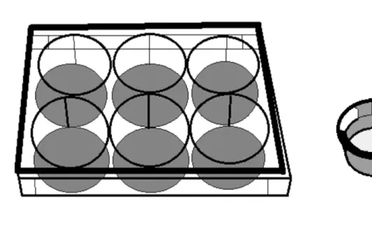
2.7 ml of serum-free DMEM media was added to each of 16 wells in 6-well plates. Semi-permeable inserts (3, 0 µm pore size cell culture insert, Falcon) were placed in eight wells for co culturing with platelets. 25x106 platelets from each donor were added to each insert in duplicates and volume adjusted to 1.7 ml with serum free DMEM. 30µl of fibrillary collagen (Chrono-par collagen, Chrono-log corp. Havertown) was finally added to activate the
platelets. The plates were placed on a vibrating platform shaker for 15 minutes followed by incubation at 37o C for 24 hours. Experimental design is shown in figure 1.
Figure 1. A 6-well plate and an insert. Fibroblasts were cultured on the bottom of the wells, and the
platelets added to an insert as indicated. Wells and inserts were combined to achieve the coculture situation.
ELISA
To detect secreted TGF-β by the platelets, a human TGF-β1 ELISA was used (optEIA™ kit BD, Becton, Dickinson and Company). An amount of 100 µl of medium from the 16 wells, were assayed as described:
At day one the microwells were coated with 100µl per well of Capture Antibody which was diluted in coating buffer. The plate was sealed and incubated overnight at 4o C.
At day two the plate was aspirated and washed three times with 300µl of washing buffer (PBS with 0,05% Tween 20). After the last wash, the plate was inverted and blotted on paper to
remove any residual buffer. The wells were blocked with 200µl Assay Diluent (PBS with 10% FBS) and incubated at room temperature for 1 hour. Meanwhile the 100 µl from the 16 samples were activated (with respect to TGF-β) with HCL (1:25) for one hour. Thereafter the plate was washed/aspirated. The samples were then neutralized with NaOH (1:25). Eight TGF-β standards were prepared by pipetting 8000pg/ml from stock solution together with 300µl Assay diluent into sex test tubes labeled with the different concentrations. A serial dilution was performed by adding 300µl of each standard to the next tube with vortexing between each transfer. One extra test tube served as the zero standard (0pg/ml).
100 µl of standard, sample and control were pipetted into the appropriate wells. The plate were sealed and incubated in room temperature for 2 hours. They were washed/aspirated with 300µl in total 5 washes. Afterwards, 100µl working detector (Detection Antibody 1:250 + streptavidin-HRP reagent 1:250) were added to each well. The plate was sealed and incubated for 1 hour at room temperature. After the incubation the plate was washed/aspirated with total of 7 washes. For the next step 100µl of substrate solution (Tetramethylbenzidine TMB and Hydrogen Peroxide, The BD Pharmingen™ TMB Substrate Reagent Set) were added to each well. The plate was incubated without plate sealer for 30 minutes at room temperature in the dark. After 30 minutes, a stop solution (H2SO4) of 50ml were added to each well. The
Absorbance was read at 450nm and 540nm within 30 minutes after stopping reaction. Statistical Analysis
TGF-β results are shown as mean concentration +/- SEM. For comparison between control group and donor group, a student t-test was made to test for significant difference (p<0,05).
Results
Appearance of fibroblasts after 24 hours of co-culturing with platelets
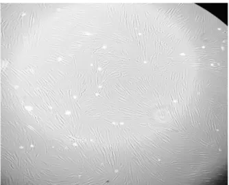
By microscopic inspection after 24 hours of co-culturing with platelets, the fibroblast appeared to show a growth difference in comparison of growing alone. With platelets, cultures of fibroblasts appeared denser (Fig 2 and 3).
Figure 2. Fibroblast growth inside the well without platelets after 24 hours of co-culturing with
DMEM and collagen.
Figure 3. Fibroblast growth inside the well with platelets after 24 hours of co-culturing with DMEM
and collagen.
Levels of TGF-β in the co-culture medium
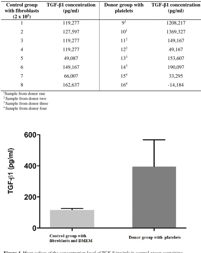
After measuring the absorbance in ELISA, the mean concentration of TGF-ß (pg/ml) was calculated for each sample. The mean concentration for the control group was 114+/- 12 pg/ml) and for the donor group containing platelets (398+/- 174 pg/ml(Fig 4). The values for the individual samples are shown in Table I. Difference between the groups were, however, not statistically significant (p=0.13).
Table I. Concentration of TGF-ß in pg/ml are shown for each sample in control group containing
DMEM and fibroblasts and the donor group containing platelets, DMEM and collagen.
Control group with fibroblasts
(2 x 10⁵)
TGF-β1 concentration (pg/ml)
Donor group with platelets TGF-β1 concentration (pg/ml) 1 119,277 91 1208,217 2 127,597 101 1369,327 3 119,277 112 149,167 4 119,277 122 49,167 5 49,087 133 153,607 6 149,167 143 190,097 7 66,007 154 33,295 8 162,637 164 -14,184
1Sample from donor one 2 Sample from donor two. 3 Sample from donor three 4 Sample from donor four
Figure 4. Mean values of the concentration level of TGF-ß (pg/ml) in control group containing
fibroblasts and DMEM, and in Donor group containing platelets, measured by ELISA. Error bars show standard error of the means.
Discussion
At the first attempt of activating the platelets, Krebs-Ringer phosphate buffer (KRG) was used for resuspension of platelets before transfer to wells together with Ca2+, needed to activate the
platelets. As a platelet agonist, denatured collagen was added, which, caused a gel-like cover
after 24h of incubation. Thus, the co-culture resulted in a unsuccessful attempt where the cells did not survive. By optimizing the method, it took in total three times to establish a coculture with fibroblasts that survived and with no gelation of added collagen. This optimization included to substitution of KRG to DMEM, adjusting the Ca2+ concentration, and exchange the denatured collagen to fibrillar collagen. In the last attempt, eight wells were covered with inserts, fibrillary collagen added to the inserts, and extra Ca2+ omitted (DMEM contains in itself >1 mM Ca2+). In this milieu fibroblasts appeared healthy over a 24 hour period. Inspection of fibroblast after 24h of co-culturing with platelets indicated that the fibroblasts exhibited a greater proliferation in comparison to the wells in absence of platelets. Cultures of fibroblast appeared denser. (Fig 2 and 3). By ELISA, TGF-β levels did not show a significant difference between control and platelet cocultures (p=0.13). However, there was a trend towards higher TGF-β concentration in platelet cocultures (Fig 4, Table I). Thus, even though each sample differed a lot in amount of TGF-β, they indicated a higher level of growth factor expression as a group. Possible causes that affects the samples can vary, but a probable reason is that the platelet samples were pre-activated and aggregated into pellets before activation with collagen and start of cocultures. Especially, this problem was encountered for donor four, which also expressed low amounts of TGF-β. We also suspect that some pipetting errors may have occurred when transferring platelets to the inserts or in the handling of ELISA. For the ELISA performance, one can optimize and avoid further errors by improving preparations such as: Calibrating pipettes, ensure sufficient incubation time and reagents being warmed to room temperature and observe that buffer & diluents are proper. It is also important to reassure a complete seal of plate and make sure that adequate reagent volumes are being added. Also, as indicated above, very careful resuspensions of platelet pellets are also called for, in order not to preactivate the platelets.
In further trials, one can use the method of measuring fibroblast proliferation with metabolic labeling, which would support our hypothesis that TGF-β has an effect on fibroblasts. Since fibroblast are important in the proliferation phase of the wound healing, the interaction between TGF-β would be important since it stimulates fibroblast to produce collagen and
other components of ECM, including the structural proteins, adhesive proteins and
proteoglycans [12]. Since the antibodies used in this ELISA kit only detects TGF-β, we could use other detection methods to look for other growth factors such as PDGF, bFGF, VEGF and other important secreted factors [4]. In other words, this collective family of growth factors should be monitored since they all have important roles in the healing process. This latter should be complemented with relevant fibroblast responses at the gene and protein levels (e.g. production of ECM proteins).
Conclusion
In conclusion, our experiment support the previously notion that platelets have a growth promoting effect on fibroblasts. Although the TGF-β levels varied a lot, it is likely that improvement of the experimental procedure will result in higher levels of this cytokine as well as other factors. Measuring various responses from the fibroblasts at gene and protein levels will further validate the relevance for the use of platelets in e.g. chronic wounds.
References
1, Barsotti MC, Losi P, Briganti E, Sanguinetti E, Magera A, Al Kayal T, et al. Effect of platelet lysate on human cells involved in different phases of wound healing. PLoS One.2013;8(12):1–11.
2, Lindemann S, Tolley ND, Dixon D a., McIntyre TM, Prescott SM, Zimmerman G a., et al. Activated platelets mediate inflammatory signaling by regulated interleukin 1β synthesis. J Cell Biol. 2001;154(3):485–90.
3, Manuscript A. NIH Public Access. Changes. 2012;29(6):997–1003.
4, Lynch SE, Colvin RB, Antoniades HN. Growth factors in wound healing. Single and synergistic effects on partial thickness porcine skin wounds. J Clin Invest.
1989;84(2):640–6.
5, Poniatowski Ł a., Wojdasiewicz P, Gasik R, Szukiewicz D. Transforming Growth Factor Beta Family: Insight into the Role of Growth Factors in Regulation of Fracture Healing Biology and Potential Clinical Applications. Mediators Inflamm [Internet]. Hindawi Publishing Corporation; 2015;2015:1–17. Available from:
http://www.hindawi.com/journals/mi/2015/137823/
6. Penn JW, Grobbelaar AO, Rolfe KJ. The role of the TGF-β family in wound healing, burns and scarring: a review. Int J Burns Trauma [Internet]. 2012;2(1):18–28.
Available from:
http://www.pubmedcentral.nih.gov/articlerender.fcgi?artid=3415964&tool=pmcentrez &rendertype=abstract
7, Hinz B. The extracellular matrix and transforming growth factor-β1: Tale of a strained relationship. Matrix Biol [Internet]. Elsevier B.V.; 2015; Available from:
http://linkinghub.elsevier.com/retrieve/pii/S0945053X15001055
8, Lynch SE, Nixon JC, Colvin RB, Antoniades HN. Role of platelet-derived growth factor in wound healing: synergistic effects with other growth factors. Proc Natl Acad Sci U S A. 1987;84(21):7696–700.
9, Matsumoto S, Tanaka R, Okada K, Arita K. The Effect of control-released basic fibroblast growth factor in wound healing: histological analyses and clinical application. Prsgo. 2013;1–9.
10, Shi H-X, Lin C, Lin B-B, Wang Z-G, Zhang H-Y, Wu F-Z, et al. The anti-scar effects of basic fibroblast growth factor on the wound repair in vitro and in vivo. PLoS One [Internet]. 2013;8(4):e59966. Available from:
http://www.pubmedcentral.nih.gov/articlerender.fcgi?artid=3615060&tool=pmcentrez &rendertype=abstract
11, Bao P, Kodra A, Tomic-canic M, Golinko MS, Ehrlich HP, Brem H. The Role of Vascular Endothelial Growth Factor in Wound Healing. J Surg Res. 2009;153(2):347– 58.
12, Kendall RT, Feghali-Bostwick C a. Fibroblasts in fibrosis: Novel roles and mediators. Front Pharmacol. 2014;5 MAY(May):1–13.
13, Givens K T. et al. Proliferation of Human Ocular Fibroblasts. Investigative Ophthalmology & Visual Science. Vol. 31. No. 9. September 1990
14, Yolken RH. Enzyme-linked immunosorbent assay (ELISA): a practical tool for rapid diagnosis of viruses and other infectious agents. Yale J Biol Med. 1980;53(1):85–92.