Hamstring muscle strain
49
0
0
Full text
(2) From the Department of Molecular Medicine and Surgery, Karolinska Institutet, Stockholm, Sweden. HAMSTRING MUSCLE STRAIN. Carl Askling. Stockholm 2008.
(3) © Carl Askling, 2008 ISBN 978-91-7357-519-5 Printed by 2008. Gårdsvägen 4, 169 70 Solna. 2.
(4) ABSTRACT Background: Acute hamstring strains are common injuries in different sports. They are often serious, causing long rehabilitation times and a proneness for re-injury. Preliminary observations indicate that the injuries can be of at least two types, one occurring during highspeed running and the other during motions where the hamstring muscles reach extreme lengths. Aims: To investigate the possible existence of different types of acute hamstring strains in two specific athletic groups, namely sprinters and dancers, as well as the generality of these findings in other sports. Methods: In the first project, 18 sprinters and 15 dancers with acute first time hamstring strains were prospectively included. All subjects were examined, clinically and with magnetic resonance imaging (MRI), on 4 occasions after injury: at day 2-4, 10, 21 and 42. The followup period was 2 years. In the second project, 30 subjects from 21 different sports were prospectively included. All subjects were examined clinically and with MRI. The follow-up period lasted until the subjects returned to sport or finished their sport activity due to the injury. Results: All sprinters sustained their injuries during competitive high-speed running. In contrast, all dancers encountered their injuries during slow-speed stretching type of exercises. The initial loss of strength and flexibility was significantly greater in sprinters than in dancers. At 42 days after injury, both groups could perform more than 90% of the test values of the uninjured leg. All the sprinters’ injuries were primarily located in biceps femoris long head, whereas the dancers’ injuries were mainly (87%) involving the proximal free tendon of semimembranosus. For the sprinters, involvement of the proximal free tendon, as estimated by MRI, and proximity to the ischial tuberosity, as estimated both by palpation and MRI, were associated with significantly longer time to return to pre-injury level. In the dancers, there were no significant correlations between clinical or MRI parameters and time to return to per-injury level. The time to pre-injury level was significantly longer (median 50 weeks, range 30-76) for the dancers compared to the sprinters (16, 6-50). In the second project, all injuries occurred during movements reaching a position with combined extensive hip flexion and knee extension. They were all located close to the ischial tuberosity and 83% involved the proximal free tendon of semimembranosus. Fourteen subjects (47%) decided to end their sport activity and for the remaining 16 subjects the median time back to sport was 31 (range 9-104) weeks. There were no significant correlations between clinical and MRI parameters and time to return to sport. Conclusions: There seems to be a link between the injury situation and the two types of acute hamstring strain in sprinters and dancers with respect to clinical findings, injury location, muscles and tissues involved, and time to return to pre-injury level. Proximity of the injury to the ischial tuberosity, as estimated both by palpation and MRI, is associated with longer recovery time. Also in other sports, an injury situation where the hamstrings reach extensive lengths caused a specific injury to the proximal posterior thigh similar to that described in dancers. Due to the prolonged recovery time associated with this type of injury, correct diagnosis based on history, clinical and MRI investigation, and adequate information to the athletes are essential. Key words: hamstring injuries, MRI, palpation, recovery time. 3.
(5) ÖNSKAN: att få uppleva en ”Lidnersk knäpp”. 4.
(6) TABLE OF CONTENTS LIST OF PUBLICATIONS. 7. INTRODUCTION Injury situation Injury location Prognosis. 9 9 9 11. AIMS. 15. METHODS Subjects Inclusion/exclusion criteria Clinical examination MRI investigation Follow-up Statistical analysis. 17 17 18 18 20 21 22. RESULTS Clinical examination MRI investigation Follow-up. 25 25 28 31. DISCUSSION Generality of results Possible injury mechanisms Clinical implications Future perspectives Recommendations. 33 33 33 35 36 39. SVENSK SAMMANFATTNING. 41. UPPSKATTNING & TACK. 43. REFERENCES. 45. STUDIES I – IV. 5.
(7) 6.
(8) LIST OF PUBLICATIONS This thesis is based on the following original papers, which are referred to in the text by their Roman numerals: I.. Askling C, Saartok T, Thorstensson A. Type of acute hamstring strain affects flexibility, strength and time back to pre-injury level. The British Journal of Sports Medicine 40: 40-44, 2006.. II.. Askling CM, Tengvar M, Saartok T, Thorstensson A. Acute first-time hamstring strains during high-speed running. A longitudinal study including clinical and MRI findings. The American Journal of Sports Medicine 35: 197-206, 2007.. III. Askling CM, Tengvar M, Saartok T, Thorstensson A. Acute first-time hamstring strains during slow-speed stretching. Clinical, magnetic resonance imaging, and recovery characteristics. The American Journal of Sports Medicine 35: 1716-1724, 2007. IV. Askling CM, Tengvar M, Saartok T, Thorstensson A. Proximal hamstring strains of stretching type in different sports. Injury situations, clinical and magnetic resonance imaging characteristics, and return to sport. The American Journal of Sports Medicine, 2008 (in press).. 7.
(9) 8.
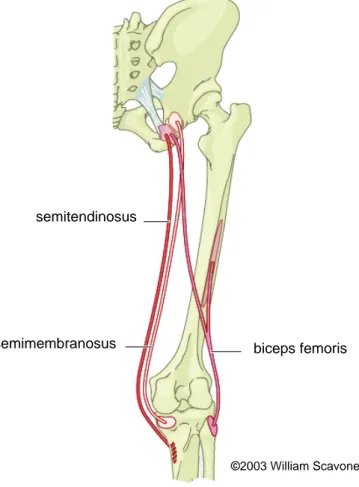
(10) INTRODUCTION Hamstring muscle strains are common in sports and are often causing extended absence from training and competition. Objective documentation of the occurrence of hamstring injuries is available primarily in Australian Rules football, European football and rugby, where a high prevalence of hamstring strains, 12-15 % of all injuries, has been documented in epidemiological studies.6,16,38,49,53 Additionally, in modern elite European football, there seems to be a shift towards an increasing representation of muscle strain injuries in general and of hamstring strains in particular; muscle strains now being as common as, or even more frequent than joint sprains.20 Several studies have indicated that hamstring strains are frequent also in track and field, especially among sprinters and jumpers, but there is only one epidemiological study, on a relatively small survey of Swedish club-level sprinters, showing that about 50 % encounter a hamstring strain injury during a period of one year.31 Injury situation It is a general belief that hamstring strains occur primarily during activities with extreme demands on speed and power, such as high-speed running.6,24,39,53 However, a recent study on dancers suggested that hamstring strains can occur also in slow-speed stretching exercises.3 A closer look at the epidemiological studies reporting injury situations in team sports reveals that hamstring strains were encountered not only in high-speed running, but also, in some cases, in stretching and kicking movements with large joint excursions.6,13,14,53 Moreover, in the study on rugby players,6 it was shown that the injuries resulting from kicking were the most severe, causing the longest absence from matches. Extended recovery periods were also reported by the dancers with hamstring strains occurring during stretching.3 In a case study on one sprinter and one dancer, not only differences in injury situation and recovery time were shown, but also in injury location, in terms of muscles as well muscle-tendon regions involved in the injury, as evidenced by both palpation and magnetic resonance imaging.4 Thus, possible connections are indicated between injury situation and injury location on the one hand and recovery time on the other. Systematic studies exploring such connections have, however, not yet been done. Injury location Anatomy. Knowledge about the specific anatomy is essential when investigating and trying to understand injuries to the musculo-skeletal system, not least hamstring strains. The collective term “hamstrings” refers to four muscles located in the posterior compartment of the thigh: semitendinosus (ST) and semimembranosus (SM) medially, and biceps femoris, long head (BFlh) and short head (BFsh) laterally (Figure 1). Proximally BFlh and ST arise from a common overall origin on the ischial tuberosity,33,34 but independent origins can be identified for these two muscles on the lateral one-quarter of the medial portion of the ischial tuberosity.52 Variation between individuals can occur, and muscle fascicles from ST can even be found to arise from the medial border of the tendon of BFlh.52 The proximal tendon of SM passes lateral and deep in relation to those of BFlh and ST, to insert on the lateral part of the upper half of the ischial tuberosity.52 Furthermore, the four hamstring muscles differ from each other with respect to muscle architecture, e.g. fascicular length, physiological crosssectional area, length of the proximal and distal free tendons and extent of the intramuscular tendons.52 In relation to injury proneness, it is noteworthy that the proximal free tendon of SM is about twice as long as that of BFlh and ten times longer than the tendon of ST.52 Consequently, the muscle fascicles of SM are much shorter than those of ST and BFlh, which should make SM less apt to withstand extensive extension.30,52 Innervation to the ST, SM and BFlh is provided by the tibial division of the sciatic nerve and the BFsh is innervated by the common peroneal division of the sciatic nerve. Mechanically, the ST, SM and BFlh can, 9.
(11) actively or passively, cause extension or counteract flexion at the hip and cause flexion or counteract extension at the knee, respectively, depending on the situation. The hamstring muscles can also influence abduction-adduction and twisting movements at these joints. The mechanical outcome will be task specific and depend on, among other things, the actual degree of activation of agonist and antagonist muscles around the two joints.. semitendinosus. semimembranosus. biceps femoris. ©2003 William Scavone. Figure 1. The anatomy of the hamstring muscle group shown schematically. Semitendinosus, semimebranosus and biceps femoris long head, all have their origins at the ischial tuberosity, whereas biceps femoris short head originates along the linea aspera and the lateral supracondylar line of femur. Distally, semitendinosus inserts on the medial surface of the superior tibia, semimembranosus on the posterior part of the medial condyle of the tibia, whereas both heads of biceps femoris have a common insertion on the lateral side of the fibular head.. 10.
(12) Detection/diagnosis. The diagnosis, hamstring strain, is commonly based on a clinical examination, which usually includes questions about the injury situation combined with palpation to detect local pain and loss of function. It is often difficult to acutely examine a hamstring strain because of pain and tenderness, so the general recommendation is to follow up the acute inspection with a new clinical examination the day after the injury occurred. Suspected total ruptures should immediately be referred to hospital care. There are different opinions as to the value of a clinical examination when it comes to predicting the duration of the time needed to return to sports. A clinician-determined site of injury, classifying the posterior thigh into upper, middle, or lower regions, has, for example, been shown to be of no assistance in making a prognosis for acute posterior thigh injuries.47 On the other hand, clinical examination including registration of pain and deficit in range of motion was shown to be useful in predicting the recovery time after acute hamstring strains.41 At present, there are no studies systematically following acute hamstring strains over time with repetitive clinical examinations, including documentation of injury situation, observation of function, location of pain by palpation, measurements of strength and flexibility, and correlating the findings with the actual time to return to sport. Magnetic Resonance Imaging (MRI) offers a means of non-invasively determining the location and extent of an injury. Because of its multi-planar capability and high sensitivity to detect the edema that results from soft-tissue injury, MRI is the technique of choice for imaging muscles and tendons.8,41 Powerful software makes parallel viewing of multiple images possible as well as quantification of distances, areas and volumes. Hamstring strains can range from microscopic foci of myofibrillar disruption, which may be beyond the resolution of MRI to total ruptures with extensive edema.26 Since MRI investigations are both expensive and demand expert evaluation, simpler clinical assessment methods would be preferable. Such methods have, however, to be validated with parallel MRI-investigations. In Table 1 an overview is presented of previous studies on hamstring strains verified by imaging techniques, listed with respect to sample size, imaging time after injury, muscles and regions involved and time back to sport. Six of the 12 studies included were done on Australian football players, which is not surprising since hamstring strains is the most common injury in that sport resulting in the highest number of days lost during a season. The study with the largest sample size (n = 154) is a retrospective study and in the other studies the sample size is moderate to low and the inclusion criteria are not strict. In most of the studies MRI was performed within 10 days after injury and showed that biceps femoris long head was the most frequently injured muscle. It was also clearly shown that a majority of the injuries were situated proximally and involved the muscle-tendon junction. The time back to sport was only reported in 8 of the studies and ranged from 3 – 6 weeks, but the criteria for return to sport were not presented clearly and/or differed between studies. As mentioned above, an increased number of hamstring strains have been documented in epidemiological studies in European football, but there is no information on injury situation and no imaging data available showing the injury location. Prognosis Making accurate predictions of recovery time is essential with all injuries, not least in the case of hamstring strains, where prolonged rehabilitation times and re-injuries are common.8,15,29,41,43 As mentioned above, there are indications that the injury situation as well as its location are factors of importance to consider when making estimates of time back to sport. It has been. 11.
(13) Table 1. Overview of studies on hamstring strains in sport verified by imaging techniques. Author, year. Sample, subjects. Garrett et al. 198913. 8 college athletes. Pomeranz et al. 199340. 14 professional athletes. Speer et al. 199344. 17 athletes different levels 17 athletes different sports 15 athletes (10 track and field) 30 Australian football players 154 athletes - majority football players 68 Australian football players 42 Australian football players. MRI or CT within 4 days MRI or CT 1 week – 3 months MRI within 5 days. Gibbs et al. 200415. 17 Australian football players. MRI within 3 days. SchneiderKolsky et al. 200641 Koulouris et al. 200727. 40 Australian football players 31 Australian football players. MRI within 3 days. Brandser et al. 19955 De Smet 12 at al. 2000 Slavotinek et al. 200243 Koulouris et al. 200325 Verrall et al. 200347 Connell et al. 20048. Imaging time after injury CT scan within 2 days MRI within 8 days. MRI within 6 days MRI and/or US within 10 days MRI within 6 days MRI within 3 days. MRI within 3 days. Muscle(s) injured. Region(s) involved. Time back to sport. 5 BFlh 2 BF+ST 1 SM 6 BF 5 SM 1 ST 2 QF 11 BF 4 SM 2 ST na. 8 proximal. 6 weeks mean. 5 proximal 7 middle 2 distal 6 MTJ na. 6 weeks mean. majority proximal. na. 11 BFlh 3 ST 1 SM 21 BFlh 9 ST. 9 PMTJ 6 DMTJ. na. 28 MTJ. 4 weeks median. 124 BF 21 SM 9 ST 49 BF 14 ST 5 SM 35 BF 3 SM 2 ST 2 ta 8 BF 5 BF+ST 2 ST 2 SM na. 98 MTJ. na. na. 4 weeks mean. 22 MTJ. 4 weeks median. na. 3 weeks mean. na. 3 weeks mean. 16 MTJ. 4 weeks mean. 26 BFlh 3 SM 2 ST. na. na = not available, CT = computed tomography, US = ultrasound, QF = quadratus femoris, MTJ = muscle-tendon junction, PMTJ = proximal muscle-tendon junction, DMTJ = distal muscle-tendon junction, ta = tandem injury i.e. more than one muscle involved. 12.
(14) shown that the size of the injury8,15,40,43 is coupled to the time back to pre-injury level. In addition, it has been speculated that also the injury site in terms of specific muscle in the hamstring muscle synergy13 and region in the muscle-tendon complex4 could be of importance. At the present time, there is only one study that has followed the progression of MRI characteristics over time after acute hamstring strains.8 That study showed that the longitudinal extent of the edema, as measured with MRI, was the best predictor for the amount of time needed until an athlete (Australian football) could return to competition. Recent research has indicated that results of clinical tests may be as useful in predicting rehabilitation time as MRI data, at least in the case of minor and moderate hamstring injuries in Australian football players.41 Strength and flexibility may be critical for performance and are often tested and used for establishing criteria for return to full activity.9,10,11,18,19,50 However, there are no studies showing that the initial decrements in muscle strength and/or flexibility are correlated with the time back to sport after a hamstring strain. The scientific support for the often-used textbook recommendation to return to sport when the strength and flexibility of the injured leg are back to 90 – 95 % of the uninjured leg is weak.19 It could be speculated that athletes in team sports, such as football, can return to play without being entirely back to their pre-injury level, whereas, at least in some individual sports such as sprinting, top athletes would need much closer to full function.37 However, there appear to be no consensus guidelines or agreed-upon criteria for safe return to sport following muscle strains that completely eliminate the risk for recurrence and maximize performance.. 13.
(15) 14.
(16) AIMS • To investigate the possible existence of different types of acute hamstring strains in two specific athletic groups, namely sprinters and dancers, as well as the generality of these findings in other sports. • To systematically follow the first 6 weeks after acute first-time hamstring strains in sprinters and dancers, with respect to injury situation, injury location and extent of the injury, recovery of strength, flexibility and function, as well as possible relationships between clinical and MRI findings and time to return to sport; follow-up period 2 years.. 15.
(17) 16.
(18) METHODS Subjects The sprinters and dancers for Study I-III were recruited in response to information given to all major track and field clubs in Sweden via the Swedish Athletic Association, and to dance schools and employers of dancers in the Stockholm area via the Swedish Society of Dance Medicine. During the recruitment of subjects for Study I-III, the main investigator (CA) was contacted via phone or e-mail by several athletes from different sports. In some cases they described an injury pattern similar to that of the dancers, namely that the injury occurred in a situation where the hamstrings reached extensive length by a combination of hip flexion and knee extension. When we realized that this type of injury appeared to exist rather frequently in various sports, we decided to collect data also from those subjects (Study IV) in parallel with our ongoing studies on well-defined sprinters and dancers. A description of the subjects included in Study I-IV is presented in Tables 2-4. Table 2. Characteristics, median values and ranges, of the sprinters included in Study I and II.. Age (years) Body height (m) Body mass (kg) Personal best 100m (s). Sprinters Women (8) Men (10) 22 (20-28) 19 (15-23) 1.76 (1.59-1.80) 1.78 (1.66-1.82) 65 (53-69) 70 (61-84) 12.7 (12.4-13.4) 11.2 (10.5-11.7). Table 3. Characteristics, median values and ranges, of the dancers included in Study I and III.. Age (years) Body height (m) Body mass (kg) Student dancers (n) Professional dancers (n). Dancers Women (14) 20 (16-24) 1.67 (1.58-1.80) 60 (48-68) 12 2. Men (1) 23 1.73 65 1 0. Table 4. Characteristics, median values and ranges, of the subjects from different sports included in Study IV.. Age (years) Body height (m) Body mass (kg) International level (n) National level (n) Recreational level (n). Subjects from different sports Women (22) Men (8) 23 (16-53) 27 (18-36) 1.69 (1.59-1.76) 1.78 (1.70-1.92) 58 (48-67) 74 (64-90) 12 6 5 1 5 1. 17.
(19) Inclusion/exclusion criteria To be included in Study I-III, the sprinters and dancers had to present a history of first-time acute sudden pain from the posterior thigh when training, competing or performing. The clinical examination 2 days post-injury had to reveal distinct pain when palpating the hamstring muscle, local pain when performing a passive straight leg raise (SLR) test and an increased pain when adding a voluntary isometric hamstring contraction during that test. In all subjects included in Study I-III, the subsequent MRI investigation had to confirm the suspected injury. Exclusion criteria were: verified or even suspected earlier hamstring strain in the same leg, extrinsic trauma to the posterior thigh (contusions), ongoing or chronic low back problems, pregnancy and total rupture or avulsion of one, two or all three hamstring muscles (determined by MRI). To be included in Study IV, the subject had to present a distinct history of acute sudden pain from the posterior thigh, occurring in a position with combined extensive hip flexion and knee extension, during training, competition or performance. The time from the injury to the clinical examination and MRI investigation varied, but was not allowed to exceed 12 months. The clinical examination was the same as for Study I-III, with the exception that the pain upon palpation and in the SLR tests had to be at the area of origin of the hamstring muscles on the ischial tuberosity or slightly (0-5 cm) below. In all subjects included in the study, the subsequent MRI investigation had to confirm the suspected injury. Exclusion criteria comprised an unclear injury situation, earlier hamstring strain at the same side during the last 12 months before the present injury, total rupture or avulsion of one, two or all three hamstring muscles (determined by MRI), extrinsic trauma to the posterior thigh, ongoing or chronic low back problems and pregnancy. Clinical examination All subjects in Study I-III were tested on four occasions, 2, 10, 21 and 42 days post-injury. In Study IV the time from the injury to the examination varied, but was less than 12 months. Palpation Palpation of the rear thigh was performed with the subjects prone and the knee extended. First, the origin of the hamstrings on the ischial tuberosity was identified. The subjects were then asked to activate their hamstrings by performing an isometric contraction with manual resistance against the heel and no flexion at the knee. The palpation started approximately 5 cm cranial to the hamstring origin and continued without interruption to the respective muscle insertions on the lower leg. The point where the subject noted the highest pain upon palpation was marked and the distance between this point and the palpated ischial tuberosity was measured. Hip flexibility The hip flexion test (Figure 2) combined a passive unilateral straight leg raise test (SLR) with pain estimation according to the Borg CR-10 scale. The subjects were placed supine with the pelvis and contralateral leg fixed with straps. A standard flexometer (Myrin®, Follo A/S, Norway, sensitivity 2o) was placed 10 cm cranial to the base of the patella. The foot was plantar flexed and the investigator slowly (approximately 30o.s-1) raised the leg with the knee straight until the subject estimated a 3 (“moderate pain”) on the Borg CR-10 scale (0 = no pain and 10 = maximal pain). The hip flexion angle at this point was recorded, and the greatest angle of three repetitions was taken as the test result for Range of Motion (ROM). Values of the injured leg were expressed as a percentage of the uninjured leg for comparisons within and between groups. No warm-up preceded the flexibility measurements.. 18.
(20) Figure 2. Hip flexion ROM test.. Knee flexion strength Isometric knee flexion strength was measured with the subject in a prone position and the pelvis and the contralateral leg fixed (Figure 3). A dynamometer (Bofors KRG-4 T10®, Nobel Elektronik, Karlskoga, Sweden, range 0-4 kN) was placed at the ankle, perpendicular to the lower leg. The foot was in plantar flexion and the knee in an extended position. Three maximal voluntary isometric knee flexion contractions were performed, each with gradually increasing effort. Each contraction lasted 3 s with 30 s of rest in-between. The highest force value was taken as the test result for strength. Values of the injured leg were expressed as a percentage of the uninjured leg for comparisons within and between groups. No warm-up preceded the strength measurements.. Figure 3. Knee flexion strength test.. 19.
(21) MRI investigation The injured sprinters and dancers were to undergo four consecutive MRI investigations, at 4, 10, 21 and 42 days after the incident of acute hamstring strain. The MRI investigation in the mixed group of Study IV was done on one occasion within 12 months after the injury. All MRI investigations were performed on a 1.0 Tesla superconductive MRI unit (Magnetom Expert®, Siemens, Erlangen, Germany). The subjects were positioned supine on a commercial phased array spine coil. First, sagittal and frontal STIR images were obtained with a large field of view from at least 5 cm above the ischial tuberosity to the knee. Then transversal, T1weighted, T2-weighted, and STIR images were obtained covering the entire damaged area on the longitudinal images. Frontal and transversal images included also the uninjured side for comparisons at all four occasions. The thickness of the slices for all sequences was 5 mm with a 0.5 mm gap. A muscle was considered as injured when it contained high signal intensity, as compared with the uninjured side, on the STIR images. A tendon tissue was considered as injured if it was thickened and/or had an intra-tendinous high signal and/or a collar of high signal intensity around it on the STIR images. Each injury was allocated to one or more of six different regions within the muscle-tendon complex (Figure 4).. 1. PT. 3. PMB. 2. PMTJ. 4. DMTJ. 5. DMB. 6. DT. Figure 4. Schematic drawing representing the muscle-tendon complex of the long head of biceps femoris (Study II) and semimembranosus (Study III), showing the six different regions used when analysing the injury location and tissues involved: 1. proximal tendon (PT), i.e. free tendon proximal to muscle fiber attachment, 2. proximal muscle-tendon junction (PMTJ) defined as the proximal intramuscular tendon and attached muscle fibers, 3. proximal muscle-belly (PMB), i.e. muscle proximal to the midpoint of the whole muscle-belly, 4. distal muscle-tendon junction (DMTJ), defined as the distal intramuscular tendon and attached muscle fibers, 5. distal muscle-belly (DMB), i.e. muscle distal to the midpoint of the whole muscle-belly, and 6. distal tendon (DT), i.e. free tendon distal to muscle fiber attachment.. 20.
(22) The size of the injury was measured on the images as the maximal length (cranio-caudal extent), width (medio-lateral extent) and depth (antero-posterior extent). Width and depth were not measured in Study IV. An attempt to estimate the volume of the injury was made by assuming that the injury had a shape of a rotational ellipsoid, i.e. volume | length x width x depth x 0.5. The most cranial pole of the injury was identified and its cranio-caudal distance to the most caudal part of the ischial tuberosity was measured; hereafter this distance will be referred to as “distance to tuber” (Figure 5). For the sprinters the cross-sectional area of the injured muscle, as a percentage of the total cross-sectional area, was calculated at the level where the injury had the largest absolute cross-sectional distribution in the muscle.. Figure 5. Distance between the most cranial pole of the edema and the ischial tuberosity is shown as the double-headed arrow in 2 different types of injury. The edema starts caudal to tuber in the left and cranial to tuber in the right MR-image.. Follow-up On the first test occasion (2 days after injury) each sprinter and dancer in Study I-III had to make a self-estimate of his/her time back to pre-injury level. They then all received the same standardized three-part progressive rehabilitation program. The three parts were distributed at the first, second and third clinical examination, respectively. After the last examination, 6 weeks post-injury, continued rehabilitation was administered by the respective athlete’s physician and/or physical therapist. The subjects were asked to note the week when they could train, compete or perform at their pre-injury level, i.e. competing at similar best-times for the sprinters and being able to train and perform without any restriction for the dancers. If re-injury occurred, the athletes were to contact the main investigator (CA) immediately by phone. Follow-up contacts by phone were made by the main investigator at 3, 12 and 24 months after the occurrence of the initial injury, and then any symptoms or problems from the previously injured hamstring muscle were noted. In Study IV, the follow-up period lasted until the subject returned to sport or decided to give up his/her sport activity (maximal follow-up period was set to 2 years). The subjects were asked to register the first full week when they could train and/or perform in their sport again and if they still had any symptoms at that time, and give that information by phone to the main investigator. If the subjects decided to stop with their sport, they were to inform the main investigator directly and a clinical examination was then made. The subjects´ 21.
(23) rehabilitation was administrated by their respective physician and/or physiotherapist and was not controlled as a part of the study. Statistical analysis An overview of the statistical procedures employed in this thesis is presented in Table 4. In all cases the STATISTICA program, version 6.0, 7.0 and 8.0 (StatSoft®, USA) and SPSS program, version 11.0 and 15.0 (Statistical Package for the Social Sciences®, USA) were used for the analysis. The level of significance was set at P < 0.05 and tendencies were identified at 0.05 P < 0.01. Shapiro-Wilk’s W test was applied to examine normality in the distribution of data. Table 5. Overview of the statistical analyses.. Descriptive statistics. Study I. Study II. Study III. Study IV. •. •. •. •. •. •. •. •. •. •. Mean ± 1 standard deviation (SD). •. Mean ± 95% confidence interval (CI) Median, range. •. Analytic statistics ANOVA. •. Mann-Whitney U test Tukey test. •. Student´s t test. •. Pearson’s correlation coefficient Spearman rank order correlation. •. Intraclass correlation coefficient (ICC). •. Fischer´s exact test. •. •. •. •. •. • •. In Study I, to estimate the test-retest reliability of the hip flexibility test and the isometric knee flexion strength test, the values for the uninjured leg on the first and second test-occasions, 8 days apart, were used to calculate the intra-class correlation (ICC) of the measurements with 95% confidence interval (CI). Repeated measures ANOVA with post-hoc Tukey test was used to detect statistical differences in ROM and strength over time. Spearman rank order correlation was calculated between subjects’ test results and time back to pre-injury level. To compare the groups with respect to functional observations, Fischer’s Exact Test was employed. Student’s t test was used to detect significant differences between the groups concerning their self-estimated time to return to pre-injury level. In Study II, repeated measures ANOVA with post-hoc Tukey test was used to reveal statistical differences in MRI parameters over time. Mann-Whitney U test was used to detect statistical differences between groups with involvement of different regions of the muscletendon complex and time to return to pre-injury level. Pearson’s rank order correlation was calculated between subjects’ MRI parameters, palpation data and time to return to pre-injury level.. 22.
(24) In Study III, repeated measures ANOVA with post-hoc Tukey test was used to detect statistical differences in MRI parameters over time. Pearson’s rank order correlation was calculated between subjects’ MRI parameters, palpation data and time to return to pre-injury level. In Study IV, Spearman rank order correlation was calculated between subjects’ MRI parameters, palpation data and time to return to sport. Mann-Whitney U test was used to detect statistical differences in age, gender and level of performance between the group of subjects returning to sport and the group who did not, and in time back to sport for elite vs. recreational level, and team vs. individual sports, respectively. Ethical approval Approval for Study I-IV was granted from the Ethical Committee of the Karolinska Institutet, Stockholm, Sweden (99-121).. 23.
(25) 24.
(26) RESULTS Clinical examination All sprinters reported that they had encountered their injuries during high-speed competition sprinting. In contrast, all dancers got their injuries in slow-speed stretching type of exercises, mainly during warming-up or cooling-down. All 18 sprinters were forced to stop their sprinting directly when the injury occurred, but only 6 of the 15 dancers had to interrupt their activity. On the first test occasion, within 2 days after injury, 15 sprinters were using crutches, but none of the dancers. None of the sprinters could walk on plain ground without pain, compared with 7 of the dancers. On the fourth test, 6 weeks post-injury, all sprinters could jog without pain, whereas 3 of the dancers still had problems. All injuries in the mixed group of Study IV occurred during movements reaching a position with combined extensive hip flexion and knee extension. The median time for the clinical examination was 12 weeks (range 1-51w) after the injury. The major part, 21 injuries (70%), occurred during training, competition or performance and the rest during warming-up or cooling down.. Hip flexibility, % of the uninjured leg. Hip flexibility The results from the sprinters and dancers showed a large decrease in range of motion (ROM) at the hip of the injured leg in both groups on the first test occasion (Figure 6). Over the subsequent 6 weeks, the subjects gradually recovered the ROM of their injured leg, but it was still significantly less in both groups on the last test occasion (Figure 6). The difference in ROM between the two legs was significantly larger for the sprinters than the dancers on the. 100 87% * 80. 81% *. 94% *. 91% *. 91% *. 91% * 81% *. 60. 63% *. Dancers Sprinters. 40 20 0 0 2. 10. 21. 42. Days after injury Figure 6. Mean values for hip ROM of the injured leg compared with the uninjured leg in sprinters (n = 18) and dancers (n = 15) at the four test occasions. *Denotes a significant difference (p < 0.05) between the injured and uninjured leg.. 25.
(27) first test occasion. In neither sprinters nor dancers were there any correlations between the decrease of hip ROM in the injured leg at the first test occasion and time back to pre-injury level.. Knee flexion strength, % of the uninjured leg. Knee flexion strength Both sprinters and dancers demonstrated significantly lower strength in the injured leg than in the uninjured leg on the first test occasion (Figure 7). On the following test occasions this difference decreased gradually. On the two last test occasions, there was no significant difference in strength between legs in the dancers, whereas significant differences remained for the sprinters (Figure 7). The decreases in strength of the injured leg on the first, second and third test occasions were significantly larger for the sprinters than for the dancers. In neither sprinters nor dancers were there any correlations between the decrease of knee flexion strength in the injured leg at the first test occasion and time back to pre-injury level.. 100. 92% *. 97%. 97%. 80% * 80. 94% *. 87% * 70% *. 60. Dancers Sprinters. 40 39% * 20 0 0 2. 10. 21. 42. Days after injury Figure 7. Mean values for knee flexion strength of the injured leg compared with the uninjured leg in sprinters (n = 18) and dancers (n = 15) at the four test occasions. * Denotes a significant difference (p < 0.05) between the injured and uninjured leg.. 26.
(28) Palpation The sprinters noted their highest pain upon palpation at a significantly more distal location than the dancers. The subjects in the mixed group of Study IV showed similar values as the dancers (Table 6). The total length of the painful area was significantly longer for the sprinters compared with the dancers at the first test occasion, but not at the last test occasion (Table 6). There was a significant correlation between the location of the point of highest pain during palpation at the first occasion and the time back to pre-injury level for the sprinters (r = 0.695, P = 0.004), that is, the more cranial the location, the longer the time back. A corresponding correlation was not seen in the other groups of subjects. Table 6. Mean values ±1 SD (and ranges) for the distance from the point of maximal pain to the ischial tuberosity and the total length of the painful area upon palpation at the first and fourth tests, 2 days and 6 weeks after injury, in sprinters and dancers, and for the subjects of the mixed group in Study IV (average time after injury: 12 weeks).. Palpation variable. 1st test sprinters. 1st test dancers. 4th test sprinters. 4th test dancers. Study IV mixed group. Distance to tuber, cm. 12±6 (5-24) *. 2±1 (1-3). 11±8(4-24) *. 3±2 (0-5). 2±1 (0-5). Total length, cm. 11±5 (5-24) *. 5±2 (2-9). 5±2 (2-8). 4±3 (2-10). na. na, measurement not available * Denotes a significant difference (p < 0.05) between the sprinters and the dancers.. 27.
(29) MRI investigation Typical examples of acute MRI findings (MRI performed within 4 days after the injury occurrence) from a sprinter, a dancer and an individual (tennis player) from the mixed group in Study IV are shown in Figures 8-10.. Figure 8. Sprint injury involving biceps femoris caput longum.. Figure 9. Dance injury involving the proximal free tendon of semimembranosus.. Figure 10. Stretching-type injury in a tennis player (Study IV) involving the proximal free tendon of semimembranosus.. 28.
(30) All sprinters had their primary injury located in BFlh (cf. Figure 8). Eight sprinters had a secondary injury, 7 in ST and 1 in BFsh. In contrast, the muscle most prone for injury in the dancers and the subjects from the mixed group of study IV was SM (cf. Figure 9 and 10). These stretch-type of injuries were, however, often complex with involvement also of other muscles (Table 7). Table 7. Muscles involved in the different individual injuries. Listed is the number of subjects in each category, sprinters (18), dancers (15) and the mixed group of study IV (30), with injuries to a specific muscle or combination of muscles.. Study II sprinters. Study III dancers. Study IV mixed goup. 10 BFlh 1 BFlh, BFsh 7 BFlh, ST. 2 SM 7 SM, QF 4 SM, QF, AM 1 QF 1 QF, AM. 16 SM 1 SM, BFlh 1 SM, ST 3 ST, BFlh 2 SM, ST, QF 3 SM, QF 2 SM, QF, AM 1 AM 1 QF, AM. BFlh, biceps femoris long head; BFsh, biceps femoris short head; SM, semimembranosus; ST, semitendinosus; QF, quadratus femoris; AM, adductor magnus. The location of the primary injury, in BFlh in sprinters and in SM in dancers, in relation to different regions of the muscle-tendon complex is presented in Figure 11. Six of the sprinters’ injuries (33%) involved the proximal free tendon, whereas all injuries to the SM in the dancers (n = 13) and in the mixed group (n = 25) involved its proximal free tendon. Time to return to pre-injury level for the sprinters with injuries involving the free proximal tendon of BFlh (n = 6) was significantly longer compared to that of the sprinters with injuries not involving the proximal free tendon of BFlh (n = 12).. 29.
(31) PT = 6. PT = 13. PMB = 0. PMB = 0 PMTJ = 12. PMTJ = 2. DMTJ =6. DMTJ =0. DMB = 2. DT = 2. DMB = 0. DT = 0. Figure 11. Number of sprinters (left) and dancers (right) with injuries in different muscletendon regions; PT, proximal tendon; PMTJ, proximal muscle-tendon junction; PMB, proximal muscle-belly; DMTJ, distal muscle-tendon junction; DMB, distal muscle-belly; DT, distal tendon.. The edema of the sprinters’ injuries started significantly more caudally, both at the first and last MRI investigation, compared with the dancers’ injuries at corresponding times (Table 3). As in the dancers, the injury of the mixed group generally started cranially to the ischial tuberosity (Table 8). The injury edema was longer in the sprinters than in the dancers (Table 8). In the sprinters, a correlation analysis showed that the shorter the distance was from the most cranial pole of the injury to tuber, established by MRI at the first investigation, the longer the time back to pre-injury level (r = 0.544, P = 0.004). A corresponding correlation was not seen in the other groups of subjects. Table 8. Mean values ±1 SD (and ranges) for two MRI parameters at the first and fourth tests, 4 days and 6 weeks after injury, in sprinters and dancers, and for the subjects of the mixed group in Study IV (average time after injury: 13 weeks). st. st. th. th. MRI variable. 1 test sprinters. 1 test dancers. 4 test sprinters. 4 test dancers. Study IV mixed group. Distance to tuber, mm. 67±71* (-21-218). -23±8 (-34--11). 102±79* (17-240). -13±15 (-33-22). -30±16 (-66-0). Length, mm. 187±74* (60-346). 98±50 (27-172). 90±60* (0-229). 31±15 (11-55). 71±58 (10-214). Negative distance means that the edema started cranially to the ischial tuberosity. * Denotes a significant difference (p < 0.05) between the sprinters and the dancers.. 30.
(32) Relative number of subjects (%). Follow-up The time back to pre-injury level was significantly longer for the dancers than for the sprinters, median values being 50 weeks (range 30 – 76w) and 16 weeks (range 6 – 50w), respectively (Figure 12). On the last test occasion, 6 weeks post-injury, none of the dancers, and only two of the sprinters, were able to participate fully in their respective sport according to their own judgement. One dancer decided to end her dancing career 68 weeks after the initial injury due to chronic symptoms from her hamstring strain. During the 2-year follow-up, 3 sprinters (17%) encountered re-injuries of their hamstrings (8, 9 and 20 months after the first injury, respectively) and 2 of them had to finish their athletic careers. The re-injuries occurred after that they had reached their pre-injury level.. 100 80 60 40 20 sprinters dancers. 0 0. 10. 20. 30. 40. 50. 60. 70. 80. Time back to pre-injury level (weeks) Figure 12. Relative number of subjects, sprinters and dancers, plotted against the time, in weeks, it took for each individual to return to pre-injury level of performance (n = 18 for the sprinters and n = 14 for the dancers).. In the mixed group of Study IV, 14 subjects (47%) decided to finish their sport careers due to chronic symptoms from their hamstring injury. The decision to quit in this group was taken after a median time of 63 weeks (range 26 – 104w). All the subjects who decided to stop their sport participation showed signs of injury at the clinical examination performed shortly after the decision, e.g. palpation pain close to the ischial tuberosity, pain in a passive straight leg raise test, and an increased pain with addition of an isometric hamstring contraction during that test.. 31.
(33) The median time back to sport for the remaining 16 subjects in Study IV was 31 weeks (range 9-104w). At the time when they decided to go back to their sport, 2 reported no symptoms, whereas 14 reported a variety of persisting problems, such as feeling of insecurity (n = 14), pain when sitting (10), pain when putting force demands on hamstrings during lengthening (stretching) (8) and a need for extra attention on warming-up before performance (8). There were no statistically significant differences between the group (n=16) that returned to sport compared with the group (n=14) that did not, with respect to age, gender or level of performance. The time back to sport was significantly longer for the subjects on recreational level (median 62 weeks, range 40-104w, n = 4) compared with the subjects on elite level (25 weeks, 9-80w, n = 12). There were no significant differences in the time back to sport between subjects in individual sports (n = 14) versus team sports (n = 2).. 32.
(34) DISCUSSION In this project it has been possible to identify and characterize two distinctly different types of proximal hamstring strain, one occurring during high-speed running and mainly involving the biceps femoris long head, the other during movements leading to extreme lengthening of the hamstrings and most often involving the semimembranosus. The type of hamstring injury was shown to have profound effect on the time to return to sports. Generality of results Even if the sample sizes were relatively small, the results are strengthened by the fact that they were obtained from two specific and rather homogenous and well-defined categories of athletes, sprinters and dancers. Furthermore, the inclusion criteria were rigorous and a careful registration of injury history ensured true first-time hamstring injuries, which disqualified many potential subjects. It is important to realize that there are many differences among subjects with hamstring strains, which makes it difficult to generalize the results to hamstring strains in other sports. However, although the high-speed injury type was documented in a group of sprinters, it should be possible to extrapolate the results to other sports where hamstring injuries occur at high running speed.6,25,53 Our results are in line with previous studies showing that biceps femoris long head is the most commonly injured hamstring muscle in high force/high speed sports.8,12,27,43 Our observation that seven of the eight secondary injuries occurred in the semitendinosus is also in accordance with earlier studies on other types of “power” athletes reporting a combined injury of biceps femoris and semitendinosus as the most common “tandem injury”.12,25 The stretch-type of injury was established in a group of dancers with strains occurring during well-controlled slow movements to extreme joint positions. The presence of a similar type of injury in a mixed, less-controlled, group of athletes emphasised the generality of the injury situation with extreme movements, albeit not necessarily slow, as a common denominator. Also in the literature, occasional observations on similar injury situations have been reported for various sports, but the injury location has not been documented.6,53 Possible injury mechanisms Before discussing possible reasons for hamstring injuries in general and specific injury types in particular, it is important to recognize that in-depth information on injury mechanisms cannot be obtained on humans in vivo, since experiments cannot be designed to systematically inflict injury on healthy subjects. Therefore, in the only case where a hamstring injury has occurred in a situation where scientific data were collected, the information gained is rhapsodic and inconclusive.17 Still, some general speculations can be made based on extrapolations from anatomy, biomechanics and motor control and application of simulation models. The data on injury situation and location clearly indicate the presence of two specific mechanisms for the sprint- and stretch-type of hamstring injury. Both appear to be strain injuries, i.e. occur as a consequence of excessive elongation of certain vulnerable structures within the muscle-tendon complex. The circumstances under which the elongation occurs differ markedly between injury types. The high-speed running type of injury is believed to occur at the end of the swing phase and/or in the beginning of the stance phase.17 Biomechanical estimations of overall muscle length changes have indicated that there is an elongation of the hamstrings (except biceps femoris short head) in these phases of the step cycle. However, since neither the hip nor the knee joint is at extreme angles, the elongation is probably far from reaching maximum lengths. This is more likely to occur during extreme hip. 33.
(35) flexion movements with extended knee, e.g. in sagittal splits and high kicking. Simulated length changes based on a computer model (AnyBody Technology A/S®, Aalborg, Denmark) showed that during a sagittal split the lengthening is about double that of the lengthening present during a sprint step (unpublished data). The model, which is based on the anatomy of a 50th percentile European male, indicated a lengthening of up to 15 cm, or about 1/3 of the resting length, for the extreme hip flexion occurring in sagittal split. To our knowledge, there are no experimental data in the literature to verify these findings for hip flexions beyond 90º. The sprinthigh-speed running-type of hamstring strain is predominantly occurring in the biceps femoris long head, whereas the stretching-type most often involves the semimembranosus. The reasons for this specificity are not evident. Biomechanical modelling of overall length changes in individual hamstring muscles during running have indicated that the long head of biceps femoris is somewhat more elongated than the semitendinosus and semimembranosus during the end of the swing phase.7,46 However, the difference was small and hard to envision as a major cause of injury specificity. The current stage of biomechanical model development does not allow for distinguishing between length changes in individual hamstring muscles during extreme movements, such as sagittal splits. The high-speed running-type of hamstring strain in the biceps femoris long head primarily involved the muscle-tendon junction. This is most often the case with muscle strain injuries and the junction between muscle and tendon appears to be the weak link.13,14,22,51 It is important to remember that the muscle-tendon junction is complex with extensive intramuscular tendon branching in the hamstring muscles, not least in the biceps femoris long head. In contrast, the stretching-type of injuries in the semimembranosus involved the free tendon. Why this is the case is difficult to explain, but a contributing factor might be the complex anatomy of the proximal hamstrings, with the semimembranosus tendon running underneath those of biceps femoris long head and semitendinosus.52 This might expose the semimembranosus tendon for mechanical wear from the actions of the other two muscles. Another anatomical feature to consider when evaluating reasons for the specific injury proneness of semimembranosus for stretching-type injuries is its relatively short muscle fascicle length,52 which should make it more vulnerable to extreme elongation.30,52 A further critical issue to evaluate is whether the muscle is activated or not at the time of injury. As mentioned, it is suggested that the high-speed running-type of injury occurs at the end of swing and/or early in the stance phase, i.e. when the hamstrings are activated.32,35 In the stretching-type of injury, the activation of hamstrings is assumed to be lower and in the dancers the muscles are intended to be passive. Although these circumstances have not been verified, they give rise to the hypothesis that muscle-tendon junction injuries primarily are related to strains caused by forces applied on/by a contracted muscle, whereas injuries to the free tendon would occur when the muscles are mainly passive. In the latter case, the forces would be transmitted via the tendon and passive structures surrounding the muscle fibres and it might be that the tendon has the poorer resistibility. It is an intriguing fact that most hamstring strains occur in the proximal part of the muscle/tendon complexes.5,12,25 It indicates that a possible explanation of the injury mechanism should involve details in the local architecture and anatomy of the hamstrings in the proximal part. Also pointing to injury mechanisms related to the hip joint is the fact that hamstring injuries rarely occur in the short head of the biceps femoris, which spans only the knee joint. It seems plausible that, at least for the stretching-type of injury, interaction. 34.
(36) between the hamstrings and other muscles, such as the quadratus femoris, could cause additional tension of the hamstrings in the proximal part. Apart from apparent differences in injury situation, there are also similarities in that both high-speed running- and stretching-type of hamstring strains occur during well-accustomed movements, which have been repeated an infinite number of times by the athlete. At least in the elite athletes, both sprinting and stretching are performed at or close to the limit. Thus, it cannot be excluded that a sudden small deviation in muscle activation, movement and/or load might result in surpassing of the limit and give rise to an injury. However, the consistency in the injury pattern for the respective injury still presents a puzzle. Clinical implications The studies included in this thesis clearly demonstrate the existence of two types of hamstring strains, distinguished by different injury situations. Whether acute hamstring strains occur during high-speed running- or stretching-type of exercises had a clear impact on the acute loss of function and performance in strength and flexibility tests, injury location determined by palpation and MRI, as well as time to return to sport. The hamstring strains occurring in sprinters during maximal speed running caused a more marked acute decline in function and a faster initial recovery than those occurring in extreme positions during stretching-type of exercises in dancers. Speculations on the reasons for this include differences in the tissues involved and a much larger intramuscular edema in sprinters than in dancers. Acknowledging the errors involved in calculations of volume of the edema, it is still noteworthy that the edema in the sprinters had, on average, twice the volume of that of the dancers at the first test occasion and four times the volume at the test 6 weeks after injury. Interestingly, there were no correlations between time to return to pre-injury level and the magnitude of the acute loss of hip flexibility or knee flexion strength in sprinters and dancers. This has to be taken into account when evaluating the prognosis and prescribing rehabilitation. Based on the results from dancers and the mixed group of athletes, in cases of injuries to the proximal posterior thigh, the clinician should request a detailed description from the patient about the etiology of the strain and also perform a careful palpation. MRI can confirm the injury during a long period (in Study IV as long as 12 months after injury) and should always be done when a total rupture is suspected. Initially, most athletes, as well as trainers and coaches, do not realize that this type of strain is a serious injury, in some cases even career ending (one dancer in Study III and 14 of 30 athletes in the mixed group), and therefore neglect to seek acute or early medical advice. It is essential that the athlete suffering an acute stretching-type injury to the upper posterior thigh gets relevant information from the medical staff about the risk of prolonged rehabilitation time, even if the initial symptoms appear minor, since unrealistically optimistic information will only reinforce the disappointment and frustration of the injured athlete. A typical scenario may be that the injured person comes to the clinic several months after the injury occurrence, complaining about pain when executing movements reaching extreme joint positions. Many of these athletes also have pain from the area of the ischial tuberosity and proximal posterior thigh when sitting, especially on hard surfaces and in fixed positions. In Study III (dancers), 60 % experienced that sitting was a major problem, which provoked pain at the last clinical examination 6 weeks post-injury. In Study IV (mixed group), 63% of the athletes reported pain when sitting at the time when they return to their sport (median time 31 weeks). The poor coupling between the initial clinical symptoms and functional impairments and the severity of the hamstring injury in terms of recovery time brings up the issue of injury. 35.
(37) classification. Traditionally, muscle injuries, including hamstring strains, have been classified based on the clinical impairment that they bring about.22,23,28 The current classification system, routinely used in clinical practice, identifies mild, moderate and severe injuries based on the clinical impairment they cause. Mild (first degree) strain is thought to represent a tear of only few muscle fibres with minor swelling and discomfort accompanied by no or minimal loss of strength and restriction of movement. Moderate (second degree) strain involves greater damage to the muscle with a clear loss of function (ability to contract). Severe (third degree) strain represents a complete rupture of the muscle-tendon unit resulting in a total loss of muscle function. Evidently, this clinical classification of acute hamstring muscle strains is not precise and its ability to predict the time to return to sport appears limited. In our studies we have shown that the distance between the point of maximal palpation pain and the ischial tuberosity was significantly correlated with the time to return to sport, the closer to tuber the longer the time to return. A more precise classification of hamstring strains could be achieved by MRI investigation and should be based on the location (distance to tuber) and size of the edema and involvement of the free tendon. It is of practical relevance to note that similar levels of correlation with recovery time were present for the simple palpation measure as for the measurements derived from MRI. Future perspectives Rehabilitation There are no studies describing what kind of rehabilitation program subjects suffering different types of posterior thigh strains should perform. At present, there is only one study available investigating the effect of different rehabilitation programs after acute hamstring strains on time to return to sport and recurrence of injuries.42 The results indicated that a rehabilitation program focusing on functional progression and core stability leaves an athlete less prone to recurrent injury than a more traditional program emphasizing strength and flexibility. However, major limitations with the study were the low number of subjects in each group (13 and 11) and that the injuries were not documented with respect to injury type, location and size. Large prospective randomised studies are needed comparing and evaluating different rehabilitation programs after different types of well-defined hamstring strains in a continued effort to provide the best evidence-based practice in the management of hamstring strains. Recently, we have initiated an investigation comparing different rehabilitation programs for proximal hamstring strains, defined with clinical examination and MRI. The two types of hamstring strains described in this project require different approaches when planning rehabilitation. In the case of the high-speed running-type of injuries, it is common that the injured athlete experiences a considerable improvement 4 – 10 days after the injury occasion with respect to pain, strength and flexibility. This is a potentially dangerous feeling because the healing process is in its initial stages and the risk for overexertion is evident since the injured tissue is less able to absorb energy. It can be recommended to start the rehabilitation with concentric muscle actions and then proceed with eccentric slow-speed exercises. Slow jogging without pain or limping can be allowed early in the rehabilitation process, whereas high-velocity eccentric conditioning is an essential component of the later part of the rehabilitation. For the stretching –type of injuries it is important to directly inform the athlete that the rehabilitation is likely to be prolonged, even though the initial symptoms are relatively mild in terms of pain and decrement in function. The athletes can do quite demanding rehabilitation training early on, as long as pain-provoking exercises are avoided. One interesting observation made during the course of our studies was that passive stretching seems to. 36.
(38) provoke the stretch-type of injuries by increasing pain. In an ongoing study of rehabilitation after surgical repair of total proximal hamstring muscle ruptures, we decided not to include passive stretching in the rehabilitation process and only have the patients perform dynamic exercises for restoring the normal range of motion. Preliminary results show that flexibility could be restored close to that in the uninjured leg using that paradigm (unpublished data). In a recent study on healthy subjects, it was indicated that hamstring muscles length could be increased through a process of active movement that did not involve stretching.45 The benefit of stretching, in general, has been questioned after acute hamstring strains42 as well as after long-standing adductor pain,21 where rehabilitation programs not including stretching seem to be more effective. A central question for all rehabilitation is when to stop, i.e. when is the athlete ready to go back to his/her sport? There is no consensus regarding criteria for safe return to sport following hamstring muscle strains that would minimize the risk for recurrence and maximize performance.36,37 No single test, clinical examination or imaging investigation is regarded as the golden standard.36 For example, a recent study showed that a significant number of Australian football players had persistent hamstring abnormalities on imaging even after successful return to play 6 weeks after injury.8 A common approach has been to allow the athlete to return once flexibility and strength are back to 90-95% compared with the uninjured side and functional activities can be performed without pain, but there is limited scientific data to support these strategies.19,36 In our studies on sprinters and dancers, after the initial sixweek period post-injury, both groups could perform, on average, more than 90% of the uninjured side in flexibility and strength tests. Still, all of the subjects in both groups (except two sprinters) stated that apprehension and fear from overexerting their injured muscles prohibited them from exposing themselves to pre-injury levels of performance. These results highlight the question regarding the usefulness of objective criteria based on tests versus the subjective feeling of aptitude and “security”. The re-injury rate is high after acute hamstring strains,9,10,36 which indicates that these injuries are underestimated and probably not healed as fast as the medical personnel, coaches, trainers, and athletes are lead to believe. In our studies on sprinters and dancers, both groups greatly underestimated the time that it would take to come back to pre-injury level. Such an underestimation is dangerous and could probably lure the athletes to premature over-exertions and re-injuries. Tests commonly used for predicting safe return to pre-injury levels of performance involve measuring flexibility and strength. However, the scientific evidence for the usefulness of such tests is limited. In our studies on sprinters and dancers, the results of passive straight leg raise and static strength tests did not correlate well with the time needed to return to pre-injury levels of performance. Strength and flexibility tests are often complemented with functional field tests of maximal ability, e.g. accelerations, decelerations, high speed running, changing directions, and return to sport is allowed if all tasks can be completed without pain or obvious deviations from normal performance. Performing maximally always includes a risk of provoking re-injury and the criteria for return to sport become rather vague. An ambition of our ongoing studies is to develop and evaluate a controlled test, where the hamstrings are lengthened during an active movement, as a valid and reliable indicator of the healing status of the injured hamstring muscles.. 37.
(39) Prevention Preventing hamstring strains is the ultimate goal both from medical and performance perspectives, but there are few prospective randomised studies addressing this issue. Most of the studies showing preventive effects of training interventions have applied a multi-modal design,48 or are not exclusively directed towards the hamstring muscles.18 Whereas prospective randomized intervention studies using flexibility training are lacking, preventive effects of strength training, per se, have been indicated in two recent Scandinavian studies. A prospective randomised study on Swedish elite male football players showed that 10 weeks of preseason specific hamstring strength training with eccentric overload could reduce hamstring strains during the subsequent competitive season.2 However, the number of subjects in the intervention and control groups was relatively low and the injuries were all clinically classified as minor without verification by MRI. A study on Icelandic and Norwegian male football players1 reported preventive effects of a training program applying specific eccentric hamstring strength training using an exercise consisting of successive forward leaning in a kneeling position with the feet secured. The fact that there was no randomisation done, that the injuries were not verified by MRI, and that there was no control of how often and how well the exercises actually were carried out, reduces the impact of the study. In addition, warm-up stretching was performed, which might have contributed to the observed effects of the program. Considering the importance of preventive measures and the limited number and quality of the studies available today in this field, there is a definite need for further randomised wellcontrolled prospective studies. Furthermore, in light of the findings presented in this thesis, different preventive paradigms should be contemplated depending on the type of hamstring injury predominating in the particular sport under consideration.. 38.
(40) Recommendations based on results and experiences from this project:. ĺ establish injury type - injury situation ĺ take all acute hamstring strains seriously - underestimation is too common ĺ even if the acute symptoms are small the recovery time could be long (stretch-type) ĺ even if the acute symptoms are marked the recovery time could be short (sprint-type) ĺ be careful after 4-10 days post-injury when the symptoms decrease (sprint-type) ĺ palpation can give important information about the prognosis - distance to tuber ĺ MRI can confirm the severity of the injury during a long period after the occurrence ĺ MRI showing that the proximal free tendon is involved - red flag ĺ MRI can give important information about the prognosis – distance to tuber ĺ the two types of strains require different approaches when planning rehabilitation. 39.
(41) 40.
(42) SVENSK SAMMANFATTNING Bakgrund: Akuta hamstringsskador är vanliga inom många idrotter. De är ofta svåra att rehabilitera och återfallsskador är vanliga. Preliminära observationer indikerar att det existerar minst två olika typer av hamstringsskador, en som uppstår vid hög löphastighet och en annan som uppstår när stor rörlighet krävs och hamstringsmuskulaturen utsätts för extrem förlängning. Syfte: Det övergripande syftet med avhandlingsarbetet var att undersöka om det existerade olika typer av hamstringsskador i två specifika grupper av idrottare, sprinters och dansare, och att utreda om dessa skadetyper också förekommer i andra sporter. Metod: I det första projektet inkluderades 18 sprinters och 15 dansare med akut förstagångs hamstringsskada prospektivt. Samtliga försökspersoner (fp) undersöktes såväl kliniskt som med MRI vid 4 tillfällen: 2-4, 10, 21 och 42 dagar efter skadetillfället. Uppföljningstiden för samtliga fp var 2 år. I det andra projektet inkluderades 30 fp från 21 olika idrotter prospektivt. Samtliga fp undersöktes kliniskt och med MRI. Uppföljningstiden varade tills de återgick till sin sport eller valde att sluta beroende på besvär från skadan. Resultat: I det första projektet visade det sig att samtliga sprinters skadade sig under tävling vid hög löphastighet medan dansarna i huvudsak skadade sig när de stretchade under uppvärmning eller nedvarvning. Initialt försämrades styrka och rörlighet signifikant mer i sprintgruppen jämfört med dansgruppen. Dock uppvisade båda grupperna en återhämtning till mer än 90% av det friska benets värden för såväl styrka som rörlighet 42 dagar efter skadeuppkomsten. Samtliga av sprinternas skador var primärt lokaliserade till biceps femoris långa huvud medan dansarnas i huvudsak (87%) var lokaliserade till semimembranosus proximala fria sena. För sprintgruppen visade det sig att en skada som involverade biceps femoris proximala sena, kartlagt med MRI, samt vars avstånd till tuber ossis ischii (ju närmare tuber desto sämre prognos), avläst med såväl palpation som med MRI, var associerad med signifikant längre återgångstid till samma idrottsliga nivå som innan skadan. För dansgruppen fanns ingen korrelation mellan kliniska fynd eller MRI-parametrar och tiden tillbaka till idrott. Tiden tillbaka till idrott var signifikant längre för dansarna (median 50 veckor, variationsbredd 30-76) jämfört med sprintrarna (16, 6-50). I det andra projektet uppstod alla skador under rörelser som krävde stor höftflexion med samtidig knäextension. Samtliga skador var lokaliserade i nära anslutning till tuber ossis ischii och 83% involverade den proximala fria senan till semimembranosus. Fjorton (47%) av idrottarna beslutade sig för att sluta med sin sport på grund av besvär från skadan och för de resterande 16 idrottarna var mediantiden tillbaka till idrott 31 veckor (variationsbredd 9-104). Det fanns inga signifikanta korrelationer mellan kliniska fynd eller MRI-parametrar och tiden tillbaka till respektive idrott. Slutsats: Det förefaller finnas ett samband mellan skadesituationen och de två typerna av akuta hamstringsskador hos sprinters och dansare med avseende på kliniska fynd, skadelokalisation, involverade muskler, och tiden åter till samma nivå som innan skadan. Ju närmare skadan var belägen tuber ossis ischii, mätt med såväl palpation som MRI, desto längre var tiden för återgång till idrott. Hamstringsskador inom andra idrotter som uppstår på liknande sätt som hos dansarna, dvs under stor förlängning av hamstrings, uppvisar ett liknande skademönster. På grund av den långa rehabiliteringstiden för denna skadetyp är det viktigt att en korrekt diagnos ställs tidigt, baserad på skadesituation, klinisk och MRI undersökning, samt att adekvat information ges till den skadade. Nyckelord: hamstringsskador, MRI, palpation, återgång. 41.
(43) 42.
(44) UPPSKATTNING & TACK (inte sällan det mest lästa avsnittet i en avhandling) Alf Thorstensson (AT), mästerlig ordbehandlare som under stundom gör skrivandet av artiklar till något riktigt njutbart. Har under hela resan, från start till mål, tagit det ansvar och givit den handledning som jag som doktorand behövt. MMVG. Magnus Tengvar, skarp blick kombinerat med pedagogisk talang – oerhört värdefull insats under hela avhandlingsarbetet - gillar PEPS vilket är ytterligare en mycket tung merit Tönu Saartok, många uppslag blev det, några var riktigt bra Ingemar Wedman, statistik är ju lite komplicerat, Ingemar förklarade Klas Östberg, DN-galor och malt på det hemtrevliga ”stället” Anna Bjerkefors, en riktig klippa på ”labbet” – nu vet jag att väggen är tunn Pierre Egholt, videobearbetning – smittande gott humör Kjartan Halvorsen, trots sitt ursprung riktigt sympatisk – fina modeller Karl Daggfeldt (herr D), bra lunch-snack genom åren särskilt på HP Sven Jönhagen, anatomi i verkligheten på ”mycket” nära avstånd – pågående studie Sophiahemmet, magnetkameraavdelningen, för ovärderlig hjälp och vänligt bemötande från all personal genom åren, speciellt tack till Lena Johansson, Rolf Magdalinski, Per Olof Hamsten, Bertil Maschmann och Eva Svedberg Försökspersonerna, för avsättning av tid och seriös inställning Försökspersonsinsamlare, kliniker, mottagningar, kollegor, läkare, naprapater, tränare, studenter, föräldrar och alla andra som bidragit till att presumtiva försökspersoner har tagit kontakt med undertecknad. Utan stort inflöde av försökspersoner inga studier av den här karaktären. BMC-labbet, Kristjan Oddsson (från ”slätta”, fina sommarfester), Jan Seger, Hans Rosdahl, Johnny Nilsson, Eva Andersson, Maria Ekblom, Lee Nolan och P-O GIH, särskilt ”vaktmästeriet” och ”biblioteket” som arbetar med en bra inställning = ingenting är svårt eller omöjligt. Särskilt imponerar ”Tobbe” med sin robusta tejpning. KI – idrottsmedicin, Marita Harringe, Per Renström (”goa” återhämtningsmöten), Anna Frohm, Cecila Fridén (ANOVA support), Annette Heijne, Anna Jansson, Lars Weidenhielm, Suzanne Werner och Linda. 43.
(45) FeelGood GrevTurgatan, avspänd stämning och högt i tak blandat med många professionella aktörer är ett vinnande koncept. Annika P, Karin W och Ulf G tackar jag särskilt för några riktigt ”nära naturen upplevelser” och Anette för bl.a. bra idé till H-test. Sussie F styr skutan med säker hand och ”Repan” förstår vad ”nedom S” innebär, kan det bli bättre? Helena Lundh, som tillsammans med Ann Ahlbom gjorde ett stort arbete med enkätstudien på Balettakademien Jon Karlsson, som bl.a. möjliggjorde genomförandet av träningsstudien på fotbollsspelare i Göteborg, när det begav sig Marketta Henriksson och Jan Ekstrand, goda råd till och från Björn Sveen, för den medicinska satsningen inför friidrotts VM-95 – konceptet lever vidare Svenska Friidrottsförbundet, Jenny Jacobsson, Sverker Nilsson, Toralf Nilsson och Anders Rydén som alla supportat på olika sätt Ekonomisk support, GIH, Sophiahemmet, CIF Mikael Fransson, 15 år fyllda av resor världen runt med friidrotten – starka minnen Gunilla Brorson-Collin, du tog fram kompassen och pekade ut lämplig riktning OJA, många fina upplevelser Vänner, ingen nämnd ingen glömd, förutom Christian ”Melvin Bobo Slacke” Carlsson för alla garv och mera allvarliga samtal från lumpen och framåt MOR, ett härligt humör och mycket humor. 44.
Figure


+7


Outline
Related documents
Previously the insulin used for treatment was isolated from pancreatic insulin producing cells from pigs, today the insulin used for treatment is human insulin and synthesised
• Page ii, first sentence “Akademisk avhandling f¨ or avl¨ agande av tek- nologie licentiatexamen (TeknL) inom ¨ amnesomr˚ adet teoretisk fysik.”. should be replaced by
46 Konkreta exempel skulle kunna vara främjandeinsatser för affärsänglar/affärsängelnätverk, skapa arenor där aktörer från utbuds- och efterfrågesidan kan mötas eller
The increasing availability of data and attention to services has increased the understanding of the contribution of services to innovation and productivity in
Av tabellen framgår att det behövs utförlig information om de projekt som genomförs vid instituten. Då Tillväxtanalys ska föreslå en metod som kan visa hur institutens verksamhet
Närmare 90 procent av de statliga medlen (intäkter och utgifter) för näringslivets klimatomställning går till generella styrmedel, det vill säga styrmedel som påverkar
På många små orter i gles- och landsbygder, där varken några nya apotek eller försälj- ningsställen för receptfria läkemedel har tillkommit, är nätet av
While firms that receive Almi loans often are extremely small, they have borrowed money with the intent to grow the firm, which should ensure that these firm have growth ambitions even
