ACTA
Digital Comprehensive Summaries of Uppsala Dissertations
from the Faculty of Medicine
998
Mechanisms and Dynamics
of Carbapenem Resistance in
Escherichia
coli
Dissertation presented at Uppsala University to be publicly examined in B42, BMC, Husargatan 3, Uppsala, Thursday, 5 June 2014 at 09:00 for the degree of Doctor of Philosophy (Faculty of Medicine). The examination will be conducted in English. Faculty examiner: Ph. D. Josep Casadesús (University of Seville).
Abstract
Adler, M. 2014. Mechanisms and Dynamics of Carbapenem Resistance in Escherichia coli.
Digital Comprehensive Summaries of Uppsala Dissertations from the Faculty of Medicine
998. 51 pp. Uppsala: Acta Universitatis Upsaliensis. ISBN 978-91-554-8950-2.
The emergence of extended spectrum β-lactamase (ESBL) producing Enterobacteriaceae worldwide has led to an increased use of carbapenems and may drive the development of carbapenem resistance. Existing mechanisms are mainly due to acquired carbapenemases or the combination of ESBL-production and reduced outer membrane permeability. The focus of this thesis was to study the development of carbapenem resistance in Escherichia coli in the presence and absence of acquired β-lactamases. To this end we used the resistance plasmid pUUH239.2 that caused the first major outbreak of ESBL-producing Enterobacteriaceae in Scandinavia.
Spontaneous carbapenem resistance was strongly favoured by the presence of the ESBL-encoding plasmid and different mutational spectra and resistance levels arose for different carbapenems. Mainly, loss of function mutations in the regulators of porin expression caused reduced influx of antibiotic into the cell and in combination with amplification of β-lactamase genes on the plasmid this led to high resistance levels. We further used a pharmacokinetic model, mimicking antibiotic concentrations found in patients during treatment, to test whether ertapenem resistant populations could be selected even at these concentrations. We found that resistant mutants only arose for the ESBL-producing strain and that an increased dosage of ertapenem could not prevent selection of these resistant subpopulations. In another study we saw that carbapenem resistance can even develop in the absence of ESBL-production. We found mutants in export pumps and the antibiotic targets to give high level resistance albeit with high fitness costs in the absence of antibiotics. In the last study, we used selective amplification of β-lactamases on the pUUH239.2 plasmid by carbapenems to determine the cost and stability of gene amplifications. Using mathematical modelling we determined the likelihood of evolution of new gene functions in this region. The high cost and instability of the amplified state makes
de novo evolution very improbable, but constant selection of the amplified state may balance
these factors until rare mutations can establish a new function.
In my studies I observed the influence of β-lactamases on carbapenem resistance and saw that amplification of these genes would further contribute to resistance. The rapid disappearance of amplified arrays of resistance genes in the absence of antibiotic selection may lead to the underestimation of gene amplification as clinical resistance mechanism. Amplification of β-lactamase genes is an important stepping-stone and might lead to the evolution of new resistance genes.
Keywords: carbapenem, antibiotic resistance, fitness cost, ESBLs, penicillin-binding proteins,
gene amplification
Marlen Adler, Department of Medical Biochemistry and Microbiology, Box 582, Uppsala University, SE-75123 Uppsala, Sweden.
© Marlen Adler 2014 ISSN 1651-6206 ISBN 978-91-554-8950-2
Not everything that counts can be counted, and not everything that can be counted counts.
–Albert Einstein
List of Papers
This thesis is based on the following papers, which are referred to in the text by their Roman numerals.
I Adler M, Anjum M, Andersson DI, Sandegren L. (2012) Influence of
acquired β-lactamases on the evolution of spontaneous carbapenem resistance in Escherichia coli. J Antimicrob Chemother 68: 51-59 II Tängdén T, Adler M, Cars O, Sandegren L, Löwdin E. (2013)
Fre-quent emergence of porin-deficient subpopulations with reduced car-bapenem susceptibility in extended-spectrum-β-lactamase-producing
Escherichia coli during exposure to ertapenem in an in vitro
pharma-cokinetic model. J Antimicrob Chemother 68(6):1319–1326
III Adler M, Anjum M, Andersson DI, Sandegren L. Mutations in PBP2,
PBP3 and AcrB contribute to high-level carbapenem resistance in
Escherichia coli. Manuscript
IV Adler M, Anjum M, Berg OG, Andersson DI, Sandegren L. (2014)
High fitness costs and instability of gene duplications reduce rates of evolution of new genes by duplication-divergence mechanisms. Mol
Biol Evol doi:10.1093/molbev/msu111
Contents
Introduction ... 9Antibiotics ... 10
Resistance to antibiotics ... 11
Cost of resistance ... 13
β-lactam antibiotics ... 14
Carbapenems ... 18
β-lactam resistance ... 20
Role of outer membrane proteins in resistance ... 20
Inactivation by β-lactamases ... 22
Inactivation by Carbapenemases ... 25
ESBL plasmid pUUH239.2 ... 26
Gene duplication and amplification ... 28
Dynamics ... 28
Mechanism of formation and loss of GDA ... 30
Evolution of new genes by duplication-divergence ... 30
Escherichia coli and Klebsiella pneumoniae as pathogens ... 32
Present Investigations ... 33
ESBL-plasmid influences evolution of carbapenem resistance ... 33
Ertapenem resistance due to pUUH239.2 in a pharmacokinetic model ... 34
Altered PBPs and drug efflux cause high-level carbapenem resistance ... 35
Cost and instability of GDA limit evolution of new genes ... 36
Concluding Remarks ... 38
Future Perspectives ... 39
Deutsche Zusammenfassung ... 41
Acknowledgments ... 44
Abbreviations
ARDB Antibiotic resistance gene database
bp Base pair
DNA Deoxyribonucleic acid
E. coli Escherichia coli
ESBL Extended spectrum β-lactamase
EUCAST European committee on antimicrobial susceptibility testing GDA Gene duplication and amplification
HGT Horizontal gene transfer HMM High-molecular mass
IAD Innovation-amplification-divergence IS Insertion sequence
K. pneumoniae Klebsiella pneumoniae
kb Kilo base pair LMM Low-molecular mass Mb Mega base pair
MIC Minimal inhibitory concentration
P. aeruginosa Pseudomonas aeruginosa
PBP Penicillin-binding protein PFGE Pulsed-field gel electrophoresis
qRT-PCR Quantitative real-time polymerase chain reaction rhs Recombination hot spot
RNA Ribonucleic acid
S. typhimurium Salmonella enterica serovar Typhimurium strain LT2 S. aureus Staphylococcus aureus
S. cattleya Streptomyces cattleya
SIR Sensitive-intermediate-resistant
Introduction
The discovery of antibiotics is often referred to as the greatest achievement of the 20th century. Together with the implications of germ theory by Louis Pasteur (1864), antibiotics are one of the main contributors to the increased human life expectancy of today. The introduction of antibiotics revolution-ized medicine and we are relying on antibiotic therapy for treatment of inju-ries and minor infections, but also for deep surgery, transplantation, chemo-therapy, neonatal care and prosthetic surgery. However, resistance to antibi-otic exposure was observed already before the clinical introduction of these compounds and quickly developed into a serious health care problem. We underestimated the genetic capacity of microbes to evolve and spread re-sistance genes among themselves and were not aware of the vast amount of resistance genes that are naturally present after millions of years of microbial evolution (more than 23,000 resistance genes, ARDB – antibiotic resistance gene database, http://ardb.cbcb.umd.edu/). Bacteria benefitted from the mis-use of antibiotics, not only as over the counter drugs but also for growth promotion in livestock, and we witnessed evolution at its best during less than 100 years of antibiotic therapy. Fortunately, not all predicted resistance genes found their way into the genomes of potential pathogenic bacteria or have spread sufficiently to reach epidemic magnitudes. It is therefore imper-ative to describe, understand and predict microbial resistance mechanisms to limit their spread and develop strategies to prolong the life span of antibiot-ics.
In this thesis I studied the mechanisms by which Escherichia coli can de-velop resistance to carbapenems, a group of last resort β-lactam antibiotics aimed for treatment of otherwise multi-resistant bacteria, in the laboratory and at concentrations typically found in patient serum during treatment. De-tailed studies of a frequent bacterial adaptation mechanism provided addi-tional understanding of the evolution of new resistance genes. The results presented in this thesis may be important to decide antibiotic treatment re-gimes and also give insights into bacterial evolution in general.
Antibiotics
Antibiotics (Greek anti, “against”, bios, “life”) are chemicals that are used clinically to treat bacterial infections. Most antibiotics are initially derived from natural substances produced by microorganisms such as bacteria and fungi to inhibit the growth of other microbes. This effect can be by growth inhibition (bacteriostatic) or killing of other microbes (bactericidal). In addi-tion to the great number of naturally occurring antibiotics identified, semi-synthetic drugs have been developed through chemical modification of natu-ral products. Natunatu-ral and semi-synthetic products make up the largest pro-portion of antibiotics in therapeutic use today, but three classes of truly syn-thetic antibiotics also exist: sulfa drugs, quinolones and oxazolidinones. Depending on their chemical characteristics, antibiotics exert their effect on differing sets of bacterial species. Narrow-spectrum antibiotics affect a lim-ited range of bacterial species, whereas broad-spectrum antibiotics affect a wide range of microbes including Gram-positive and Gram-negative bacte-ria. The development of antimicrobial drugs has greatly enhanced the control of infectious diseases and facilitated the improvement of advanced invasive medicine.
The most important characteristic to predict the outcome of antimicrobial therapy is the specific antibiotic concentration that is necessary to inhibit growth of a pathogen. This is called the minimal inhibitory concentration (MIC) and can be determined by three methods: i) In disc diffusion tests, antibiotics diffusing from a paper disc cause a zones of growth inhibition and the size of this zone can be compared to reference strains or related to treatment outcome. ii) Growth of bacteria in broth with serial dilutions of antibiotics can be used to determine the MIC (Andrews, 2001). iii) Commer-cially available ‘Etests’ offer a gradient of antibiotic concentration on a plas-tic strip. The antibioplas-tic diffuses off the strip causing a zone with growth inhibition. The inhibiting concentration can be read from the printed scale as the lowest concentration that inhibits growth. Static or kinetic time-kill ex-periments cannot be used to measure the MIC, but are useful to study the effects of stable or varying antibiotic concentrations on bacterial survival. The so-called SIR-system (sensitive-intermediate-resistant) uses two empiri-cal determined antibiotic concentrations to predict the outcome of antimi-crobial therapy. These breakpoint concentrations are called clinical break-points. The outcome of antibiotic treatment is unclear if the MIC of the in-fecting bacterium exceeds the first clinical breakpoint and pathogens that can grow in the presence of antibiotic concentrations above the second clini-cal breakpoint are referred to as resistant. The European committee on anti-microbial susceptibility testing (EUCAST) is working to update and unify clinical breakpoints throughout Europe.
To selectively kill bacterial cells while not harming host cells, clinically useful antibiotics must target essential bacterial pathways not present or
sufficiently different from those present in eukaryotic cells. Known targets involve: i) DNA replication, ii) RNA synthesis, iii) Protein synthesis, iv) The cell membrane, and v) Cell wall synthesis (Walsh and Walsh, 2003; Kohanski et al., 2010). DNA replication is inhibited by fluoroquinolones. These antibiotics induce DNA breaks and blockage of the DNA replication forks by inhibiting topoisomerase II and IV. Trimethoprim and sulfamethox-azole inhibit two essential enzymes in folic acid metabolism and thereby block nucleotide biosynthesis. Rifamycins bind to the β-subunit of the DNA-dependent RNA polymerase and inhibit RNA transcription. Inhibition of the ribosome impedes protein synthesis. Macrolides, lincosamides, strepto-gramins, amphenicols and oxazolidinones block the 50S ribosomal subunit and tetracyclines and aminoglycosides block the 30S ribosomal subunit. Antibiotics such as fosfomycin and bacitracin inhibit the synthesis of cell wall precursors. Cell wall biosynthesis is targeted by glycopeptides and β-lactams. Glycopeptides such as vancomycin bind to the D-alanyl-D-alanine peptidoglycan tail making it inaccessible for both transglycosylation and transpeptidation. β-lactams bind to the enzymes responsible for transpepti-dation, the transpeptidases, and form inactive enzyme complexes. This pre-vents effective peptidoglycan cross-linking. The focus of this thesis will be on β-lactams. Members of this potent class of antibiotics and their mode of action will be discussed in more detail in later chapters (Page 14).
Resistance to antibiotics
In the environment microbes have evolved together for millions of years, releasing substances that serve to provide growth advantages over neigh-bouring microbes (among other functions). Consequently, microbial com-munities were exposed to naturally produced antibiotic substances long be-fore their clinical introduction. Resistance genes are thought to have evolved from enzymes with low binding affinity or moderate catalytic activity against an antibiotic, or originate from antibiotic producing bacteria. Studies have shown that the resistome (all antibiotic resistance genes and their pre-cursors in pathogenic and non-pathogenic bacteria) confers resistance to all known antibiotics, even those that environmental bacteria were never ex-posed to. It is even suggested that environmental organisms are by default drug resistant (Wright, 2007). This is threatening because genetic material can be transferred between organisms and species. The introduction of anti-biotics into clinical use conferred strong selective advantage to pathogens that were able to incorporate natural resistance genes into their genome.
DNA can be mobilized and transferred between organisms and species by horizontal gene transfer (HGT). HGT is proposed to be a major driving force in bacterial evolution and transformation, conjugation, transduction, and other mechanisms are known (Popa and Dagan, 2011). The uptake of DNA
mation. Some bacteria, for example Streptococcus pneumoniae are naturally competent and able to take up exogenous DNA (Johnsborg and Håvarstein, 2009). Conjugation is the process of DNA transfer via cell-to-cell contact. Here a so-called pilus from the donor cell establishes cell contact, initiating transfer of plasmid DNA or other mobile genetic elements (Frost and Koraimann, 2010; Wozniak and Waldor, 2010). The transfer of DNA be-tween viruses and bacteria is called transduction. Occasionally, bacterial viruses may package and transfer parts of the bacterial genome to a different cell. The potential for DNA mobilisation together with selective pressure from antibiotics in the environment and hospital settings, provide a strong possibility for the spread of resistance genes throughout microbial popula-tions.
Antibiotic resistance can also be acquired through de novo mutation of genes in the bacterial chromosome. Spontaneous mutations, such as nucleo-tide substitutions, frameshifts, deletions, inversions and amplifications, oc-cur as consequences of exogenous DNA damaging agents, endogenous agents (such as damaged bases), or DNA polymerase errors (Rosche and Foster, 2000). Efficient repair mechanisms have evolved to limit the number of spontaneous mutations, because the majority of mutations will be disad-vantageous and potentially lethal. These mechanism include the DNA poly-merase proofreading function, methyl-directed mismatch repair, the nucleo-tide excision repair systems, and various base excision repair systems and recombinational repair (Lindahl and Wood, 1999). For evolution to occur a certain amount of genetic diversity is needed, because in rare cases cells acquire beneficial mutations that confer a selective advantage over other cells in the population. In the presence of antibiotics, resistant mutants will be able to grow whereas sensitive cells will be inhibited or killed, leading to an enrichment of resistant cells in the population.
To date resistance to all available antibiotics can be observed in patho-genic bacteria. A main mechanism is the decreased uptake of antibiotics into the bacterial cell. Many small antibiotics, for instance β-lactams, enter the cell through water filled transport channels called porins (Pagès et al., 2008; Delcour, 2009). Decreases in the number of channels, changes of porin structure or expression of a different kind of porin (shift from general porins to specific porins, or from porins with wide diameter to porins with smaller diameter) can lead to resistance (Fernández and Hancock, 2012). After crossing the membrane, antibiotics and other toxic compounds can be recog-nized and exported out of the cell. Numerous transporters spanning the inner membrane or transporter complexes spanning both the inner and outer mem-brane can extrude antibiotics from the cytoplasm or periplasm (Piddock, 2006a; Piddock, 2006b). Furthermore, degradation or modification of the antibiotic can reduce the effective antibiotic concentration and lead to re-sistance and here hydrolysis of the β-lactam ring by β-lactamases is one of
the most important examples (Jacoby and Munoz-Price, 2005; Queenan and Bush, 2007; Livermore, 2008). Resistance can also be achieved by modifica-tion or replacement of the antibiotic target so it is not longer recognized by the drug (Hiramatsu et al., 2001; Lambert, 2005; Yamachika et al., 2013). More recently altered expression of target genes and amplification of re-sistance genes has been associated with rere-sistance development (Sandegren and Andersson, 2009; Paulander et al., 2010).
Cost of resistance
In the majority of cases mutations that confer resistance are associated with a cost in the absence of antibiotics. It is not always known which process confers the cost, and several processes may be involved simultaneously. One can imagine costs from changes in essential proteins that lead to lower pro-tein activity, from the use of the cell’s replication machinery to replicate resistance conferring plasmids, or from changes in the expression of regula-tory processes (Andersson and Levin, 1999; Andersson and Hughes, 2010). These costs can affect how well resistant bacteria are transmitted or cleared from the host, or affect their ability to compete with non-resistant cells. Costs are sometimes dependent on the environment and can vary with pH, temperature, osmolarity and nutrient availability. Thus, identifying fitness costs under standard laboratory conditions is not always straightforward, and repeated tests under a range of conditions may be required. Costs can be determined in different ways. For example, one can compare the maximum exponential growth rate of a mutant with that of the wild type. This gives valuable information about the growth potential of the mutant. Another more comprehensive method is to compete isogenic strains under different growth conditions, taking into account additional components, such as time spent in the lag phase, utilization of resources and survival during the stationary phase. Using technology such as flow cytometry and fluorescently labelled bacteria a large sample of bacterial population can be analysed, reducing experimental error and allowing the detection of differences in fitness costs as low as 0.3% (Lind et al., 2010). Competition of isogenic strains in exper-imental animals such as mice would represent an environment closer to that present in the human host, but this method is ethically questionable.
In addition, resistance mechanisms may also come with a low cost, none at all or even confer a fitness benefit (Luo et al., 2005; Ramadhan, 2005; Criswell et al., 2006; Kunz et al., 2012). However, in these cases it should be considered carefully, whether appropriate conditions were tested.
Can resistance be reversed?
In an environment free of antibiotic selection pressure, fitness costs present growth disadvantages for resistant mutants and they are typically outcom-peted by non-resistant strains. However, subsequent compensatory mutations are readily selected and can decrease the cost associated with resistance. The resistance phenotype might be lost during the process of acquiring fitness compensating mutations. However, compensatory mutations often arise in different regions of the chromosome and both resistance and compensatory mutation can be present in the evolved strain. The combination of both mu-tations might make reversion of resistance genetically disadvantageous, be-cause the compensatory mutation alone may now confer a fitness cost. Also, several resistance conferring mechanisms can be acquired together, on plas-mids or other transferable elements and are then genetically linked. Given selection pressure for one of the linked resistance genes and depending on linkage distance, they can be co-selected and resistance will not be lost. Ad-ditionally, it has been demonstrated that the environment is often not antibi-otic free, but that low levels of antibiantibi-otics are present (Kümmerer, 2009). Contamination with antibiotics result not just from use of antibiotics as hu-man medication, but also from their widespread veterinary and agricultural application (Aarestrup, 2005; Cabello, 2006). Alarmingly, resistance muta-tions can be selected for at these low levels and already existing mutants are able to outcompete wild type bacteria at very low antibiotic concentrations
in vitro (Gullberg et al., 2011). All these factors limit the reversibility of
antibiotic resistance and stress the importance of actions to avoid develop-ment of resistance in the first place.
β-lactam antibiotics
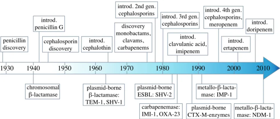
The first β-lactam, penicillin, was discovered by Alexander Fleming in 1928 (Fleming, 1929). He performed a number of studies but failed to realise its potential as treatment against bacterial infections. In 1940 Chain et al. demonstrated the efficiency of penicillin as a systemic chemotherapeutic agent (Chain et al., 1940), which led to the development of penicillin for clinical use in humans and fuelled the discovery of additional antibiotics. In many cases the introduction of drugs into clinical use was quickly followed by the appearance and spread of bacterial resistance mechanisms (Figure 1). In the case of penicillins, penicillin-degrading enzymes were identified even before the clinical introduction of penicillin G (benzylpenicillin) (Abraham and Chain, 1940).
Figure 1. Timeline of key events 80 years after β-lactam discovery. The time of discovery and clinical introduction of β-lactam antibiotics are depicted above the arrow. Events beneath the arrow mark key events in the evolution of β-lactam re-sistance.
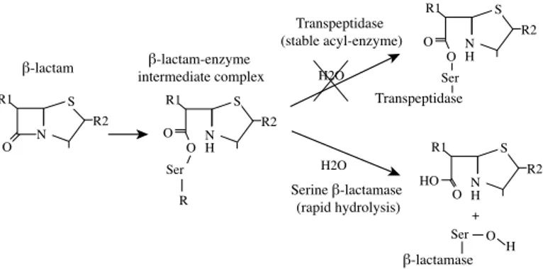
β-lactam antibiotics inhibit bacterial cell wall biosynthesis. Cell wall synthe-sis is mediated by transpeptidases, transglycosylases and carboxylases, which build N-acetylglucosamine and N-acetylmuramic acid peptidoglycan chains and cross-link them to form peptidoglycan. Naturally, transpeptidases react with the peptide D-alanyl-D-alanine side chain to form an acyl-enzyme intermediate. The structure of transpeptidases excludes water molecules from the active site to allow reaction of the acyl-enzyme intermediate with the amine group of the neighbouring chain. In the presence of β-lactams, the β-lactam ring is mistaken by the transpeptidases for a yet to be cross-linked peptidoglycan side chain and consequently they bind to it. The transpepti-dases are therefore called penicillin-binding proteins (PBPs). As illustrated in figure 2, the PBPs will form the acyl-enzyme intermediate complex with a β-lactam, but with the exclusion of water and no amine group in the proxim-ity the transpeptidase is trapped in this inactive form. Stabilisation of the peptidoglycan layer and growth of the cell will be significantly reduced and depending on the bound PBP this can lead to cell lysis (Walsh and Walsh, 2003). 1930 1940 1950 1960 1970 1980 1990 2000 penicillin discovery introd. penicillin G cephalosporin discovery discovery monobactams, clavams, carbapenems introd. clavulanic acid, imipenem introd. 3rd gen. cephalosporins introd. 4th gen. cephalosporins, meropenem chromosomal introd. 2nd gen. cephalosporins introd. ertapenem introd. doripenem 2010 introd. cephalothin
N O R1 R2 Ser O H N O R1 R2 N R1 R2 H H O O O Transpeptidase Ser H2O R O N R1 R2 H O H β-lactamase + β-lactam Transpeptidase (stable acyl-enzyme) Serine β-lactamase (rapid hydrolysis) H2O β-lactam-enzyme intermediate complex S S Ser S S
Figure 2. Reaction of β-lactam antibiotics with transpeptidase (penicillin-binding protein) or serine-β-lactamase. The conformation of transpeptidases excludes water from the active site and the enzyme stays inactive and bound to the β-lactam. Water molecules can freely access the active site of serine-β-lactamases and the antibiotic is rapidly hydrolysed.
PBPs are divided into low-molecular mass (LMM) and high-molecular mass (HMM) PBPs. The LMM PBPs in E. coli (PBP4-7, PBP4b, PBP6b, AmpH) are involved in cell separation, peptidoglycan maturation and recycling (Popham and Young, 2003; Sauvage et al., 2008). These enzymes are not essential, but studies have shown that they are needed to maintain uniform cell shape and deletion of at least three LMM enzymes caused significant morphological defects in E. coli cells (Nelson and Young, 2001). There are five HMM PBPs in E. coli. PBP1a and PBP1b are the major peptidoglycan synthetases with transglycosylase and transpeptidase activity. Deletion of one of these enzymes is tolerated but deletion of both is lethal. PBP1b has been shown to interact with PBP3, suggesting a role in localisation or regu-lation of cell division (Bertsche et al., 2006). PBP1c is less well studied. It is similar to PBP1a and 1b in molecular weight but only has transglycosylase activity (Schiffer and Höltje, 1999). It cannot compensate for deletion of both PBP1a and 1b. PBP2 and PBP3 are monofunctional transpeptidases. PBP2 (encoded by mrdA) is essential for cell elongation and inhibition by PBP2-specific β-lactams such as mecillinam leads to the formation of spher-ical cells (Spratt, 1975). PBP3 (encoded by ftsI) is the main protein in the cell division complex and polymerises the septal peptidoglycan. When in-hibited cells grow as filaments (Sauvage et al., 2008). Each PBP has a spe-cific “binding-pattern” to various β-lactam antibiotics; penicillins are bound more strongly by PBP1a and PBP3; cephalosporins and monobactams are bound by PBP3; mecillinam and carbapenems are bound strongest by PBP2 (Curtis et al., 1979; Bush et al., 1987; Davies et al., 2008; Koga et al., 2009).
β-lactams are the best studied and most widely used class of antibiotics. They owe this to their strong bactericidal activity, low toxicity and synergis-tic effect with other classes of antibiosynergis-tics. The common feature of all
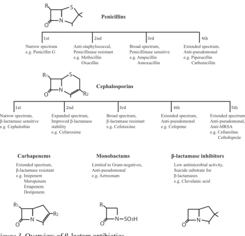
β-lactams is a 4-membered amide ring, the β-lactam ring. This ring is fused to other 5- or 6-membered rings or can stand alone, depending on the group of β-lactam. Attributes of newly developed generations of β-lactams reflect a constant struggle to stay ahead of bacterial resistance development. Mem-bers of β-lactams include:
i) Penicillins. The β-lactam ring is fused to a 5-membered sulphur ring system and this group of β-lactams is produced by Penicillium and
Aspergil-lus species of fungi. To date there are four generations of penicillins (Figure
3). The first generation is only active against Gram-positive species and following generations were developed against Gram-negative organisms. Increased stability against penicillinases is characteristic for later genera-tions as well as the good activity against Pseudomonas aeruginosa in the newest generation (Walsh and Walsh, 2003).
ii) Cephalosporins. The 4-membered β-lactam ring is fused to a 6-membered sulphur ring system. In the 1940s Giuseppe Brotzu isolated a fungus called Cephalosporidium acremonium that was later found to pro-duce a number of different cephalosporins and further studies by E. T. Abra-ham and others led to the development of cephalosporins for clinical use (Muñiz et al., 2007). Worldwide, cephalosporins are prescribed the most among the β-lactams. Modifications of the C3 and C7 side chains of the third generation cephalosporins prevent binding of β-lactamases. Fourth generation cephalosporins show greater activity against Gram-negative bac-teria, including P. aeruginosa and the fifth generation even has activity against methicillin-resistant Staphylococcus aureus (MRSA) (Walsh and Walsh, 2003).
iii) Monobactams. Their monocyclic structure with the β-lactam ring not fused to another ring presents a departure from the usual 2-ring structure of other β-lactams. Monobactams are not effective against Gram-positive bac-teria but show good antimicrobial activity against Gram-negative bacbac-teria, including P. aeruginosa. Aztreonam is the only commercially available monobactam.
iv) β-lactamase inhibitors. With the emergence of β-lactamases the need for β-lactamase inhibitors grew. These are not antibiotics but substances that target β-lactamases and are administered together with a β-lactams to protect them from degradation. Olivanic acid was discovered and shortly thereafter clavulanic acid in 1976, which became the first clinically available β-lactamase inhibitor (Papp-Wallace et al., 2011).
v) Carbapenems. The first carbapenem was thienamycin from
Streptomy-ces cattleya and all other carbapenems are derived from it, starting with
imipenem, which became clinically available in 1985. Carbapenems are the focus of this thesis and members of this group of β-lactams are described in more detail in the following chapter (Page 18).
Figure 3. Overview of β-lactam antibiotics.
Carbapenems
Carbapenems are part of the β-lactam class of antibiotics. They have a 4-membered β-lactam ring fused to a thiazolidinic 5-4-membered ring (Figure 4) and were discovered in the 1970s. The first carbapenem, thienamycin from
S. cattleya, was not pursued for clinical use due to its instability in aqueous
solutions. Imipenem was discovered shortly after and is more stable in solu-tion. It needs to be administered with another compound, cilastatin, to pro-tect it from degradation by human renal dehydropeptidase I (DHP-1). Later additions to the group of carbapenems have a 1-β-methyl substituent at C1 (marked in bold in figure 4) making them stable against DHP-1 degradation. The hydroxyethyl group in trans-configuration at C6 of all carbapenems confers stability to serine-β-lactamases (Moellering et al., 1989; Nicoletti et
al., 2002; Papp-Wallace et al., 2011). Because of this stability, carbapenems
are primarily used for treatment of severe infections caused by otherwise resistant bacteria such as extended spectrum β-lactamase-producers.
N O Penicillins Cephalosporins Extended spectrum, Meropenem 1st Extended spectrum, 4th 2nd 3rd N O S R 1st 5th
Extended spectrum, Extended spectrum, 4th 2nd 3rd N O R1 S R2 R1 R2 N O R SO3H N O Carbapenems Monobactams
Figure 4. Chemical structure of carbapenems. (a) Imipenem, (b) Meropenem, (c) Ertapenem, (d) Doripenem.
All carbapenems bind strongly to penicillin-binding protein 2 (PBP2) in E.
coli and show varying affinity to PBP3 and other PBPs. Imipenem binds
most strongly to PBP2, followed by PBP1a and 1b and has a weak affinity for PBP3. Ertapenem binds equally strongly to PBP2 and PBP3, whereas meropenem binds strongly to PBP2 but somewhat weaker to PBP3. Both drugs bind well to PBP1a and 1b. Doripenem binds strongly to PBP2, but shows only weak binding to PBP1a, 1b and PBP3 (Yang et al., 1995; Kohler
et al., 1999; Davies et al., 2008; Koga et al., 2009). The high affinity of
carbapenems to these essential PBPs is what facilitates their rapid bacteri-cidal effect and very broad spectrum, as they are effective against Gram-positives, Gram-negatives and both aerobes and anaerobes of these groups. The interaction with PBPs takes place on the cell surface of Gram-positive bacteria and in the periplasmic space of Gram-negative bacteria. In the latter, carbapenems must cross the outer membrane through water filled channels, called porins, to access the PBPs. Efficient penetration though porins is due to their zwitterionic properties (Kattan et al., 2008) and make carbapenems very potent. Once in contact with the PBPs, carbapenems will trap these enzymes in an inactive state and the peptidoglycan will weaken.
N OH O S CH3 1 2 3 4 5 6 7 N H O N N OH OH O O S N O N H H O OH OH O N OH O S 1 2 3 4 5 6 7 OH O NH H N H CH3 (a) (c) (b) N OH OH O O S CH3 N H N H NH S 2 O O (d) 1 2 3 4 5 6 7 1 2 3 4 5 6 7
β-lactam resistance
The use of β-lactams led to clinical resistance developing as early as the 1930s (Abraham and Chain, 1940; Hedge and Spratt, 1985; Näsvall et al., 2012; Yamachika et al., 2013). Today, there are four described mechanisms by which bacteria can become resistant to β-lactams:
i) The outer membrane of Gram-negative bacteria serves as a natural bar-rier for β-lactams, restricting the influx of antibiotics more than the Gram-positive cell wall (Walsh and Walsh, 2003). Lowering the number of outer membrane porins can further decrease the membrane permeability. Outer membrane porins are the entryway of β-lactams into the cell and the number of porins ultimately influences the antibiotic susceptibility of the cell.
ii) Efflux pumps can recognize β-lactams in the periplasm and export them back to the cell surface. This can lower the drug concentrations within the cell and confer resistance. Decreased membrane permeability and in-creased efflux often act synergistically to significantly increase resistance (Malléa et al., 1998; Källman et al., 2009).
iii) Overproduction and modification of target PBPs or acquisition of novel PBPs may render bacteria insensitive to β-lactams. Examples are the overexpression of PBP5 in Enterococcus hirae (Zapun et al., 2008) or ac-quisition of PBP2a through the staphylococcal cassette chromosome mec in
Staphylococcus aureus. No E. coli clinical isolates have been found to
ex-press modified PBPs but in vitro studies show that mutant PBP2 and PBP3 can be selected during β-lactam exposure (Hedge and Spratt, 1984; Yama-chika et al., 2013).
iv) Hydrolysis of β-lactam antibiotics by β-lactamases and car-bapenemases is the main cause for resistance development worldwide. Below I address the role of outer membrane porins, β-lactamases and car-bapenemases in β-lactam resistance development in more detail, as these factors are of special importance for this thesis.
Role of outer membrane proteins in resistance
Permeability of the outer membrane significantly affects the entry of β-lactams into the bacterial cell (Nikaido, 2003; Lartigue et al., 2007; Pagès et
al., 2008). β-lactams enter through the major non-specific outer membrane
porin proteins, called OmpC and OmpF in E. coli, that form water filled channels for the transport of nutrients. Porins are excessively regulated to enable survival under widely different condition and environmental factors such as osmolarity, temperature, pH, oxidative stress, acetyl phosphate, cer-tain toxins, antibiotics as well as the bacterial growth phase influence porin expression (Pratt et al., 1996).
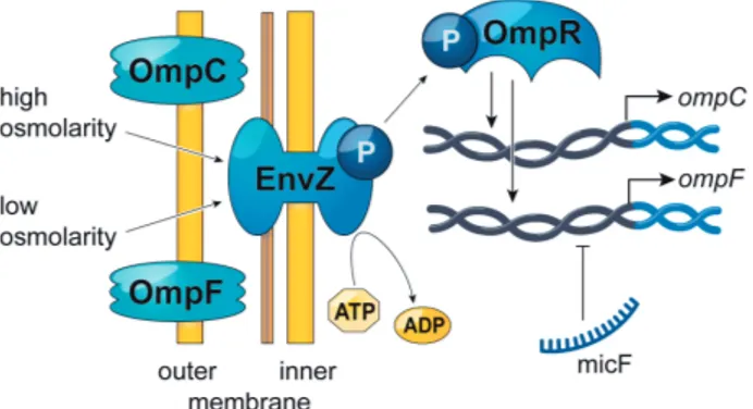
The main regulator of ompC and ompF in E. coli is the two-component regulatory system EnvZ-OmpR (Figure 5). EnvZ senses external osmolarity, is phosphorylated at residue His243 and passes this information on to OmpR by phosphorylation of Asp55 (Forst, Delgado, and Inouye, 1989b; Forst, Delgado, and Inouye, 1989a). The level of phosphorylated OmpR (OmpR-P) is controlled by EnvZ phosphatase activity. At low external osmolarity only a small amount of OmpR is phosphorylated, whereas at high osmolarity the phosphatase activity of EnvZ decreases and additional OmpR-P becomes present in the cytoplasm (Aiba and Mizuno, 1990). Phosphorylation of OmpR improves its DNA-binding activity and results in tandem-binding to regions upstream of the ompC and ompF promoters (Harlocker et al., 1995). These regions are three 20-basepair units located at -100 to -38bp from
ompC (C1, C2 and C3) and -100 to -39bp from ompF (F1, F2 and F3) with
an additional fourth ompF-binding site (F4) at -380 to -361bp. There is a distinct hierarchy in binding of these sites where the F1 and C1 sites are bound first. This increases the OmpR-P affinity to subsequent sites. The F2 site is bound next and at this point OmpF can be expressed, but not OmpC. Binding of the F3 site happens almost simultaneously with the F2 site, but higher concentrations of OmpR-P are required for binding of C2 and C3. At increased levels C2 and C3 are bound and OmpC is expressed. The F4 site is also bound at these high OmpR-P concentrations but this binding instead blocks OmpF expression by a DNA loop formation. This complex binding pattern (F1, C1 > F2, F3 > C2 > C3, F4) regulates coupled expression of OmpC and OmpF.
Expression of ompF is also regulated by the sRNA MicF. MicF binds to the translation-initiation region of the ompF mRNA and represses expres-sion. Expression of micF in turn is regulated by a number of transcriptional regulators in response to osmolarity, temperature, acids or antibiotics, in-cluding OmpR and MarA (Pratt et al., 1996; la Cruz and Calva, 2010).
The pores of OmpC and OmpF have different diameters (Nikaido and Rosenberg, 1983) and the observed regulation mainly serves to adapt to the osmolarity of the surrounding medium and to evade toxic molecules, such as bile salts or antibiotics (Nikaido, 2003). The smaller pore of OmpC is main-ly expressed in high osmolarity environments where the presence of toxins is more likely. Low osmolarity environments may contain less toxic com-pounds and here the larger pore of OmpF is advantageous to allow more general influx of scarce nutrients.
Figure 5. Main regulators of ompC and ompF gene expression.
Porins are the entryway for β-lactams and other hydrophilic antibiotics, e.g. tetracycline, chloramphenicol and fluoroquinolones into the bacterial cell (Nikaido, 2003). Any changes that affect this route can lead to increased tolerance to these drugs. Loss-of-function mutations in the porin sequence (Doumith et al., 2009) or mutations in the porin regulatory genes (Paper I) will decrease the number of channels. Cells can also replace the large gen-eral porins by channels with narrower pores or substrate specific channels (Doménech-Sánchez et al., 1999; García-Sureda et al., 2011). It is argued that porin-deficiency alone leads to only small increases in drug tolerance and that synergy with efflux complexes (Nikaido, 2001) or inactivation through β-lactamases (Nikaido, 1989) are necessary for high level re-sistance.
Little is known about regulation of OmpK35 and OmpK36, the main pore proteins in Klebsiella pneumoniae (homologues to OmpF and OmpC in E.
coli), but homologues of EnvZ and OmpR are present. In K. pneumoniae
porin loss in combination with expression of β-lactamases has frequently been associated with resistance to β-lactams and carbapenems (Doménech-Sánchez et al., 1999; Woodford et al., 2007; Doumith et al., 2009).
Inactivation by β-lactamases
One of the major resistance mechanisms conferring β-lactam resistance is hydrolysis of the β-lactam ring by β-lactamases (Figure 2). β-lactamases may have evolved from penicillin-binding proteins (PBPs) (Massova and Mobashery, 1998). Both kind of enzymes recognise the β-lactam ring and form the acyl-enzyme intermediate, but only β-lactamases allow water mol-ecules to enter their active site. This allows rapid hydrolysis of the β-lactam and the β-lactamase is free to hydrolyse more β-lactam molecules. In con-trast, PBPs exclude water from their active site and remain inactively bound to the β-lactam. Penicillinase was the first β-lactamase discovered (Abraham and Chain, 1940) and in 1983 was followed by the discovery of the first plasmid-encoded extended spectrum β-lactamase (ESBL) in Gram-negative
bacteria (Knothe et al., 1983). There are two main types of β-lactamases, serine-lactamases with a serine residue at the active site and metallo β-lactamases, which employ zinc ions as a co-factor. β-lactamase nomencla-ture is complicated and the names can refer to the organism from which the enzyme was first isolated (e.g., KLUA from Kluyvera ascorbata), the place of isolation (e.g., NDM from New Delhi metallo-β-lactamase) or the name of a patient (e.g., TEM from patient Temoneira). A collection of more exam-ples has been made by Jacoby (Jacoby, 2006). The enzymes are currently grouped according to two main classification schemes. Ambler’s molecular classification is based on amino acid sequence (Ambler, 1980). Here Class A, C and D enzymes use serine in their active site and Class B enzymes requires zinc ions to hydrolyse the β-lactam ring. The functional classifica-tion scheme of Bush assigns β-lactamases into groups 1-4 and uses a more phenotypical approach, taking substrate and inhibitor profiles into account (Bush, 1989). Functional group 4 has been omitted in a recent update of this classification because these enzymes were assigned to other groups as more information became available (Bush and Jacoby, 2010).
β-lactamases can hydrolyse all penicillins and early cephalosporins. ESBLs have evolved in response to newer β-lactam antibiotics and the name reflects the expanded substrate spectrum of these enzymes. In addition to the substrate of broad-spectrum β-lactamases, these enzymes can hydrolyse third and forth generation cephalosporins and monobactams. They are most often found in K. pneumoniae and E. coli, but also in Acinetobacter
bau-mannii, P. aeruginosa, Salmonella typhimurium, Enterobacter sp., Proteus sp., Serratia sp. and others (Jacoby and Munoz-Price, 2005). Giske et al.
(Giske et al., 2009) recently proposed a new nomenclature for ESBLs, in an attempt to increase comprehension in the clinic and for the broader public. Here ESBLs of the molecular Class A (ESBLA) and other Classes (miscella-neous ESBLs,ESBLM) are separated from ESBLs with activity against car-bapenems (ESBLCARBA).
Described below are β-lactamases important for the work performed in this thesis.
TEM-enzymes
The β-lactamase TEM-1 was identified in 1963 and was named after patient Temoneira from which the first sample was obtained (Salverda et al., 2010). It is a member of the molecular Class A and functional group 2b. TEM-1 can hydrolyse penicillins and early cephalosporins and used to be responsible for 90% of the ampicillin resistance in E. coli (Bradford, 2001). Now CTX-M-type enzymes are more prominent and widespread (Naseer and Sundsfjord, 2011). All TEM-enzymes are plasmid borne derivatives of TEM-1, and there are 217 TEM-enzymes described (http://www.lahey.org/Studies/). TEM-3 (a TEM-2 derivative) was reported in 1988 to be the first TEM-enzyme
dis-tive against penicillins and broad-spectrum cephalosporins with higher activ-ity against ceftazidime than cefotaxime (Bush’s group 2be, Giske’s ESBLA class) and some derivatives are inhibitor resistant ESBLs (Bush’s group 2br). Other derivatives are known to have a substrate spectrum similar to 1 but are not inhibited by clavulanic acid. The mobilisation of TEM-genes is associated with Tn3-like structures (Salverda et al., 2010). The rap-id worldwrap-ide spread of TEM-enzymes during the 1960s and 1970s world-wide is only comparable to recent global advances of CTX-M-type enzymes.
OXA-enzymes
The OXA-1 β-lactamase belongs to the molecular Class D and functional group 2d, it utilizes serine in its active site and is poorly inhibited by clavu-lanic acid. It is active against ampicillin and cephalothin and was named after its high hydrolytic activity for oxacillin and cloxacillin (oxacillinase). There are currently 404 OXA-derivatives (http://www.lahey.org/Studies/) but members of the group are extremely diverse, sharing only 20% sequence similarity (Bradford, 2001). It has been shown that OXA-genes originate from P. aeruginosa (Cantón et al., 2008) and have mobilised to plasmids at least three times, millions of years ago (Barlow and Hall, 2002). They can also be found in E. coli, S. typhimurium, K. pneumoniae and Proteus
mirabi-lis (Naas and Nordmann, 1999). OXA-ESBLs are derived from OXA-10 and
OXA-2 and several enzymes have been reported to be sensitive to clavulanic acid (Rasmussen et al., 1994; Philippon et al., 1997).
CTX-M-enzymes
CTX-M-enzymes were named for their preferred activity against cefotaxime over that of other oxyimino-β-lactams (Gniadkowski, 2008) and their place of isolation, Munich (German), in the late 1980s (Matsumoto et al., 1988; Bauernfeind et al., 1990). CTX-M-enzymes belong to the Ambler Class A, and Bush’s functional group 2be, and ESBLA according to Giske’s classifi-cation. They are mainly found in K. pneumoniae and E. coli but originate from genomes of environmental Kluyvera sp. (Bonnet, 2004). Currently there are 151 CTX-M derivatives (http://www.lahey.org/Studies/) clustered into 5 groups based on amino acid homology: CTX-M cluster 1A, 1B, 2, 8, 9 and 25 (Bonnet, 2004; Naseer and Sundsfjord, 2011), with the CTX-M-1 cluster including some of the major clinical β-lactamases. There are approx-imately 226 nucleotide mutations between the different groups and their most recent common ancestor (Barlow et al., 2008). Their efficient world-wide spread is believed to be caused by an unusually frequent mobilization, eight times, compared with other class A β-lactamases (Barlow et al., 2008) and it is believed that this mobilisation occurred recently, because some CTX-M-gene sequences on plasmids still exactly match the sequence of some Kluyvera chromosomal genes. The rate of mobilisation and spread of CTX-M enzymes has led to the present CTX-M-pandemic (Cantón and
Coque, 2006). CTX-M-15 is a member of the CTX-M-1A (Naseer and Sundsfjord, 2011) cluster originating from the chromosome of Kluyvera
ascorbata (Rodríguez et al., 2004). Specific point mutations allow
CTX-M-15 to hydrolyse ceftazidime at high levels (Bonnet, 2004). The first plasmid described to encode CTX-M-15 was pC15-1a (Boyd et al., 2004). Together with other enzymes from the CTX-M-1, CTX-M-2 and CTX-M-9 clusters, CTX-M-15 has been captured by ISEcp1-like insertion sequences (Bonnet, 2004). These IS elements can mobilize adjacent DNA by recognizing a vari-ety of DNA sequences. It is proposed that the promoter of ISEcp1-like ele-ments provides higher expression levels for CTX-M genes in Enterobacteri-aceae than in Kluyvera species (Poirel et al., 2003).
Inactivation by Carbapenemases
The strong selective pressure posed on bacteria by the use of carbapenems has facilitated the emergence and spread of carbapenemases in β-lactamase classes A, B and D in clinical settings (Nordmann et al., 2011; Cantón, Akóva, et al., 2012). Carbapenemases are defined as β-lactamases with ac-tivity against most β-lactams and measurable acac-tivity against carbapenems (Livermore, 1992; Queenan and Bush, 2007) and can naturally be encoded on the chromosome, acquired to the chromosome, or be plasmid encoded. Just like other β-lactamases, these enzymes can be grouped according to their amino acid sequence in the Ambler classification, depending on sub-strate and inhibition profile in Bush’s functional groups and in Giske’s new-ly proposed simplified scheme. Currentnew-ly, carbapenemases with the greatest clinical relevance in Europe are KPC-enzymes, OXA-carbapenemases and enzymes of the metallo-β-lactamase group. Below, I shortly describe these carbapenemases as their clinical importance makes them interesting for this thesis.
Serine-carbapenemases
KPC-derivatives (K. pneumoniae carbapenemase) belong to Ambler class A, Bush’s group 2f and Giske’s ESBLCARBA-A and are mainly encoded on plasmids, which has facilitated their worldwide spread. Currently there are 18 KPC-derivatives (http://www.lahey.org/Studies/).
OXA-carbapenemases are clustered into nine subgroups (Queenan and Bush, 2007; Walther-Rasmussen and Høiby, 2007), which share 40 to 70% sequence similarity and are only remotely related to Class D β-lactamases. Many OXA-type carbapenemases hydrolyse oxacillin at much lower rates than Class D oxacillinases and have only low hydrolytic activities against imipenem and meropenem. Bacteria expressing these enzymes often require additional mechanisms to provide clinical resistance levels. 23, OXA-40, OXA-48 and OXA-58 are the only plasmid borne OXA-type
car-bapenemases. It is possible that the mobile ancestors of OXA-carbapenemases disappeared long ago.
Metallo-β-lactamases
VIM-enzymes (Verona integron-encoded metallo-β-lactamase, 41 deriva-tives) and IMP-enzymes (active on imipenem, 48 derivaderiva-tives) were initially found in P. aeruginosa and are associated with class 1 integrons (Nordmann and Poirel, 2002). The VIM and IMP-type enzymes have the largest num-bers of enzyme derivatives among the metallo-β-lactamases and outbreaks with bacteria producing these have been reported worldwide. However, the greatest clinical impact has been made by NDM-enzymes (New Delhi metal-lo-β-lactamase, 11 derivatives) (Yong et al., 2009). NDM-1 was first report-ed in 2009, since then it has spread to most parts of the world and is most often found in K. pneumoniae and E. coli. It is associated with diverse plas-mids and infections were initially connected to the Indian subcontinent, where NDM-enzymes are very frequent in the human population and can even be found in the environment (Nordmann et al., 2011).
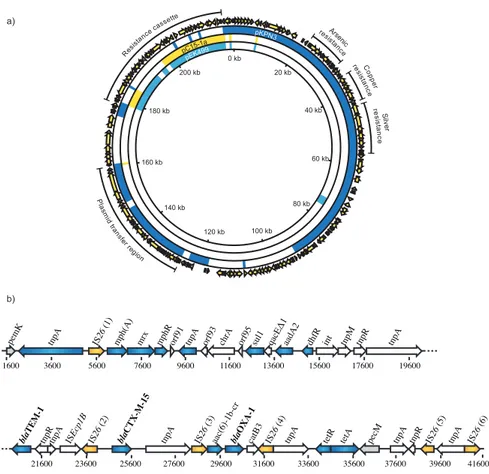
ESBL plasmid pUUH239.2
Plasmids are circular DNA molecules that replicate independently of the bacterial chromosome. They frequently encode proteins for their own trans-fer, but also virulence factors, novel metabolic proteins or proteins for anti-biotic resistance. They are not essential for bacterial survival, but can be advantageous under certain conditions. The 220 kilo base pairs (kb) plasmid pUUH239.2 was isolated from the K. pneumoniae clone that caused the first major ESBL outbreak in Scandinavia (Lytsy et al., 2008) in 2005 at Uppsala University Hospital, Uppsala, Sweden. It has been fully sequenced and ana-lysed (Sandegren et al., 2011).
Figure 6. pUUH239.2. (a) Complete view of pUUH239.2 and alignment to pKPN3, pC15-1a and pEK499. (b) Simplified pUUH239.2 resistance cassette with resistance genes depicted in blue and IS26 elements in yellow.
The plasmid is a recombinant formed from the plasmid backbone of pKPN3 from K. pneumoniae and a multidrug resistance cassette commonly found in IncFII plasmids, like pEK499 (Woodford et al., 2009), and pC15-1a (Boyd
et al., 2004) from E. coli (Figure 6a). The pKPN3 backbone encodes
pro-teins required for plasmid maintenance, transfer, and resistance to arsenic, copper and silver. The size of the resistance cassette is 41 kb and it encodes resistance against macrolides [mph(A), mrx, mphR(A)], chromate (chrA), sulfonamides (sul1), aminoglycosides (aadA2), aminoglyco-sides/fluoroquinolones [aac(6’)-1b-cr], trimethoprim (dhfrXII), β-lactams (blaTEM-1, blaOXA-1), tetracycline [tet(A), tetR], and the ESBL gene blaCTX-M-15.
It is flanked by two IS26 elements with four other IS26 elements spread out across the cassette, which have been involved in rearrangements of this re-gion. The presence of six identical IS26 elements in the resistance cassette provide regions of homology for GDA. This makes pUUH239.2 a good
200 kb 180 kb 160 kb 140 kb 120 kb 100 kb 80 kb 60 kb 40 kb 20 kb 0 kb Pla sm id tra ns fer re gion C op per resista nce Res istan ce ca ssette Arsen ic re sista nce re sis tan ce S ilv er pC15-1a pEK499 pKPN3 pecM tetR tetA IS26 (3) bla OXA-1 IS26 (1) IS26 (5) IS26 (4) IS26 (6) IS26 (2) bla TEM-1 bla CTX-M-15 pemK tnpA catB3 tnpR
aac(6)-1b-cr tnpA tnpA
tnpA tnpA ISEcp1B tnpR tnpA tnpR tnpM aadA2 dhfR int tnpA mrx mphR chrA sul1 mph(A)
tnpA orf91 orf93 orf95
1600 3600 5600 7600 9600 11600 13600 15600 23600 21600 19600 17600 29600 27600 25600 31600 33600 35600 37600 39600 41600 a) b)
tion. Studies of the fitness cost identified a 4% reduction in the growth rate of E. coli and K. pneumoniae cells carrying this ESBL-plasmid plasmid.
Gene duplication and amplification
Gene duplication and amplification (GDA) is a genetic phenomenon ob-served in all three kingdoms of life (Devonshire and Field, 1991; Romero and Palacios, 1997; Wong et al., 2007; Andersson and Hughes, 2009; Sandegren and Andersson, 2009). It provides genetic variation in popula-tions, short-term solutions to environmental challenges, and a source of ge-nome complexity. Together with de novo emergence of new genes from previously non-coding DNA sequences (Kaessmann, 2010; Tautz and Domazet-Lošo, 2011), GDA is believed to be the major contributor to gen-erating genetic diversity in all types of organisms. In medicine GDA has been shown to cause a number of human diseases (Lee and Lupski, 2006; Lupski, 2007; McCarroll and Altshuler, 2007). Understand the mechanisms and dynamics of GDA is of fundamental evolutionary interest.
Dynamics
Gene duplication and amplifications (GDAs) are very frequent in a growing bacterial population, >10-2
∼10-5 per cell per generation, depending on the gene and region of the genome (Anderson and Roth, 1981). Any region on the chromosome can be affected and the duplicated region can range from a few base pairs to several Mega base pairs (Mb) (Straus and D'Ari Straus, 1976; Anderson and Roth, 1981; Nilsson et al., 2006; Kugelberg et al., 2006; Andersson and Hughes, 2009; Sandegren and Andersson, 2009). From this data it has been estimated that at any time 10% of all cells in an unse-lected growing population have a duplication somewhere on the chromo-some (Roth et al., 1996).
The steady-state of GDA is many orders of magnitude higher than the base substitution rates determined in several locations in the bacterial chro-mosome, 10-11∼10-10 per cell per generation (Hudson et al., 2002), or for instance mutation rates for spontaneous carbapenem resistance, 10-10
∼10-7 per cell per generation (Paper I). This suggests that subpopulations with GDAs are likely enriched in response to selective pressures rather than rare mutation, assuming that GDAs can alleviate the selective pressure. Duplicat-ed genes can confer resistance during antibiotic treatment of patients (Mush-er et al., 2002; Hammond et al., 2005; B(Mush-ertini et al., 2007; Brochet et al., 2008; Sandegren and Andersson, 2009; Huang et al., 2013) and with the above-mentioned steady state frequencies of GDA it is very likely that am-plification of resistance genes are present in a population. Standard growth
conditions in clinical laboratories are usually non-selective and GDA can be lost from the population after routine clinical isolation resulting in the un-derestimation of GDA as clinically relevant resistance mechanism.
Studies need to be specifically designed to detect GDA and some tech-niques that are commonly used are transduction assays, pulsed-field gel elec-trophoresis (PFGE), next generation sequencing and quantitative real-time polymerase chain reaction (qRT-PCR) (Andersson and Hughes, 2009; Elliott
et al., 2013). In a transduction assay, an auxotrophic phenotype is caused by
insertion of an antibiotic resistance cassette and this auxotrophic mutation is subsequently moved by transduction into a recipient that is suspected to carry a duplication of this gene. The transferred auxotrophy will not be de-tectable if the inactivated gene is indeed present in at least two copies, be-cause only one copy is inactivated by the antibiotic marker. This technique is only suitable for detection of large duplicated regions. PFGE can upon di-gestion of chromosomal or plasmid DNA show changes in the banding pat-tern as compared to non-amplified DNA. Amplifications between digestion sites will cause larger DNA fragments and additional fragments will appear if the amplification covers a restriction site. Next generation sequencing detects amplification joint points as novel junction sequences (Sun et al., 2012). It also allows a rough estimate of the number of additional gene cop-ies, because the number of sequence reads covering the region will increase proportionally to the number of gene copies. qRT-PCR is useful if the dupli-cated sequence is known and then it is a valuable technique to verify the presence of the amplification in particular isolates and make precise meas-urements of the number of gene copies.
The loss rates of GDA have been determined experimentally to be as high as 0.15 per cell per generation (Roth et al., 1996; Pettersson et al., 2009) and the fitness costs of duplications in absence of selection can range from indis-tinguishable from the unduplicated wild type to as high as 20% growth rate reduction (Pettersson et al., 2009). The cost does not depend on the size of the duplication and therefore it is unlikely that it is caused by carriage and replication of additional DNA. The cost of replicating the complete genome is 2% of the energy budget of the cell (Lane and Martin, 2010) and does not explain costs larger than that for the replication of amplified DNA. The cost might be due to expression of duplicated genes and production of protein by ribosomes and the use of amino acids. In addition to that the translated pro-teins can take part in energy-requiring metabolic reactions (Koskiniemi et
al., 2012) and this can lead to decreased growth rates. Also increased
amounts of RNA or protein can affect regulatory processes and lead to im-balanced regulation for instance in a positive feedback loop (Mileyko et al., 2008).
Mechanism of formation and loss of GDA
Two mechanisms have been described for the initial duplication formation depending on whether RecA is required or not. Non-equal homologous re-combination requires RecA and long directly oriented sequence repeats, e.g. ribosomal RNA operons (Anderson and Roth, 1981), rearrangement hot spot (rhs) sequences (Lin et al., 1984), insertion sequences (IS) (Nichols and Guay, 1989; Nicoloff et al., 2007) or transposable elements. This leads to the duplication of the region between the repeat sequences and a novel joint sequence. The importance of RecA has been demonstrated recently (Reams
et al., 2010), but a recA mutant, essentially unable to perform homologous
recombination, only experiences ten-fold lower duplication rates. This indi-cates that a substantial amount of duplication events occur in a RecA-independent manner and that other mechanisms must be capable of duplica-tion formaduplica-tion. These mechanisms include strand slippage during DNA rep-lication or repair, pairing of single stranded regions at the reprep-lication fork and erroneous ligation of DNA ends by DNA gyrase (Trinh and Sinden, 1993; Lovett et al., 1993; Ikeda et al., 2004). This can result in duplication of genomic regions by recombination between very short repeats or without repetitive sequences.
The initial duplication formation event is the rate-limiting step for GDA and further amplification may be achieved by selection for increased gene dosage. This mechanism involves RecA-mediated homologous recombina-tion between the large repeats provided by the initial duplicarecombina-tion. It is also possible to form amplifications without the initial duplication. Here a rolling cycle replication-type mechanism could generate a large tandem array in a single generation (Petit et al., 1992).
With regard to the loss of GDA, RecA-mediated homologous recombina-tion is the main mechanism and many studies showed that recA mutarecombina-tions can stabilize GDA (Hill et al., 1969; Lin et al., 1984; Haack and Roth, 1995; Andersson et al., 1998; Reams et al., 2010). In some plasmid systems how-ever, deletion between repeat sequences occurred through RecA-independent mechanisms (Matfield et al., 1985; Lovett et al., 1993).
Evolution of new genes by duplication-divergence
Gene duplications are central to many theories of evolution of new gene functions. In the model proposed by Susumu Ohno (Ohno, 1970), after du-plication, the second gene copy is released from selection pressure to per-form the initial function and free to accumulate beneficial mutations. It may eventually develop a new function, while the other copy retains the original function. This model became known as the neofunctionalization model (Force et al., 1999). One problem is that the “free copy” is subject to random drift, gene conversion or may acquire deleterious mutations that would turn
it into a pseudogene rather than acquire a new function (Lynch, 2000). An-other problem is that gene duplications often confer fitness costs (as men-tioned above) and are therefore counterselected. Thus, it is unlikely that the second copy will remain in the population in high frequencies and for suffi-ciently long to acquire rare beneficial mutations. Two mechanisms have been suggested that would help retaining the second copy. After duplication, subfunctions of the gene may be divided onto both gene copies by a series of inactivating mutations (sub-functionalization model) (Force et al., 1999; Lynch and Force, 2000). Assuming that both subfunctions are important for cell growth, both gene copies are stabilized. Secondly, GDA has been shown in response to a number of limitations, such as starvation, exposure to anti-biotics, or deleterious mutations by supplying a higher dosage of the limiting gene (Normark et al., 1977; Neuberger and Hartley, 1981; Sonti and Roth, 1989; Nichols and Guay, 1989; Hammond et al., 2005; Nilsson et al., 2006; Nicoloff et al., 2007; Paulander et al., 2007; Sun et al., 2009; Lind et al., 2010; Paulander et al., 2010; Pränting and Andersson, 2011; Nicoloff and Andersson, 2013). GDA can be a rapid, short-term solution for environmen-tal challenges or facilitate the evolution of new gene functions. If the solu-tion is a beneficial mutasolu-tion within the amplified region then GDA increases the target size for beneficial mutations. GDAs that improve growth allow the population to expand and increase the likelihood to acquire beneficial muta-tions, even outside the amplified region.
In the recently proposed innovation-amplification-divergence (IAD) model (Bergthorsson et al., 2007) selection acts at every step of the process to stabilize the duplicated copy. i) A minor secondary protein function, en-coded by the gene, is present before the duplication event (innovation). This minor function proves beneficial under new environmental circumstances (e.g., presence of antibiotic or availability of novel nutrient). ii) Selection will promote any increase in the amount of the trace activity and duplication and subsequent amplification will be enriched in the population (amplifica-tion). The amplified state will be maintained and adjusted (as far as the po-tential fitness cost allows) to meet the environmental challenge. For instance an increase in the antibiotic concentration may further increase the number of gene copies. iii) Extra gene copies increase the mutational target and the probability to acquire beneficial mutations. Selection will favour beneficial mutations as they may improve the trace activity (divergence). Improved gene copies can undergo further amplification cycles, while less beneficial copies are lost from the gene array. As more proficient copies emerge, selec-tion of GDA is relaxed on remaining copies and the gene array may shorten until one copy with improved function remains. If this improved copy has lost its original function , a copy of the original gene must remain and the cell is left with two specialist genes, encoding mainly their selected function. Other outcomes are possible, where a very proficient generalist replaces the
original copy to perform both functions, or combinations of generalists with improved genes encoding either the old or new function (Näsvall et al., 2012).
In summary, GDAs are likely to be the initial response to many selective pressures due to their high frequency in unselected bacterial populations. The intrinsic instability and potential cost however will cause GDAs to be lost rapidly. Selection can act to maintain the amplified state and allow bene-ficial mutations to accumulate until a new gene function can emerge.
Escherichia coli and Klebsiella pneumoniae as
pathogens
E. coli and K. pneumoniae are Gram-negative rod-shaped bacteria of the
family of Enterobacteriaceae. This family is of clinical importance harbour-ing many other potential pathogenic genera such as Citrobacter sp.,
Entero-bacter sp., Proteus sp., Pseudomonas sp., Salmonella sp., Shigella sp., and Yersinia sp.. E. coli and K. pneumoniae are part of the normal intestinal flora
in animals and humans and are harmless most of the time. They mainly cause infections in immuno-compromised patients (acting as opportunistic pathogens). Otherwise healthy individuals can be infected by specific strains that have acquired virulence factors or if the bacteria have breached the in-nate immunity’s defences through catheters or ventilators. Both species are the main cause of nosocomial Gram-negative bacteraemia and most often cause urinary tract infections. Some E. coli variants cause severe and even fatal diarrheal diseases. In this study we utilised E. coli MG1655. Due to its fast growth, large population sizes and fully sequenced genome, E. coli is one of the main workhorses of bacterial genetics (Blattner et al., 1997).
K. pneumoniae is the medically most relevant species of the genus Klebsiel-la. It causes community acquired pneumonia and most often urinary tract
infections. Studies of for instance resistance development are not as straight-forward as in E. coli because genetic techniques are limited. Today K.
pneumoniae producing ESBLs or carbapenemases are more frequently