R e v i e w
open access to scientific and medical research
Open Access Full Text Article
immunocytokines: a review of molecules in clinical
development for cancer therapy
Thomas List Dario Neri
Department of Chemistry and Applied Biosciences, Swiss Federal institute of Technology (eTH Zürich), Zurich, Switzerland
Correspondence: Dario Neri Department of Chemistry and Applied Biosciences, Swiss Federal institute of Technology (eTH Zürich), wolfgang-Pauli-Strasse 10, CH-8093 Zurich, Switzerland
Tel +41 44 6337401 email neri@pharma.ethz.ch
Abstract: The concept of therapeutically enhancing the immune system’s responsiveness to
tumors is long standing. Several cytokines have been investigated in clinical trials for their therapeutic activity in cancer patients. However, substantial side effects and unfavorable phar-macokinetic properties have been a major drawback hampering the administration of therapeu-tically relevant doses. The use of recombinant antibody–cytokine fusion proteins promises to significantly enhance the therapeutic index of cytokines by targeting them to the site of disease. This review aims to provide a concise and complete overview of the preclinical data and clinical results currently available for all immunocytokines having reached clinical development.
Keywords: antibodies, cytokines, preclinical
Introduction
The possibility of boosting the immune system’s activity against tumors has been the focus of pharmaceutical research for many years. Cytokines are proteins which modulate immune responses and, for this reason, have been considered for therapeutic applications. Ever since the approval in 1995 of the first recombinant cytokine
(inter-feron [IFN]-α2) for the treatment of malignant melanoma, interest in cytokines for
cancer therapy has increased.1 To date, a number of immunostimulatory cytokines,
which have shown beneficial effects in preclinical animal models of cancer and in clini-cal studies, have received marketing authorization (eg, interleukin [IL]-2 [Proleukin™,
Aldesleukin™; Novartis, Basel, Switzerland], tumor necrosis factor [TNF]-α
[Bero-mun™; Boehringer Ingelheim, Ingelheim am Rhein, Germany], interferon [IFN]-α2
[Roferon-A™; Hoffmann-La Roche, Basel, Switzerland, Intron-A™; Merck & Co., Whitehouse Station, NJ, USA], and granulocyte-macrophage colony-stimulating fac-tor [GMCSF] [Leukine™; Genzyme, Cambridge, MA, USA, Leucomax™; Novartis, Basel, Switzerland]). In addition, immunosuppressive and immunomodulatory cytok-ines (eg, IL-4 and IL-10) have been considered for treatment of rheumatoid arthritis, psoriasis, and inflammatory bowel disorders.
At present, only a handful of cytokines is in active clinical development. Tumor eradication has been achieved in models of cancer by intratumoral or peritumoral
application of cytokines or by implantation of tumor cells expressing cytokines.2–6
Yet, these techniques are not readily applicable in the clinical setting, particularly in consideration of the fact that cancer is often a disseminated disease. Systemic administration of cytokines, on the other hand, rarely results in complete cures, and dose escalation is hindered by dose-limiting toxicities (DLTs), which in turn prevent Number of times this article has been viewed
This article was published in the following Dove Press journal: Clinical Pharmacology: Advances and Applications
the administration of potentially curative regimens. These observations indicate that cytokines are potent modulators of the immune system that can eradicate tumors if high enough concentration is achieved at the site of disease.
With the introduction of monoclonal antibody engineer-ing technology and the identification of tumor-specific and accessible antigens, the targeted delivery of cytokines has become possible. Indeed, the use of antibody–cytokine fusion proteins (immunocytokines) has the potential to improve the therapeutic index of cytokines by concentrating the payload at the site of localized or disseminated disease, thus reduc-ing side effects. A prominent example is represented by the antibody-mediated targeted delivery of IL-12, which has been shown to be at least 20 times more potent than untargeted IL-12 (ie, has achieved a better therapeutic activity at less than 1/20th of the dose of the unmodified cytokine) in a mouse
model of cancer.7
While good reviews exist on the topic of immunocy-tokines, this work focuses on immunocytokines that have reached clinical development (Table 1), aiming to provide an overview of the preclinical data that has led to clinical trials
and of emerging clinical results.8–11
Formats and biodistribution
The format of the antibody moiety responsible for the selec-tive targeting of the cytokine payload to the site of disease is of great importance, as pharmacokinetic properties of the
fusion protein directly impact on performance.11 It would be
beyond the scope of this review to discuss all existing formats in detail, but some key considerations need to be presented, to provide a basis for the analysis of clinical-stage products.
Generally, antibody–cytokine fusion proteins can be divided in two categories: (1) large fusion proteins with cytokines fused to the heavy chain of full-scale antibodies (immunoglobulin [Ig]G, .150 kDa) (Figure 1A), and (2) small fusion proteins based on antibody fragments (single-chain variable fragment [scFv], diabodies, .28 kDa) (Fig-ure 1B). Different philosophies may lead to the choice of one of these two strategies. IgG-based immunocytokines are preferred if one wishes to achieve long circulatory half-lives in blood. By contrast, smaller immunocytokines may be preferred, if one wishes to keep the blood concentrations of cytokines as low as possible, in order to prevent side-effects. Interestingly, the use of antibody moieties as delivery vehi-cles is not per se a guarantee that the product will efficiently reach the site of disease in vivo. Immunocytokines may face several potential problems that could prevent in-vivo targeting. Most target antigens are located in perivascular
structures, and immunocytokines need to extravasate in order
to selectively localize at sites of disease.12 Extreme pI
(iso-electric point) values, glycosylation, large size, and trapping by a molar excess of accessible cytokine receptors are some of the molecular mechanisms which may limit the selective localization of immunocytokines and, consequently, their
therapeutic action.12–17 IgGs typically have longer
circula-tory half-life in the range of days, which is mainly due to the interaction of the Fc (fragment, crystallizable) domain
with neonatal Fc receptors.18,19 Paradoxically, Fc receptor
binding can also have a negative impact by greatly increasing
blood clearance time of immunocytokines.20 This problem
can be solved, however, by introducing mutations in the Fc
domain.21 The tissue penetration of IgGs has been shown
to be slow and heterogenous in solid tumors which are characterized by high interstitial pressure but, because of long circulatory half-life, intact immunoglobulins display
a higher tumor uptake compared with antibody fragments.22
Small formats, such as monomeric scFvs (28 kDa) may have better tumor penetration compared with IgG, but lack the avidity effects of bivalent antibodies and typically
display short retention times at the tumor site.23–25 Bivalent
recombinant antibodies of intermediate size, such as dimeric scFv fragments (also called diabodies), scFv-Fc fusions, and minibodies (small immunoprotein [SIP]), may display long residence time at the site of disease and rapid blood
clearance.26–28 At present, immunocytokines which have
progressed to clinical trials are based on antibodies in IgG format or on scFv fragments.
Mode of action of immunocytokines
Different cytokines may activate (or inhibit) different subsets of leukocytes, thus mediating various immune processes. Most pro-inflammatory cytokine payloads (eg, IL-2, TNF, IL-12) mediate the influx of leukocytes at the site of disease. However, cytokines may differ for their activity on regulatory T-cells (IL-2 being a prominent pro-inflammatory cytokine which activates them) and on other cell types. For example, TNF has been reported not only to activate the immune system, but also to be capable of direct tumor cell killing and to promote intravascular blood coagulation of small tumor capillaries. Conversely, immunosuppressive cytokines (eg, IL-10) may exert differ-ent effects on the immune system, justifying their possible use as anti-inflammatory agents.
The mechanism of action of immunocytokines is easier to study in mice than in humans, because of the possibility to examine biopsies and to perform therapy experiments
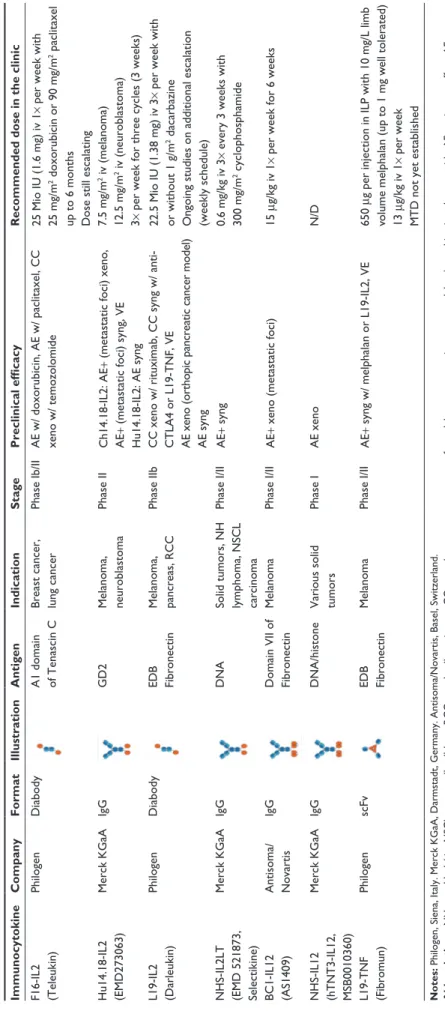
Table
1
Overview of the immunocytokines in clinical development
Immunocytokine Company Format Illustration Antigen Indication Stage Preclinical efficacy
Recommended dose in the clinic
F16-iL2 (Teleukin) Philogen Diabody A1 domain of Tenascin C Breast cancer, lung cancer
Phase ib/ ii A e w/ doxorubicin, A e w/ paclitaxel, CC xeno w/ temozolomide 25 Mio iU (1.6 mg) iv 1
× per week with
25 mg/m
2 doxorubicin or 90 mg/m 2 paclitaxel
up to 6 months Dose still escalating
Hu14.18-iL2 (e MD273063) Merck KGaA igG GD2 Melanoma, neuroblastoma Phase ii Ch14.18-iL2: A e+
(metastatic foci) xeno,
A
e+
(metastatic foci) syng,
ve Hu14.18-iL2: A e syng 7.5 mg/m 2 iv (melanoma) 12.5 mg/m 2 iv (neuroblastoma) 3×
per week for three cycles (3 weeks)
L19-iL2 (Darleukin) Philogen Diabody eDB Fibronectin Melanoma, pancreas, RCC Phase iib
CC xeno w/ rituximab, CC syng w/ anti- CTLA4 or L19-TNF,
ve
A
e xeno (orthopic pancreatic cancer model)
A
e syng
22.5 Mio
iU (1.38 mg) iv 3
× per week with
or without 1 g/m
2 dacarbazine
Ongoing studies on additional escalation (weekly schedule)
NHS-iL2LT (e MD 521873, Selectikine) Merck KGaA igG DNA
Solid tumors, NH lymphoma, NSCL carcinoma
Phase i/ii A e+ syng 0.6 mg/kg iv 3
× every 3 weeks with
300 mg/m 2 cyclophosphamide BC1-iL12 (AS1409) Antisoma/ Novartis igG Domain vii of Fibronectin Melanoma Phase i/ii A e+
xeno (metastatic foci)
15
μg/kg iv 1
× per week for 6 weeks
NHS-iL12 (hTNT3-iL12, MSB0010360) Merck KGaA igG DNA/histone v arious solid tumors Phase i A e xeno N/D L19-TNF (Fibromun) Philogen scFv eDB Fibronectin Melanoma Phase i/ii A e+ syng w/ melphalan or L19-iL2, ve 650 μg per injection in
iLP with 10 mg/L limb
volume melphalan (up to 1 mg w
ell tolerated)
13
μg/kg iv 1
× per week
MTD not yet established
Notes:
Philogen, Siena,
italy. Merck KGaA, Darmstadt, Germany. Antisoma/Novartis, Basel, Switzerland.
Abbreviations: NH, non-Hodgkin; NSCL, non-small cell lung; RCC, renal cell carcinoma; CC, complete cure; xeno, xenograft model; syng, syngeneic tumor model; w/, combination therapy with; A e, antitumor effect; A e+ , strong antitumor effect; ve
, vaccination effect; iv, intravenous administration;
iLP, isolated limb perfusion; N/D, no data available yet; MTD, maximal tolerated dose;
with selective lymphocyte depletion. The contribution of different subsets of immune cells (eg, natural killer [NK] cells and T-cells) is often model-dependent and, at least in mice, there seems to be a strong contribution from the mouse strain used.
When considering IgG-based immunocytokines, one would expect these molecules to be multifunctional, potentially leading to the crosslinking of cytokine receptor-positive cells with those components of the immune system, capable of interacting with the Fc portion of IgG. Intact antibodies can trigger antibody-dependent cellular toxicity
(ADCC) or complement dependent cytotoxicity against the
tumor cell it binds to.29,30 The relative importance of these
two mechanisms of action remains controversial.31 Finally,
it is useful to draw the analogy between immunocytokines and bispecific antibodies, since both classes of therapeutic proteins are bifunctional agents, which effectively mediate cell crosslinking. An IL-2-based immunocytokine may tether IL-2 receptor-expressing cells to the tumor cell and induce Fas/FasL (Fas/Fas-ligand)-mediated cell death in the target, a mechanism akin to the one reported for cytotoxic bispecific
antibodies.32–35 L19-IL2 F16-IL2 IgG-cytokine Hu14.18-IL2 NHS-IL2LT L19-TNF Diabody-cytokine NHS-IL12 BC1-IL12 scFv-cytokine (trimeric) VL CL VH CH1 CH2 CH3 Cytokine A B
Figure 1 Schematic representation of the protein structure of immunocytokines currently in active clinical trials. (A) immunocytokines based on antibodies in igG format. (B) immunocytokines based on antibody fragments (monomeric scFv and dimeric diabody formats).
Abbreviations: igG, immunoglobulin G; scFv, single-chain variable fragment; vL, variable domain of the immunoglobulin light chain; vH, variable domain of the immunoglobulin heavy chain; CL, constant domain of the immunoglobulin light chain; CH1, CH2, CH3, constant domains of the immunoglobulin heavy chain; TNF, tumor necrosis factor.
Current molecular targets of
immunocytokines used in the clinic
Immunocytokines have been developed against various tumor-associated antigens such as carbonic anhydrase 9 (CAIX), epidermal growth factor receptor (EGFR), epithe-lial cell-adhesion molecule (EpCAM), fibroblast activation protein (FAP), fibronectin splice variants (EDA, EDB), and tenascin splice variants. Yet only immunocytokines specificfor a few of these molecular targets have advanced to clinical trials. It is to be noted that the targets that have been explored for targeted therapy by immunocytokines encom-pass cell-surface proteins as well as components of the tumor microenvironment such as extracellular matrix proteins. In addition, two immunocytokines in clinical trials are based on an antibody which targets nuclear structures that become exposed upon cell lysis in necrotic tumors. Below, we briefly
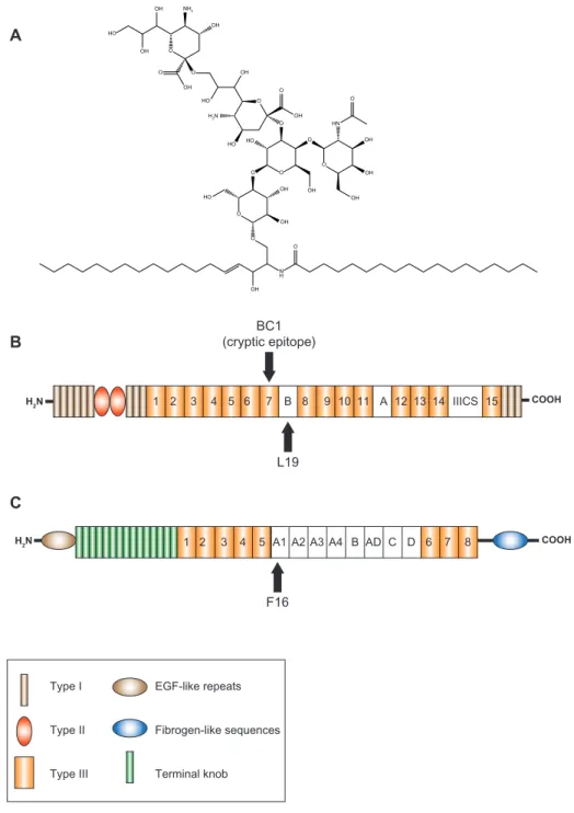
COOH L19 BC1 (cryptic epitope) COOH H2N H2N A1 A2 A3 AD 1 2 3 4 5 A4 B C D 6 7 8 F16 O N H O O HO O O OH O O OH OH OH HN O O O O O HO OH OH NH2 OH O OH HO OH H2N HO O OH HO OH OH OH Type I Type II Type III EGF-like repeats Fibrogen-like sequences Terminal knob A B C 1 2 3 4 5 6 7 B 8 9 10 11 A 12 13 14 IIICS 15
Figure 2 Schematic representation of the structures of tumor antigens targeted by immunocytokines in clinical trials: (A) fibronectin, (B) tenascin-C, and (C) disialoganglioside GD2.
Note: Arrows indicate the epitopes recognized by different antibodies. Abbreviations: eGF, epidermal growth factor; iiiCS, type iii connecting segment.
introduce the tumor antigen targets that immunocytokines in the clinic currently target.
Cell surface (disialoganglioside GD2)
Neuroblastomas, melanomas, non-B-cell lymphomas, soft tissue sarcomas, small-cell lung cancer, and some osteosarco-mas display the disialoganglioside GD2 on their cell surface (Figure 2A). In normal tissues, GD2 expression is limited to neurons, melanocytes, and peripheral pain fibers, making itan attractive tumor-associated antigen.36–41
Tumor environment
In addition to dramatic changes to the tumor cell’s genome and proteome as a result of transformation, tumors present another dimension of complexity by creating a “tumor
microenviron-ment.”42 One of the hallmarks of this microenvironment is tissue
remodeling and neo-angiogenesis. As a byproduct of these pro-cesses, disease environments can present a number of specific and accessible antigens that can be targeted therapeutically.
Fibronectin extradomain B (eDB)
Fibronectin is a large glycoprotein ubiquitous in the extracel-lular matrix of mammalian tissues and plasma. Under tissue remodeling conditions, mechanisms of alternative splicing can lead to the insertion of EDB, an extra 91-amino-acid type III
homology domain, into fibronectin (Figure 2B).43,44 In healthy
individuals, EDB is undetectable, but in many aggressive solid
tumors EDB is highly expressed around tumor vasculature.45–48
Furthermore EDB is identical in mouse, rat, rabbit, dog,
mon-key, and man.43 The high-affinity antibody L19 recognizes
EDB and has been shown to efficiently localize to tumor blood
vessels in animal models and cancer patients.26,49–53
Tenascin C A1 domain
Tenascins are glycoproteins found in the extracellular matrix of vertebrates. Like fibronectin, isoforms of tenascin can arise through alternative splicing at the site of neo-angiogenesis in tumors. In particular, the C domain of tenascin is undetectable in normal adult tissue but strongly expressed in a perivascular
pattern in brain and lung tumors (Figure 2C).44,54,55 The F16
antibody recognizes the extra-domain A1 of tenascin and has shown selective accumulation at the site of tumors and
inflammatory disorders in animals and humans.9,56
Deoxyribonucleic acid (DNA)
and necrotic tumors
Targeting the necrotic core of tumors, also known as “tumor necrotic therapy” was pioneered by the group of Alan Epstein
and relies on the antibody-based recognition of molecular components (eg, histones and DNA) released at sites of cell
death, such as necrotic areas in tumors.57–60 Anti-histone
antibodies have been used for tumor-targeting applications.61
The human NHS76 antibody was selected from phage dis-play libraries with the use of cell extracts from Burkitt’s lymphoma Raji cells and recognizes nucleic acids exposed
by necrotic tumor cells as well as metastases.62,63
IL-2-based immunocytokines
iL-2 immunology
IL-2 is a cytokine naturally produced by T-cells that stimulates the proliferation of T- and B-lymphocytes, mono-cytes, and NK cells in response to antigenic or mitogenic stimulation. Human recombinant IL-2 (Aldesleukin™/ Proleukin™) produced in bacteria has been approved for the
treatment of renal cell carcinoma and melanoma in adults.64,65
In the clinic, the agent was shown to promote complete and durable responses in a small portion (8%–10%) of patients, yet at the expense of substantial toxicities which limited the treatment to physically fit patients and to intensive care units. The most important toxicities were capillary leak syndrome,
hypotension, fever, malaise, and nausea.66–68
The contribution of NK or T-cells to the antitumor
response varies depending on the tumor model used.69,70
Despite the observed side effects, IL-2 remains the only cytokine shown to mediate cures in metastatic cancer and consequently immunocytokines based on IL-2 figure amongst
the most advanced immunocytokines in the clinic.66,71
L19-iL2 (Darleukin)
The L19 antibody was isolated from phage display libraries
as a binder of the extra-domain B of fibronectin.72 It has been
extensively used with good tumor accumulation as a vehicle for many payloads including radionuclides, photosensitizers,
coagulation factors, and cytokines.10,44,52,73–76 The L19-IL2
immunocytokine currently in Phase II clinical trials is a diabody with human IL-2 genetically fused to the C-terminus of each scFv domain, yielding a total of two IL-2 moieties per molecule.
Preclinical observations and results
L19-IL2 has both excellent tumor targeting properties in vivo, with a tumor to blood ratio of 30:1 at 24-hours postinjection, and a greatly improved therapeutic index over IL-2 cytokine alone or IL-2 fused to an antibody moiety of irrelevant specificity in a syngeneic murine teratocarcinoma
effect compared with untargeted IL-2 in an orthopic xenograft
model for human pancreatic cancer. Furthermore,79 L19-IL2
was able to completely eradicate B-cell lymphoma xeno-grafts in combination with rituximab and localized tumors in syngeneic teratocarcinoma and colon carcinoma models when combined with the blockade of anti-inflammatory receptor CTLA-4 (Cytotoxic T-Lymphocyte antigen 4) or
with another immunocytokine L19-TNF.78,80 The effector cells
responsible for the antitumor activity were identified to be NK cells in depletion experiments. It was further observed that the L19-IL2 combination therapies provided a vaccina-tion effect: cured mice re-challenged with implantavaccina-tion of the same B-cell lymphoma and colon carcinoma cell lines
did not develop tumors.77,81
Clinical results
L19-IL2 has been studied in patients with metastatic renal cell carcinoma and is currently being investigated for the treatment of melanoma and of pancreatic cancer. Patients with metastatic renal cell carcinoma are usually treated with doses of recombinant IL-2 of approximately 88–864 Mio IU/week
for a total of 6 weeks (an equivalent of 0.66 mg/kg/week).82
For the immunocytokine L19-IL2, dosage can however be reduced, as reported in the first clinical trial in humans, where the recommended dose was established at 67.5 Mio IU/week
(an equivalent of 0.05 mg/kg/week) for 4–6 weeks.83 In this
Phase I clinical trial, 83% of patients with metastatic renal cell carcinoma showed disease stabilization after two cycles of intravenous injection with L19-IL2 alone at the recom-mended dose and a median progression-free survival time
of 8 months.83 This is in line with the improved therapeutic
index of L19-IL2 compared with IL-2 alone reported in
pre-clinical therapy experiments in xenografts models.78 A later
clinical study assessed the therapeutic potential of L19-IL2 in combination with dacarbazine, by combining three L19-IL2
doses with 1 g/m2 dacarbazine. In this trial, 28% of patients
(8 of 29) with metastatic melanoma achieved an objective response, and one patient achieved a complete ongoing
response for 21 months.84 Median progression-free survival
was 14 months. The completed Phase IIa trial continues into Phase IIb (90 patients – ongoing) to assess the antitumor activ-ity in patients of the combination therapy versus dacarbazine alone. L19-IL2 is being studied in combination with gemcit-abine for the treatment of patients with pancreas cancer.
L19-IL2 was administered by infusion over 1 hour and was shown to have an in-vivo half-life of about 2–3 hours. It is not immunogenic, but injections led to transient lymphocytopenia, followed by substantial increase in the number of circulating
T-lymphocytes and NK cells after 10 days. In addition, an over-all expansion of the T-reg population was observed between
the first day and the end of the treatment cycle.83,84
L19-IL2 is well tolerated, with mild and reversible tox-icities at the recommended dose of 22.5 Mio IU/patient/day (equivalent to 1.4 mg/patient/day) in monotherapy or with dacarbazine. The absence of G3 toxicities suggests that the
dosage regimen could potentially be further increased.85,86
F16-iL2 (Teleukin)
F16-IL2 consists of the F16 monoclonal human antibody in diabody format specific for the alternatively spliced A1 domain of tenascin C, genetically fused to two molecules of human IL-2 at the C terminus of each light chain. It is being developed for the treatment of breast cancer and lung cancer
and is currently in clinical Phase Ib/II.87
Preclinical observations and results
F16-IL2 is an immunocytokine that was shown to signifi-cantly improve the antitumor potency of chemotherapeutic agents used in the clinic. Twenty micrograms of F16-IL2 in combination with a high dose of DNA intercalating agent doxorubicin (4 mg/kg) per injection resulted in a significantly improved survival time in mice bearing human breast cancer xenografts and showed a synergistic antitumor effect in com-bination with the mitotic inhibitor paclitaxel at low (1 mg/kg)
and high (5 mg/kg) dose with no observed toxicities.88
Addi-tionally, BALB/c mice bearing glioblastoma xenografts were cured of their tumors and remained tumor free for 160 days after treatment with five total administrations every third day
of 20 μg F16-IL2 in combination with 0.525 mg alkylating
agent temozolomide per injection.89 In biodistribution
stud-ies, radiolabelled F16-IL2 showed specific tumor accumu-lation of 4% ID/g at 24 hours and promoted the infiltration of leukocytes, NK cells, and macrophages into the tumor
lesions.88,89
Clinical results
F16-IL2 is currently being evaluated in Phase II clinical trials in patients with various malignancies, but with a focus on lung cancer, breast cancer, and melanoma. Because of the product’s synergy with chemotherapeutic drugs, clinical stud-ies have assessed the safety, tolerability, and recommended dose of the immunocytokine in combination with paclitaxel or doxorubicin in humans. The maximal administered doses of
doxorubicin (25 mg/m2) and paclitaxel (90 mg/m2) combined
with a recommended dose of 1.6 mg (25 Mio IU) F16-IL2 per injection (equivalent to approximately 0.02 mg/kg) over
6 months were well tolerated. Objective responses and long-lasting disease stabilization was observed in the paclitaxel combination trial in patients with non-small cell lung
carci-noma and melacarci-noma.90 In chemotherapeutically pretreated
patients with progressive non-small cell lung carcinoma and melanoma, objective responses and long-lasting disease stabilizations were observed, including two partial responses
in combination with paclitaxel.91 More recently, it has been
observed that the administration of higher doses of F16-IL2 is possible, and the product is continuing Phase Ib investiga-tions, in combination with weekly paclitaxel.
Surprisingly, pharmacokinetic analyses in patient blood
showed that the half-life was in the range of less than 1 hour,90
while the half-life was shown to be of approximately 3 hours in preclinical studies, and was not influenced by the
co-injection of doxorubicin.88
No DLTs were observed in the trial, and toxicities were mainly of grade 1 (nausea, fever, fatigue), with a few
revers-ible grade 4 toxicities.90
Hu14.18-iL2 (eMD 273063)
The ch14.18 antibody is reactive to ganglioside GD2 – a carbohydrate found on the surface of melanomas and neu-roblastomas – and contains 75% human and 25% murine
sequences.38,92 Its humanized homologue, hu14.18, contains
fully human amino acid sequences in the Fab region, with only the CDR regions being of murine origin. Both the ch14.18-IL2 and hu14.18-IL2 immunocytokine consist of an IgG antibody genetically fused to two molecules of human IL-2.
Preclinical observations and results
Fusion of IL-2 to ch14.18 did not impact the antibody’s ability to bind GD2-positive neuroblastoma cells, and ch14.18-IL2 was able to enhance ADCC-mediated
kill-ing of the tumor cells by activated lymphocytes in vitro.93
Ch14.18-IL2 was also able to stimulate proliferation of peripheral blood mononuclear cells isolated from melanoma
patients in vitro.94 In vivo, radiolabelled ch14.18-IL2
accu-mulated at the tumor site and in organs bearing experimental metastases in both a metastatic tumor xenografts model
and a syngeneic model for metastatic melanoma.70,95 Data
obtained from mouse serum ELISA (enzyme-linked immu-nosorbent assay) indicate that, surprisingly, the ch14.18-IL2 fusion protein was cleared twice as rapidly from circulation as the parental ch14.18 antibody alone, with an observed half-life of 4.1 hours for the immunocytokine. Addition-ally, ch14.18-IL2 was unstable in mouse serum in vitro and
appeared to become altered or fragmented after intravenous
injection in mice.96
Nevertheless, therapy with ch14.18-IL2 was able to markedly reduce the number of metastatic foci found in the liver of mice in a hepatic metastasis model for human neuroblastoma, where the mice were supplied with
pre-activated leukocytes.70,93,95 In syngeneic settings,
treat-ment of neuroblastoma and melanoma subcutaneous model tumors with ch14.18-IL2 mediated liver metastases eradica-tion and induced a tumor-specific vaccinaeradica-tion effect against
re-challenge with tumor cells.69,70,97,98 The antitumor effect
was originally thought to be driven by CD8+ T-cell and NK
cell populations as the therapeutic effect of ch14.18-IL2 was lost in severe combined immunodeficiency (SCID) mice. It was later shown that NK cells play a major role as the antitumor response, as the immunocytokine activity could be reduced when NK cells were depleted and enhanced with NK stimulatory agents in vivo, while the tumor infiltrating
leukocytes were mainly NK cells.99
The humanized hu14.18-IL2 immunocytokine showed modest antitumor activity in animals with a dose-dependent retardation effect followed by recurrence after several weeks. Only the combination of intravenous injections of
22 μg/day/mouse (∼70 mg/day in a human) with
continu-ous administration of IL-2 by means of a subcutanecontinu-ously
implanted osmotic pump achieved tumor eradication.99
Clinical results
Hu14.18-IL2 has progressed to Phase II clinical trials for patients with metastatic melanoma and pediatric refractory neuroblastoma. Hu14.18-IL2 is administered as a 4-hour intra-venous infusion over three consecutive days at the beginning
of each treatment cycle, for a maximum of 3 weeks.39,100–102.
In Phase I clinical trials, the maximal tolerated dose
(MTD) was established at 7.5 mg/m2/day for melanoma
patients and 12 mg/m2/day for pediatric neuroblastoma
patients.100,102 No partial or complete responses were recorded;
however, disease stabilization was achieved in 58% of mela-noma patients after the first cycle. Yet only 24% of patients
showed progression-free disease after the second cycle.102 In
a different study, using lower dosage (4 mg/m2/day), two of
nine patients with unresectable melanoma showed disease
stabilization.103 In neuroblastoma patients, no objective
response was observed.100 These observations are consistent
with the preclinical observation that tumor burden plays an important role in the outcome of therapy: mice starting treat-ment before day 8 post tumor implantation showed consider-ably fewer detectable metastases compared with mice who
started treatment after day 8.99 In Phase II, five of 23 pediatric patients with neuroblastoma had complete responses, four of
23 stable disease, and 14 of 23 progressive disease.104
Mean half-life in melanoma and neuroblastoma patient serum was 3–4 hours, which is comparable to pharma-cokinetic data obtained from mouse studies for the IL-2 component of the fusion protein, where the half-life was
4.1 hours.39,96,102,103 In contrast to the aforementioned
phar-macokinetic studies in mice where the IL-2 component was cleared while the IgG component remained detectable long after injection into the mouse, the IgG 14.18 component was undetectable in patients after clearance of the serum. Furthermore, hu14.18-IL2 was immunogenic, as 52% of
patients developed anti-idiotypic antibodies.105
The toxicities associated with hu14.18-IL2 were mainly grade 2 transient fevers and rigors that resolved upon comple-tion of the 4-hour infusions. Grade 3 hypoxia, hypotension, allergic reactions, and pain requiring morphine were dose-limiting toxicities.
NHS-iL2LT (Selectikine, eMD521873)
The NHS-IL2LT immunocytokine consists of the human-ized NHS76 antibody in IgG format fused genetically at theC-terminus to a low toxicity mutant of human IL-2 (IL2LT).62
The NHS76 antibody binds specifically to nucleic acids and
targets the necrotic core of tumors.60 Early attempts to reduce
toxicity of IL-2 by using an N88R mutant selective for the high affinity IL-2 receptor, showed improved tolerability in low dosing but unfortunately returned to a normal IL-2
associated toxicity profile upon continuous dosing.106
The low toxicity mutant IL2LT bears another mutation (D20T) which abrogates its ability to bind to intermediate-affinity but not high-intermediate-affinity IL-2 receptors found on T-cells and NK cells. Similar results to the N88R mutant were originally observed, but adjustment of the dosing protocol to intermittent rather than continuous injections found IL2LT
to be well tolerated in mouse models.107
Preclinical observations and results
NHS-IL2LT acts predominantly on human and murine acti-vated T-cells via the high-affinity IL-2 receptor, but binds only minimally (three orders of magnitude less) to the
low-affinity IL-2 receptor on naïve lymphocytes.107
NHS-IL2LT showed a striking antitumor effect in a synge-neic mouse metastatic tumor model by dramatically reducing the metastatic load in the lung and liver when administered
intravenously (80 μg/injection) for 5 consecutive days,
start-ing 4 days after intravenous injection of tumor cells.107 This
suggests that DNA-targeting immunocytokines may not only be used for the treatment of localized tumors but also for residual disease. Preclinical toxicity profiling of NHS-IL2LT is however not very consistent. In mice, there was a trend for NHS-IL2LT to be less toxic than in the wild-type IL-2 counter-part, as shown by the administration of 20 times higher doses of immunocytokine with less weight loss and fewer animal
deaths.107 This lower toxicity profile was, however, only
main-tained when therapy regimens were intermittent as described in cynomolgus monkeys treated in cycles of 21 days, with 3 consecutive days of 1-hour intravenous injections at doses of
up to 10 mg/kg, which correlated with lymphocytosis.107
Clinical results
NHS-IL2LT has been investigated as a single agent in a Phase I clinical study, which has established a recommended dose for subsequent Phase II trials and given preliminary evidence on the clinical efficacy of the immunocytokine.
Patients with localized or metastatic refractory solid tumors (including colorectal, ovarian, prostate, renal cell, and skin carcinoma) were injected with 1-hour intravenous infusion at escalating doses on 3 consecutive days every 3 weeks. A sub-group of these patients also received the immunocytokine in
combination with the 300 mg/m2 alkylating agent
cyclophos-phamide and the MTD was found to be 0.6 mg/kg.108 The
rationale behind combining immunocytokine treatment with cyclophosphamide is the potentiation of the antitumor activity
of the product by depletion of regulatory T-cells.109,110
A strong activation of T-cells but only weak activation of NK cells was recorded, which is in line with the expected activity profile of IL2LT. No tumor responses were observed, but prolonged disease stabilization in some patients was
achieved.111 The absence of an antitumor effect in this study
is, however, not entirely conclusive due to the heterogeneity of cancer types and pre-treatments in the patient population.
The increase of peak serum concentrations was dose-dependent and linear. The mean serum half-life was approximately 10 hours in patients, which is notably longer than the half-life observed in other IL-2 based
immunocytokines.100–103,108
The MTD of NHS-IL2LT, with or without cyclophos-phamide, was determined at 0.6 mg/kg in humans, with a
grade-3 skin rash being the DLT.108,111 In addition,
NHS-IL2LT had a favorable safety profile associated with typi-cal IL-2-like toxicities such as transient lymphopenia. The immunocytokine induced only mild hypotension and no vascular leak syndrome, two side-effects commonly seen in
IL-12 based immunocytokines
iL-12 immunology
IL-12 is a 70 kD glycoprotein and heterodimer consisting of the p40 and p35 moieties covalently linked via a disulfide bridge. It is produced by dendritic cells, monocytes, macrophages, neu-trophils, and B-cells in response to recognition of pathogens; for example, through toll-like receptors (TLRS). IL-12 medi-ates both adaptive and innate immunity by stimulating the
production of IFN-γ in resting and activated peripheral blood
lymphocytes, stimulating NK and T-cell effector functions and promoting MHCI (major histocompatibility complex
I) processing and presentation.112 IL-12 has also established
antitumor activity in animal models and in humans.113,114 There
is a synergistic effect between IL-12 and other cytokines, in particular the combination of IL-12 and IL-2 has been shown in animal models and clinical studies to enhance multiple
immu-nological parameters such as IFN-γ production.115,116 Early
clinical trials with IL-12 have seen responses in metastatic renal cell carcinoma, Kaposi sarcoma (50%–71% response rate), T-cell lymphoma (56%), and non-Hodgkin’s lymphoma
(21%).117 However, the systemic administration of IL-12
has also been associated with significant toxicities, which
have prevented further clinical development. Doses of 1 μg/
kg were associated with DLTs such as fever, chills, fatigue, anorexia, nausea, and elevated transaminase levels. The MTD
for recombinant human IL12 was established at 0.5 μg/kg/day
in humans.113,114 By using the L19 antibody fused to IL-12, a
therapeutic effect could be achieved at a 20-fold lower dose, as compared with free recombinant human IL12 in mouse tumor models, resulting in the infiltration of macrophages, NK
cells, and elevated IFN-γ in the tumors.7 Targeted IL-12 was
also able to potently inhibit tumor growth in three different
immunocompetent syngeneic murine cancer models.118
NHS-iL12 (hTNT3-iL12, MSB-0010360)
NHS-IL12 is a fusion protein consisting of the necrosis-specific antibody chTNT3 in IgG format genetically fused at the C-terminus to two human IL-12 cytokine molecules, whereby the p35 subunit of IL-12 is genetically fused to the3′ end of chTNT-3 heavy chain via a glycine/serine linker
and the p40 subunit – encoded separately – forms a covalent heterodimer with p35.
Preclinical observations and results
NHS-IL12 is designed to reduce toxicity associated with systemic administration of IL-12 by selectively targeting delivery to necrotic areas of solid tumors. NHS-IL12 was originally expressed in murine myeloma cells and shown
to have cytotoxic activity in vitro. When NHS-IL12 was incubated with human tumor cell lines in the presence of pre-activated human peripheral blood lymphocytes, at effec-tor to target cell ratios of 100:1, it showed approximately twofold better lytic activity over the chTNT3 (NHS) antibody alone.
The circulatory half-life of NHS-IL12 in vivo was
deter-mined to be 24 hours by injecting mice with 125I radiolabeled
fusion protein. The compound reached approximately 3% ID/g after 24 hours. A modest antitumor effect was also shown in a xenograft model with nude mice injected with pre-activated human peripheral blood lymphocytes and subcutaneously
implanted with a human prostate cancer cell line.119
It is reported that immunohistochemistry studies have demonstrated effective tumor targeting by a version of the immunocytokine containing murine rather than human IL-12 (NHS-muIL12). Furthermore, the antitumor activity of NHS-muIL12 was shown in a study with canines that had developed spontaneous solid tumors. In this study, two of eleven subjects achieved partial responses with a single
dose treatment.117
Clinical results
NHS-IL12 is currently in Phase I clinical trials for patients with solid tumors, to determine the maximum tolerated dose and optimal administration schedule. No clinical data in humans has yet been published.
BC1-iL12 (AS1409)
BC1-IL12 is a fusion protein consisting of the humanized murine BC1 antibody in IgG format, specific for the EDB-containing splice isoform of fibronectin, genetically fused at the C terminus of the Fc domain to the p35 subunit of IL-12, which in turn forms covalent dimers to the p40 subunit via a disulfide bridge. The BC1 antibody binds EDB-containing fibronectin, although its recognized epitope is different from that of L19. BC1 recognizes a cryptic epitope localized on the type III homology repeat 7 and which is masked in fibronectin
molecules lacking the EDB sequence.120
Preclinical observations and results
The murine BC-1 antibody has been successfully used to specifically target tumor vasculature in nude mice bearing human tumor xenografts and to image brain tumor mass in
glioblastoma patients.121,122 Its humanized form (huBC-1)
which is used to construct the immunocytokine BC-1-IL12, stains preferentially tumor blood vessels in human renal cell carcinoma samples.
BC1-IL12 was approximately tenfold less efficient in stimulating proliferation of human peripheral blood
mono-nuclear cells and inducing production of IFN-γ from a human
cell line in vitro as compared with free IL-12.
In xenogenic murine metastasis and subcutaneous tumor models using SCID mice, huBC1-muIL12 was efficacious in notably reducing metastatic burden. Treatment with
5–7 intravenous injections per day at doses of up to 16 μg
almost completely eradicated established experimental lung metastases. In three skin tumor models, the antitumor effect was less pronounced, and mice treated with 7 daily
intrave-nous injections at 20 μg showed tumor growth retardation
by approximately 50% without curative effect.123
A variant of the immunocytokine-containing murine IL-12 (huBC1-muIL12) was used as a surrogate to assess in-vivo plasma half-life and antitumor activity in mice, as
human IL-12 is not active in mice.124 The pharmacokinetic
profile of BC1-IL12 was only assessed by ELISA methods and the human huIL12) and murine (huBC1-muIL12) variants performed comparably. A steep alpha phase with a half-life of 0.15 hours on average was followed by a relatively long beta phase where the circulating half-life was determined to be 19 hours.
In addition, the authors report a “surprisingly low toxic-ity” profile for huBC1-muIL12, referencing the survival of mice despite an intense dosage regimen with up to seven injections per day. However, no quantitative data have been published.
Clinical results
BC1-IL12 is currently in Phase I/II clinical trials for renal cell carcinoma and malignant melanoma. In cynomolgus monkeys, an MTD of 2.5 mg/kg was determined over 8 weeks. Addition-ally, by comparison with the MTD of huIL12 in nonhuman primates and human patients, a tolerable and active dose of
150 μg/kg was estimated and a 10-fold lower starting dose was
used for dose escalation. Renal cell carcinoma and malignant melanoma patients not amenable to surgical or systemic treat-ment were injected intravenously over 30 minutes at weekly
intervals for six cycles at doses of 15 μg/kg or 25 μg/kg.
In humans, the MTD was established to be 15 μg/kg –
corresponding to a 3–5 molar increase in IL-12 compared
with the MTD determined for untargeted IL-12.113,114 Still, the
dose response was weak, with only three of 13 patients with malignant melanoma achieving tumor shrinkage and only one
achieving a sustained partial response 17 months later.125
The serum half-life of BC1-IL12 was determined to
be approximately 21.8 hours. Injection of 15 or 25 μg/kg
correlated with a sharp increase in IFN-γ and IP10 (interferon
gamma induced protein 10) plasma levels, indicative of the activation of cell-mediated immune responses. Additionally ADA (antidrug antibody) responses were seen in all patients. This had, however, no impact on the ability of BC1-IL12 to bind its target EDB, as assayed in vitro. The authors of the trial claim that there was no correlation between ADA titer
and clinical response.125
Dose-limiting toxicities such as grade 3 and 4 fatigue, anemia, and elevated transaminase levels, indicative of severe
liver damage, were observable above 15 μg/kg per week.
Some grade 2 toxicities also appeared, such as pyrexia,
fatigue, chills, headache, and vomiting.125 Overall, toxicities
of the BC1-IL12 immunocytokine were lower than those reported for IL-12 alone, providing further evidence for a loss of biological activity of the payload upon antibody fusion.
TNF-based immunocytokines
TNF immunology
TNF-α was originally identified as a serum factor that caused
necrosis of certain murine tumors in vivo.126 Human TNF is
a pleiotropic homotrimeric cytokine that exerts its antitumor activity primarily by activating the endothelium and being
toxic to neovasculature in tumors.127,128 Commercially
avail-able recombinant TNF (Beromun™) is applied clinically to reduce tumor size before surgery or therapeutically if the tumor is nonexcisable in patients with soft tissue sarcoma and melanoma. Its high associated toxicities prevent systemic
administration at doses higher than 200 μg/m2 (equivalent to
approximately 4 μg/kg).129 Thus TNF has only been approved
for application in isolated limb perfusion. In this setting, TNF has achieved complete responses (29%), partial responses (53%), and limb salvage (82%) in patients at doses of 4 mg
administered by perfusion.130,131
L19-TNF (Fibromun)
L19-TNF is composed of the L19 antibody in scFv format fused to human TNF. In solution, the immunocytokine assembles into a functional noncovalent homotrimer, forming
a trivalent antibody fusion protein.132
Preclinical observations and results
In preclinical evaluations, a variant of L19-TNF-containing murine TNF has been tested (L19-mTNF). L19-mTNF had greater toxic activity against EDB expressing mouse fibroblasts in vitro than TNF alone, and this activity could be competitively inhibited by addition of L19 antibody in excess. This is a strong argument in support of the ability
of cytokines to exert their biological activity if delivered to the site of disease. Radiolabelled L19-mTNF also localized persistently at the tumor site, with 10% ID/g at 48 hours and
a tumor to blood ratio of 700.132
In three syngeneic murine tumor models L19-mTNF had potent antitumor activity in combination with melphalan and L19-IL2. Tumor-bearing mice had 95% necrotic tumors after a single dose (1 ng/kg) of immunocytokine adminis-tered intravenously. In addition, inhibition of tumor growth was fourfold higher in mice treated with L19-mTNF than in mice treated with TNF alone. Injection of L19-mTNF
(0.7 μmol/kg) in combination with melphalan (4 mg/kg) and
L19-IL2 (1 mg/kg) had a remarkable – albeit not curative –
antitumor effect.132 The synergistic effect of melphalan with
L19-mTNF can be explained by the vasoactive properties of TNF, which may allow higher melphalan accumulation
in the tumor tissue.133
A single systemic administration of L19-mTNF and mel-phalan resulted in a high rate of complete tumor eradication in two syngeneic tumor models using fibrosarcoma and colon carcinoma cell lines. In addition, mice cured of their tumors were resistant to tumor re-challenge, effectively rejecting implants of the same cell line and another histologically
unrelated syngeneic tumor cell line.134
Clinical results
L19-TNF is currently in Phase I/II clinical trials for systemic administration in sarcoma and for isolated limb perfusion (ILP) in melanoma. The variant of the immunocytokine used in patients contains human TNF.
L19-TNF was administered as ILP (dose up to 650 μg
corresponding to 250 μg TNF) in combination with
mel-phalan (10 mg/L limb volume) in patients with metastatic melanoma, leading to objective responses in the treated limbs but not the rest of the untreated body. Fifty percent of melanoma patients achieved a complete response, which was maintained for 12 months. This is remarkable, as this
regimen replaces the standard high-dose regimen of 4000 μg
untargeted TNF with 650 μg (6.25% of the TNF dose)
tar-geted L19-TNF and achieved a comparable outcome while
being well tolerated.135
In the first in-man trial for the systemic application of L19-TNF, patients with various advanced solid tumors
received immunocytokine doses up to 13 μg/kg as three
intravenous injections in weekly cycles. While the MTD was not reached in this trial and no objective tumor responses were recorded, therapy achieved transient stable disease in
19 of 31 patients.136
In systemic application, the serum half-life of L19-TNF was 33.6 minutes, and the maximum peak serum
concentra-tion was 73–14 μg/L.136
The toxicities associated with L19-TNF when adminis-tered as ILP consisted mainly of mild nausea and fever in very few patients only. In systemic intravenous application,
toxicities associated with doses of up to 13 μg/kg were
mild and transient, consisting mainly of mild chills, nausea, and vomiting but no hematological toxicities. The DLT was grade 3 lumbar pain in one patient. The MTD found in previous trials for administration of untargeted TNF via continuous intravenous injections was considerably lower
(between 30 and 40 μg/m2, corresponding to approximately
0.7–0.9 μg/kg) than the well tolerated L19-TNF dose
(13 μg/kg, corresponding to 5 μg/kg TNF).137
Concluding remarks and outlook
In this review, we have presented an overview of preclinical and clinical data related to immunocytokines which have been inves-tigated in clinical trials for the treatment of various malignancies. From both preclinical and clinical observations, it is becoming evident that combination therapies involving immunocytokines have, as a rule, outperformed monotherapies as well as con-ventional chemotherapy, as measured by the rate of complete eradications of disease in the clinical setting. Additionally, combination of immunocytokines with antibody therapeutics was shown to potentiate ADCC by increasing infiltration ofleukocytes into the tumor mass.138,139 The clinical potential of
combining antibody–cytokine fusion proteins with other thera-peutic agents is illustrated by the following examples:
1. Pretreatment with IL-2 or TNF-based immunocytokines has been used to increase vascular leakage and increase
uptake of therapeutic agents at the tumor site.140,141
2. hu14.18-IL2 in combination with fenretinide, a cytotoxic derivative of retinoic acid, has led to a .5 years complete
response in a neuroblastoma patient.8
3. F16-IL2 in combination with doxorubicin or paclitaxel is in Phase Ib clinical trials and has led to long-lasting
disease stabilization.90
4. L19-IL2 in combination with dacarbazine has led to objective partial responses in eight of 29 patients and to one complete response in metastatic melanoma. This
therapy has now moved on to Phase II.84
5. Combination studies with F8-IL2 or KS-IL2 (the F8 and KS antibodies are specific for extra-domain A of fibronectin and epithelial cell-adhesion molecule respectively) in combina-tion with paclitaxel show better tumor accumulacombina-tion and
6. F16-IL2 in combination with temozolomide achieved complete tumor eradication in a murine glioblastoma
model.89
7. F8-IL2 potentiates the antitumor performance of the tyrosine kinase inhibitor sunitinib, leading to tumor
growth retardation in a xenograft model.144
8. L19-IL2 in combination with rituximab has led to
complete eradication of B-cell lymphoma xenografts.78
9. A bifunctional immunocytokine (KS-IL2/IL12) has synergistic and curative antitumor effect in a mouse
tumor model.145
10. Combination of L19TNF with L19-IL2 or L19-IL12 achieved tumor eradication in immunocompetent mouse
models and induced a vaccination effect.132,134,140,146
Despite seemingly antagonistic roles, cytotoxic or cytostatic chemotherapeutic agents, radiotherapy, and immunostimulatory cytokines may be good combination partners. Firstly, the lack of toxic overlap translates into nonadditive MTDs, which in turn allows higher dosing. Secondly, compounds used in che-motherapy can directly or indirectly affect the immune system by altering antigen uptake, MHC expression, antigen cross-presentation, inhibiting regulatory T-cells, increasing vascular permeability or exposing previously inaccessible tumor antigens, and thus contribute to the antitumor effect of the
immunocy-tokine.147 Preliminary radiation or immunoablation treatment
can have a favorable impact on the outcome on therapy, because immunosuppressive cells are removed and the immune system can redevelop under the influence of endogenous IL-7 and
IL-15.8,148 An example for this is the observation of increased
tumor regressions and survival in mice after radiofrequency
ablation and treatment with an IL-2 based immunocytokine.8
Some challenges still persist in the immunocytokine field. Preliminary clinical investigations, which are typically performed in late-stage refractory patients, often fail to detect potential antitumor effects, which may decrease the motivation to continue clinical studies in less pretreated patient popula-tions. There is evidence that patients who have not responded to cancer vaccine treatment, may exhibit favorable, good responses to subsequent chemotherapy. A similar effect can be hypothesized for immunocytokines, possibly in relation to
an induction and activation of antitumoral T-cells.97,149,150
Animal models of cancer may present a bottleneck for discovery. Syngeneic tumor models are genetically less vari-able than tumors found in humans. The alternative of using xenografts in nude or SCID mice injected with human leu-kocytes creates an even more artificial environment. Murine cytokines may act differently than their human homologues, and tumor growth rate in mice is faster than in humans.
Despite promising results obtained in preclinical models and encouraging clinical data, much work is still needed to see whether immunocytokines may become standard thera-peutic agents. Until now, clinical development activities have been facilitated by the ease of production and low doses per patient. In addition, fully human immunocytokines were not
immunogenic in humans.83,136 The main challenge for the
years to come relates to the possibility to induce objective and durable responses, which cannot be achieved with other therapeutic agents. In preclinical studies, immunocytok-ines often work best when combined with other drugs (eg, cytotoxic drugs or biological). The sequence dependence of the combination benefit is easy to demonstrate in mouse models of disease but much more cumbersome to investigate in clinical trials. Furthermore, dose-escalation studies may last several years if the initial dose is too low compared with the recommended dose. Improved procedures for the Phase I testing of immunocytokines (alone or in combination) will be crucially important to allow products to be investigated in those Phases (II and III), which provide more information about therapeutic activity and tolerability. Immunocytokines in clinical development have outperformed their untargeted cytokine counterparts in terms of antitumor effect, yet dose-limiting toxicities persist.
The history of anticancer therapeutic proteins is often marked by the introduction of agents into clinical practice, which are later improved by the clinical investigation of new combinations, new indications, or new regimens. The field of anticancer immunocytokines is still in its infancy, and for some of the most recent products, the recommended dose is not firmly established. Once a sufficient number of Phase II studies have been performed, with products used alone or in combination, the real clinical potential of this product class will become clearer. The fact that murine tumors, which cannot be cured by conventional chemotherapy, can be eradicated using immunocytokine-based approaches provides a strong motiva-tion for continuing clinical developments in the field.
Disclosure
Dario Neri is a cofounder and shareholder of Philochem, the company that owns the F8, L19, and F16 antibodies. The authors report no other conflicts of interest in this work.
References
1. Kirkwood JM, Butterfield LH, Tarhini AA, Zarour H, Kalinski P, Ferrone S. Immunotherapy of cancer in 2012. CA Cancer J Clin. 2012;62(5): 309–335.
2. Aoki T, Tashiro K, Miyatake S, et al. Expression of murine interleukin 7 in a murine glioma cell line results in reduced tumorigenicity in vivo. Proc
3. Barker SE, Grosse SM, Siapati EK, et al. Immunotherapy for neuroblastoma using syngeneic fibroblasts transfected with IL-2 and IL-12. Br J Cancer. 2007;97(2):210–217.
4. Jackaman C, Bundell CS, Kinnear BF, et al. IL-2 intratumoral immu-notherapy enhances CD8+ T cells that mediate destruction of tumor cells and tumor-associated vasculature: a novel mechanism for IL-2.
J Immunol. 2003;171(10):5051–5063.
5. Koshita Y, Lu Y, Fujii S, et al. Efficacy of TNF-alpha gene-transduced tumor cells in treatment of established in vivo tumor. Int J Cancer. 1995;63(1):130–135.
6. Miller PW, Sharma S, Stolina M, et al. Intratumoral administration of adenoviral interleukin 7 gene-modified dendritic cells augments specific antitumor immunity and achieves tumor eradication. Hum Gene Ther. 2000;11(1):53–65.
7. Halin C, Rondini S, Nilsson F, et al. Enhancement of the antitumor activity of interleukin-12 by targeted delivery to neovasculature. Nat
Biotechnol. 2002;20(3):264–269.
8. Gillies SD. Immunocytokines: a novel approach to cancer immune therapy. In: Lustgarten J, Cui Y, Li S, editors. Targeted Cancer Immune
Therapy. 2009:241–256.
9. Gutbrodt K, Neri D. Immunocytokines. Antibodies. 2012;1(1):70–87. 10. Pasche N, Neri D. Immunocytokines: a novel class of potent armed
antibodies. Drug Discov Today. 2012;17(11–12):583–590.
11. Kontermann RE. Antibody-cytokine fusion proteins. Arch Biochem
Biophys. 2012;526(2):194–205.
12. Niesner U, Halin C, Lozzi L, et al. Quantitation of the tumor-targeting properties of antibody fragments conjugated to cell-permeating HIV-1 TAT peptides. Bioconjug Chem. 2002;13(4):729–736.
13. Melkko S, Halin C, Borsi L, Zardi L, Neri D. An antibody-calmodulin fusion protein reveals a functional dependence between macromolecular isoelectric point and tumor targeting performance. Int J Radiat Oncol
Biol Phys. 2002;54(5):1485–1490.
14. Halin C, Niesner U, Villani ME, Zardi L, Neri D. Tumor-targeting prop-erties of antibody-vascular endothelial growth factor fusion proteins.
Int J Cancer. 2002;102(2):109–116.
15. Hemmerle T, Wulhfard S, Neri D. A critical evaluation of the tumor-targeting properties of bispecific antibodies based on quantitative biodistribution data. Protein Eng Des Sel. 2012;25(12):851–854. 16. Gafner V, Trachsel E, Neri D. An engineered antibody-interleukin-12
fusion protein with enhanced tumor vascular targeting properties. Int
J Cancer. 2006;119(9):2205–2212.
17. Ebbinghaus C, Ronca R, Kaspar M, et al. Engineered vascular-targeting antibody-interferon-gamma fusion protein for cancer therapy. Int J
Cancer. 2005;116(2):304–313.
18. Kuo TT, Aveson VG. Neonatal Fc receptor and IgG-based therapeutics.
MAbs. 2011;3(5):422–430.
19. Woof JM, Burton DR. Human antibody-Fc receptor interactions illuminated by crystal structures. Nat Rev Immunol. 2004;4(2): 89–99.
20. Gillies SD, Lan Y, Lo KM, Super M, Wesolowski J. Improving the efficacy of antibody-interleukin 2 fusion proteins by reducing their interaction with Fc receptors. Cancer Res. 1999;59(9): 2159–2166.
21. Gillies SD, Lo KM, Burger C, Lan Y, Dahl T, Wong WK. Improved circulating half-life and efficacy of an antibody-interleukin 2 immuno-cytokine based on reduced intracellular proteolysis. Clin Cancer Res. 2002;8(1):210–216.
22. Jain RK, Baxter LT. Mechanisms of heterogeneous distribution of monoclonal antibodies and other macromolecules in tumors: significance of elevated interstitial pressure. Cancer Res. 1988; 48(24 Pt 1):7022–7032.
23. Yokota T, Milenic DE, Whitlow M, Schlom J. Rapid tumor penetration of a single-chain Fv and comparison with other immunoglobulin forms.
Cancer Res. 1992;52(12):3402–3408.
24. Graff CP, Wittrup KD. Theoretical analysis of antibody targeting of tumor spheroids: importance of dosage for penetration, and affinity for retention. Cancer Res. 2003;63(6):1288–1296.
25. Milenic DE, Yokota T, Filpula DR, et al. Construction, binding prop-erties, metabolism, and tumor targeting of a single-chain Fv derived from the pancarcinoma monoclonal antibody CC49. Cancer Res. 1991; 51(23 Pt 1):6363–6371.
26. Borsi L, Balza E, Bestagno M, et al. Selective targeting of tumoral vasculature: comparison of different formats of an antibody (L19) to the ED-B domain of fibronectin. Int J Cancer. 2002;102(1):75–85. 27. Holliger P, Hudson PJ. Engineered antibody fragments and the rise of
single domains. Nat Biotechnol. 2005;23(9):1126–1136.
28. Wu AM, Senter PD. Arming antibodies: prospects and challenges for immunoconjugates. Nat Biotechnol. 2005;23(9):1137–1146. 29. Naramura M, Gillies SD, Mendelsohn J, Reisfeld RA, Mueller BM.
Mechanisms of cellular cytotoxicity mediated by a recombinant antibody-IL2 fusion protein against human melanoma cells. Immunol
Lett. 1993;39(1):91–99.
30. Alderson KL, Sondel PM. Clinical cancer therapy by NK cells via antibody-dependent cell-mediated cytotoxicity. J Biomed Biotechnol. 2011;2011:379123.
31. Dorai H, Mueller BM, Reisfeld RA, Gillies SD. Aglycosylated chi-meric mouse/human IgG1 antibody retains some effector function.
Hybridoma. 1991;10(2):211–217.
32. Baeuerle P, Reinhardt C. Bispecific T-cell engaging antibodies for cancer therapy. Cancer Res. 2009;69(12):4941–4944.
33. Lustgarten J, Marks J, Sherman LA. Redirecting effector T cells through their IL-2 receptors. J Immunol. 1999;162(1):359–365.
34. Gubbels JA, Gadbaw B, Buhtoiarov IN, et al. Ab-IL2 fusion proteins mediate NK cell immune synapse formation by polarizing CD25 to the target cell-effector cell interface. Cancer Immunol Immunother. 2011;60(12):1789–1800.
35. Buhtoiarov IN, Neal ZC, Gan J, et al. Differential internalization of hu14.18-IL2 immunocytokine by NK and tumor cell: impact on conjuga-tion, cytotoxicity, and targeting. J Leukoc Biol. 2011;89(4):625–638. 36. Chang HR, Cordon-Cardo C, Houghton AN, Cheung NK, Brennan MF.
Expression of disialogangliosides GD2 and GD3 on human soft tissue sarcomas. Cancer. 1992;70(3):633–638.
37. Svennerholm L, Bostrom K, Fredman P, et al. Gangliosides and allied glycosphingolipids in human peripheral nerve and spinal cord. Biochim
Biophys Acta. 1994;1214(2):115–123.
38. Mujoo K, Cheresh DA, Yang HM, Reisfeld RA. Disialoganglioside GD2 on human neuroblastoma cells: target antigen for monoclonal antibody-mediated cytolysis and suppression of tumor growth. Cancer
Res. 1987;47(4):1098–1104.
39. Yamane BH, Hank JA, Albertini MR, Sondel PM. The development of antibody-IL-2 based immunotherapy with hu14.18-IL2 (EMD-273063) in melanoma and neuroblastoma. Expert Opin Investig Drugs. 2009;18(7):991–1000.
40. Cheung NK, Saarinen UM, Neely JE, Landmeier B, Donovan D, Coccia PF. Monoclonal antibodies to a glycolipid antigen on human neuroblastoma cells. Cancer Res. 1985;45(6):2642–2649.
41. Modak S, Gerald W, Cheung NK. Disialoganglioside GD2 and a novel tumor antigen: potential targets for immunotherapy of desmoplastic small round cell tumor. Med Pediatr Oncol. 2002;39(6):547–551. 42. Hanahan D, Weinberg RA. Hallmarks of cancer: the next generation.
Cell. 2011;144(5):646–674.
43. Zardi L, Carnemolla B, Siri A, et al. Transformed human cells produce a new fibronectin isoform by preferential alternative splicing of a previ-ously unobserved exon. EMBO J. 1987;6(8):2337–2342.
44. Neri D, Bicknell R. Tumour vascular targeting. Nat Rev Cancer. 2005;5(6):436–446.
45. Carnemolla B, Balza E, Siri A, et al. A tumor-associated fibronectin isoform generated by alternative splicing of messenger RNA precursors.
J Cell Biol. 1989;108(3):1139–1148.
46. Kaczmarek J, Castellani P, Nicolo G, Spina B, Allemanni G, Zardi L. Distribution of oncofetal fibronectin isoforms in normal, hyperplas-tic and neoplashyperplas-tic human breast tissues. Int J Cancer. 1994;59(1): 11–16.