final publisher proof-corrections or journal pagination.
Citation for the published paper:
Nocchi, Sarah; Björklund, Sebastian; Svensson, Birgitta; Engblom, Johan;
Ruzgas, Tautgirdas. (2017). Electrochemical monitoring of native catalase
activity in skin using skin covered oxygen electrode. Biosensors and
Bioelectronics, vol. 93, p. null
URL: https://doi.org/10.1016/j.bios.2017.01.001
Publisher: Elsevier
This document has been downloaded from MUEP (https://muep.mah.se) /
DIVA (https://mau.diva-portal.org).
1 Electrochemical monitoring of native catalase activity in skin using skin covered oxygen electrode
Sarah Nocchi1,2, Sebastian Björklund1,2, Birgitta Svensson3, Johan Engblom1,2, and Tautgirdas Ruzgas1,2*
1Department of Biomedical Sciences, Faculty of Health and Society, Malmö University, SE-205 06, Malmö, Sweden.
2Biofilms - Research Center for Biointerfaces, Malmö University, SE-205 06, Malmö, Sweden 3Bioglan AB, SE-202 13 Malmö, Sweden
*corresponding author: tautgirdas.ruzgas@mah.se Abstract
A skin covered oxygen electrode, SCOE, was constructed with the aim to study the enzyme catalase, which is part of the biological antioxidative system present in skin. The electrode was exposed to different concentrations of H2O2 and the amperometric current response was recorded. The observed current is due to H2O2 penetration through the outermost skin barrier (referred to as the stratum corneum, SC) and subsequent catalytic generation of O2 by catalase present in the underlying viable epidermis and dermis. By tape-stripping the outermost skin layers we demonstrate that SC is a considerable diffusion barrier for H2O2 penetration. Our experiments also indicate that skin contains a substantial amount of catalase, which is sufficient to detoxify H2O2 that reaches the viable epidermis after exposure of skin to high concentrations of peroxide (0.5-1 mM H2O2). Further, we demonstrate that the catalase activity is reduced at acidic pH, as compared with the activity at pH 7.4. Finally, experiments with often used penetration enhancer thymol shows that this compound interferes with the catalase reaction. Health aspect of this is briefly discussed. Summarizing, the results of this work show that the SCOE can be utilized to study a broad spectrum of issues involving the function of skin catalase in particular, and the native biological antioxidative system in skin in general.
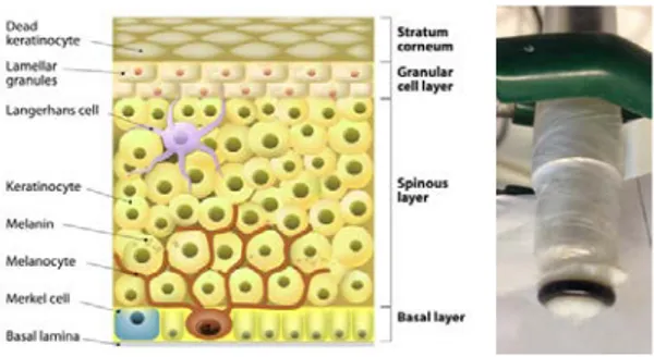
Figure: (left) Schematic presentation of layered structure of skin membrane consisting of stratum corneum (SC) and viable epidermis; (middle) a photo of oxygen electrode covered with skin membrane; (right) current of skin-covered oxygen electrode immersed in PBS, pH 7.4. Steady state baseline current (time interval: 100-200 s) is due to O2 dissolved in PBS. At 200 and 500 s H2O2 is added resulting into 0.5 and 1 mM H2O2 concentration, respectively. The observed two current steps reflect activity of skin catalase. At 800 s the catalase inhibitor NaN3 was added to the solution, resulting in the current return to the baseline level.
2 Introduction
Since the invention of biosensors in 1962 (Clark and Lyons 1962), the Clark oxygen electrode has been used to construct biosensors based on various enzymes, cells, and tissues (Turner et al. 1987). In most cases, the mentioned biological materials served as a recognition element providing specificity for the transduction function of the biosensor. In several cases the biosensor design has been used to address functions of the biomaterials itself, e.g., to monitor the activity of surface bound enzymes (Haberska et al. 2008; Ruzgas et al. 1995) or yeast cells attached at the electrode (Heiskanen et al. 2009; Spegel et al. 2007). Recently we proposed a skin covered electrode for studies of dynamics of transdermal penetration of biologically active compounds such as hydrogen peroxide, ascorbic acid, and quercetin (Gari et al. 2015; Rembiesa et al. 2015).
In this work we assessed the possibility of covering an oxygen electrode with excised pig skin membranes for investigation of native catalase function in skin. Catalase is one of the enzymes of the biological antioxidative system in skin (Pillai et al. 2005) where it removes hydrogen peroxide, H2O2, from skin by converting it to water and oxygen according to:
2𝐻𝐻2𝑂𝑂2
𝐶𝐶𝐶𝐶𝐶𝐶𝐶𝐶𝐶𝐶𝐶𝐶𝐶𝐶𝐶𝐶
�⎯⎯⎯⎯⎯� 2𝐻𝐻2𝑂𝑂 + 𝑂𝑂2 Eq. 1
H2O2 is one of the most stable forms of reactive oxygen species, ROS, which might be generated in skin by both exogenous and endogenous factors. Exogenous factors leading to elevated ROS in skin is, for example, exposure to UV irradiation, whereas chronic inflammation is an example of endogenous factor. Considering the general relevance of these examples, it is clear that monitoring the skin´s ability to detoxify ROS is of great interest in a wide range of situations including healthcare settings and development of topical products such as sun screen lotions. To the best of our knowledge, this work represents the first study where it is demonstrated that the oxygen
electrode can be utilized for electrochemical in-vitro monitoring of native catalase activity in skin. The proposed electrode design is a first step towards a simple and easy operated electrochemical tool to assess the function of native antioxidative system in skin.
As a background to help appreciate the results of this work, the skin structure is briefly described by the schematic drawing in Fig. 1a. It should be kept in mind that the structure of the skin is much more complicated, however, our simplified description is sufficient to understand the results of this work. Skin is one of the largest organ of the body and consists of several anatomically different layers as seen in Fig. 1a. The outermost layer is called the stratum corneum, SC, and is usually represented by the brick and mortar model, where the bricks represent dead cells (i.e. corneocytes), which are surrounded by a continuous lipid multilamellar matrix, i.e. the mortar (Michaels et al. 1975). Tape-stripping of the SC is often used to demonstrate that the barrier properties of the skin for transdermal penetration vanishes when the SC is stripped of (Valk and Maibach 1990). In particular, the continuous lipid lamellae matrix of SC represents a tough diffusional barrier towards transdermal penetration and thus ensures hydration homeostasis by minimizing the water loss from the body to the external environment (Iwai et al. 2012; Scheuplein and Blank 1971). Interestingly, the transdermal permeability of the skin barrier can be reversibly regulated by changing the degree of SC hydration (Björklund et al. 2010; Björklund et al. 2013). This example emphasizes the fact that the skin is a responding membrane, i.e., the membrane can change its properties following the changes of external biophysical factors (Björklund et al. 2010). Regulation of transdermal permeability as well as monitoring of enzyme activity in skin are, thus, of broad medical and industrial interest.
The results described in this work provide a number of important conclusions. Firstly, we demonstrate that the activity of native catalase
3 in skin is considerable in freshly excised pig skin
membranes and that it is possible to switch-off the catalase activity in skin by an enzyme inhibitor. Secondly, the catalase activity is high enough to detoxify transdermal penetration of H2O2 from a relatively concentrated solution of H2O2. Thirdly, from tape-stripping experiments we conclude that the catalase is primarily located in the viable epidermis and that the H2O2 penetration is significantly enhanced upon successive stripping of SC. Fourthly, the activity of native catalase is severely suppressed by changing the pH from neutral to acidic solution in contact with the SC barrier. Taken together, several of the findings in the present study are important to consider when developing topical formulations. For example, when the skin becomes exposed to UV irradiation, i.e., the conditions when ROS and H2O2 production might be elevated and functioning of catalase in skin should not be compromised. Our experiments also indicate that some penetration enhancers, i.e., components of topical formulations, can interfere with catalase reaction in skin. Material and methods
1. Material
Hydrogen peroxide (30%, 10.3 M), phosphate buffer saline (PBS, pH 7.4) in tablets, sodium citrate and sodium chloride for preparation of citrate buffer saline (CBS, pH 4.0 consisting of 10 mM sodium citrate and 150 mM NaCl), thymol, and azide were purchased from Sigma Aldrich. Oxygen electrode, constructed using 5 µm thick Teflon membrane from DuPont Fluoropolymers (Detroit, MI, USA), 250 µm Pt melted in glass and internal Ag/AgCl reference, was purchased from UAB “OPTRONIKA”, Vilnius, Lithuania. All solutions have been prepared by using deionised water purified by Milli-Q system (Merck Millipore, Billerica, USA) with resistivity of 18.2 Ω cm.
2. Preparation of skin membranes
Fresh pig ears were obtained from a local abattoir and stored at -80°C if not immediately
used. To prepare skin membranes, fresh or defrosted pig ears were rinse with cold water and cut into strips with a scalpel. Outer part (stratum corneum side) of the strip was shaved and approximately 500 µm thick skin membranes were sliced with a dermatome. The resulting skin stripes were punched out to make circular membranes with 16 mm in diameter. If not immediately mounted on the oxygen electrode, the membranes were kept in the fridge (+4°C) on a filter paper soaked with PBS. Skin membranes, prepared as described, were used within two weeks.
3. Preparation of skin covered oxygen electrode (SCOE)
The platinum cathode surface of the oxygen electrode was polished using alumina suspension (1 µm alumina, Buehler, Lake Bluff, IL) and rinsed with deionized water. The body of the electrode was filled with saturated KCl solution and covered with 5 µm Teflon membrane. This constituted the Clark type oxygen electrode. This electrode was then covered with the skin membrane as shown in Fig. 1b.
Fig. 1. (left) Schematic presentation of layered structure
of skin membrane consisting of stratum corneum (SC) and viable epidermis (Creative Commons (Attribution 3.0) from freedesignfile.com) and (right) a photo of oxygen electrode covered with skin membrane.
4. Amperometric monitoring of catalase activity in skin
The skin membrane covered oxygen electrode (Fig. 1b) was dipped into an electrochemical cell filled with 20 mL PBS (pH 7.4) or CBS (pH 4.0) buffer. Electrochemical measurements were performed using a CompactStat
4
potentiostat from IVIUM Technologies (Eindhoven, The Netherlands). The oxygen electrode was connected to the potentiostat in two electrode configuration by applying -0.7 V vs Ag/AgCl/KClsat on Pt cathode of the oxygen electrode. After the baseline current was stabilized, a defined amount of H2O2 was
pipetted into the electrochemical cell, which increased the reduction current of the oxygen electrode. In all cases, the solution was continuously mixed with a magnetic stirrer and all measurements were conducted at room temperature (+21oC).
Results and discussion
To access the activity of native catalase in skin we employed split-thickness membranes (approx. 500 µm thickness) and mounted them on the tip of the Clark-type oxygen electrode resulting in the skin covered oxygen electrode, SCOE, as shown in Fig. 1b. Upon immersion of the SCOE in a solution of PBS a stable baseline current is generated (Fig. 2, time interval 100-200 s). The baseline current is related to the O2 diffusion from the PBS solution, across the skin membrane, to the oxygen electrode and is to a great extent determined by the O2 concentration of the PBS solution. This was confirmed by reducing the O2 concentration by passing N2 gas through the PBS solution, which resulted in that the baseline current approached zero (data not shown). As can be seen from Fig. 2a, stepwise addition of H2O2 into PBS results in an increase of the electrode current due to the increase of O2 at the electrode surface. The increase of O2 is caused by H2O2 diffusion into skin where O2 is generated due to the catalase reaction specified in Eq. 1. To confirm that the native catalase was responsible for the generation of O2 from the added H2O2, a few crystals of the catalase inhibitor, sodium azide (NaN3), were added into the PBS at time 800 s (Fig. 2). It is clear that the NaN3 addition resulted in that the electrode current returned to the baseline level (Fig. 2, time interval 800-1400 s), which implies total inhibition of the native catalase activity. Taken together, the data in Fig. 2 demonstrate
that the activity of native catalase in skin can be studied by the proposed SCOE setup (Fig. 1b). Below we illustrate the versatility of this setup by a few key experiments and discuss how the SCOE can be utilized to address important questions related to skin healthcare and cosmetic applications.
Fig.2. Current of skin-covered oxygen electrode
immersed in PBS, pH 7.4. Steady state baseline current (time interval: 100-200 s) is due to O2 dissolved in PBS. At 200 and 500 s H2O2 is added resulting into 0.5 and 1 mM H2O2 concentration, respectively. The observed two current steps reflect activity of skin catalase as specified by Eq. 1. At 800 s the catalase inhibitor NaN3 was added to the solution, resulting in that the current returned to the baseline level.
The SC is one of the toughest biological barriers, which protects the body from the external environment as well as from dehydration due to minimal transepidermal water loss. It is well established that the SC is the diffusion limiting barrier in transdermal drug delivery (Scheuplein and Blank 1971). Permeability of SC can be increased by skin hydration (Björklund et al. 2010; Björklund et al. 2013), exploiting penetration enhances (Pham et al. 2016; Williams and Barry 2004) or simply removing this barrier by tape stripping (Valk and Maibach 1990). It has been shown that approximately one layer of corneocytes/keratinocytes is removed per single tape stripping application, but it should be noted that the cells may originate from various depths because of furrows in the skin (Molen et al. 1997). Nevertheless, repeated stripping of the SC results in a thinner and more defective skin barrier and after about 20 repetitions of this procedure SC usually
5 contains regions where the barrier is
completely removed (Molen et al. 1997; Valk and Maibach 1990). To understand the barrier properties of SC with respect to H2O2 penetration we investigated the effect of tape stripping on the current of the SCOE. The experiments were done by repeating the following experimental cycle: (i) amperometric registration of the response of the SCOE to 0.5 mM H2O2, and (ii) tape stripping without removing the skin membrane from the electrode. The electrode response to H2O2 addition after different number of tape stripping of skin is demonstrated in Fig. 3.
Fig.3. Amperometric response of skin-covered oxygen
electrode (SCOE) to H2O2 before and after tape stripping. A: (A1) Amperometric response to two consecutive additions of H2O2 into PBS solution resulting into 0.5 and
1 mM H2O2 concentrations, respectively. (A2)
Amperometric response of the electrode after 20-time tape stripping of skin membrane to the addition of H2O2
into PBS solution resulting into 0.5 mM H2O2
concentration. (B) The dependence of steady-state amperometric current on the tape-stripping number. The current is calculated as a difference between the electrode current in the presence of H2O2 minus the
current in the absence of H2O2 in buffer solution (baseline current).
As can be seen in Fig. 3a, 20 applications of the tape strip method resulted in approximately an eigth-fold increase in the current response of the SCOE to 0.5 mM H2O2. The electrode response increases gradually with the number of tape strip applications, as demonstrated in Fig. 3B. As expected, after around 18-20 tape strip repetitions the current approaches a high and stable value, which corresponds to the minimal resistance of diffusional transport through the viable skin that is left once the skin´s SC barrier is removed. Tape stripping results in approximately 10 times higher response of the electrode to H2O2. Several important conclusions can be drawn from the experiments conducted with SCOE in combination with the tape-stripping methodology. Firstly, SC acts as a substantial diffusion barrier even for such a small molecule as H2O2. The observation that the response of the electrode increases substantially with the number of tape stripping indicates that skin hosts a substantial activity of catalase. In other words, H2O2 penetrating from a 0.5 - 1 mM H2O2 solution into the viable epidermis and underlying viable tissue will be converted to O2 and thus detoxified by native skin catalase. To understand the relevance of these conclusion it can be mentioned that 0.1-1 mM H2O2 concentrations have been reported to promote wound healing or to be toxic to cells (Loo et al. 2011; Schreml et al. 2010). Peroxide containing antimicrobial creams and bleaching solutions might contain between 1 % (300 mM) to 5 % (1500 mM) of H2O2 (Chan and Maibach 2008). The experiments summarized in this paragraph demonstrate that the SCOE can be used to study barrier properties of skin for optimizing topical products containing H2O2.
In development of topical products, such as creams and lotions, certain pH values of the formulation are chosen. In some cases the choice is motivated with the aim to match the assumed skin pH, or to increase the topical or transdermal delivery, or simply to increase the
6 stability of the formulation for storage
purposes. We have assessed the effect of pH on the activity of catalase in skin by recording the amperometric current response of the SCOE to 0.5 mM H2O2 at different pH ranging from 3.5 to 8.0. Typical experimental data are summarized in Fig. 4. As can be seen the response of the electrode is considerably reduced at lower pH implying that the activity of catalase in skin is noticeably suppressed at acidic pH. The stepwise return to pH 7.4 recovers some activity, however, the activity is lower as compared to the initial measurements. This observation can probably be explained by inactivation of catalase due to exposure to H2O2 at acidic pH. However, this conclusion is restricted by the fact that the skin membrane may change between each experiment. The fact that the current response is decreased for the repeated measurement at the same pH implies irreversible changes of the skin membrane. However, it should be mentioned that the reduction of the electrode response at acidic pH shown in Fig. 4 is considerably more pronounced if compared to the loss of response after repeated measurements at pH 7.4 (data not shown). The general conclusion is that the catalase activity in skin is considerably lower at acidic pH. However, since the effect of pH on the rate of transdermal penetration of H2O2 across SC was not evaluated in these experiment this conclusion is associated with some uncertainty.
Fig.4. Amperometrically measured steady-state current
of the skin covered oxygen electrode (SCOE) at 0.5 mM of H2O2 in buffer solutions of different pH. Phosphate buffer
saline with pH 8.0, 7.4 and 6.5 and or citrate buffer saline with pH 4.5 and 3.5. The current is calculated as a difference between the electrode current in the presence of H2O2 minus the current in the absence of H2O2 in buffer solution (baseline current).
It should be kept in mind that the SCOE is an in-vitro system and that the relevance of the observed pH effect on skin catalase activity might be questionable for the in-vivo situation where the pH of healthy viable epidermis should be constant and close to 7.4. However, a local reduction of the pH may be possible in cases of metabolic disturbances or diseases, including acute or chronic inflammation and cancer (Kato et al. 2013).
From the in-vitro measurements, summarized above, it is clear that acidic pH of topical formulations might reduce the activity of skin catalase, i.e. suppress the function of biological antioxidative system. This conclusion should have a significant interest and relevance in case when a formulation with low pH is applied on skin parts possessing elevated ROS production, e.g., in vitiligo disease (Schallreuter et al. 2001). This discussion about the function of catalase in skin leads us to a very complex and medically relevant area addressing interaction of formulations with the biological antioxidative system in skin. The potential use of the skin-covered electrode setup to address some relevant questions in this context is demonstrated below.
Topical formulations are mixtures of chemical compounds with different functions, such as moisturizer, emollient, thickener, etc. The compounds that increase the rate of transdermal delivery are called penetration enhancers. Ideally they are supposed to be biologically “inert”, i.e., not bioactive, and only act to change the physicochemical permeability properties of the skin barrier. By using skin-covered electrode for measurements of skin catalase activity we looked at the effect of the well know penetration enhance thymol (Pham et al. 2016). The chemical structure of thymol and the amperometric response of the
skin-7 covered electrode to thymol, in the presence of
H2O2, are presented in Fig. 5.
Fig. 5. Amperometric response of skin-covered electrode
to two consecutive addition of H2O2 resulting in 0.5 and 1 mM H2O2 concentration in PBS. Thymol was dropped into the electrochemical cells in amount of few flakes (thymol is sparingly soluble in water solution).
As can be seen from Fig. 5, two consecutive additions of H2O2 into PBS results into two similar amperometric current responses of the SCOE. The results indicate that catalase in skin is active and generates O2 from the H2O2, which penetrates through SC. At time 1000 s the thymol is added into the reaction mixture, resulting in a decrease of the electrode current. This is not what should be expected for the compounds, which is an extremely potent penetration enhancer (Pham et al. 2016). In other words, it is expected that thymol should increase the SC permeability and thus increase the H2O2 flux, leading to the increased electrode current. Nevertheless, even though the experimental results presented in Fig. 5 contradict the expected outcome, the results can sill be explained. As seen from the chemical structure of thymol, it contains a phenolic group. Phenolic structures can be oxidized by H2O2 catalyzed by catalases and peroxidases (Dunford 2010). In this case, the catalase will not generate O2 at the same rate as the peroxidase reaction consumes H2O2. Taking this reaction into consideration, the amperometric response of the skin-covered electrode to thymol in the presence of H2O2 is logical. Concluding, the experiments with thymol demonstrate that some compounds present in topical formulations can interfere with the biological antioxidative system in skin.
Since the compounds present in formulations have been clinically tested, in the majority of the cases this is probably not a problem. However, in cases when H2O2 is generated in skin, e.g., at UV irradiation and in cases of acute or chronic skin inflammations, their effect on antioxidative system of skin might be questionable. Finally, it should be emphasized that the proposed setup, based on the skin-covered oxygen electrode, can be useful not only to measure catalase activity in skin but also provides a tool to address more complex functions of biological antioxidative system in skin.
Conclusions
In this work we demonstrate that the skin-covered oxygen electrode (SCOE) allows investigation of catalase activity in skin. The setup comprises a skin membrane placed on the Clark type oxygen electrode. The catalase activity and function in skin is monitored based on the current response of the oxygen electrode. The current is due to H2O2 penetration through SC and subsequent catalytic generation of O2. By tape-stripping the SC we demonstrate that skin contains a substantial amount of catalase, which is sufficient to detoxify H2O2 that reaches the viable epidermis after exposure of skin to 0.5-1 mM H2O2. Our results also indicate that catalase activity is reduced at acidic pH as compared to the activity at pH 7.4. Some reservation must be made since the effect of pH, solely, on catalase activity is difficult to separate from the pH dependence of H2O2 permeability through SC. Nevertheless, we argue that the observed pH dependence might be relevant to in-vivo situations when local pH is reduced and ROS generation is elevated, e.g., by disease in skin. The experiments with penetration enhancer, thymol, shows that the SCOE can also be exploited to study broad spectrum of questions relevant to understand functioning of biological antioxidative system in skin.
8 Acknowledgements
Financial support from The Knowledge Foundation (SB, JE), The Swedish Research Council (TR) and The Gustaf Th. Ohlsson Foundation (JE and TR) are greatly acknowledged. SN thanks ERASMUS program for scholarship.
REFERENCES
Björklund, S., Engblom, J., Thuresson, K., Sparr, E., 2010. A water gradient can be used to regulate drug transport across skin. J. Controlled Release 143, 191-200.
Björklund, S., Ruzgas, T., Nowacka, A., Dahi, I., Topgaard, D., Sparr, E., Engblom, J., 2013. Skin membrane electrical impedance properties under the influence of a varying water gradient. Biophys J. 104(12), 2639-2650.
Chan, H.P., Maibach, H.I., 2008. Hydrogen peroxide, bleaching, and skin: an overview. Cutaneous & Ocular Toxicol. 27, 307-309. Clark, L.C.J., Lyons, C., 1962. Electrode systems for continuous monitoring in cardiovascular surgery. Ann. NY Acad. Sci. 102, 29-45.
Dunford, H.B., 2010. Peroxidases and Catalases: Biochemistry, Biophysics, Biotechnology and Physiology, 2nd ed. Wiley-Blackwell, New Jersey.
Gari, H., Rembiesa, J., Masilionis, I., Vreva, N., Svensson, B., Sund, T., Hansson, H., Morén, A.K., Sjöö, M., Wahlgren, M., Engblom, J., Ruzgas, T., 2015. Amperometric in vitro monitoring of penetration through skin membrane. Electroanalysis 27, 111 - 117. Haberska, K., Svensson, O., Shleev, S., Lindh, L., Arnebrant, T., Ruzgas, T., 2008. Activity of lactoperoxidase when adsorbed on protein layers. Talanta 76(5), 1159-1164.
Heiskanen, A., Spégel, C., Kostesha, N., Lindahl, S., Ruzgas, T., Emnéus, J., 2009. Mediator-assisted simultaneous probing of cytosolic and mitochondrial redox activity in living cells. Anal. Biochem. 384, 11–19.
Iwai, I., Han, H., Hollander, L.d., Svensson, S., Öfverstedt, L.-G., Anwar, J., Brewer, J., Bloksgaard, M., Laloeuf, A., Nosek, D., Masich, S., Bagatolli, L.A., Skoglund, U., Norlén, L.,
2012. The Human Skin Barrier Is Organized as Stacked Bilayers of Fully Extended Ceramides with Cholesterol Molecules Associated with the Ceramide Sphingoid Moiety. J. Invest. Dermatol. 132, 2215-2225.
Kato, Y., Ozawa, S., Miyamoto, C., Maehata, Y., Suzuki, A., Maeda, T., Baba, Y., 2013. Acidic extracellular microenvironment and cancer. Cancer Cell International 13(89), 1-8.
Loo, A.E.K., Ho, R., Halliwell, B., 2011. Mechanism of hydrogen peroxide-induced keratinocyte migration in a scratch-wound model. Free Rad. Biol. Med. 51, 884-892. Michaels, A.S., Chandrasekaran, S.K., Shaw, J.E., 1975. Drug permeation through human skin. Theory and in vitro experimental measurement. AIChE J. 21(5), 985-996.
Molen, R.G.v.d., Spies, F., Noordende, J.M.v.t., Boelsma, E., Mommaas, A.M., Koerten, H.K., 1997. Tape stripping of human stratum corneum yields cell layers that originate from various depths because of furrows in the skin. Arch. Derm. Res. 289(9), 514-518.
Pham, Q.D., Björklund, S., Engblom, J., Topgaard, D., Sparr, E., 2016. Chemical penetration enhancers in stratum corneum — Relation between molecular effects and barrier function. J. Controlled Release 232, 175-187. Pillai, S., Oresajo, C., Hayward, J., 2005. Ultraviolet radiation and skin aging: roles of reactive oxygen species, inflammation and protease activation, and strategies for prevention of inflammation-induced matrix degradation – a review. Int. J. Cosmetic Sci. 27, 17-34.
Rembiesa, J., Gari, H., Engblom, J., Ruzgas, T., 2015. Amperometric monitoring of quercetin permeation through skin membranes. Int. J. Pharmaceutics 496, 636-643.
Ruzgas, T., Gorton, L., Emnéus, J., Marko-Varga, G., 1995. Kinetic models of horseradish peroxidase action on a graphite electrode. J. Electroanal. Chem. 391, 41-49.
Schallreuter, K.U., Moore, J., Wood, J.M., Beazley, W.D., Peters, E.M.J., Marles, L.K., Behrens-Williams, S.C., Dummer, R., Blau, N., ThoÈny, B., 2001. Epidermal H2O2 Accumulation Alters Tetrahydrobiopterin
9 (6BH4) Recycling in Vitiligo: Identi®cation of a
General Mechanism in Regulation of All 6BH4-Dependent Processes? J. Invest. Dermatol. 116(1), 167-174.
Scheuplein, R.J., Blank, I.H., 1971. Permeability of the skin. Physiol. Rev. 51(4), 702-747. Schreml, S., Landthaler, M., Schaferling, M., Babilas, P., 2010. A new star on the H2O2rizon of wound healing? Exp. dermatol. 20, 229-231. Spegel, C.F., Heiskanen, A.R., Kostesha, N., Johanson, T.H., Gorwa-Grauslund, M.-F., Koudelka-Hep, M., Emneus, J., Ruzgas, T., 2007. Amperometric Response from the Glycolytic versus the Pentose Phosphate Pathway in Saccharomyces cerevisiae Cells. Analytical Chemistry (Washington, DC, United States) 79(23), 8919-8926.
Turner, A., Karube, I., Wilson, G.S., 1987. Biosensors: Fundamentals and Applications. Oxford University Press, Oxford.
Valk, P.G.v.d., Maibach, H.I., 1990. A functional study of the skin barrier to evaporative water loss by means of repeated cellophane-tape stripping. Clin. Exp. Dermatol. 15(3), 180-182. Williams, A.C., Barry, B.W., 2004. Penetration enhancers. Adv. Drug Deliv. Rev. 56(5), 603-618.