1
Homotrimeric dUTPases
Principles of Catalysis
and
Inhibitor Design
Doctoral ThesisLorena González Palmén
School of Pure and Applied Natural Sciences Faculty of Natural Sciences and Engineering
University of Kalmar, Sweden 2009
Akademisk avhandling som för avläggande av doktorsexamen i biokemi vid Naturvetenskapliga institutionen vid Högskolan i Kalmar kommer att offentligt försvaras i Västergårds hörsal (N2007), Smålandsgatan 24, Kalmar, fredagen den 27 februari, kl. 10.00.
Opponent: Dr. Dolores González-Pacanowska. Institute of Parasitology and Biomedicine "López-Neyra" (IPBLN), Consejo Superior de Investigaciones Científicas, Parque Tecnológico de Ciencias de la Salud, Avenida del Conocimiento, s/n, 18100-Armilla, Granada, Spain.
2 Organization: Document name: University of Kalmar DOCTORAL DISSERTATION
School of Pure and Applied Natural Sciences Date of issue:
SE-391 82 Kalmar, Sweden 2009-01-19 Sponsoring organization:
Author: Lorena González Palmén
University of Kalmar, Sweden and FLÄK (The Research School in Pharmaceutical Science) Lund, Sweden Title and subtitle: Homotrimeric dUTPases: Principles of Catalysis And Inhibitor Design
Abstract:
The ubiquitous enzyme dUTPase hydrolyzes dUTP into dUMP and pyrophosphate, preventing DNA fragmentation and cell death due to accumulation of dUTP. Inhibitors of dUTPase could serve as drugs in the treatment of cancers and infectious diseases. This thesis presents five studies. A mutational study on the Escherichia coli dUTPase (S72A) provides new insights about the catalytic principles of the homotrimeric dUTPases. A model is presented in which transition state formation is associated with a rotation of the conserved Ser72 side chain. The model can explain the strict order of deamination and hydrolysis catalyzed by the bifunctional dCTP deaminase:dUTPases. The S72A/D90N double mutant is currently investigated. Preliminary data indicate that this form preserves the binding properties of the S72A mutant but is completely inactive, making it attractive for structural studies. In the remaining studies we compare the binding of substrate analogues to the human, the E. coli and the equine infectious anemia virus (EIAV) homotrimeric dUTPases. One study concerns 2´,3´-dideoxy-UTP (ddUTP) and shows that removal of the 3´-hydroxyl group increases KM, ten times with the cellular dUTPases and fifty times with the viral dUTPase, but does not affect kcat with any of these enzymes.
Another study concerns the inhibitory effects of 3´-azido-2´,3´-dideoxy-UTP. This derivative binds to the bacterial dUTPase but not to the other forms making it a potential lead for the development of antibacterial dUTPase inhibitors. Yet another study investigates two uracil derivatives. Both compounds are found to inhibit the human, the bacterial but not the viral dUTPase. The inhibition is shown to be competitive.
Key words: dUTPases, kinetics, ground state destabilization, inhibitors Classification system and/or index terms (if any):
Supplementary bibliographical information:
Language: English
ISSN and key title: 1650-2779
ISBN: 978-91-85993-19-2 Recipient’s notes:
Number of pages: 97 Price: Security classification: Distribution by:
Lorena González Palmén, School of Pure and Applied Natural Sciences, University of Kalmar, Sweden
I, the undersigned, being the copyright owner of the abstract of the above-mentioned dissertation, hereby grant to all reference sources to publish and disseminate the abstract of the above-mentioned dissertation.
3
4
“Människan kan inte upptäcka nya oceaner förrän hon har fått modet att förlora kusten ur sikte”.
Okänd
Front Cover Illustration: View down the active site of E. coli dUTPase in complex with the substrate analogue α,β-imido-dUTP⋅Mg (PDB 1RN8). Color codes: Carbon: yellow, Nitrogen: blue, Oxygen: Red, and Phosphorus: Orange. The figure was produced using PyMol (Delano Scientific, 2003).
Copyright © Lorena González Palmén, 2009 Printed by: Högskolans tryckeri i Kalmar
ISSN 1650-2779 ISBN 978-91-85993-19-2
5
CONTENTS
LIST OF PUBLICATIONS... 7
ABBREVIATIONS ... 9
1. INTRODUCTION...11
DNA-Synthesis and Nucleotide Metabolism ... 11
Enzyme Inhibitors as Drugs ...14
The Thesis: A Brief Presentation ...14
1.1 dUTPASES... 16
Homotrimeric Dimeric and Monomeric dUTPases ...16
The dCTP Deaminase:dUTPases...18
1.2 THE REACTION CATALYZED... 19
The In-line Nucleophilic Attack...19
Conformation of the Phosphate Moiety...21
1.3 PRINCIPLES OF CATALYSIS... 22
Definition of a Catalyst...22
Transition State Theory ...22
Enzymes as Catalysts ...23
The Active Site and Ground State Destabilization...24
Transition State Analogues...25
Enzyme Kinetics ...25
1.4 dUTPASE INHIBITORS ... 27
The Role of Enzyme Functional Groups in Catalysis...27
The Role of Substrate Functional Groups in Catalysis ...28
Finding dUTPase Inhibitors ...28
2. EXPERIMENTAL APPROACH ... 31
6
2.1 KINETIC MEASUREMENTS...31
2.1.1 STEADY STATE MEASUREMENTS ... 32
The pH-indicator Technique ...32
Data Acquisition...32
Data Treatment...33
Using Enzyme Intrinsic Fluorescence as Signal ...34
Parameter Evaluation...35
2.1.2 SUBSTRATE BINDING KINETICS... 36
Viscosity Effects Approach ...36
Pre-Steady State Measurements...37
2.2 EQUILIBRIUM BINDING STUDIES... 39
Frontal Chromatography...39
2.3 PROTEIN STABILITY ... 40
Thermal Stability...40
Chaotropic Stability ... 41
3. RESULTS AND DISCUSSION... 43
3.1 MUTATIONAL STUDIES ... 44
A Dual Role of the Strictly Conserved Serine...44
An extended Catalytic Model ...46
Properties of the S72A/D90N Double Mutant...48
3.2 MODIFIED SUBSTRATES ... 48
The Substrate Analogue ddUTP ...48
Inhibition Studies with 3´-Azido-ddUTP...50
3.3 URACIL DERIVATIVES ...51
Triskelion Shaped Uracil Derivatives... 51
4. CONCLUDING REMARKS ... 53
ACKNOWLEDGEMENTS... 55
7
LIST OF PUBLICATIONS
This thesis is based on the following publications, referred to in the text by their roman numerals. All published papers are reproduced with permission from the original publisher.
I. A Double Role for a Strictly Conserved Serine: Further Insights into the dUTPase Catalytic Mechanism
L. González Palmén, K. Becker, L. Bülow, and J.O. Kvassman.
Biochemistry 2008, 47, 7863–7874.
II. Changing the 3´-OH of dUTP to -H affects KM exclusively in the
reaction with homotrimeric dUTPases: Differing effects and mechanistic implications
L. Gonzalez Palmén, and J.O. Kvassman. Manuscript ready for submission.
III. Differential Inhibition of Homotrimeric dUTPases by the 3´-Azido Derivative of Dideoxy-UTP
L. González Palmén and J. O. Kvassman. Manuscript ready for submission.
IV. Inhibition of Homotrimeric dUTPases by Triskelion Shaped Uracil Derivatives
L. González Palmén, J. T. Stivers, and J.O Kvassman. Manuscript.
V. A Double Mutant of the E. coli dUTPase with Tighter Substrate Binding But No Activity
L. González Palmén, K. Becker, L. Bülow, and J.O. Kvassman. Manuscript.
8
Other publications I have contributed to, outside the scope of this thesis:
Gazzaniga, S., González, L., Mantovani, A., Vecchi, A., and Wainstok, R. (2004) Isolation and molecular characterization of a mouse renal microvascular endothelial cell line. In Vitro Cell. Dev. Biol. Animal 40, 82–88.
Aldén, A., González, L., Persson, A., Christensson, K., Holmqvist, O., and Ohlson, S. (2007) Porcine platelet lysate as a supplement for animal cell culture. Cytotechnology 55, 3–8.
9
ABBREVIATIONS
DNA Deoxyribonucleic acid
ADP Adenosine diphosphate
GDP Guanosine diphosphate
UDP Uridine diphosphate
CDP Cytidine diphosphate
dUTP Deoxyuridine triphosphate
dUMP Deoxyuridine monophosphate
dUDP Deoxyuridine diphosphate
PPi Pyrophosphate
dTTP Deoxythymidine triphosphate
EIAV Equine Infectious Anaemia Virus
dCTP Deoxycytidine triphosphate
ddUTP Dideoxyuridine triphosphate
Azido-ddUTP 3′-Azido-dideoxyuridine triphosphate
GuHCl Guanidinium Hydrochloride
11
1. INTRODUCTION
DNA Synthesis and Nucleotide Metabolism
All organisms have evolved systems to defend themselves against pathogens and toxins. On a molecular level, various enzymes work together to neutralize harmful substances. Sometimes natural metabolites are harmful if accumulated in the cell, and this is the case with deoxyuridine triphosphate, dUTP. The present thesis concerns the ubiquitous enzyme deoxyuridine triphosphatase or dUTPase (EC 3.6.1.23), which removes dUTP through hydrolysis and which thereby, contributes to the production of error free DNA at cell replication. The dUTPases exists in three oligomeric forms, monomeric, dimeric and homotrimeric. Most organisms, including humans and the majority of bacteria and viruses express the homotrimeric C3-symmetric form of the dUTPase, which is also at the focus of this thesis.
DNA is made up of deoxyribonucleotides, each composed of a nitrogenous base, a sugar and a phosphate group. The bases in DNA are adenine, (A), guanine, (G), cytosine, (C), and thymine, (T), which are present in the respective 5´-deoxy-ribonucleotide triphosphate (dNTP). Cell proliferation depends ultimately on the duplication of the chromosomal DNA of the mother cell. This, in turn, requires access to the DNA building blocks, dATP, dCTP, dGTP and dTTP, in sufficient quantities and appropriate proportions. The DNA building blocks are generated through 2´-deoxygenation of the RNA metabolites, ADP, GDP, UDP and CDP, a step catalyzed by the ribonucleotide reductase enzyme. The generated dNDPs are phosphorylated to the corresponding dNTPs, by NDP kinases [1]. Thus, dUTP is present in the nucleotides pool, and it can be incorporated into DNA, which leads to strand breakage and cell death [2-5]. Because DNA polymerase cannot distinguish between dTTP and dUTP [6,7], the incorporation of uracil into DNA depends on the dTTP/dUTP ratio. A key component in nature’s solution to this problem is the enzyme dUTPase which catalyzes the hydrolysis of dUTP into dUMP and pyrophosphate [8,9], according to:
dUTP + H2O → dUMP + PPi
This reaction not only removes dUTP from the DNA polymerase substrate pool, but feeds the cell with dUMP, the precursor for synthesis of dTTP via the thymidylate synthase reaction.
12
The degree to which dUTP is used, instead of dTTP, as a substrate in the replication process is determined by the concentration ratio between these two nucleotides. The key position with respect to DNA-replication occupied by the dUTPase enzyme is illustrated in Fig. 1.1.
Figure 1.1 DNA replication and dUTPase activity. The potentially fatal incorporation of dUTP into DNA instead of dTTP is determined by the dUTP/dTTP ratio in the nucleotide pool. Points of drug intervention, practiced or potential, are indicated.
Thymidylate Synthase (TS), which catalyzes the methylation of deoxyuridylate (dUMP), into thymidylate (dTMP), is an important target of the chemotherapeutic agents used in cancer treatment. The TS reaction is the sole de novo source of dTMP for DNA replication and repair. The methyl group transferred to uracil is provided by 5,10-methylene tetrahydrofolate. This has led to the development of antifolate-based antitumor drugs with specific inhibition of TS [10], as well as to the fluoropyrimdine class of anticancer agents. Inhibitors of dihydrofolate reductase, such as methotrexate, deplete the tetrahydrofolate pools necessary for the TS reaction, and indirectly block dTMP formation [11,12]. The other group of anticancer drugs include fluorouracil [13], which is converted in vivo into fluorodeoxyuridylate (FdUMP) which irreversibly inhibits the TS. The inhibition of TS depletes the cell of dTTP and results in accumulation of dUMP, subsequently phosphorylated by mono and dinucleotide kinases into dUTP [14]. The resulting DNA damage and cytotoxicity has been termed ‘thymineless cell death’ [15]. Because inhibition of dUTPase has similar consequences, the dUTPases have emerged as potential targets for drugs against cancer and infectious diseases
[16-13
20]. In cancer treatment, dUTPase inhibitory drugs could alternatively be administered to enhance the effects of the TS inhibitors. The two drug-target points are indicated in Fig. 1.1.
The ribonucleotide reductase reaction and subsequent phosphorylation is not the only source of dUTP, and the dUTPase reaction is not the only provider of dUMP. In certain bacteria dUTP is also formed through the deamination of dCTP, a reaction catalyzed by dCTP deaminases. The dCTP deaminases have been found in enterobacteria [21], mycobacteria [22], and archaeabacteria [23,24]. Some of these enzymes are bifunctional and catalyze also the hydrolysis of the dUTP product into dUMP and pyrophosphate [22-24]. Another route for dUMP formation, which is present in higher organisms, is the deamination of dCMP by dCMP deaminases. However in enterobacteria, which lack dCMP deaminases, the main route of dUMP formation is through the action of dCTP deaminases and dUTPases. The different pathways of dUMP formation are summarized in Fig. 1.2
Figure 1.2 The pathways for formation of dUMP, the precursor for synthesis of dTTP. In enterobacteria, cytosine deamination occurs only by dCTP deaminases, which makes the dUTPase the only source of dUMP.
In recent years the number of crystal structures that have been resolved for dUTPases has increased dramatically [25-30]. The structural information should help in the development of drugs intended for dUTPase inhibition. It may also form the basis for functional studies aiming at a better understanding of how nature, by carefully varying a common theme, has created efficient catalysts for the hydrolysis of dUTP into dUMP and pyrophosphate. On a broader level such studies may increase our understanding of enzyme catalysis in general, a field still open to debate.
14
Besides the pioneering structural study on the E. coli dUTPase [25], two structural studies in particular [29,31] have contributed to our understanding of the catalytic mechanism operated by the homotrimeric and monomeric dUTPases. The two studies confirm what was already deduced regarding the mechanism in the first structural [25] and functional [32] reports on the E. coli dUTPase, and provide new information on the structural basis for a carboxylate assisted in-line nucleophilic attack by the substrate water on the α-phosphorus of dUTP. One of the studies [29], indicates a role in catalysis for a serine residue, which is strictly conserved at the active site of the homotrimeric and monomeric dUTPases.
Enzyme Inhibitors as Drugs
Enzymes are proteins with a catalytic function. They control the flow of metabolites in the catabolic and anabolic paths of the cell and without them, virtually none of the reactions in the living organism would proceed at an appreciable rate. But enzymes also promote the growth of tumors and pathogenic organisms, which makes some of them suitable targets for drug intervention. Drugs against retroviral enzymes, (reverse transcriptase inhibitors), are in use for the treatment of HIV [33], and new directions are emerging for the development of drugs against glycogen synthase kinase-3, one of the enzymes involved in protein aggregation that causes Alzheimer’s disease [34,35]. Other drugs are designed to inhibit glycogen phosphorylase, which has been linked to diabetes type 2, and may lead to antihyperglycaemic drugs [36]. Thus, drug design, based on the structural and functional properties of enzymes, is one of the leading tendencies in Biomedical Sciences, and promise to provide cures for many common diseases. In this process, the developments in genetic engineering and protein structure determination are of invaluable importance.
The Thesis: A Brief Presentation
The present thesis is based on a series of functional studies which have provided insights into the catalytic mechanism of the homotrimeric dUTPases, as well as into the ligand binding properties of dUTPases from various sources.
In Paper I we report how the substitution of hydrogen for the β-hydroxyl group of a strictly conserved serine affects substrate binding and catalysis in the E. coli dUTPase reaction. This work provides not only insights into the dUTPase mechanism, but gives experimental support to the catalytic theory which considers ground state destabilization as part of the process together with transition state stabilization.
In Paper II we have investigated the role of the 3´-hydroxyl group of the dUTP ribose moiety in substrate biding and catalysis. This was done by testing
3´-15
dideoxy-UTP (ddUTP) as a substrate. To identify differences between the active sites of various dUTPases, the investigation was broaden to include a comparison of the homotrimeric dUTPases from three different sources, namely the human, the E. coli bacterium and the equine infectious anaemia virus (EIAV). With the ordinary substrate the three enzymes exhibit the same catalytic efficiency but with the dideoxy form, the EIAV dUTPase is seven times less efficient than the cellular forms.
In Paper III the importance of various substitutions at the 3´-carbon of dUTP is further investigated. The substrate derivative 3´-azido-dUTP, which is analogous to a drug used in the treatment of HIV, is tested for inhibition of the dUTPases studied in Paper II. The results prove azido-dUTP to be a dUTPase inhibitor, and the first reported to be highly specific for a bacterial dUTPase.
In Paper IV we investigate two non-nucleotide and triskelion shaped compounds [37] for inhibition of our target dUTPases. Both substances are shown to inhibit the human and E. coli dUTPases, but not the viral form.
In Paper V we combine two mutations at the active site of the E. coli dUTPase. Both mutations affect strictly conserved residues, and when introduced individually, each of them has drastic and well characterized consequences for the substrate binding and catalytic properties of the enzyme. The double mutant is devoid of catalytic activity but binds the substrate much stronger than the native enzyme. The results support conclusions drawn in Paper I and make the complex of the double mutant with substrate attractive for structural studies.
Thus, the properties of dUTPases obtained from three different sources have been investigated. The methods used are diverse, and some of them applied for the first time in the study of the dUTPases. The latter include the variation of medium viscosity to probe the kinetics of substrate binding, and frontal chromatography, on immobilized dUTPase.
All together, this thesis contributes to the understanding of the catalytic function of the homotrimeric dUTPases, and of how these enzymes, when obtained from various species, differ with respect to binding of the substrate, substrate analogues and uracil derivatives. This information may prove valuable for the design of general and species-specific dUTPase inhibitors.
16
1.1 dUTP
ASESHomotrimeric, Dimeric and Monomeric dUTPases
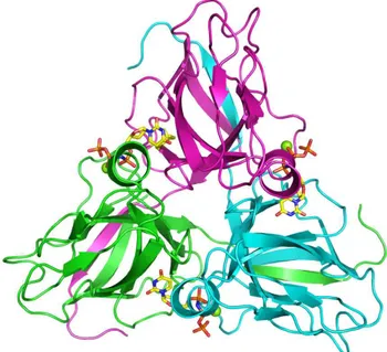
The subunits of the homotrimeric dUTPase are related by the symmetry of a threefold rotation axis (Fig. 1.3). The substrate binding sites are in pockets created at the subunit interfaces. In the apo enzyme, the pockets are lined by four conserved sequence motifs [25,38]. Motifs I, II and IV are located to one subunit and Motif III to the other. A fifth conserved motif is located at the C-terminus. The C-terminus constitutes a mobile part of the subunit, a ‘flexible arm’, which wraps around the trimer to let motif V complete the active site by covering the bound substrate [29]. The C-terminus is usually not resolved in structural studies due to its flexibility [31], and it is not visible in the structure of the homotrimeric E. coli dUTPase, shown in Fig. 1.3. Complete immobilization of motif V over the active site has been observed only in a complex of the human dUTPase with the α,β-imido-dUTP⋅Mg substrate analogue [29].
The three active sites are independent in substrate binding and catalysis [32]. Despite considerable sequence differences among the various homotrimeric dUTPases, they have virtually identical substrate pockets, a fact that makes the design of species-specific inhibitors a difficult task.
Figure 1.3 The E. coli dUTPase with the substrate analogue α,β-imido dUTP⋅Mg (PDB 1RN8). The last 15 C-terminal residues are not resolved on this structure. Color codes: Carbon: yellow, Nitrogen: blue, Oxygen: Red, and Phosphorus: Orange. The figure was produced using PyMol (Delano Scientific, 2003).
17
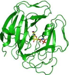
Dimeric dUTPases have been identified in the protozoans Trypanosoma cruzi [39,40] (Fig. 1.4), and Leishmania major [41], as well as in the bacteria Campylobacter jejuni [42]. The dimeric dUTPases show no homology with the trimeric forms, and they catalyze the reaction by a different mechanism [40,42]. In contrast, the monomeric dUTPases, which only occur in the herpes viruses such as the Epstein-Barr virus (EBV), have a single active site (Fig. 1.5) that mimics in detail that of the trimeric dUTPases [30].
Trypanosoma cruzi is a parasite that causes the Chagas disease, wide spread in Central and South America. Inhibitors of the parasite dUTPase, may provide a solution to this problem.
Figure 1.4 Dimeric Trypanosoma cruzi dUTPase in complex with dUDP (PDB 1OGK). The subunits are marked in different colors. The ligand color code is the same as in Fig. 1.3, except C: grey. The structure was generated with PyMol (Delano Scientific, 2003).
18
Figure 1.5 Epstein-Barr Virus monomeric dUTPase in complex with the α,β-imido dUTP (PDB 2BT1). The ligand color code is the same as in Fig. 1.3. The figure was created with PyMol (Delano Scientific, 2003).
The dCTP Deaminase:dUTPases
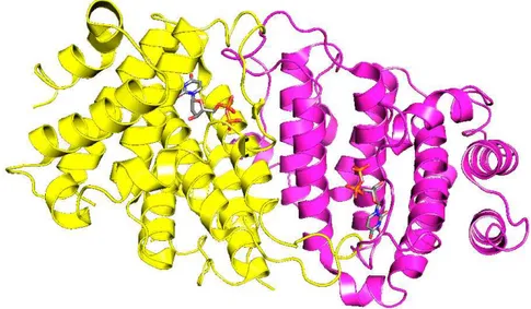
In gram negative bacteria, the dCTP deaminases, which catalyze the deamination of dCTP to dUTP, provide for a route of dUTP formation additional to dehydrogenation of UTP, catalyzed by the ribonucleotide reductase. The dCTP deaminases have a core and active site architecture homologous to that of the dUTPases [21-23]. Interestingly, except for motif V, the conserved sequence motifs that create the active sites of the dUTPase are found in structurally equivalent positions in the bifunctional dCTP deaminase [23]. Recently, the dCTP deaminase of the thermophile Methanocaldococcus jannaschii (Fig. 1.6) was reported to be a bifunctional enzyme, which in addition to deamination of the pyrimidine ring, in a second step catalyses the hydrolysis of the intermediate dUTP product [43,44]. The list of organisms that expresses such bifunctional dCTP deaminases has expanded to include the M. tuberculosis pathogen [22].
In Paper I, we presented a catalytic model for dUTPases, which appears to solve the question why, in the reaction catalyzed by the bifunctional dCTP deaminases, deamination of the pyrimidine ring must precede hydrolysis of the phosphate moiety.
19
Figure 1.6 The homotrimeric bifunctional Methanocaldococcus jannaschii dCTP deaminase-dUTPase in complex with dUTP and Mg2+ (PDB 2HXD). The figure was created with
PyMol (Delano Scientific, 2003).
1.2 T
HER
EACTIONC
ATALYZEDThe In-line Nucleophilic Attack
The enzyme dUTPase belongs to the family of hydrolases (EC 3). As the name indicates the hydrolases catalyze the hydrolysis of a chemical bond, which can be represented by the following generic reaction:
A-B + H2O → A-OH + B-H
The substrate of the dUTPase reaction is dUTP. Its hydrolysis into dUMP and pyrophosphate can occur by a nucleophilic attack by water on either the α- or β- phosphorus. However, experiments with O18 enriched water showed that the hydrolysis of dUTP by the homotrimeric E. coli dUTPase, takes place by a nucleophilic attack on the α-phosphorus (G. Rogers et al. personal communication).
20
In consistence with this finding, several molecules of water were found in close proximity of the α-phosphate in the crystal structure of the E. coli dUTPase in complex with dUDP and Mg2+[45]. The structure of the monomeric dUTPase from Epstein-Barr virus, suggested that this enzyme operates by the same mechanism as that used by the homotrimeric group [30]. In contrast, the dimeric dUTPases catalyze the reaction by a different mechanism, where the attack occurs on the β-phosphorus [40,42].
Irrespective of which phosphorus is attacked by the water molecule the reaction can proceed either by an in line attack, where the nucleophile attacks opposite to the leaving group or by an adjacent attack, where the nucleophile attacks on the side where the leaving group resides [46].
Sufficient evidence has accumulated to show that the monomeric and trimeric dUTPases catalyze a direct in-line nucleophilic attack by water on the α-phosphorus with displacement of dUMP and pyrophosphate. This reaction proceeds via formation of a penta-coordinated transition state:
Because the nucleophilic water molecule needs to get rid of at least one of its protons, this reaction is open for base catalysis. Structural information showed that a strictly conserved aspartate (D90, in E. coli dUTPase) is in position to activate the suggested catalytic water, by attracting one of its protons [29,31,32].
In analogy with peptide hydrolysis by the serine proteinases, the dUTPase reaction should also be promoted if the negative charge that develops around the α-phosphate in the transition state is balanced by the enzyme. The β-hydroxyl group
21
of the strictly conserved serine (Ser72, in E. coli dUTPase) at the opposite side of the reaction center compared to D90, has been proposed to play this role in dUTPase catalysis [29]. This proposal was based on the hydrogen bond observed between the hydroxyl of the conserved serine and the nitrogen that bridges the α- and β-phosphorus of the α,β-imido-dUTP⋅Mg substrate analogue in complex with the M. tuberculosis dUTPase. Results presented in Paper I indicate that the analogue acts donor in this hydrogen bond. However, in the complex with dUTP⋅Mg the corresponding hydrogen bond could be formed with the serine β-hydroxyl group as donor, such that it would balance the negative charge of the transition state. As an alternative, the charge balancing act of the serine could be directed towards a non-bridging oxygen of the α-phosphorus, as indicated here:
The strictly conserved nature of the catalytic aspartate and serine residues indicates that they have the same function throughout the homotrimeric dUTPases and dCTP deaminase:dUTPases, as well as in the related monomeric dUTPases.
The role of the conserved serine in catalysis, together with an extended model of how this strictly conserved residue may direct the catalytic steps by a rotation of its side chain, is explained in detail in Paper I, and expanded on in section 3.1.
Conformation of the Phosphate Moiety
Structural studies on dUTPases in complex with substrate or substrate analogues have indicated that the phosphate moiety of the ligand can adopt two conformations, which differ with respect to the C3′-C4′-C5′-O5′ torsion angle. A torsion angle of 60° results in the gauche conformation whereas an angle of 180° characterizes the trans conformation:
22
In complexes of the human, and feline immunodeficiency virus (FIV) dUTPases [26,27], formed in the absence of Mg2+, the phosphate moiety adopts the trans conformation, whereas in structures of complexes of the E. coli and M. tuberculosis dUTPases, formed with dUTP⋅Mg or α,β-imido-dUTP⋅Mg the gauche conformation was observed. The gauche conformation appears to be stabilized by Mg2+ and is considered to be the catalytically active conformation [29].
1.3 P
RINCIPLES OFC
ATALYSISDefinition of a Catalyst
A catalyst is a substance that increases the rate of a reaction without being affected itself. It could be a pure element (Pt), a compound substance (MnO2), or it could be a more complex molecule such as a protein. Proteins with catalytic activity are called enzymes.
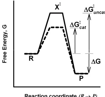
Transition State Theory
According to the transition state theory, the reactant(s) and product(s) of a chemical reaction are separated by an energy barrier. The height of this barrier represents the activation energy (∆G‡) of the reaction. The activation energy is the amount of energy each mol of reactant must acquire to reach the transition state (X‡), a strenuous form of the reactant which collapses instantaneously to form product (Fig. 1.7). The activation energy determines the rate of the reaction through:
23
1)
-(1
e
A
k
RT ∆G‡ −−−−====
Here R is the gas constant, T the absolute temperature and A is a constant specific for the reaction in question. A catalyst functions by reducing the height of the barrier allowing more molecules to pass from reactants to products per unit of time.
Figure 1.7 A schematic energy profile for the reaction of R to P. The reaction coordinate shows the progress of the reaction as it proceeds from reactant (R) to product (P). The energy axis shows the potential acquired by the reactant at the various states of completion of the reaction. The transition state is marked by X‡, the free energy of the reaction
denoted ∆G and the activation energy of the catalyzed and uncatalyzed reactions, ∆G‡ cat
and ∆G‡
uncat, respectively.
Enzymes as Catalysts
Enzymes are far more efficient than the catalysts employed in industry and transportation and they operate in aqueous solutions at low temperatures. With an enzyme as catalyst the yield is determined solely by the equilibrium position of the reaction catalyzed. With conventional catalysts the yield of the desired product is usually reduced through the occurrence of one or several competing side reactions.
24
The Active Site and Ground State Destabilization
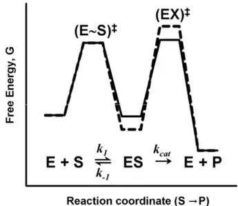
Part of the secret behind the extraordinary catalytic properties of enzymes is that an enzyme has a preorganized site to which the substrate binds in a fixed position relative to functional groups of the enzyme. However, binding of the ground state to a perfectly matching binding site would stabilize this state of the substrate and impede catalysis. Natures answer to this problem is the formation of an ‘active site’, that is an enzyme substrate pocket which is complementary to the transition state of the reaction catalyzed, rather than to the substrate or product of the same reaction. This concept was proposed by Michael Polanyi in 1921, and by Haldane in 1930. In 1946 Linus Pauling suggested that an enzyme must be complementary to the transition state of a reaction, in order to catalyze it. With such a binding site, it follows that the intrinsic binding energy is used in full for tight binding of the transition state, but only in part for binding of the substrate. The surplus binding energy is instead used to distort the bound substrate along the reaction coordinate. To account for this phenomenon we must include formation of the enzyme substrate complex ES in the energy profile for enzyme catalysis (Fig. 1.8).
Figure 1.8 Schematic energy diagrams for an enzyme catalyzed reaction.
The two profiles in Fig. 1.8 show that the difference in chemical potential between enzyme bound substrate ES and the corresponding transition state (EX)‡, i.e., the activation energy ∆G‡ of the reaction catalyzed, can be reduced both through destabilization of the ground state and stabilization of the transition state.
25
Stabilization of the transition state has been found experimentally to be the role of individual enzyme active site groups [31]. As indicated in Fig. 1.8, reduction of the activation energy can be achieved also through destabilization of the substrate ground state by the enzyme [47]. Ground state destabilization has been suggested to contribute to the extraordinary catalytic power (catalysis factor = 1017) of the enzyme orotidine 5´-monophosphate [48,49]. Nevertheless, destabilization of the ground state as part of catalysis, has been under debate, due to the scarce of experimental evidence.
Results we presented in Paper I associates both transition state stabilization and destabilization of the ground state with the strictly conserved serine at the active site of the homotrimeric dUTPases.
Transition State Analogues
In the context of enzyme complexes, stabilization means tighter binding, and the transition state of the reaction is the specie most stabilized by the enzyme. It follows that transition state analogues should be efficient enzyme inhibitors. As such they could serve as leads for the development of drugs. To build a transition state analogue we must first characterize the transition state. For this purpose, both enzyme structural and functional studies are necessary. Structural studies may define the active site. Functional studies are required to determine the specific role of the functional groups that line the active site and this, in turn, may provide information on the nature of the transition state.
Enzyme Kinetics
Despite the purposive less than perfect match between substrate and enzyme, experimental conditions which saturate the enzyme with substrate are easily achieved (although this may rarely occur in vivo). Enzyme saturation was observed long before the structure of an enzyme was known. It led Leonor Michaelis and Maud Menten to suggest in 1913 a simple scheme [50] known as the Michaelis-Menten scheme, to account for the activity of enzymes:
k1 kcat
E + S
⇌
⇌
⇌
⇌
ES → → → E + P → k-1Scheme 1
The first step in the Michaelis-Menten scheme is the reversible formation of the enzyme-substrate complex, ES, which is directly followed by its decomposition into free enzyme and product. This reaction scheme accounts for the behaviour of the vast majority of enzyme catalyzed reactions in experimental situations at which
26
the product can be neglected. In reality, the first step is followed by the chemical transformation of the enzyme bound substrate to enzyme bound product (EP), and dissociation to give product and free enzyme. Thus, a more realistic scheme would be:
E + S
⇌
⇌
⇌
⇌
ES⇌
⇌
⇌
⇌
EP⇌
⇌
⇌
⇌
E + P Scheme 2Scheme 2, takes into account the fact that the enzyme operates (with the same efficiency) in both directions of the catalyzed reaction. However, it is not necessary to take the reverse reaction into account to explain the behaviour of enzymes which are studied in the absence of significant amounts of product. In the case of the dUTPases studied here, and at the experimental conditions chosen, this simplification is valid over the complete reaction.
According to Scheme 1 the rate of an enzyme catalyzed reaction is given by Eqn. (1-2):
2)
-(1
[S]
[S]
M maxK
V
v
++++
====
Here, Vmax represents the maximum velocity that the reaction can achieve at a
specific total concentration of enzyme CE and it is defined as:
3) -(1 E cat max k C V ====
The catalytic constant, kcat, is the first order rate constant for the forward decay of
the enzyme substrate complex, also known as the turn-over number. It is expressed in units of s-1.
According to Eqn. (1-2), KM equals the concentration of substrate that results in v = Vmax/2. In terms of rate constants it is defined as:
4)
-(1
1 cat 1 M
k
k
k
K
====
−−−−++++
If kcat<< k-1, we have:5)
-(1
K
k
k
K
d 1 1 M≈≈≈≈
====
−−−−It can be seen from Eqn. (1-2) that if [S] is increased above KM, v approaches Vmax. At the other end, with [S] << KM, the rate equation reduces to:
27
K
V
v
M max6)
-(1
[S]
≈≈≈≈
and the decay of the substrate becomes exponential (first order). At the latter condition, CE is approximately equal to [E] the concentration of free enzyme, so
7)
-(1
[E][S]
K
k
v
M cat====
This makes kcat/KM the limiting second order rate constant for product formation
as [S] approaches zero. The upper limit of kcat/KM is restricted by diffusion to be
108-109 s-1 M-1 at 25°C. The kcat/KM ratio is alternatively named the specificity constant or the catalytic efficiency.
1.4 dUTP
ASEI
NHIBITORSThe Role of Enzyme Functional Groups in Catalysis
Regarding the dUTPases we are well supplied with structural information. In particular, the structure of the apo form of several dUTPases as well as their complexes with the substrate analogue α,β-imido-dUTP⋅Mg are known [25-31]. In this perspective, functional studies are lagging behind.
Site-directed mutagenesis has proven to be an effective tool in elucidating the contributions to catalysis and binding of individual groups in the active site of enzymes [46]. It was recently practiced on the E. coli dUTPase to show that the strictly conserved motif III aspartate, i.e., D90, provides transition state stabilization, but has no effect on substrate binding [31]. The information obtained in comparative kinetic studies (regarding the effect of the mutation on kcat and KM,
and further, on the individual rate constants of the system) may tell us if the functional group that was modified contributes to transition state stabilization or ground state destabilization. Ground state destabilization appears to be inherent in enzyme catalysis but direct experimental evidence, is scarce, a fact that enhances the importance of our first study (Paper I).
28
The Role of Substrate Functional Groups in Catalysis
The distinction between binding and catalysis discussed above for enzyme functional groups is valid also for groups carried by the substrate. The concept of substrate assisted catalysis, where a substrate functional group participates directly in catalysis to increase kcat, is a topic much discussed in literature [51-53].
For sterical reasons UTP is excluded from the active site of the dUTPases, where its 2´-hydroxyl group would interfere with the strictly conserved tyrosine of the loop which connects β-strands 5 and 6 [29] (Tyr93 in E. coli dUTPase). The hydroxyl group remaining at the 3´-position of dUTP is however accommodated by the enzyme in a position from which it could play a role in both binding and catalysis. In Paper II we study the effect of removing the 3´-hydroxyl group of dUTP on its hydrolysis by three homotrimeric dUTPases, namely the E. coli, the human and the EIAV forms.
Finding dUTPase Inhibitors
Due to their crucial role in keeping uracil out of DNA, described in the introduction, the dUTPases have emerged as potential targets for drugs against cancers, parasites, viruses and bacteria [16-20]. Recent attempts at finding an inhibitor, which is specific for the dUTPase of the malaria parasite (Plasmodium falciparum), have resulted in three reports [54-56]. It is essential that a dUTPase inhibitor intended to cure an infectious disease does not bind to the dUTPase of the host, because of the anticipated cytostatic effect. Specific binding is of less importance in the case of cancer treatment, unless this can be cured by fighting an infection. An in silico approach was applied by Ladner and co-workers to find inhibitors of the human enzyme [57]. Stivers et al. synthesized a library of triskelion shaped uracil derivatives and tested them as inhibitors of the enzyme uracil DNA glycosidase and the human dUTPase respectively. Two of these compounds showed inhibitory effects on the latter enzyme [37].
The design of dUTPase inhibitors is a current approach in finding treatments against parasitic-caused diseases such as malaria and Chagas disease, as well as in the development of new chemotherapeutic agents for cancer. Drugs inhibiting the human enzyme could potentially be used as standalone drugs or in combination with the thymidylate synthase inhibitors. A major difficulty in developing drugs against dUTPases of pathogens lies in the fact that the homotrimeric dUTPases, which include viral, bacterial and the human dUTPases, have almost identical substrate pockets, which bind the substrate with high affinity (KM≈10-7 M). To address this problem, we have studied the binding of various analogues of dUTP to dUTPases of different origin. This was done in an attempt at disclosing differences in binding that could guide the design of pathogen-specific drugs. In
29
this effort, we have studied the effect of replacing the 3´-hydroxyl group of the substrate by hydrogen (Paper II), or by the azido group (Paper III), on the binding to the human, the E. coli and the EIAV dUTPases.
To extend the search for dUTPase inhibitors to include non-substrate based compounds, we have performed studies on the inhibition of the same three enzymes by uracil containing triskelion shaped compounds (Paper IV). These studies were made in collaboration with Dr. Stivers, who kindly provided two compounds, previously reported to inhibit the human dUTPase [37].
31
2. EXPERIMENTAL APPROACH
Production and Purification of dUTPases
I have produced all the dUTPases used in this work, according to standard protocols [58] and purified them using established techniques reported previously for dUTPases [59]. These procedures are well explained in the respective report, and shortly summarized below.
As starting material we used the plasmids containing the gene (dut) for the respective dUTPase. The plasmids containing the human and EIAV genes, respectively, were kindly provided by Dr. Per Olof Nyman. On our request, the plasmids for E. coli dUTPase with the single (S72A) and double (S72A/D90N) mutations were produced and sequenced by Kristian Becker at Lund University, using the plasmid with the original bacterial dut gene as a starting point.
The plasmids were purified and transformed into E. coli BL21 competent cells. Cultures were grown based on single colonies, selected by antibiotic resistance according to standard protocols. The cultivated cells were lysed and the dUTPase was extracted and purified using a combination of affinity chromatography, fractional precipitation and gel filtration. The purified proteins were analyzed by SDS-PAGE [60] and activity assays.
The following section describes in more detail the procedures used for the functional characterization of the purified dUTPases.
2.1 K
INETICM
EASUREMENTSThe steady-state kinetic parameters Vmax (kcat) and KM are usually determined in
multiple turn over experiments, in which substrate is used in large excess over enzyme and a single measurement typically lasts over a minute or more. The determination of the magnitude of individual rate constants, i.e., k-1 and k1, usually
rely on the use of a rapid reaction instrument, such as a stop-flow spectrophotometer, which allow the reactions to be studied in the milliseconds time range. With enzyme in excess over substrate the reaction is restrained to a single turn over.
32
2.1.1
S
TEADY-S
TATEM
EASUREMENTSThe pH-Indicator Technique
The dUTPase reaction is optically transparent (there is no difference in absorbance between substrate and products over the accessible wavelength range), and for this reason it cannot be studied directly by spectrophotometric techniques. However, the reaction releases up to two protons per dUTP hydrolyzed, depending on the pH and metal ion concentration:
dUTP⋅⋅⋅⋅Mg + H2O → dUMP + PPi ⋅⋅⋅⋅Mg + n H+ (n = 0-2)
Data Acquisition
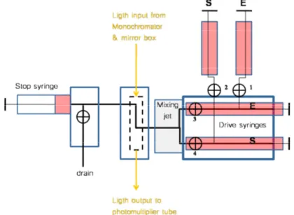
By including a suitable pH-indicator in the reaction medium, product formation in the dUTPase reaction can be monitored indirectly (in a spectrophotometer) at a wavelength at which the acidic or basic component of the indicator absorbs at maximum. To avoid pH-changes triggered by temperature changes or carbon dioxide uptake it is essential to reduce the reaction time and to seclude the medium from the atmosphere. This makes the stop-flow apparatus the ideal instrument (Fig. 2.1), because it is designed for fast measurements and the reaction takes place in a closed compartment.
Figure 2.1 The stop-flow spectrophotometer, a simplified scheme. The contents of the drive syringes are pushed pneumatically through the mixer and reaction chamber to reach the stop syringe. Measurement starts at, or just prior to, the flow stops. In our system, the dead-time, i.e., the average age of the reaction mixture at flow stop, is 3 milliseconds.
33
The stop-flow machine we use is an old but updated Durrum D-100 instrument. The detector consists in a digital oscilloscope device which stores the voltage, which is registered over a preset reaction time, in a memory buffer for subsequent transfer to a computer.
Data Treatment
Reaction traces covering the complete hydrolysis of the dUTP substrate are collected. The signal voltage is converted to transmittance (T) and then to absorbance (A) through A = -log (T). The absorbance scale is related to one in product concentration through Eqn. (2-1) [32].
1)
-(2
[P]
q
l
n
A
i i iεεεε
∆∆∆∆
∆∆∆∆
∆∆∆∆
====
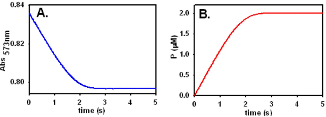
Here, n is the number of protons exchanged in the reaction, ∆εi the difference between the extinction coefficients of the basic and acidic forms of the indicator at the wavelength studied, l the optical path length, and qi is the relative buffer capacity provided by the indicator. However, the conversion of absorbance to product concentration is based on the fact the total absorbance change reflects hydrolysis of all dUTP included in the reaction mixture, as exemplified in Fig. 2.2.
Figure 2.2 Traces reflecting the hydrolysis of dUTP by the E. coli dUTPase under standard reaction conditions (dUTPase 0.2 µM, dUTP 2 µM, Mg2+ 5 mM, cresol red 50 µM, bicine
50 µM, pH 7.8). The two panels show the trace before (A) and after (B) conversion of the absorbance scale to one in product concentration. The total change in absorbance, i.e., in pH, is kept at a desirably low (and adjustable) level by the inclusion of small amounts of buffer in the reaction medium.
34
If we take the trace shown in Fig. 2.2.B that shows product (P) versus time (t), and calculate its first derivative (d[P]/dt) pointwise along the trace, we obtain the rate of the reaction, ν, supported by the corresponding concentrations of residual free substrate. By plotting the reciprocal rate (1/ν) versus the reciprocal concentration of free substrate (1/[S]), we obtain the conventional Lineweaver-Burk plot [61] displayed in Fig. 2.3. The concentration of free substrate was calculated based on the trace in Fig. 2.2B and the KM evaluated for the trace.
Figure 2.3 Lineweaver-Burk plot representing the data in Fig. 2.2B, for the hydrolysis of dUTP⋅Mg catalyzed by the E. coli dUTPase.
The plot in Fig. 2.3 demonstrates the number of initial rate measurements covered by a single complete reaction trace, and is neither intended nor used for data analysis. Its linear nature attests to the fact that the Michaelis Menten mechanism in Scheme 1, at the experimental conditions typically used here, is valid throughout the reaction.
Using Enzyme Intrinsic Fluorescence as Signal
We found the pH-indicator technique unsuitable for the kinetic characterization of the S72A mutant E. coli dUTPase (Paper I), because of the extremely low activity displayed by the mutant. Instead, we used the fact that the intrinsic fluorescence of E. coli S72A dUTPase decreases about 15% upon binding to the substrate (dUTP⋅Mg) to monitor the reaction catalyzed by the mutant. The trace in Fig. 2.4 shows the fluorescence changes associated with the hydrolysis of dUTP (10 µM) by the S72A mutant (4 µM) at saturating Mg2+. The second phase of the trace
35
represents the hydrolysis of the last enzyme bound substrate, releasing the more strongly fluorescent free enzyme. This phase is preceded by ~1.5 rounds of enzyme turn over at which the enzyme is present as the less fluorescent ES complex. The flat nature of the first phase and the abrupt transition from zero to first order kinetics indicate tight binding in the ES complex.
Figure 2.4 Hydrolysis of dUTP by the S72A E. coli dUTPase monitored through the effects of substrate binding on the intrinsic protein fluorescence. The signal is quenched when the enzyme is bound to the substrate, and recovered as this is hydrolyzed.
This method was applied to measure the activity of the S72A E. coli dUTPase under different conditions, and is explained in detail in Paper I.
Parameter Evaluation
Integrated forms of rate equations for various enzyme systems, including the Michaelis Menten equation have been derived. Such equations can form the basis for analysis of complete reaction curves as demonstrated [32]. However, from a trace like that in Fig. 2.2A, only the total concentration of substrate ([S] + [ES]) can be calculated directly. Ideally, and in particular if CE is comparable to KM, this should be converted to the concentration of free substrate before analyzed in terms of the integrated rate equation. Steady state rate equations are derived based on the assumption that [ES] is insignificant compared to [S].
The differential equations describing the rate of change of the four species in the Michaelis Menten scheme are shown in Eqns. (2-2).
36
The steady-state approximation assumes that the two central equations amounts to zero, that is, that the concentration of enzyme containing species does not change (at least not over the time the reaction is studied). With this condition the system can be solved analytically to give the Michaelis Menten rate equation. In the general case, an approximate solution can be arrived at by various numerical methods [62-63]. In principle these methods multiply the right hand side of the equations with a small time increment, and this process is repeated for the desired length of accumulated time.
The program Dynafit [64] that we have been using throughout this work, combines modern numerical methods [62-63] to solve the differential equations valid for the mechanism chosen as model, with an efficient fitting algorithm [65]. This means that as an experimentalist you are no longer restrained to a limited set of experimental conditions in order to evaluate your system. Practically any set-up is open to analysis. In addition, the program Dynafit has a simulation capability which can be used to predict, if an experiment is feasible under a specific set of conditions.
The numerical approach necessitates that the individual rate constants are used as parameters. The precision to which they can be evaluated individually depends, however, on the experimental conditions. For instance, with our most typical set up, we can evaluate Vmax (kcat if the enzyme concentration is known) and KM, but
not k-1 or k1 (or one of them, provided the other is known).
2.1.2
S
UBSTRATEB
INDINGK
INETICSIn order to obtain estimates of the rate constants for substrate binding (k-1 and k1)
to the dUTPases studied, three alternative methods were used.
Viscosity Effects Approach
If k-1 and kcat in the Michaelis Menten reaction are of comparable magnitudes, the
kinetic constants k1 and k-1 can be determined from the effects of medium
viscosity on the kinetics of the reaction [66,67]. We used sucrose as viscous agent and monitored the reactions by the pH indicator method (Fig. 2.5). The
d[S]/dt = -k1 [S][E] + k-1 [ES]
d[E]/dt = -k1 [S][E] + k-1 [ES] + kcat [ES]
d[ES]/dt = k1 [S][E] - k-1 [ES] - kcat [ES]
d[P]/dt = kcat [ES]
2) -(2
37
dependence of KM/kcat on the relative viscosity ηrel, defined as the ratio of η, the
viscosity of the sucrose solution, to η0, the viscosity of water, at the same
temperature, is given by Eqn. (2-3):
3)
-(2
k
η
k
k
k
k
K
1 rel 1 cat 1 cat M====
−−−−++++
If KM/kcat is plotted versus ηrel, Eqn. (2-3) predicts that the data points should
adhere to a straight line. The slope of the line equals 1/k1 and the intercept on the
y-axis equals k-1/(kcat k1).
Complete traces for the hydrolysis of dUTP by the E. coli dUTPase were collected at increasing viscosity of the reaction medium (Fig. 2.5A), obtained by the inclusion of sucrose (0-50 %, w/w). The kcat and KM evaluated for each trace were
used to determine k1 and k-1 with Eqn. (2-3) (Fig. 2.5B, upper line). A parallel
series of measurements, serving as a control of method, were done using EIAV dUTPase as catalyst. For this enzyme, k1 and k-1 have been determined previously
by a direct method [68].
Figure 2.5 A) Reaction traces for the hydrolysis of dUTP⋅Mg, catalyzed by the E. coli dUTPase, obtained at (from left to right) increasing concentrations of sucrose. B) Analysis of data obtained for E. coli (●) and EIAV (▼) dUTPases, respectively, according to Eqn. (2-3).
Pre-Steady-State Measurements
In the case of the S72A mutant, characterized in Paper I, a favourable signal and relatively slow kinetics enabled direct determination of k1 by stop-flow fluorimetry,
38
By changing indicator (to bromocresol purple) and accessory buffer (to MES), we could apply the pH-indicator method to compare the rate of substrate binding to the E. coli wild type and S72A mutant dUTPase, respectively at pH 6 (Fig. 2.6). In addition to the lower pH, the concentration of Mg2+ was reduced to 20 µM. Under such conditions, the substrate will be mostly metal free and protonated. In binding to the dUTPase, the substrate picks up a magnesium ion and the proton is expelled:
E + (dUTP⋅⋅⋅⋅H)3- + Mg2+ + H2O → (E⋅⋅⋅⋅H2O⋅⋅⋅⋅dUTP⋅⋅⋅⋅Mg)2- + H+
This accounts for the indicator absorbance decrease observed in the pre-steady-state phase of the reaction with both forms of the enzyme (Fig 2.6). The overall reaction is best described by:
(dUTP⋅⋅⋅⋅H)3- + H2O →
→ (dUMP⋅⋅⋅⋅H)- + n(PPi ⋅⋅⋅⋅H2)2- + (1-n)( PPi ⋅⋅⋅⋅H)3- + (1-n)H+
where n is a fraction. This means that a 1-n fraction of a proton is released in each enzyme turn over, as demonstrated by the ‘steady-state’ absorbance decrease in the trace obtained with the wild type enzyme (Fig. 2.6, left panel).
Figure 2.6 Kinetics of formation of the enzyme-substrate complex of the E. coli dUTPase A) wild type B) S72A mutant. (Figure from Paper I, reproduced with permission).
As the concentration of substrate approaches that of enzyme active sites, the scene becomes dominated by the enzyme substrate complex:
39 (E⋅⋅⋅⋅H2O⋅⋅⋅⋅dUTP⋅⋅⋅⋅Mg)2- + H+ →
→E + (dUMP⋅⋅⋅⋅H)- + n(PPi ⋅⋅⋅⋅H2)2- + (1-n)( PPi ⋅⋅⋅⋅H)3- + (1-n)H+ Mg2+
The first order decomposition of this complex is associated with uptake of n protons as demonstrated (Fig. 2.6, left panel inset).
The time constant r associated with the pre-steady-state absorbance change, can be evaluated by non-linear regression using,
A = A0 +
νννν
A t + ∆∆∆∆A e-r t (2-4)Here, A is the absorbance at time t, ∆A the amplitude of the exponential phase of the reaction, A0 the final absorbance extrapolated to zero reaction time, and νA the steady state rate of absorbance change.
Evaluation of the time constant r in Eqn. (2-5), and knowledge of KdUTP⋅Mg, the Kd for the dUTP⋅Mg complex, as well as of k-1 and kcat, allows the estimation of k1
based on the following equation:
[[[[
]]]]
5) -(2 Mg 1 2 cat 1 Mg dUTP dUTP 1 k k K C k r ++++ ++++ ++++ ==== −−−− ++++ ⋅⋅⋅⋅where CdUTP, is the initial concentration of substrate.
2.2 E
QUILIBRIUMB
INDINGS
TUDIESFrontal Chromatography
Ligand binding studies were performed with the enzyme immobilized to the matrix in a small column. Information on the strength of binding is obtained by comparing the distance between the ligand fronts when a binding and inert ligand, respectively, are flushed through the column. This technique is named frontal chromatography [69] and has not previously been reported applied to a dUTPase system. A major advantage with this technique is that the column can be used over and over again and that it does not depend on a differential signal. It was applied to measure binding of the following ligands to the S72A E. coli mutant dUTPase:
40
dUTP, ddUTP, (both in the absence of metal ions) α,β-imido-dUTP, dUMP, dTTP, dCTP. As inert control we used ATP.
The volume (V) passing through the chromatography system in front of a substance that binds to the immobilized enzyme will be larger than the corresponding volume (V0) for a non-binding substance (i.e., ATP). The difference depends on the ligand concentration Clig, the Kd and the number of immobilized binding sites NE, according to the following relationship:
6)
-(2
C
K
N
V
V
lig d E 0====
++++
−−−−
Elution profiles obtained with this technique, testing the binding of metal-free dUTP to the S72A E. coli dUTPase, using ATP as reference, are shown in Fig. 2.7.
dUTP(µM) 0 5 10 15 V -V o ( m l) 0.0 0.4 0.6 0.8 Ve (ml) 0 3 4 5 A b s 2 5 4 0 15 30 ATP 4µM dUTP 6uM
Figure 2.7 Frontal Chromatography of dUTP on immobilized S72A E. coli dUTPase. The effect on V-V0 of the concentration of dUTP, is shown in the main panel. The elution
profiles of ATP and dUTP at a single concentration are displayed in the inset. (Figure from Paper I, reproduced with permission).
2.3 P
ROTEINS
TABILITYThermal Stability
The structure of most proteins is sensitive to changes in the environment, such as pH, ionic strength and temperature. If such changes result in loss of the
three-41
dimensional structure of the protein, it is called denaturation. The denaturated protein, does not necessarily imply a completely unfolded state, it could consist of several partially folded states.
Profiles showing the dependence of some physical property of the protein, such as fluorescence, on an agent, such as temperature, can be used to compare the stability of a wild type enzyme with that of a mutated form. A thermal transition occurs in the E. coli dUTPase around 50 ◦C [70]. Data in Fig. 2.8 indicates that this transition occurs at a higher a temperature in the S72A E. coli dUTPase.
Figure 2.8 Thermal transition curves for the E. coli dUTPase wild type (●) and S72A mutant (■) forms. (Figure from Paper I, reproduced with permission).
Chaotropic Stability
Another method for testing protein stability uses chaotropic agents, such as urea or the guanidinium ion, to induce denaturation of proteins. The E. coli wt dUTPase was reported to be 50% unfolded at a concentration of GuHCl of about 1.4 M [70]. To investigate the relative stability of the wild type and S72A E. coli dUTPases (Paper I), we decided to monitor continuously the unfolding of the two proteins at various concentrations of the chaotropic agent (1.5 – 4 M). This was because we wanted to compare the properties of the intact forms of the two proteins, that is, their unfolding. The signal used was, as in the thermal transition study, the intrinsic fluorescence of the two proteins. In Fig. 2.9 the kinetics of unfolding each protein, triggered by exposing them to GuHCl (2 M), is shown.
42
Figure 2.9 Time courses of guanidinium induced denaturation of the E. coli dUTPase wild type and S72A mutant forms. The concentration of the chaotropic agent was 2 M.
43
3. RESULTS AND DISCUSSION
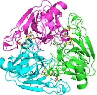
The α,β-imido-dUTP⋅Mg binds to the E. coli dUTPase in the same way as dUTP⋅Mg binds to the D90N mutant of the same enzyme (Fig 3.1). One of the differences observed is a rotation of the side chain of serine 72, strictly conserved in motif II among the dUTPases and dCTP deaminases. In the complex with the true substrate, the β-hydroxyl of S72 points inward in the intersubunit cleft to approach the pyrimidine ring. In the complex with the analogue, the β-hydroxyl has rotated away from the pyrimidine to form a hydrogen bond to the nitrogen that bridges the α- and β-phosphorus.
Figure 3.1 Views down the active site of the E. coli D90N single mutant (left panel, PDB 1SYL) and wild type dUTPase (PDB 1RN8), respectively (green surface: subunit 1 with motif III; violet surface: subunit 2 with motif I, II and IV; yellow surface: part of motif V in the distal subunit 3). The D90N form holds the substrate dUTP⋅Mg and the latter the
α,β-imido-dUTP⋅Mg analogue. Except for the rotation of the S72 side chain, a partial immobilization of motif V is observed in the complex with the α,β-imido-dUTP⋅Mg substrate analogue (in yellow).
A identical arrangement exists in the complex of the M. tuberculosis dUTPase with the α,β-imido substrate analogue [29], and led to the proposal that S72 stabilizes the transition state by balancing negative charge at the α,β-bridging oxygen of the latter.
44
3.1 M
UTATIONALS
TUDIESA Dual Role for the Strictly Conserved Serine
To test the role of the conserved serine in motif II in substrate binding and catalysis, we replaced its β-hydroxyl by hydrogen. This was done by mutating Ser72 in the E. coli dUTPase to an alanine. A chromatographic assay indicated that kcat of
the mutant was reduced to 0.008 s-1. This corresponds to an increase in the activation energy of the catalytic step of more than (ln(kcat/k´cat)RT =) 16 kJ mol-1
compared to the wild type enzyme (the prime is used to denote constants for the mutant). The low k´cat represents a 725-fold reduction compared to kcat (5.8 s-1)
and confirms a catalytic role for S72. With such a low activity we reasoned that it should be possible to determine K´M by using the S72A mutant as a competitor for substrate in the standard assay of the wild type enzyme. With this technique, we had previously confirmed that substrate binding to the D90N mutant (kindly provided by Prof. Vertessy) is characterized by a KM of 0.5 µM, as previously reported [31].
Figure 3.2 Competition for dUTP⋅Mg (2 µM) between the wild type and S72A E. coli dUTPases. The concentration of the former was 0.18 µM. The dashed black lines were obtained after inclusion of the mutant in the reaction mixture at 0.8, 1.2 and 1.6 µM, respectively (top to bottom). The grey dashed line was simulated and shows the course the reaction would take at 0.8 µM of the mutant if K´M is identical to KM (0.21 µM).