LICENTIA TE THESIS IN ODONT OL OG Y ULRIKE SC HÜTZ-FR ANSSON MALMÖ UNIVERSIT
FIXED
MANDIBUL
AR
RET
AINERS
ULRIKE SCHÜTZ-FRANSSON
FIXED MANDIBULAR
RETAINERS
A controlled 12-year follow-up
© Copyright Ulrike Schütz-Fransson, 2018 Illustrations: Ulrike Schütz-Fransson ISBN 978-91-7104-935-3 (print) ISBN 978-91-7104-936-0 (pdf) ISSN 1650-6065
ULRIKE SCHÜTZ-FRANSSON
FIXED MANDIBULAR
RETAINERS
A controlled 12-year follow-up
Department of Orthodontics
Faculty of Odontology
Malmö University, Sweden 2018
Publikationen finns även elektroniskt på: http://muep.mau.se/handle/2043/25665 Publikationen finns även elektroniskt,
”There is nothing permanent, except change” Heraclitus of Ephesus 6th Century B.C.
CONTENTS
PREFACE ... 11 THESIS AT A GLANCE ... 12 ABSTRACT ... 13 Paper I ...14 Paper II ...14PAR Index evaluation ...15
Key conclusions and clinical implications ...15
POPULÄRVETENSKAPLIG SAMMANFATTNING ... 16
Konklusioner i delarbete I ...17
Konklusioner i delarbete II ...17
Konklusion i PAR Index utvärderingen ...17
Klinisk betydelse ...18
INTRODUCTION ... 19
Background ...19
Retention methods ...22
Mandibular anterior segment stability ...25
Mandibular anterior segment in untreated patients ...26
Biology during tooth movement and histological changes during orthodontic relapse ...26
Peer Assessment Rating ...30
SIGNIFICANCE ... 32
AIMS ... 34
Paper I ...34
HYPOTHESIS ... 35
Paper I ...35
Paper II ...35
PAR Index evaluation ...35
SUBJECTS AND METHODS ... 36
Subjects ...36
PAR Index evaluation ...39
Ethical considerations and consent ...40
Methods ...40
Papers I and II ...40
Paper II ...42
PAR Index evaluation ...43
Statistical analysis ...43
RESULTS ... 45
Paper I ...45
Paper II ...48
PAR Index evaluation ...51
DISCUSSION ... 56
Irregularity of mandibular anterior segment ...56
Causative factors of relapse ...59
Irregularity of mandibular anterior segment in untreated cases ...59
Overjet and overbite ...60
Cephalometric outcomes ...60
Gender ...62
Extractions versus non-extractions ...62
Retention for how long time? ...63
Failures and retainer breakages ...64
Unexpected effects of fixed retainers ...65
PAR Index evaluation ...69
Maintenance of fixed retainers ...70
Methodological aspects...71
Future research...72
CONCLUSIONS ... 73
Key conclusions and clinical implications ...74
ACKNOWLEDGEMENTS ... 75
REFERENCES ... 78
PREFACE
The following papers, on which this thesis is based, are referred to in the text as Paper I and Paper II.
I. Schütz-Fransson U, Lindsten R, Bjerklin K, Bondemark L. Twelve-year follow-up of mandibular incisor stability: Comparison between two different bonded lingual orthodontic retainers. Angle
Orthod. 2017; 87:200-208.
II. Schütz-Fransson U, Lindsten R, Bjerklin K, Bondemark L. Mandibular incisor alignment in untreated subjects compared with long-term changes after orthodontic treatment with or without retainers. Accepted for publication by Am J Orthod Dentofacial Orthop. March 2018.
These papers are reprinted with kind permission from the copyright holders, The Angle Orthodontist and the American Journal of
THESIS A
T A GL
AN
CE
THE SI S A T A GL A NCE Study Purpose Study design SampleMain outcome parameter
Main findings I To
compare the long
-term
outcome 9 years after removal of two different types of f
ixed
retainers used for stabilis
ation of
the mandibular anterior segment;; canine
-to -canine retainer and Twistflex retainer Retrospective Controlled Longitudinal 64 children Little’s Irregularity Index (LII) Both a can ine -to
-canine retainer and
a T wistflex retainer 3- 3
can be recommended
, as
bo
th are
equally effective when
they are in place.
None of the retention types prevent long
-term
changes of mandibular incisor
irregularity
after
removal of the retainers.
II
To
analys
e the dental and
skeletal changes
12 years after
treatment
in patients treated with
fixed orthodontic appliances, with or without retention appliances and to compare the
changes with untreated subjects Retrospective Controlled Longitudinal 130 children Little’s Irregularity Index (LII) No differences were found in Little’s Irregularity Index
12 years after treatment
for the
mandibular incisors
between the
group that had
a retainer and the
group
that had no
retainer
after treatment
.
In the untreated group
, LII
increased over time
,
but not to the same extent as in the treated groups.
Correction of overjet and overbite was stable long
-term. To ev aluate the orthodontic
treatment outcome after treatment and long
-term
, 12
years after treatment
for two
different retainer gro
ups and one
non -retention group Retrospective Controlled Longitudinal 94 children PAR Index
Twelve years after treatment, the mean reduction in PAR
score was over 70 per cent
for
the groups who had a mandibular retainer after treatment
.
The
non
-retention group had a reduction of 66
per cent. The cases that were ‘greatly improved’ and/or ‘improved’ 12
years after treatment were 56
per
cent
in the non
-retention group, 64
per cent in
the canine
-to
-ca
nine retention group and 60
per
cent in the Tw
istflex retainer group.
ABSTRACT
A fixed retainer is a retention appliance that is often used after orthodontic treatment. For the mandibular incisors, two types of fixed retainer are commonly used – a canine-to-canine retainer bonded only to the canines or a Twistflex retainer bonded to each of the mandibular incisors and canines.
The increase in mandibular incisor irregularity seems to be a continuous process throughout life even in untreated patients. The natural physiological changes of aging causes the types of changes that occur after orthodontic treatment and the removal of a retainer. There are few long-term studies that compare patients who have had a mandibular fixed retainer with those patients who have not had any retention appliance after treatment, and then compare the treated patients with untreated subjects.
The overall aims of this thesis were to compare and evaluate two different mandibular fixed retainers and to compare orthodontically treated cases with those untreated long-term. The mandibular anterior region was evaluated.
This thesis is based on two studies, and a PAR Index evaluation is presented in the frame story.
The retrospective longitudinal study of Paper I focuses on the dental casts and lateral head radiographs of patients who had received either a canine-to-canine retainer or a Twistflex retainer after treatment. The study measured different variables, where Little’s Irregularity Index was the main outcome measure. The measurements were taken
at four different occasions, with the last registration occurring 12 years after treatment (i.e. nine years after the removal of the retainer). Paper II is also a retrospective longitudinal study, but with three different groups. One group received a fixed mandibular retainer, one group did not receive any retention appliance after treatment, and the third group was composed of untreated subjects. Measurements were taken on dental casts and lateral head radiographs at four different occasions to analyse dental and skeletal changes 12 years after treatment. Here, Little’s Irregularity Index was also the main outcome measure.
The PAR Index evaluation was conducted to get an overall assessment of the orthodontic treatment outcome and not only to evaluate the mandibular anterior region, both directly after treatment and in the long-term for two different retainer groups and one non-retention group.
The following conclusions were drawn:
Paper I
• Both the canine-to-canine retainer and the Twistflex retainer can be recommended, as both are equally effective during the retention period.
• None of the retention types prevent long-term changes of mandibular incisor irregularity or available space for the mandibular incisors after removal of the retainers.
Paper II
• There were no differences found 12 years after treatment in Little’s Irregularity Index for the mandibular incisors between the group that had a retainer and the group that had no retainer after treatment.
• In the untreated group, Little’s Irregularity Index increased over time but not to the same extent as in the treated groups. • The crowding before treatment did not explain the crowding
• The use of fixed mandibular retainers for two to three years does not appear to prevent long-term changes.
• The overjet and overbite were stable long term.
PAR Index evaluation
• Twelve years after treatment, the mean reduction in PAR score was over 70 per cent for the groups who had a mandibular retainer after treatment while the non-retention group had a PAR score reduction of 66 per cent.
• The cases that were ‘greatly improved’ and/or ‘improved’ 12 years after treatment were 56 per cent in the non-retention group, 64 per cent in the canine-to-canine retention group and 60 per cent in the Twistflex retainer group.
Key conclusions and clinical implications
For retention of the mandibular incisors after orthodontic treatment, the canine-to-canine retainer and the Twistflex retainer can both be recommended, as they are equally effective during the retention period.
The use of fixed mandibular retainers for two to three years does not appear to prevent long-term changes.
In untreated cases the mandibular incisor irregularity will increase over time but not to the same extent as in treated cases where the retainer has been removed.
Patients must be informed about the development of natural physiological changes that appear of the mandibular incisors through aging. If the patient wants to constrain natural development and changes, lifelong retainers are needed.
POPULÄRVETENSKAPLIG
SAMMANFATTNING
Efter att ha genomgått tandregleringsbehandling (ortodontibehand-ling) måste tänderna ofta fixeras i sina nya lägen, eftersom de har en tendens att vilja flytta sig tillbaka till sina ursprungliga lägen (recidiv). Därför behöver tänderna hållas kvar efter behandling med t.ex. en metalltråd (retainer) som limmas på baksidan av framtänderna. För underkäkens framtänder finns det vanligen två olika typer av reta-inrar, antingen cuspid-retainern, som är styvare och limmas enbart på hörntänderna eller Twistflex-retainern, som är mer flexibel och limmas till samtliga framtänder och hörntänder.
Ökad trångställning av underkäkens framtänder är en kontinuerlig fysiologisk process som sker under hela livet både bland obehandlade och behandlade patienter. Det finns få långtidsuppföljningar som har jämfört patienter som har haft en retainer i underkäksfronten med individer utan retainer efter behandling, och dessutom jämfört behandlade med obehandlade individer.
Licentiatexamen är baserad på följande studier:
I en retrospektiv kontrollerad studie av studiemodeller samt profil-röntgenbilder var syftet i Studie I att i ett långtidsperspektiv jämföra cuspid-retainern med Twistflex-retainern ur stabilitetssynpunkt.
I studie II var syftet att jämföra ortodontiskt behandlade patienter med och utan retainer efter behandling med obehandlade, vad gäller underkäkens framtänder.
I ramberättelsen utfördes dessutom en PAR Index utvärdering (Peer Assessment Rating) för att analysera det totala behandlingsre-sultatet och inte bara utvärdera förändringarna av underkäksfram-tänderna. Utvärderingen gjordes direkt efter behandling samt 12 år efter behandling, för grupperna med olika retainrar samt gruppen utan retention.
Konklusioner i delarbete I
Sex och 12 år efter tandregleringsbehandling, och i genomsnitt 9,2 år efter att retainrarna hade avlägsnats kunde följande resultat och slutsatser dras:
• Både cuspid-retainern och Twistflex-retainern kan rekommenderas eftersom båda är lika effektiva under retentionsperioden.
• Ingen av retainrarna kunde förhindra trångställning av underkäkens framtänder på lång sikt efter att retainrarna har tagits bort.
Konklusioner i delarbete II
En jämförelse mellan patienter som fått en bondad retainer med patienter utan någon retention efter behandling och en obehandlad grupp, gav följande slutsatser:
• Det förelåg inga skillnader mellan gruppen som hade haft en retainer och gruppen utan retainer, 12 år efter behandling med avseende på trångställningsgraden av underkäkens framtänder. • I den obehandlade gruppen ökade också trångställningen av
underkäkens framtänder över tid men inte lika mycket som i de behandlade grupperna.
Konklusion i PAR Index utvärderingen
• Tolv år efter behandling är förbättringsprocenten fortfarande bra men lägre än direkt efter behandling. Detta visar att förändringar har skett för många variabler i bettet. Detta gäller för samtliga 3 grupper.
Klinisk betydelse
För att retinera underkäksframtänderna efter tandreglerings-behandling är cuspid-retainern och Twistflex-retainern likvärdiga.
Hos obehandlade individer ökar också trångställningen av underkäksframtänderna, dock inte lika mycket som hos behandlade patienter, där retainern har tagits bort.
Om patienten vill ha jämna tänder livet ut och därmed förhindra de förändringar som sker genom naturlig fysiologisk utveckling, behöver retentionstrådarna sitta kvar hela livet.
INTRODUCTION
Background
In Sweden, all children and adolescents receive free dental care until they are 22 years old, with the exception of some counties where the cut-off age is 24 years. There are several studies (Linder-Aronson et al. 2002; Holm 2005; Bjerklin et al. 2012) that have shown various prevalence of objective treatment need in Sweden. In most of the studies, 30–35 per cent of the children, adolescents, and young adults in this age range are offered free orthodontic treatment to correct their malocclusions.
The goal for orthodontic treatment is to achieve good function, good aesthetics and stable occlusion. Later, it is important to retain the treatment result to avoid relapse, and this is a great challenge in orthodontic treatment.
Relapse can be defined as the tendency of treated teeth to return to their former positions. Back in 1936, Mershon explained, “You can move teeth to where you THINK they belong; NATURE will place them where they best adapt themselves to the rest of the organism” (Mershon 1936).
The tendency of treated teeth to move back to their former position was already known by Coleman back in 1865 in England. A year later, Marvin described the physiological reasons for retention, and in 1881, James W. Smith designed the first retainer in the USA (Weinberger 1926). This retainer consisted of a vulcanite plate with a bar to keep the maxillary incisors in place. Since then, much has changed in the concept of how teeth should be retained. In 1887, Edward H. Angle suggested that a retaining appliance should hold
the teeth so firmly that there will be no movement to disturb or in any way interfere with the new bone formation (Angle 1887).
Riedel shared his ideas on stability and proposed nine retention rules (Riedel 1960). In the nine rules, he claims that
1) teeth tend to return to their former positions,
2) mandibular arch form should be maintained during orthodontic treatment,
3) a proper diagnosis based on determining the cause of malocclusion is invaluable,
4) overcorrection of malocclusion is a safety factor in retention, 5) occlusion is an important factor in retention,
6) bone and adjacent tissues must be allowed to reorganise around the newly positioned teeth for some length of time, 7) placing the mandibular incisors upright over basal bone will
result in a more stable correction of a malocclusion,
8) corrections carried out during periods when the patients are growing are less likely to relapse, and
9) the farther teeth have been moved the less the likelihood of relapse.
Retention is central for establishing long-term stability. Thus, after orthodontic treatment, the retention period has to be taken into consideration in the treatment plan, which involves considering how to retain and for how long. Therefore, it is important to understand the relapse process. Orthodontic relapse is a term to describe the post-treatment adaptation of the dentoalveolar system to changes in the mechanical conditions caused by the withdrawal of orthodontic forces. Usually, this change is distinguished between fast relapse, which occurs during the remodelling of the periodontal ligament, and slow
relapse, which refers to changes later in life, i.e. natural physiological
changes, (Thilander 2000). These later changes cannot be separated from normal age-related changes that appear independently whether orthodontic treatment has been carried out or not.
The amount and nature of relapse is unpredictable and variable. In most studies, we do not find any clinical, biometric or cephalometric variables that can predict the post-retention crowding. This is because relapse is considered as a multifactorial process. Among the factors that may cause relapse following orthodontic tooth movement are
tooth size, arch form, abnormal muscle function, occlusal stress, patient age, length of retention, mandibular rotation, apical base, craniofacial growth and mainly, the contraction of displaced fibrous tissue rearrangement that may be observed even after several years. In a study by Glenn et al. (1987), eight years post-retention, out of retention for a minimum of three years in non-extraction cases, the mandibular incisor mesiodistal and faciolingual dimensions were not associated with either pre-treatment or post-treatment incisor crowding. Another study with extraction cases, 5.1 years post-treatment and a fixed mandibular retainer for mean 1.6 years, showed that the mandibular-incisor-crown morphology was not significantly correlated with the amount of mandibular-anterior-crowding relapse (Freitas et al. 2006a).
If a malocclusion is caused by muscular or other soft tissue dysfunctions (i.e. cheek/lip/tongue pressure), and if the correction of this malocclusion is performed without any alteration in muscular or dysfunctional behaviour, then there will be an obvious risk of relapse (Proffit 1986).
Other important factors for long-term stability are a good occlusion, unchanged intermolar and intercanine distance (Proffit 2013). Changing the original dental arch form during treatment and the expansion of the transverse distance between the canines and molars increase the risk of relapse (de la Cruz et al. 1995). In cases with small apical bases, expansion treatment should be avoided to reduce the risk of relapse. The position of the incisors is recommended to be within normal range, i.e. the incisors have to be positioned over the apical base.
Also, the growth pattern of an individual patient is an important factor. Orthodontic corrections are likely to relapse during periods of growth, which include the eruption of teeth. It seems that the pattern of late mandibular growth is one contributor to the crowding tendency (Nanda & Nanda 1992). Moreover, there are researchers who claim that early treatment of malocclusions provides greater final stability (Kerosuo et al. 2013).
A mesially acting force emanating from the back of the dental arch is another possible factor (Samspon 1995), but the theory that pressure from the developing third molars causes late incisor crowding has not been confirmed. The presence of third molars does not appear
to produce a greater degree of mandibular anterior crowding after the cessation of retention than that which occurs in patients with third molar agenesis (Kaplan 1974). A study examined 51 subjects with intact lower arches and bilateral third molars present at the ages of 13 and 18 years. It was considered that the third molars are one of the causes of late mandibular arch crowding (Richardson 1989). The principal conclusion from another study was that the removal of third molars to reduce or prevent late mandibular incisor crowding could not be justified (Harradine et al. 1998). In addition, a review study concluded that long-term studies in untreated individuals do not suggest evidence of a cause and effect relationship between third molars and late mandibular incisor crowding. Thus, asymptomatic and pathology-free third molars should not be extracted to prevent post-retention crowding or to prevent late mandibular crowding in untreated individuals (Sumitra & Arundhati 2005).
Periodontal force is a potential cause of relapse (Reitan 1967; Johnston & Littlewood 2015). The periodontium exerts a continous force on the mandibular dentition and this force acts to maintain the contacts of approximating teeth in a state of compression. This force is increased after occlusal loading and may help to explain long-term crowding of the mandibular anterior teeth, physiologic drifting of teeth, and maintenance of posterior dental contacts after interproximal wear (Southard et al. 1992).
Genetic factors must also be considered, as it also constitutes a factor during retention following treatment. For instance, the influence caused by hereditary factors may be observed in certain cases in which overlapping by one or both lateral incisors are observed in the patient as well as in his/her mother or father (Reitan 1969).
One reliable predictor of relapse has been found by (Renkema et al. 2008 and 2011). They reported that the number of retainer failures, i.e. the number of detachments and/or breakages of the retainer, contributed to the relapse.
Retention methods
‘Retention’ is derived from the Latin verb, retinere, which means ‘to hold back or maintain in place’.
Retention is necessary in cases where
• the supporting tissues of the teeth need to be reorganised around the teeth in their new position
• there is a (neuro)muscular imbalance, as the teeth may be in an inherently unstable position after treatment
• there is continued bone growth and remodelling after orthodontic treatment
• persistent unwanted habits could be present
To prevent relapse after orthodontic treatment, a fixed retainer was advocated 45 years ago (Knierim 1973), when enamel etching and modern adhesive systems became more common. Prior to that, a lingual wire soldered to canine bands for the fixed mandibular retainer had been used. Various combinations of stainless steel or beta titanium alloys were used on rectangular or round wires that were either braided or twisted and in various sizes ranging from 0.016 to 0.032-inch. Later, a multi-strand 0.0175-inch wire bonded on six anterior teeth was used (Zachrisson 1977). The next generation of fixed retainers was introduced in 1982. These were composed of heavier, flexible, round multi-strand 0.032-inch wires bonded only to the canines. Unfortunately, this heavy multi-strand wire caused an increase of plaque accumulation and reduced wearing comfort (Årtun & Zachrisson 1982). Further clinical experience resulted in a new generation of fixed retainers constructed on a plaster model from a smooth stainless-steel wire (0.030 to 0.032-inch) sandblasted at both ends to increase composite retention (Zachrisson 1995).
Another retention method is the vacuum-formed retainer such as the Essix retainer (Sheridan et al. 1993). This retainer can be used alone or in combination with bonded retainers.
Interproximal enamel reduction, or interproximal stripping of the mandibular anterior teeth has been shown to be an alternative strategy for preventing relapse. Often, it is done in combination with over-correction of rotated teeth. In a study by Aasen and Espeland (2005), 56 patients were treated with over-correction of rotated teeth and systematic enamel reduction of the approximal surfaces in the mandibular anterior region. In 45 per cent of the patients, the change in Little’s Irregularity Index was less than 0.5 mm three years
post-treatment, indicating that this treatment approach may be an alternative strategy to mandibular fixed retainers.
In another study by Edman Tynelius et al. (2015) five years out of retention, three different retention strategies were compared: bonded canine-to-canine retainer, positioner, and stripping of the mandibular anterior teeth. The results showed that all three retention methods gave equally favourable clinical results.
Surgical peri-incision of the supra-alveolar tissue (i.e. fiberotomy) has been proposed as a method to prevent relapse of rotated incisors. This method has been combined with serial reproximation early in treatment, six months after debond, and further on post-retention (Boese 1980). In a prospective study, the surgical procedure appeared to be more effective in alleviating pure rotational relapse than in labiolingual relapse (Edwards 1988). In addition, patients treated with circumferential supracrestal fiberotomy one week before debond were compared with a control group with no fiberotomy. In the group treated with fiberotomy, no significant increase of the irregularity index was noted, but in the control group, a significant increase of irregularity index was noted for both maxillary and mandibular anterior segment (Taner et al. 2000).
The two most common retention methods for the mandibular anterior segment are bonded retainers, either a Twistflex retainer bonded to all incisors and the canines or a rigid retainer bonded to the canines only (Zachrisson 1997; Proffit 2013). They are independent of patient cooperation, except for tooth brushing, nearly invisible, and easy to fabricate, but they need regular check-ups. Many studies have shown that bonded retainers are an efficient and reliable retention appliance in the long-term (Al Yami et al. 1999; Segner & Heinrici 2000; Zachrisson 2007; Booth et al. 2008; Renkema et al. 2011). Thus, these retainers are widely used, but few studies have evaluated the long-term effectiveness. Moreover, a Cochrane review (Littlewood et al. 2016) revealed that only two randomized controlled trials assessed the mandibular anterior segment long-term five and six years after treatment (Bolla et al. 2012; Edman Tynelius et al. 2015). It can also be pointed out that few studies have analysed the long-term outcome after removal of the retainers (Edman Tynelius et al. 2015), and in many studies, the retainers are still in place at the follow-up, five years post-treatment (Renkema et al. 2008 and 2011).
Mandibular anterior segment stability
Most of the studies on retention and relapse that have been published refer to the mandibular anterior arch. The first published studies considered the intercanine distance a key factor for the stability of the mandibular anterior segment. It was published in 1944 by McCauley who stated that the two mandibular dimensions – molar width and canine width – are of such an uncompromising nature that one should establish them as fixed quantities (McCauley 1944). Many studies have later also shown that increasing the intercanine distance during treatment will later end up with a reduction in intercanine distance and increased mandibular incisor crowding after the follow-up period (Fastlicht 1970; Sondhi et al. 1980; Uhde et al. 1983; Zachrisson 1997).
When comparing extraction and non-extraction cases, some studies have shown a greater reduction in intercanine distance in cases treated with extractions (Shapiro 1974; Gardner & Chaconas 1976), while another study showed a larger mandibular intercanine dimension in extraction cases after treatment (Gianelly 2003).
The studies by Little and co-workers at the University of Washington in Seattle, where they studied more than 600 treated cases for more than 35 years, have given us much knowledge about mandibular incisor crowding, both in treated cases and in normal occlusions without treatment (Sinclair & Little 1983; Little 1990 and 1999). The conclusions from their studies are that the mandibular arch length progressively decreases and the intercanine distance also decreases in both treated and untreated cases. There is a tendency towards mandibular incisor crowding over time that continues at least up to the age of 40. There were no statistics to predict which cases would relapse and which would remain stable (Little 1999). Park and co-workers studied arch length changes over time in adolescents and adults. They found that arch length and intercanine width showed statistically significant decreases over time in both groups. Over the 16-year-long post-treatment period, adolescents showed a significantly greater increase in mandibular incisor irregularity than adults (Park et al. 2010).
Mandibular anterior segment in untreated patients
Increased mandibular incisor irregularity has also been reported to be a continuous process throughout life in untreated patients (Eslambolchi et al. 2008; Tsiopas et al. 2013; Bondevik 2015). Continuous changes of the dental arches occur from the primary until the adult period, with individual variations. This change could be explained as a biological migration of the dentition, which will result in anterior crowding, especially in the mandible. Crowding depends on the relationship between the size of the teeth and the dimensions of the dental arches (Thilander 2009). The length, width, and depth of the jaws, as well as the size of the teeth, are all integrated parts of this equation.
The natural physiological changes that appear in the dental arches throughout aging are similar to those after orthodontic treatment once the retainers have been removed, but are they of the same amount? Are the post-treatment changes a result of relapse or are they part of the normal aging process? Studies have compared mandibular irregularity in treated and untreated patients long-term and found that normal, untreated patients showed similar physiological changes as treated cases (Little 1999).
Thilander (2000) found that late changes occurring during the post-retention period could not be distinguished from normal aging processes that occur regardless of whether a person has been treated orthodontically or not. Freitas et al. (2013) studied three groups: one treated with four premolar extractions, one treated non-extraction, and one untreated group. They concluded that the post-treatment change of the mandibular anterior crowding of the treated extraction cases was greater than the mandibular crowding caused by physiologic changes in the untreated group. On the contrary, another study that compared extraction and non-extraction cases with untreated subjects concluded that the long-term development of mandibular anterior crowding was unfavourable in subjects treated without extractions (Jonsson & Magnusson 2010).
Biology during tooth movement and histological changes
during orthodontic relapse
The stability of tooth position is determined by the principal fibres of the periodontal ligament (PDL) and the supra-alveolar gingival fibre
network. These fibres contribute to a state of equilibrium between the tooth and the soft-tissue envelope. Orthodontic tooth movement will cause disruption of the PDL and the gingival fibre network, and a period of time is required for reorganisation of these fibres after orthodontic treatment.
Newly formed bone tissue, the adjacent bone tissue, and the fibre bundles of the periodontal membrane must be allowed to reorganise around the newly positioned teeth. Histologic evidence shows that bone and the tissue around the teeth that have been moved are altered, and it takes a considerable amount of time before complete reorganisation occurs.
Orthodontic relapse starts very fast and slows down until a stable situation is attained. The time at which half of the relapse takes place, i.e. the time it takes for half of the total amount of collagen to be replaced, varies among species, areas, and applied forces from less than one day to approximately 11 days. The total amount of immediate relapse ranges from 30 to 90 per cent of active tooth movement (Maltha & Von den Hoff 2017).
Histological studies have shown that the process of orthodontic relapse is similar to active tooth movement. The pressure side during active tooth movement can be considered the new tension side during orthodontic relapse. The initial reaction is physical due to the strain of the extracellular matrix of the PDL and the alveolar bone. This strain leads to a fluid flow in the PDL and the canaliculi of the alveolar bone, and later to cell strain of the periodontal fibroblasts, osteoblasts and osteocytes. This will then lead to cascades of molecular events and finally to the synthesis of many neurotransmitters, cytokines, growth factors, and colony-stimulating factors. The tooth movement is then facilitated by local stimulation of extracellular matrix synthesis or degradation and bone deposition or resorption (Maltha & Von den Hoff 2017).
The PDL, which is about 0.25 mm wide, joins the root cementum with the lamina dura or the alveolar bone. In the coronal direction, the PDL is continuous with the lamina propria of the gingiva and is separated from the gingiva by the collagen fibre bundles, which connect the alveolar bone crest with the root.
The principal fibres of the PDL associate into the following groups: • Alveolar crest fibres
• Horizontal fibres • Oblique fibres • Apical fibres • Interradicular fibres
The connective tissue of the gingiva consists of 60 per cent collagen fibres (including elastic-like oxytalan fibres), five per cent are fibroblasts and 35 per cent are vessels, nerves and matrix. The collagen fibres can usually be divided into four groups:
• Dentogingival fibres • Circular fibres • Transseptal fibres • Dentoperiosteal fibres
The tissue reactions in the gingiva differ from those in the PDL, which is important for the stability of an acquired tooth position (Thilander 2000). The remodelling of gingival connective tissue (collagenous and elastic fibres) is not as rapid as that of the PDL, as the supra-alveolar fibres are not anchored in a bone wall that is readily remodelled. The reason for the slow remodelling of the supra-alveolar tissues is probably related to the quality of particular fibre groups whose main function is to protect the alveolar process and conserve tooth position and interproximal contact (Rygh 1995). The difference in turnover rate between gingival and periodontal fibres might be related to the fact that the gingival fibres are generally only embedded in the root cementum and not in the alveolar bone (Thilander 2000).
Within four to six months, the collagenous fibre networks within the gingiva have normally completed their reorganisation, but the elastic supracrestal fibres remodel extremely slowly and can still exert forces capable of displacing a tooth at one year after removal of the orthodontic appliance (Proffit 2013).
The reorganisation or remodelling of the periodontal ligament occurs over a three to four month period. Several factors are essential for the re-establishment of an adequate supporting apparatus during and after tooth movement, and conversely, for a possible lack of stability after treatment:
• the main remodelling of the periodontal ligament takes place near the alveolar bone and is different on the tension side, and • remodelling of the fibrous system on the tension side is related to the direction of pull on the tooth resulting in the production of new fibres only in that direction (Rygh 1995).
The experimental studies assessed by Reitan 1959 and 1967 found that there is no rearrangement of the fibrous structures in the marginal region of the tooth after a retention period of 28 days. Even after 232 days, the free fibre bundles of the labial and lingual surfaces of the root will remain stretched and displaced. Complete rearrangement in the middle and apical region of the tooth can be seen after 232 days. The most persistent relapse tendency is caused by the structures related to the marginal third of the root, whereas little relapse tendency exists in the area adjacent to the middle and apical thirds.
Much of the cellular activity in a bone consists of removal and replacement at the same site – a process called remodelling. Bone remodelling is a process where osteoclasts and osteoblasts work sequentially in the same bone remodelling unit. The osteoclasts resorb the bone by a strictly coordinated process, whereas the osteoblasts produce new bone of the same amount as the osteoclasts have resorbed.
The adult skeleton is renewed by remodelling every ten years (Mangolas 2000). In the time span of one year, 10 per cent of the skeleton is replaced by new bone, and every ten years, we get a new skeleton. In children and adolescents, the bone tissue is converted faster but no exact time periods can be found in the literature. During childhood and adolescence, bones are sculpted by a process called modelling, which allows for the formation of new bone at one site and the removal of old bone from another site within the same bone. Bones are shaped or reshaped by independent action of osteoblasts and osteoclasts (Langdahl et al. 2016). This process allows individual bones to grow in size and to shift in the space.
During derotation of teeth, the compressed gingiva that rotates with the tooth during the rotation movement will form an increased amount of elastic fibres. These fibres exert pressure on the tooth leading to relapse after the release of retention (Redlich et al. 1999).
It is possible to prevent relapse of rotated teeth by the surgical procedure of fiberotomy, which disconnects the compressed gingiva from the tooth thus preventing relapse (Taner et al. 2000).
Other fibres in the PDL may also play a role for the relapse tendency, namely, the presence of oxytalan fibres. Oxytalan fibres can be classified as belonging to the elastic fibre family. It has been claimed that stress induced by orthodontic force application leads to the increased amount, size and length of oxytalan fibres. This suggests a mechanical function for the oxytalan fibre network in the PDL. The oxytalan fibres may take up to six years to remodel (Kharbanda & Darendeliler 2016). Oxytalan fibres develop simultaneously with the root and the vascular system within the PDL. A close association between oxytalan fibres and the vascular system also remains later in life, suggesting a role in vascular support. Further research is required to clarify the exact mechanical function and possible role of oxytalan fibres in orthodontic tooth movement(Strydom et al. 2012).
Peer Assessment Rating
In orthodontics, it is important to objectively assess whether or not an improvement of the orthodontic treatment has been achieved in terms of overall alignment and occlusion. Therefore, to evaluate orthodontic treatment outcome, the Peer Assessment Rating (PAR) Index is often used (Richmond et al. 1992a). The PAR Index was developed to provide a single summary score for all the occlusal anomalies, which can be found in a malocclusion. The PAR Index is claimed to offer uniformity and standardisation in assessing the outcome of orthodontic treatment and is thereby used as an evaluation instrument for the orthodontist to measure his/her own quality as well as measuring the overall quality in larger samples. The PAR Index comprises 11 components: right segment, upper-anterior segment, upper-left segment, right segment, lower-anterior segment, lower-left segment, right buccal occlusion, left buccal occlusion, overjet, overbite and centreline. The score provides an estimate of how far a case deviates from normal alignment and occlusion.
The difference between the before and after treatment scores reflects the degree of improvement, which can be judged as ‘greatly improved’, ‘improved’ or ‘worse/not improved’. The mean reduction
in PAR Index should be as high as possible, e.g. greater than 70 per cent. At least a 30 per cent reduction in PAR score is the minimal requirement for a case to be judged as ‘improved’ (Richmond et al. 1992b). If the score is less than 30 per cent after treatment the case is found to be ‘worse or not improved’.
A change of 22 points or greater in a weighted PAR score is required for a case to be considered ‘greatly improved’. Weightings have been derived for individual components from validation studies in which panel assessments serve as the “gold standard”. The weighted score for each of the components are combined to form a single summary score.
SIGNIFICANCE
It can be noted that there are few long-term studies that have been performed more than five years after removal of the mandibular retainer, and furthermore, long-term follow-up studies conducted more than 10 years after treatment are lacking, and therefore, desirable. For this purpose, a unique material of both treated and untreated subjects with records dating back more than 10 years after orthodontic treatment at the Department of Orthodontics in Jönköping, Sweden, was used in the studies of this thesis. As these patients were treated with two different fixed mandibular retainers in addition to a group of patients who never got a retention appliance in the mandibular anterior region, this was an opportunity to analyse the cases. It became possible to compare the two most commonly used types of fixed retainers focusing, in particular, on the retainers’ ability to stabilise the mandibular anterior segment. The results of the studies are considered to help orthodontists when deciding which fixed retainer type to choose in order to avoid or minimise relapse.
Another challenge was to compare treated patients with aged-matched untreated subjects long-term regarding the mandibular incisor alignment, to find out what has been caused by relapse and what has been caused by natural physiological changes. To gain more insight into this problem could result in being able to make more clear what is a normal development of the occlusion and tooth positions and what is a relapse when explaining these to both patients and other professionals. Perhaps this could even lead to the patients’ greater understanding about how natural physiological changes may occur thus avoiding misplaced blame on the orthodontist for a relapse.
In the frame story of this thesis, a PAR evaluation was also carried out to get an overall assessment of the orthodontic treatment outcome and not only to evaluate the mandibular anterior region.
AIMS
Paper I
• To compare the long-term outcome 9 years after the removal of two different types of fixed retainers used for stabilisation of the mandibular anterior segment.
Paper II
• To analyse the dental and skeletal changes in patients treated with fixed orthodontic appliances, with or without retention appliances, and to compare the changes with a group of untreated subjects. In particular, mandibular incisor irregularity was studied.
PAR Index evaluation
A further aim was to conduct a Peer Assessment Rating evaluation, which is presented in the frame story of this thesis.
The main purpose with the evaluation was to get an overall assessment of the orthodontic treatment outcome and not only to evaluate the mandibular anterior region. Firstly, the degree of improvement in malocclusion was determined after orthodontic treatment and also long-term, in terms of ‘greatly improved’, ‘improved’ or ‘worse/not improved’. Secondly, the pre-treatment weighted PAR score was compared with the weighted PAR score after treatment. In addition, the pre-treatment, weighted PAR score was compared with the weighted PAR score 12 years after treatment between the group without retention, the group with canine-to-canine retainer, and the group with Twistflex retainer.
HYPOTHESIS
Paper I
There would be no difference in mandibular incisor stability between the two different mandibular retainers in a long-term perspective.
Paper II
The long-term mandibular incisor irregularity for the treated group without retainer is greater than for the group with retainer. Both groups who have undergone orthodontic treatment had a higher amount of irregularity than the untreated group.
PAR Index evaluation
The mean percentage reduction in weighted PAR score should be greater than 70 per cent in each of the three groups, after orthodontic treatment. Furthermore, 12 years after treatment, at least 70 per cent of the cases in each group should be ‘greatly improved’ and/or ‘improved’.
SUBJECTS AND METHODS
Subjects
In Paper I, the study comprised 64 children who had their orthodontic treatment performed 1980–1995 at the Department of Orthodontics at the Institute for Postgraduate Dental Education in Jönköping, Sweden. The patients received orthodontic treatment for Class II malocclusions, large overjet, crowding and/or deep bites.
The sample was divided into two groups based on which type of fixed mandibular retainer was used after treatment. Twenty-eight of the patients received a canine-to-canine retainer (0.028-inch spring hard wire) bonded to the canines (Group 1), and 36 patients had a bonded Twistflex retainer (0.0195-inch) which was bonded to all mandibular incisors and canines (Group 2). Both types of retainers were custom-made in the laboratory and bonded with composite (Figure 1).
Figure 1
Canine-‐to-‐canine retainer Twistflex retainer
Figure 1
Canine-‐to-‐canine retainer Twistflex retainer
To participate in the study, long-term records were required. No interproximal enamel reduction or circumferential supracrestal fiberotomy was performed in either group. In both groups, extractions were carried out prior to treatment for the same number of patients (i.e. 64 per cent extraction cases and 36 per cent non-extraction cases in each group).
The orthodontic treatment consisted of fixed edgewise appliances (0.018-inch) in both jaws.
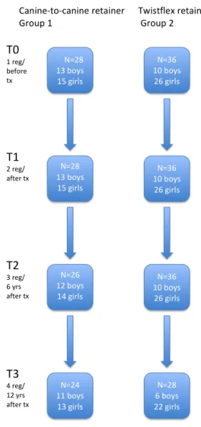
Figure 2 presents a flow chart of the patients in Paper I.
N=28 13 boys 15 girls N=36 10 boys 26 girls N=36 10 boys 26 girls N=28 13 boys 15 girls N=36 10 boys 26 girls N=28 6 boys 22 girls N=26 12 boys 14 girls N=24 11 boys 13 girls T0 1 reg/ before tx T1 2 reg/ after tx T2 3 reg/ 6 yrs after tx T3 4 reg/ 12 yrs after tx
Canine-‐to-‐canine retainer Twistflex retainer Group 1 Group 2
In Paper II, three different groups were included with a total of 130 children: two groups who received orthodontic treatment (105 patients) and one untreated group (25 subjects). Long-term records were required for participation in the study. Sample exclusions were single arch treatment, cleft lip and/or palate, agenesis and extraction of anterior teeth. This was a retrospective material, and no cases have been added or excluded after applying the inclusion criteria.
One of the groups who had undergone orthodontic treatment with fixed appliances in both jaws received a fixed mandibular retainer after treatment. In total, there were 64 patients, and they were all patients from Paper I. This group received either a canine-to-canine retainer or a Twistflex retainer (Group 1, retention group). The mean retention time was 2.7 years (SD 1.50).
The other group consisted of 41 patients with similar orthodontic treatment as the retention group, but they did not receive any retainer in the mandibular arch at all (Group 2, non-retention group).
The decision to leave the treated patients without retention in the mandible was made by the orthodontists who treated the patients.
All of the treated cases had a removable appliance in the maxilla for retention. No interproximal enamel reduction or circumferential supracrestal fiberotomy had been performed in the groups who had orthodontic treatment.
The third group – the untreated subjects – was comprised of 25 cases that were age-matched with the subjects in the two other groups (Group 0, untreated group).
The indications for orthodontic treatment for the 105 patients in Paper II were the same as in Paper I. All participants in Paper II were also recruited from the Department of Orthodontics in Jönköping, Sweden, between 1980 and 1995.
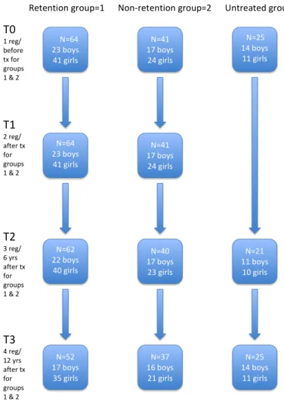
Twelve patients were excluded from the study, as the records did not meet the requirements and they were all from Group 2. In particular, there were missing registrations, especially at the last registration. Figure 3 shows a flow chart of the subjects in Paper II.
N=64 23 boys 41 girls N=41 17 boys 24 girls N=25 14 boys 11 girls N=41 17 boys 24 girls N=64 23 boys 41 girls N=40 17 boys 23 girls N=21 11 boys 10 girls N=37 16 boys 21 girls N=25 14 boys 11 girls N=62 22 boys 40 girls N=52 17 boys 35 girls T0 1 reg/ before tx for groups 1 & 2 T1 2 reg/ after tx for groups 1 & 2 T2 3 reg/ 6 yrs after tx for groups 1 & 2 T3 4 reg/ 12 yrs after tx for groups 1 & 2
Retention group=1 Non-‐retention group=2 Untreated group=0
Figure 3. Flow chart of the patients in Paper II.
PAR Index evaluation
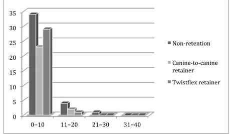
To evaluate the orthodontic treatment outcome from before (T0) to after treatment (T1) and long-term from T0 to 12 years after treatment (T3), measurements were done on dental casts from the studies in Papers I and II. In this analysis, 94 cases were evaluated: 39 cases with no retention in the mandibular anterior segment, 25 cases with canine-to-canine retainer after treatment, and 30 with Twistflex retainer after treatment. Unfortunately, dental casts from
11 cases were not available of which, 2 were distributed to the non-retention group, 3 to the canine-to-canine group, and 6 to the Twistflex retainer group.
Ethical considerations and consent
The Ethics Committee of Linköping, Sweden, approved the protocol (2014/381-31).
Each patient and parent received verbal information at start of the treatment about the possibility of using their study models in future studies. There was no need for any written information according to the Ethics Committee.
Methods
Papers I and II
Measurements were performed on dental casts using a sliding digital caliper (Mitutoyo 500-171 Kanagawa, Japan) with an accuracy of 0.01 mm.
The measurements were made at four occasions: T0 before treatment, T1 immediately after orthodontic treatment (i.e. at the start of retention), T2 six years after treatment (i.e. mean 3.6 years after the retainer was removed), and T3 12 years after treatment (i.e. mean 9.2 years after removal of the retainer).
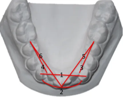
The main outcome measure was Little’s Irregularity Index, LII (Little 1975) – the summed displacement of the anatomic contact points of the mandibular anterior teeth (Figure 4). Other measurements performed on dental casts were intercanine width (cusp tip to cusp tip of the mandibular canines), intercanine perimeter distance (arch perimeter length between the mesial contact points of the canines), available mandibular incisor space (intercanine perimeter distance minus summed tooth width for the four mandibular incisors), two different lateral arch lengths (from the mesial contact point of the mandibular first molar to the mesial contact point of the canines and to the mesial contact point of the central incisors), overjet, and overbite (Figure 5). In Paper II, one of the lateral arch lengths (to the central incisors) was not measured.
Figure 4. Little’s Irregularity Index = A+B+C+D+E
Measured variables 1.Intercanine width 2.Intercanine arch perimeter distance 3.Lateral arch length le7 6-2 4.Lateral arch length right 6-2 5.Lateral arch length le7 6-central 6.Lateral arch length right 6-central 1 2 3 5 4 6
Figure 5. Measured variables
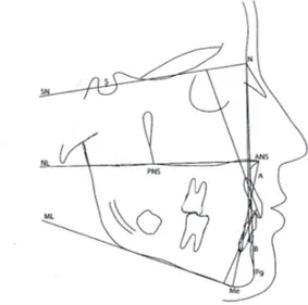
On lateral head radiographs the sagittal and vertical relationships between the jaws and the incisor inclination were evaluated. The cephalometric reference lines and points were assessed according to Björk (1947) and Solow (1966) (Figure 6). The measurements on the lateral head radiographs were made to the nearest half-degree or 0.5 mm with correction for enlargement.
SNA ̊ SNB ̊ ANB ̊ SN/ML ̊ ML/NL ̊ SN/NL ̊ U1/NL ̊ L1/Apg mm L1/ML ̊ Interincisal angle ̊
Figure 6. Cephalometric reference lines and points
All registrations were made by one examiner (USF). At T2 and T3, no retention appliance was in place. As a result, a blinded evaluation was possible, i.e. the examiner was unaware to which group the patients belonged or if the casts were taken at T2 or T3.
Also in Paper I, the bonding failures for the different retainers were obtained from the patients’ records.
Paper II
For the untreated group, dental casts and lateral head radiographs were available for the corresponding ages at T0, T2 and T3, but not at T1. In the retention group, no retention appliance was in place at T2 and T3.
The untreated control material consisted of subjects from two PhD dissertations: Infraocclusion of primary molars (Kurol 1984) and Ectopic eruption of maxillary first permanent molars (Bjerklin 1994). From these dissertations, it is clear that in cases with infraocclusion of primary molars with permanent successors, the growth and development of the dentition is normal. Also, cases with the reversible type of ectopic eruption of the maxillary first permanent molars show a normal growth and development of the dentition regarding eruption of the first, second and third permanent molars.
The subjects had Class I occlusion without any other malocclusions and registrations were performed for the above reasons.
PAR Index evaluation
The same examiner (USF) did the evaluation using a PAR ruler to take the measurements. The individual scores were summed to obtain an overall total score representing the degree a case deviates from normal alignment and occlusion. A score of zero indicates good alignment and occlusion whereas higher scores indicate increased levels of irregularity and deviation in occlusion.
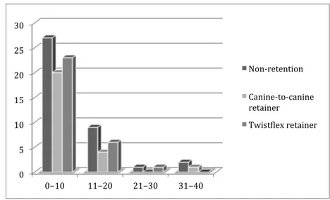
The overall score was recorded on pre-treatment (T0), post-treatment (T1), and 12 years post-post-treatment dental casts (T3) for the three groups (non-retention, canine-to-canine retainer, and Twistflex retainer). The weighted PAR scores were calculated according to the British system of weights and measures (Richmond et al. 1992a). Points reduction and percentage reduction in the weighted PAR scores were calculated for each of the three groups.
Statistical analysis
Sample size calculation
In Paper I, with its two groups, the sample size estimation was performed and based on an alpha significance level of 0.05 and a beta of 0.1 to achieve 90 per cent power to detect a clinically meaningful difference of 1.5 mm (SD 1.5) of Little’s Irregularity Index. The calculation revealed that 21 patients in each group were sufficient.
In Paper II, with its three groups, the sample size estimation was based on a significance level of 0.05 and 80 per cent power to detect a clinically meaningful difference of 1.5 mm (SD 1.5) in Little’s Irregularity Index. The estimation revealed that 22 patients in each group were sufficient.
The sample was normally distributed according to the Kolmogorov-Smirnov test.
Descriptive statistics
In Papers I and II, the arithmetic means and standard deviations (SD) were calculated for each variable at group level at times corresponding
to pre-treatment (T0), end of active treatment (T1), six years after treatment (T2) and 12 years after treatment (T3).
For the PAR Index evaluation, the arithmetic means and standard deviations (SD) were calculated for each group at pre-treatment (T0), end of active treatment (T1) and 12 years after treatment (T3).
Differences between and within groups
Significant differences in means in groups and between groups were assessed by one-way analysis of variance (ANOVA) using the IBM Statistical Package for Social Sciences (version 22.0; SPSS Inc., Chicago, Illinois, USA). P-values less than 5 per cent (P<0.05) were considered statistically significant. When significant differences were found between groups, the Bonferroni correction was used.
In Paper II, a regression analysis was also assessed to evaluate whether Little’s Irregularity Index (LII) at T3 was dependent on LII at T0 and to relate mandibular incisor inclination (L1/ML) to changes in LII.
Error of the method
In Papers I and II, the same examiner (USF) measured on two separate occasions with at least a four-week interval 24 randomly selected cases, which included both the dental casts and the lateral head radiographs. The mean error of the measurements according to Dahlberg’s formula (Dahlberg 1940) for the linear variables was mean 0.1 mm. The largest measurement error was 0.5 mm for intercanine width, 0.5 mm for intercanine perimeter distance, and 0.5 mm for left lateral arch length. Error measurements for the cephalometric angular variables were, on average, 0.8°. The greatest measurement error was noted for the maxillary incisor inclination, 3.3°.
A paired t-test showed no significant differences between the two series of records in most of the measurements, except for left lateral arch length (range min -0.1 to max 0.5), available space (range min -0.3 to max 0.2), tooth width 32 (range min -0.1 to max 0.1), tooth width 41 (range min -0.1 to max 0.1) and L1/Apg (range min -0.1 to max 1.3). The systematic error was within the aforementioned boundaries.
45
RESULTS
Paper I
Distribution according to age at the four registration occasions can be seen in Figure 7. There were no significant differences in age at the four registration occasions between the two groups (Paper I, Table 1, p. 201).
The orthodontic treatment started (T0) at a mean age of 12.5 years (SD 1.47) in Group 1 (canine-to canine retainer) and 13.2 years (SD 4.18) in Group 2 (Twistflex retainer).
The treatment lasted, on average, 2.6 years (SD 0.94) for Group 1 and 2.9 years (SD 1.76) for Group 2. Twelve years after treatment (T3), the post-retention time was 9.3 years (SD 2.79) for Group 1 and 9.1 years (SD 1.86) for Group 2. At T3, most of the patients were aged between 25 and 30 years (Paper I, Table 2, p. 202).
Figure 7.
Mean ages at the four different registration occasions for the two retainer groups Registration occasions Age
Figure 7. Mean ages at the four different registration occasions for the two retainer groups
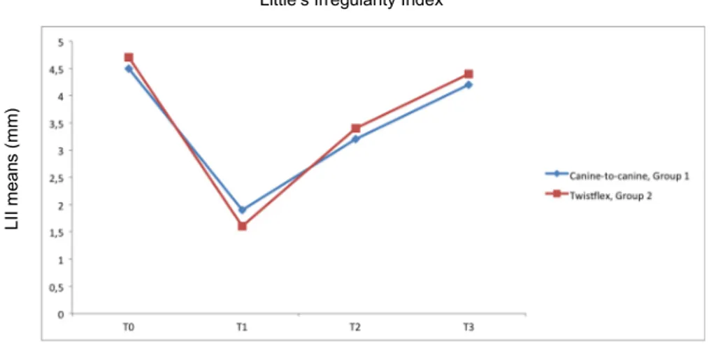
Little’s Irregularity Index (LII)
LII was the main outcome measure. It showed that there were no significant intergroup differences. However, within each group, several significant differences were found between the four registrations. LII was 4.5 mm at T0 for Group 1 and 4.7 mm for Group 2. Nine years after retention (T3), LII was 4.2 mm for Group 1 versus 4.4 mm for Group 2 (Figure 8). 0 0,5 1 1,5 2 2,5 3 3,5 4 4,5 5 T0 T1 T2 T3 Canine-‐to-‐canine Twist5lex LII m ean s ( m m ) Fig ure 8 . LII (m ean ) a t th e fo ur di ffere nt r egi str atio n o cca sio ns
Little’s irregularity Index
Registration occasions
Figure 8. LII (mean) at the four different registration occasions
The available space
The available space for the mandibular anterior segment showed similar results as LII. For both groups, the available space in the mandibular anterior segment increased after treatment. Six and 12 years after treatment, the available space had decreased in both groups, and for Group 1, it was almost equivalent to that before treatment, with no significant intergroup differences (Paper I, Table 3, p. 204).
Overjet and overbite
In both groups, the overjet and overbite were reduced after treatment and stayed almost the same throughout the observation period. Thus, the overjet was reduced from mean 6.3 mm in the group with a
0 0,5 1 1,5 2 2,5 3 3,5 4 4,5 5 T0 T1 T2 T3 Retention Non-‐retention Untreated 0 2 4 6 8 10 12 14 16 0−10 11−20 21−30 31−40 Non-‐retention Canine-‐to canine retainer Twist5lex retainer Time LII m ean s ( m m )
Little’s Irregularity Index
Figure 11. Pre-‐treatment PAR score categories for the three groups
Figure 12. Post-‐treatment PAR score categories for the three groups
Nu m be r o f c as es PAR score
canine-to-canine retainer to 3.2 mm after treatment. Twelve years after treatment, the mean overjet was 3.7 mm. The corresponding measures for the group with Twistflex retainer were 7.5 mm at T0, 3.3 mm at T1 and 4.0 mm at T3.
Overbite went from mean 3.7 mm in the canine-to-canine retainer group to 2.5 mm after treatment and from 3.5 mm in the Twistflex retainer group to 2.4 mm. At T3, the mean overbite was 2.9 mm in both groups.
There were no significant differences between the two groups for overjet and overbite (Paper I, Table 3, p. 204).
Intercanine width
The intercanine width was almost the same in both groups before and after treatment. It decreased from T0 to T3, with about 1 mm in both groups.
Arch length
The arch length measures, both to the canine and to the central incisor, decreased after treatment for Group 1, 1.3-1.5 mm, and for Group 2, 0.5-0.9 mm. This includes some extraction-cases. It continued to decrease six and 12 years after treatment in both groups (Paper I, Table 3, p. 204).
Tooth width
There was a small but significant difference in tooth width of the mandibular right lateral incisor between Groups 1 and 2.
Cephalometric variables
There were no significant intergroup differences for any of the cephalometric variables at the four different occasions (Paper I, Table 4, p. 205).
Extraction versus non-extraction
According to LII, there was no difference in mandibular incisor stability between the patients who had extractions prior to treatment and those who had no teeth taken out.
Bonding failures
Bonding failures were found in 32 per cent of the patients who had a canine-to-canine retainer versus 44 per cent in the group with Twistflex retainer, but this difference was not statistically significant. In some patients, the Twistflex retainer came loose more than once.
Paper II
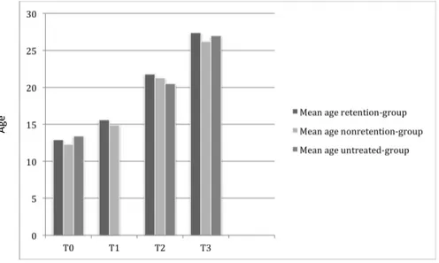
The mean ages at the four registration occasions for Paper II can be seen in Figure 9.
No significant age differences were found at the four registration occasions between the three groups, but there was a larger number of girls in the two treated groups compared to the untreated group (Paper II, Table 1).
Figure 9.
Mean ages at the four different registration occasions for the three groups Age Registration occasions Figu re 10. LII (m ean ) a t the fo ur di ffrer ent re gist rat ion occ asio ns
Figure 9. Mean ages at the four different registration occasions for the three groups
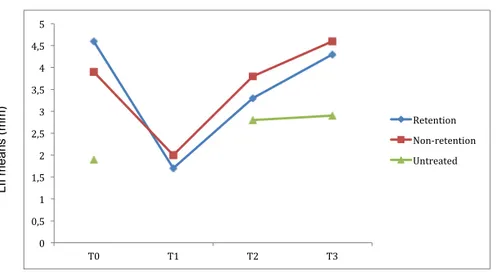
Little’s Irregularity Index
Before treatment (T0), there were no significant differences according to Little’s Irregularity Index between the groups with and without retention wires (Paper II, Table 2).
Twelve years after treatment, no significant differences were found in Little’s Irregularity Index between the retention and
non-retention group. However, significant differences were found between the untreated and two treated groups at T3 (Figure 10) (Paper II, Table 2). The untreated group showed less LII at T0 and at the last registration. No registrations were available at T1 for the untreated group. 0 0,5 1 1,5 2 2,5 3 3,5 4 4,5 5 T0 T1 T2 T3 Retention Non-‐retention Untreated 0 2 4 6 8 10 12 14 16 0−10 11−20 21−30 31−40 Non-‐retention Canine-‐to canine retainer Twist5lex retainer Time LII m ean s ( m m )
Little’s Irregularity Index
Figure 11. Pre-‐treatment PAR score categories for the three groups
Figure 12. Post-‐treatment PAR score categories for the three groups
Nu m be r o f c as es PAR score
Figure 10. LII (mean) at the four different registration occasions
The multiple regression analysis showed that LII at T2, six years after treatment, was the only variable that could explain LII at T3 (P=0.000). The incisor irregularity 12 years after treatment could not be related or predicted to LII before treatment.
Available space
The retention group had mean -2.0 mm available space at T0 compared with the non-retention group, which had -0.8 mm, and this was statistically significant. Compared with the untreated group, the available space was significantly less for treatment groups, but after 12 years, no significant differences were found between the three groups.
Intercanine width
Expansion of the intercanine width during orthodontic treatment was avoided in the groups that had orthodontic treatment and thus, no increase in intercanine width was seen.
No differences in intercanine width were found between the three groups at baseline or throughout the observation period.
Arch length
In the untreated group, the lateral arch length was significantly larger compared with the two treatment groups at all registrations. Some extraction cases were included in the treated groups. On average, the lateral arch length for the untreated group was 1.6–3.9 mm larger at all registrations, and in all groups, the arch length was decreased from T0 to T3 (Paper II, Table 2).
Tooth width
The four mandibular incisors showed no statistically significant differences in tooth width between the three groups and over time.
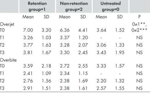
Overjet and overbite
At T0, there were statistically significant differences in overjet between the untreated group and the two treatment groups, P<.01 for the retention group and P<.001 for the non-retention group.
Overjet was reduced during orthodontic treatment in the two treatment groups, and at T1, it was almost the same as the untreated group had at T0, 3.6 mm, (SD 1.52) (Table 1).
Overbite was also reduced during treatment in the two treatment groups. Six and 12 years after treatment, both overjet and overbite stayed nearly the same in all three groups, and there were no statistically significant differences between the groups (Table 1).