D o c t o r a l D is s e r ta t io n i n o D o n t o lo g y M a r ia P iH l M a l M ö U n iV e r s it y MalMö Högskola
Maria PiHl
MicroBial BioFilMs on
Peritoneal Dialysis
catHeters
isbn 978-91-7104-123-4 M ic r o B ia l B io F il M s o n P e r it o n e a l D ia ly s is c a t H e t e r s© Maria Pihl, 2011 Photo: Maria Pihl ISBN 978-91-7104-123-4 Holmbergs, Malmö 2011
Malmö University, 2011
Faculty of Odontology
Department of Oral Biology
MARIA PIHL
MICROBIAL BIOFILMS ON
PERITONEAL DIALYSIS
Publication also available in electronic format at, www.mah.se/muep
Till Magnus
”I hear and I forget. I see and I remember. I do and I understand.”
Confucius
CONTENTS
LIST OF PAPERS ... 9 ABSTRACT ... 10 POPULÄRVETENSKAPLIG SAMMANFATTNING ... 13 ABBREVIATIONS ... 15 INTRODUCTION ... 16 Peritoneal dialysis ...16Peritoneal dialysis-related infections ...18
Single species peritonitis ...18
Polymicrobial peritonitis...19
Biofilm-related peritoneal dialysis infections ...20
Microbial biofilms ...21
Biofilm formation ...22
Microbial interactions ...26
Quorum sensing ...29
Biofilm-related infections in humans ...33
The peritoneal cavity ...35
Eradication of biofilms ...35
Bacterial species involved in biofilm mediated diseases ...37
Staphylococcus epidermidis ...37
Pseudomonas aeruginosa ...39
AIMS ... 42
SIGNIFICANCE ... 43
MATERIALS AND METHODS ... 44
Bacterial strains and media ...44
Bacterial identification ... 45
Culturing ... 45
Gene sequencing ... 46
Fluorescence in situ hybridisation ... 47
Bacterial visualisation ... 48
Fluorescence ... 48
Confocal laser scanning microscopy ... 50
Bacterial characterisation ... 52
Protein desorption ... 52
Gel electrophoresis ... 53
Protein and polysaccharide detection ... 54
Detection of other cellular substances ... 55
Surface characterisation ... 56
Contact angle measurement ... 56
Atomic force microscopy ... 57
Devices created for this thesis ... 58
Flattening device ... 58
Peritoneal dialysis catheter flow cell system ... 58
RESULTS ... 61
Paper I - Occurrence of bacteria on catheters in patients undergoing peritoneal dialysis ... 61
Paper II - Differential effects of Pseudomonas aeruginosa on biofilm formation by different strains of Staphylococcus epidermidis. ... 64
Paper III - Effects of clinical isolates of Pseudomonas aeruginosa on Staphylococcus epidermidis biofilm formation. ... 66
Paper IV - Biofilm formation by Staphylococcus epidermidis on peritoneal dialysis catheters and the effects of extracellular products from Pseudomonas aeruginosa. ... 68
Additional results ... 71 DISCUSSION ... 74 Surfaces ... 74 Catheter coatings ... 75 Bacterial adhesion ... 76 Microbial interactions ... 78
In vivo and in vitro conditions ... 79
CONCLUDING REMARKS ... 83 ACKNOWLEDGEMENTS ... 84 REFERENCES ... 86
9
LIST OF PAPERS
This thesis is based on the following papers, which will be referred to in the text by their roman numerals.
I: Maria Pihl, Julia R. Davies, Ann-Cathrine Johansson, Gunnel Svensäter. Occurrence of bacteria on catheters in patients under-going peritoneal dialysis. Submitted.
II: Maria Pihl, Julia R. Davies, Luis E. Chávez de Paz, Gunnel Svensäter. Differential effects of Pseudomonas aeruginosa on biofilm formation by different strains of Staphylococcus epider-midis. FEMS Immunol Med Microbiol 2010 59(3): 439-56
III: Maria Pihl, Luis E. Chávez de Paz, Artur Schmidtchen, Gunnel Svensäter, Julia R. Davies. Effects of clinical isolates of
Pseudomonas aeruginosa on Staphylococcus epidermidis biofilm formation. FEMS Immunol Med Microbiol. 2010 59(3): 504-12
IV: Maria Pihl, Anna Arvidsson, Marie Skepö, Julia R. Davies, Gunnel Svensäter. Biofilm formation by Staphylococcus epider-midis on peritoneal dialysis catheters and the effects of extracellu-lar products from Pseudomonas aeruginosa. Manuscript.
10
ABSTRACT
When the kidneys are failing the blood needs to be purified artifi-cially. This can be done using peritoneal dialysis, in which a cathe-ter is surgically inserted into the peritoneal cavity, through the ab-dominal wall, and used to infuse or withdraw dialysis fluid.
When handling the catheter, touch contamination may occur and this can result in bacterial access to the peritoneal cavity, formation of biofilms and subsequent infections. Peritonitis, an often very painful inflammation of the peritoneum, is the major cause of morbidity in peritoneal dialysis patients and often leads to hospi-talisation during treatment. Thus, the benefits of reducing the number on infections would be huge, with better quality of life for the patients and a reduction in costs for society.
The aim of this thesis was to study bacterial colonisation on cathe-ters from peritoneal dialysis patients as well as to study biofilm formation and microbial interactions on catheters in vitro.
In a clinical study we investigated peritoneal dialysis catheters re-moved from patients due to renal transplantation (15 catheters) or infections (2 catheters). Bacterial colonisation was detected on 14 of the 17 catheters, as seen using confocal laser scanning micros-copy or microbiological cultures, although the bacterial load was rather low. The most common species found on the catheters were
Staphylococcus epidermidis and Propionibacterium acnes, both of which are low-virulent, commensal skin bacteria. Although usually low-virulent, S. epidermidis is the most common cause of
medical-11
device related infections, and can cause persistent infections that are hard to eradicate.
Due to its common presence in peritoneal dialysis catheter-related infections, S. epidermidis was further studied, together with an-other common opportunistic pathogen, Pseudomonas aeruginosa, often associated with more severe peritonitis cases which require catheter removal. These bacteria were cultured together and, inter-estingly, we found the number of S. epidermidis cells constantly declined compared to the P. aeruginosa cells, even when they were originally present in equal amounts, suggesting that P. aeruginosa
dispersed the S. epidermidis biofilms. This dispersal effect of P. aeruginosa on S. epidermidis biofilms was seen for clinical and laboratory strains of both species.
When S. epidermidis biofilms were exposed to cell-free growth me-dium of P. aeruginosa biofilms, the same effect could be seen, demonstrating that it was due to extracellular substance(s). Several extracellular substances of P. aeruginosa were investigated for their dispersal effect, including proteases, a quorum sensing-related ho-moserine lactone, the toxin pyocyanin and polysaccharides. For various reasons, all were excluded as the potentially active sub-stance except the polysaccharides. This result was further sup-ported when the cell-free growth medium of P. aeruginosa was fractionated and studied for S. epidermidis dispersal effect. The material with the greatest effect on S. epidermidis was a fraction rich in polysaccharides as shown by PAS staining. This pool con-tained no detectable mannose or galactose residues, and may corre-spond to the glucose-rich polysaccharide product of the pel gene cluster. Other fractions, including a galactose- and mannose-rich polysaccharide possibly corresponding to the psl gene product as well as one that was protein-rich were also active but to a lesser ex-tent. Other bacterial polysaccharides have exhibited antibacterial effects, probably due to altered cell integrity, and possibly the po-tential P. aeruginosa polysaccharide might function in a similar way. In addition to functioning as a dispersal agent of established biofilms, the P. aeruginosa supernatant also seemed to inhibit
12
biofilm formation by preventing adsorption of proteins to the catheter surface.
In the future, identification of the substances responsible for these effects would allow them to be used in new strategies to prevent formation or eradicate biofilms from peritoneal dialysis catheters, thus reducing the number of infections.
13
POPULÄRVETENSKAPLIG
SAMMANFATTNING
Då njurarna slutar fungera produceras ingen urin, och avfallspro-dukterna stannar kvar i blodet. Detta tillstånd kallas uremi (”urin i blodet”) och leder till döden om det inte behandlas. Det finns två artificiella sätt att rena blodet på, hemodialys och peritonealdialys. I peritonealdialys fylls bukhålan med dialysvätska, som drar ut slaggprodukter och överflödigt vatten från blodet, genom bukhin-nan (peritoneum). För att kunna fylla och tömma buken på dialys-vätska behövs en kateter (slang), som opereras in genom bukväg-gen. Om bakterier, till exempel från huden, kommer in i bukhålan
kan de fästa till kateterytan och bilda en biofilm. Biofil-mer kan ge upphov till infek-tioner i bukhålan som leder till bukhinneinflammationer, vilket kan vara mycket smärtsamt. För att ge patien-terna en bättre livskvalitet och minska sjukvårdskostnaderna är det önskvärt att reducera antalet bukhinneinflammationer. Syftet med vårt arbete har varit att få fram kunskap som bidrar till en bättre förståelse av biofilm-relaterade infektioner i buken.
Vi studerade 15 katetrar uttagna från patienter som fick en ny nju-re (och därmed inte hade några infektioner) och 2 som var uttagna på grund av infektioner. Att inte ha några symptom på infektion betydde dock inte att katetern var fri från bakterier, och på 12 av dessa 15 katetrar hittade vi bakterier. Mängden bakterier var dock
Biofilm: Bakterier som lever på en yta. Ofta är bakterierna inkapslade i ett slem som de själva producerat.
14
ganska liten och det verkade som om bakterierna och immunför-svaret hade uppnått en balans, där de fungerade tillsammans. De vanligaste bakterierna som hittades var hudbakterier, som i norma-la fall är harmlösa när de förekommer på huden. En av dessa bak-terier var Staphylococcus epidermidis, och denna studerade vi vida-re, tillsammans med en bakterie kallad Pseudomonas aeruginosa.
Pseudomonas aeruginosa hittas normalt inte hos människor och är inte farlig för friska individer, men om immunförsvaret är nedsatt kan den ta chansen och orsaka infektioner, ofta i samband med implantat eller lungsjukdomen cystisk fibros.
När vi odlade Staphylococcus epidermidis och Pseudomonas aeru-ginosa tillsammans i en biofilm upptäckte vi något intressant, näm-ligen att Pseudomonas aeruginosa tog över och att Staphylococcus epidermidis försvann från ytan. Detta upptäckte vi senare berodde på en substans som Pseudomonas aeruginosa producerade och pla-cerade utanför själva cellen, dvs. en extracellulär produkt. Extra-cellulära produkter kan till exempel vara det socker-rika slem som omger bakterierna i biofilmen och skyddar dem mot kroppens im-munförsvar, eller enzymer som bryter ned näringsämnen i omgiv-ningen så att bakteriecellen kan ta upp det.
När vi studerade de extracellulära produkterna närmare kunde vi isolera en fraktion som var rik på polysackarider, dvs. socker, och som fick Staphylococcus epidermidis att lossna från ytor. Dessutom verkade den socker-rika lösningen kunna förhindra inbindning av se-rumproteiner, vilka i sin tur påverkar den kommande infästningen av bakterier. Substansen är inte identifierad än, men skulle alltså kun-na vara en typ av socker.
I framtiden vill vi rena fram substansen och identifiera den, för att förhoppningsvis kunna använda den för att förhindra biofilmbild-ning på peritonealdialys-katetrar och därmed minska antalet infek-tioner hos peritonealdialys-patienter.
15
ABBREVIATIONS
AFM Atomic force microscopy AHL N-acyl homoserine lactone AI Autoinducer AMP Antimicrobial peptide CLSM Confocal laser scanning microscopy CMC Critical micelle concentration CoNS Coagulase negative staphylococci FISH Fluorescence in situ hybridisation HD Haemodialysis
HSL Homoserine lactone
ISPD International Society for Peritoneal Dialysis PAS Periodic acid/Schiff’s reagent
PD Peritoneal dialysis
PIA Polysaccharide intercellular adhesin
PSM Phenol-soluble modulin
PQS Pseudomonas quinolone signal
16
INTRODUCTION
Peritoneal dialysis
The inner environment of the body is tightly regulated, maintaining physiological levels of ions, pH etc. in a process called homeostasis. The kidneys are vital for maintaining homeostasis by the excretion of excess water, ions, acids and bases, and the removal of waste products, including urea and creatinine. In addition to the excre-tory function, the kidneys also have a secreexcre-tory function and pro-duce the hormones, renin, involved in blood pressure regulation, and erythropoietin, EPO, involved in the erythrocyte production, and transforms vitamin D to an active form.
If the kidneys are failing, electrolyte, fluid, acid-base and hormone imbalances arise, and waste products and excess water accumulates in the blood. This condition is called uraemia (“urine in the blood”) and is characterised by a wide range of symptoms, such as, confusion, cramps, nausea, hypertension, itching, infertility, weight loss, acidosis etc. (Almeras & Argiles 2009). If left untreated urae-mia causes death.
Uraemia can be treated by two dialysis modalities, haemodialysis (HD) and peritoneal dialysis (PD). In haemodialysis, the blood is filtered in a machine whereas, in peritoneal dialysis purification of the blood takes place across the peritoneum. A catheter is surgi-cally inserted through the abdominal wall into the peritoneal cavity (Fig. 1) which is subsequently infused with dialysis fluid, often us-ing glucose as the osmotic agent. With time, waste products and excess water are filtered from the peritoneal capillary blood across
the peritoneum and into the peritoneal cavity, i.e. the peritoneum is thus being used as a dialysis membrane. The cleaning of the blood involves the diffusion of molecules from lower to higher concentra-tions, convective transport (the movement of molecules by fluid motion) and osmosis (the transport of solvents from a region of high solute concentration to a region of low solute concentration via a semipermeable membrane allowing only the solvent to pass) (Gokal & Mallick 1999).
At the end of 2008, there were 8029 patients in active treatment for uraemia in Sweden, 4462 with a functioning renal transplant, 2719 on haemodialysis and 848 on peritoneal dialysis (Svenskt njurregister 2009). In 1996, there were more than 105000 PD pa-tients worldwide, constituting around 15% of the dialysis popula-tion of the world (Gokal & Oreopoulos 1996).
In haemodialysis, the dialysis take place a few times a week at a medical facility and trained personnel are present, but treatments are scheduled and transportation must be arranged. This therapy is more expensive than peritoneal dialysis since PD patients perform the dialysis by themselves in their homes, although it must be done 7 days a week.
Figure 1: In peritoneal dialysis a catheter is surgically inserted through the abdominal wall into the peritoneal cavity. Dialysis fluid is infused, allowed to dwell for a few hours and then removed.
18
Peritoneal dialysis-related infections
When comparing haemodialysis and peritoneal dialysis, the obvi-ous advantages of PD are the higher relative freedom for the pa-tients and the lower cost, but the major drawback is the risk of in-fections such as peritonitis. Peritonitis is an inflammation of the peritoneum and a leading cause of morbidity and technique failure in PD patients (Woodrow et al. 1997; Pérez Fontán et al. 2005). Other infections related to PD are exit site and tunnel infections (Bernardini 2004; Aslam et al. 2006). At the catheter entry into the skin, exit site infections can be seen whereas tunnel infections are related to the subcutaneous part of the catheter.
Patients with peritonitis suffer from symptoms such as abdominal discomfort, ranging from diffuse to painful, fever, vomiting, chills, constipation, and delirium, but patients can also be symptom-free (Dasgupta et al. 1991; Johnson et al. 2003; Nasser et al. 2005). The peritoneal fluid can often be straw-coloured and cloudy from fibrin (Dasgupta et al. 1991). Patients manifesting at least two of the three symptoms, fever, abdominal pain and cloudy dialysate, should receive a dialysate test. A result of 100 white blood cells/µl with at least 50% polymorphonuclear neutrophils supports the diagnosis of peritonitis (Keane et al. 2000).
The average incidence of peritonitis in the PD population has changed over time and varies depending on factors such as coun-try, centre, PD technique and patient category, ranging from more than 2 episodes per patient and year to less than 1 episode every fourth year (Scalamogna et al. 1990; Golper et al. 1996; Kim et al.
2004; Pérez Fontán et al. 2005).
Single species peritonitis
In PD-related peritonitis, bacteria often enter the peritoneal cavity by touch contamination or via the exit site, but can in some cases move through the bowels (Cameron 1995). The most common or-ganisms causing peritonitis are coagulase-negative staphylococci (CoNS) including Staphylococcus epidermidis, accounting for 28-41% of all peritonitis episodes, whereas Staphylococcus aureus
19
2002; Kim et al. 2004; Pérez Fontán et al. 2005; Mujais 2006). Other Gram-positive bacteria, such as, streptococci, Enterococcus
and Corynebacterium can also cause peritonitis (Troidle et al.
1998; Finkelstein et al. 2002). Although less common, peritonitis caused by Gram-negative bacteria, such as Klebsiella, Escherichia coli, Pseudomonas and Enterobacter, are generally more severe, leading to more hospitalisations, catheter removals, PD dropouts and deaths (Bunke et al. 1997; Troidle et al. 1998; Krishnan et al.
2002). Fungal peritonitis rates are 2-15% and up to 20% of the infections are culture negative (Warady et al. 2000; Johnson et al.
2003; Prasad et al. 2004). Strict anaerobes are rarely encountered as causes of peritonitis, constituting only 2-3 % of the isolated mi-croorganisms (de Fijter et al. 1994; Troidle et al. 1998; Zelenitsky
et al. 2000). In the literature, some of the anaerobes described are
Bacteroides fragilis, Veilonella, Clostridium perfringens, Peptococ-cus and Propionibacterium (Sombolos et al. 1986; Alfa et al. 1997; Catchpole et al. 1997; Castillo et al. 1999) whereas facultative an-aerobes as a group are frequently encountered in peritonitis, for example the above mentioned Klebsiella, E. coli and Enterobacter
in addition to Acinetobacter, Serratia, Proteus and Neisseria (de Fijter et al. 1994; Zelenitsky et al. 2000; Finkelstein et al. 2002). The presence of Pseudomonas, fungi and anaerobes in the dialysis fluid may identify patients in need of earlier catheter removal (Szeto et al. 2002) and the ISPD recommend early catheter removal in Pseudomonas infections (Keane et al. 2000).
Polymicrobial peritonitis
Polymicrobial peritonitis, defined as two or more organisms iso-lated from cultures, make up 6-11% of all peritonitis cases (Holley
et al. 1992; Kiernan et al. 1995; Kim & Korbet 2000; Krishnan et al. 2002; Szeto et al. 2002; Barraclough et al. 2010). A study by Szeto et al (2002) showed 79% of polymicrobial peritonitis epi-sodes to be caused by two organisms, 17% to be caused by three organisms and 4% to be caused by four organisms. Of all po-lymicrobial peritonitis cases 21-28% are caused by pure Gram-positive bacteria, 9-23 % of pure Gram-negative bacteria and 24-44% of mixed Gram-positive and Gram-negative bacteria (Kier-nan et al. 1995; Kim & Korbet 2000; Szeto et al. 2002;
Barrac-20
lough et al. 2010). A mixture of bacteria and fungi constituted 9-19% of the polymicrobial peritonitis episodes (Holley et al. 1992; Kim & Korbet 2000; Barraclough et al. 2010). Anaerobes have been estimated to constitute 7% of the polymicrobial peritonitis episodes (Szeto et al. 2002), indicating these to be more common as polymicrobial colonisers compared to single species colonisers. Polymicrobial peritonitis is commonly worse than single species peritonitis (Krishnan et al. 2002; Barraclough et al. 2010; Jarvis et al.), but pure Gram-positive polymicrobial peritonitis result in sig-nificantly lower risks of catheter removal, PD dropout and death than pure negative or mixed positive and Gram-negative polymicrobial peritonitis (Szeto et al. 2002; Barraclough
et al. 2010).
Biofilm-related peritoneal dialysis infections
Biofilm formation on PD catheters has been suggested as a possible cause of relapsing or recurrent peritonitis (Evans & Holmes 1987) and several studies show biofilm formation on removed catheters (Marrie et al. 1983; Dasgupta et al. 1986; Dasgupta et al. 1987; Gorman et al. 1994; Troidle & Finkelstein 2006). In addition to bacteria, macrophages, leukocytes, mesothelial cells, fibrillar ma-trix and lipoid droplets have also been found on catheters (Swartz
et al. 1991).
Microbial biofilms on PD catheters removed due to infections can be found both on the external and the luminal sides (Dasgupta
et
al.
1987; Gormanet al.
1994). Normally the dialysis effluent is used to isolate infecting bacteria, but it might not be an exact cor-relation to what is actually on the catheter, indicating the impor-tance of patient catheter studies. Dialysis fluid, with glucose as the most common osmotic agent, and peritoneal fluid provide the nec-essary nutrients for bacterial growth (Dasgupta et al. 1988). Naturally, much focus has been placed on patients with infections, but microbial biofilms can also be found on PD catheters from pa-tients without clinical signs of infections (Fig. 2) (Dasgupta et al.21
virulent chronic infection in balance with the host immune system that only gives rise to peritonitis when disturbed (Cameron 1995). These biofilms, from patients without infections, were scraped with a sterile scalpel blade and streaked on nutrient agar. The results revealed the pres-ence of S. epidermidis and other coagulase-negative staphylococci, S. aureus, En-terobacteriacae, streptococci and others (Dasgupta et al.
1986; Dasgupta et al. 1987; Gorman et al. 1994). How-ever, these studies did not fo-cus on identifying bacterial species on the catheters and there is a lack of information whether the characteristics of these bacterial isolates differ compared to bacteria isolated from catheter-related infection sites.
Microbial biofilms
Bacteria living on surfaces form communities resulting in slimy mats, or biofilms, such as those found on rocks in streaming rivers. A biofilm can be defined as “a structured community of bacterial cells enclosed in a self-produced polymeric matrix and adherent to an inert or living surface” (Costerton et al. 1999). They are highly hydrated and in addition to cells also contain 73-98% non cellular material, consisting of e.g. polysaccharides, proteins, nucleic acids and lipophilic compounds (Lawrence et al. 2003). The formation of a biofilm with extracellular polymers demands considerable en-ergy output by the biofilm microflora, but the benefits can be sig-nificant, such as protection from harmful environments, the ability to stay in a nutrient rich environment and increased metabolic effi-ciency as different species can help each other (Jefferson 2004).
Figure 2: Presence of bacteria does not always mean that signs of infec-tions are present.
Biofilm formation
Biofilms develop and mature as bacteria attach to the surface and start forming extracellular polymers, such as proteins, polysaccha-rides (Lawrence et al. 2003) and DNA (Whitchurch et al. 2002), and undergo different stages with changes in bacterial phenotype and gene expression (Sauer et al. 2002), for example increased an-tibiotic resistance (Uckay et al. 2009). The stages of biofilm forma-tion involve initial attachment to the surface, formaforma-tion of micro-colonies and maturation into matrix-enclosed biofilms, from which cells can also detach and migrate to other areas and form new biofilms (Costerton et al. 1999), see figure 3.
Figure 3: Dynamics of multi-species biofilm formation
On medical implants, proteins will immediately cover the surface and form a conditioning film, to which bacteria can attach, and mature into a biofilm. As a result of bacterial growth and production of different substances, gradients occur and contribute to the formation of various micro-niches, which enhance the phenotypic variation of bacteria.
23 Protein adsorption
When an implant is placed in the body it will immediately be cov-ered by proteins and this protein coat is the surface bacteria will meet. Therefore, protein adsorption is important for the subse-quent biofilm formation.
Adsorbing and desorbing proteins go through several steps; trans-port to the surface, attachment, structural rearrangement, detach-ment, transport away from the surface and possibly another struc-tural rearrangement. Normally desorption from the surface is very low for proteins (Norde 2003).
The adsorption of proteins is governed by the Gibbs energy, and for spontaneous adsorption to occur the change in Gibbs energy must be negative (Box 1). The adsorption process can involve sev-eral types of forces, such as, van der Waals forces, hydrogen bonds, electrostatic interactions and hydrophobic interactions.
Box 1
Gibbs energy: G = H - TS H= Enthalpy, the internal energy T = Temperature
S = Entropy, the degree of disorder
Soluble proteins are often globular in shape, and the hydrophobic amino acids are internalised, whereas the hydrophilic, polar amino acids are externalised to the outer surface and interact with rounding water. However, if the protein is denatured on the sur-face the hydrophobic amino acids are exposed.
DLVO theory
The DLVO theory (named after Derjaguin, Landau, Verwey and Overbeek) describes the stability of colloids in suspension and ex-plains why some colloids are stable while others aggregate.
24
According to the DLVO theory colloid stability is determined by attractive van der Waals forces and electrostatic repulsion due to electrical double layers, formed by overlapping counter ions clouds of charged surfaces (Fig. 4A). When two colloidal particles
ap-proach each other electro-static repulsion becomes evident as their double layers begin to interfere. To overcome this, energy is required and the maxi-mum energy value is re-quired when the surfaces are almost touching, whereas it is zero when there are no interferences between the double lay-ers. The net interaction curve combining the re-pulsive and attractive force curves is described by Gibbs interaction en-ergy, G.
G = GE +GVDW
The point of maximum repulsive energy is called the energy barrier and in-dicates the stability of the colloid system. In a stable colloidal dispersion there is repulsion between the particles preventing them from aggregating. To be able to aggregate, the ki-netic energy of two collid-ing particles must be large enough to overcome the
Figure 4: A) The DLVO theory describes the stability of colloids, but has some limi-tations when applied to bacteria (B) as they are not flat particles with uniform charge. B adapted from Jones & Isaacson (1983).
25
energy barrier. The secondary minimum accounts for reversible adhesion.
The extended DLVO theory of van Oss (1986) also includes hy-drophobic attraction and hydrophilic repulsion in addition to the van der Waals forces and the electrostatic interactions.
Theoretically, bacteria should not be able to attach to a surface as they do not posses enough kinetic energy to overcome the energy barrier (Jones & Isaacson 1983). However, there are some assump-tions made in the DVLO theory that do not apply to bacteria, such as particles are assumed to have flat surfaces and uniform charges. In reality, the bacterial surface is not flat but full of protruding, po-tential adhesins, in addition to the associated extracellular poly-mers, that can overcome the potential barriers and firmly adhere the bacterium to the surface (Jones & Isaacson 1983) (Fig. 4B).
Bacterial adhesion
Adhering bacteria must first get close to the surface. The surface approach can be made using several mechanisms, including Brownian motion, sedimentation, convective transport and active transport by flagellar motion. Turbulent flow can also force bacte-ria close to the surface, overcoming the energy barrier. (Palmer et al. 2007)
There are two main theories of bacterial attachment to solid sur-faces. The first one, developed after adhesion studies of Marshall et al (1971) involve a two-step process of a primary reversible adhe-sion due to electrostatic forces, van der Waals forces and hydro-phobic interactions that may be explained by the extended DLVO theory, as described above. In the first phase, bacteria are found in the secondary minimum and still exhibit Brownian motion and can easily by washed away from the surface. Adhesive molecules on cell-surface structures such as pili and fimbriae can probably cross the energy barrier and reach the primary minimum and bind to the surface. This is an irreversible adhesion and the bacteria no longer exhibit Brownian motion and cannot easily be washed away.
26
The second theory of bacterial adhesion was proposed by Busscher and Weerkamp (1987) in a three-step process over different dis-tances between solid surface and bacteria. They propose at large distances (>50nm) only attractive van der Waals forces are active and non-specific interactions, such as, surface-free energy and charge are dominating. At intermediate distances (10-20 nm), van der Waals forces are present and an increasing amount of electrostatic interaction results in a secondary minimum. With time these interactions become more irreversible due to rearrangements on the bacterial cell surface. At small separation distances (<1.5 nm), the potential energy barrier must be overcome, which can be done by protruding adhesins on the bacterial cell surface that mediate a firm, irreversible adhesion by specific interactions. At physiological pH bacteria often have a net negative surface charge, with charges mainly originating with carboxyl, phosphate and amino groups in the cell wall structure. If the ionic strength of the medium is increased more ions are available to shield the charges and neutralise the surface (Palmer & Sternberg 1999). The hydrophobicity of bacteria has in some studies been indicated as an important factor for cell adhesion with more hydrophobic bacteria being better able to adhere both to hydrophobic and hy-drophilic solid surfaces (van Loosdrecht et al. 1987; Gorman et al.
1997; Liu et al. 2004), whereas other studies indicated it was not that important for adhesion (Sorongon et al. 1991; Flint et al.
1997). However, the differences may be related to species, strain, surrounding medium and the solid surface used.
Microbial interactions
Natural biofilms can consist of hundreds of bacterial species and these are involved in a range of physical, metabolic and molecular interactions (Fig. 5). These interactions can provide benefits for the bacteria, including a broader habitat range for growth, increased metabolic diversity and efficiency and enhanced resistance to stress (Marsh 2005).
27
Dental plaque consists of hundreds of species, several of which adhere to the enamel or other bacteria in a mutualis-tic way. For example, neither of the early colonisers Streptococ-cus oralis and Actino-myces naeslundi could form monoculture biofilms in a flow cell with saliva, but when co-cultured biofilms were formed (Palmer et al. 2001). Also, Fuso-bacterium nucleatum
was shown not to form monoculture biofilms
in vitro, but when co-cultured with A. naeslundi biofilms were formed (Periasamy et al. 2009). Studies have shown that F. nuclea-tum could coaggregate with many oral bacteria and act as a bridge between early and late colonisers.
In both single and mixed species biofilms, oxygen is respired in the upper layers, establishing anaerobic regions in the lower parts where anaerobes can thrive even if the biofilm is flushed with aer-ated liquid (Stewart & Franklin 2008). F. nucleatum and Porphy-romonas gingivalis belong to the anaerobic oral microflora, but the ability of F. nucleatum to reduce oxygen is very high for an aner-obe, possibly due to superoxide dismutase. When cocultured with
P. gingivalis it could take care of the oxygen enabling the anaerobic
P. gingivalis to grow (Diaz et al. 2002).
In mature biofilms, concentration gradients of metabolic substrates and products form, creating various microniches suitable for dif-ferent bacterial species or phenotypes. Sometimes metabolic inter-mediates or waste products produced by one species can be used as
Symbiosis (close partnership between spe-cies)
Mutualism: Association between organisms where both species benefit.
Commensalism: Association between organ-isms where one species benefits and the other is unaffected.
Parasitism: Association between organisms where one species benefits and the other is harmed.
Non-symbiotic relationship
Synergism: Association of organisms where both species benefit but the association is not necessary for survival.
Antagonism: Association between species arising from (aggressive) competition.
substrate by another species (Stewart & Franklin 2008). For ex-ample, isobutyric acid produced by P. gingivalis is used as nutrient by Treponema denticola, whereas succinic acid produced by the latter is used as nutrient by P. gingivalis (Grenier 1992).
Saliva consists of complex proteins and glycoproteins which re-quires the combined actions of enzymes from different species to be
degraded. Thus, bacteria can inter-act synergistically and metabolise complex host molecules that one bacterium alone could never digest (Marsh 2005). Not all interac-tions between bac-teria are beneficial. Competition be-tween bacteria oc-curs, such as the dispersal of S. epi-dermidis biofilms by P. aeruginosa described in pa-pers II-IV of this thesis. P. aerugi-nosa produce sev-eral antimicrobial substances, some of which are described below. Bacteriocins are proteinacious bacte-ricidal substances produced by bacteria and they inhibit the growth of closely related strains or species. Antibiotics are also produced by some bacteria (Huddleston et al. 1997). Other competitive fac-tors include hydrogen peroxide that inhibits growth of other bacte-ria and short-chain fatty acids, for example lactic acid that can
Figure 5: Microbial interactions can be beneficial or detrimental, for both or one of the participating species.
29
lower pH and thereby make the environment unsuitable for certain bacteria (Hojo et al. 2009).
Quorum sensing
Bacteria can communicate directly by sending out signal molecules, but they can also sense changes in their environment (cues) caused by surrounding bacteria. Chemical cues are often metabolic prod-ucts and the bacterium producing it may not intend to communi-cate. (Jakubovics 2010)
Quorum sensing, QS, is the “language” of molecular signalling in biofilms and was first discovered in two marine bacteria, Vibrio fischeri and Vibrio harveyi. It is a mechanism by which bacteria regulate specific genes as an answer to a certain concentration of a signalling molecule, which is dependent on cell population density (Diggle et al. 2008) i.e. it only occurs at high enough cell densities. Quorum sensing is involved in the regulation of several traits such as bioluminescence, virulence, biofilm formation, sporulation, and mating (Bassler 2002). There are three typical systems, used both for intra species and inter species communication.
Most Gram-negative bacteria utilise acylated homoserine lactones (AHLs) as autoinducers and have a QS system homologous to that of V. fischeri, which lives in symbiosis with several eukaryotic hosts, producing light in specialised light organs by use of the en-zyme luciferase. The V. fischeri luciferase production is regulated by five structural genes (luxCDABE) and two regulatory genes (luxR and luxI). The LuxI protein, a synthase, is responsible for production of the autoinducer homoserine lactone which can freely diffuse out of the cell. When in sufficient concentration, the autoinducer will bind to the regulatory protein, LuxR, and this complex then activates transcription of luxICDABE, producing both more LuxI synthase and luciferase, and repressing expression of luxR. (Miller & Bassler 2001)
Gram-positive bacteria often use short peptides as autoinducers. Generally, a peptide precursor is translated and cleaved to the autoinducer peptide, which is actively transported out of the cell
30
via ABC transporters (ATP-binding cassette). From the outside of the cell, the autoinducer peptide interacts with a two component sensor kinase that starts a phosphorylation cascade leading to phosphorylation and activation of the regulator protein. The regu-lator protein then activates transcription of its target genes. (Miller & Bassler 2001)
The third system was originally found in V. harveyi and has simi-larities to both the Gram-negative and Gram-positive QS systems. There are two parallel QS parts involved in regulation of luciferase related bioluminescence in V. harveyi. Both parts are composed of a sensor (sensor 1, LuxN, and sensor 2, LuxQ) and an autoinducer (AI-1 and AI-2). AI-1 is a homoserine lactone (HSL) whose synthe-sis is regulated by the LuxM protein. AI-2 is derived from the pre-cursor 4,5-dihydroxy-2,3-pentanedione (DPD), the product of the LuxS protein. The two sensors, LuxN and LuxQ, both have kinase and response regulation domains. At low cell densities, i.e. in the absence of the autoinducers, they autophosphorylate and transfer the phosphate to the phosphotransferase protein, LuxU, that fur-ther transfers the phosphate group to the response regulator LuxO. In its phosphorylated form LuxO indirectly repress transcription of
luxCDABE by preventing translation of LuxR (which is not a member of the V. fischeri LuxI/LuxR protein family and does not detect homoserine lactones) that activate transcription of luciferase. At high cell densities, the autoinducer concentrations are high and when complexed to LuxN or LuxQ the kinases change to phosphatases and drain phosphate out of the system, from LuxO. Dephosphorylated LuxO cannot repress LuxR translation and, thereby, luxCDABE is expressed and luciferase can produce light. (Federle 2009)
The luxS gene, producing AI-2, is widely spread and is found in more than half of all sequenced bacterial genomes (Federle 2009) and AI-2 is a known inter-species signalling molecule (Jakubovics 2010). However, AI-2 is not the only inter-species communication molecule. For example, in the cystic fibrosis lung P. aeruginosa
AHLs can alter expression of virulence factors in S. aureus (Popat
31 Quorum sensing in Pseudomonas aeruginosa
In P. aeruginosa, there are two main, autoinduced quorum sensing systems based on N-acyl homoserine lactones, the las and rhl sys-tems. Quorum sensing controls 1-7% of the genes in P. aeruginosa
(Whiteley et al. 1999; Smith & Iglewski 2003b) and is involved in biofilm formation and virulence, such as production of elastases, pyocyanin, catalase, superoxide dismutase, rhamnolipids and exotoxin A (Smith & Iglewski 2003a). Both systems consist of a transcription regulator (LasR and RhlR) and a synthase protein (LasI and RhlI), which controls synthesis of an autoinduced signal-ling substance, N-(3-oxododecanoyl)-L-homoserine lactone (C12
-HSL) and N-butyryl-L-homoserine lactone (C4-HSL) (Smith &
Iglewski 2003b). The homoserine lactones interact with the regula-tor gene products (LasR and RhlR) to form complexes, which acti-vate the synthases and other target genes (Venturi 2006). The two systems are not independent from each other and the LasR-C12
-HSL complex also regulates rhlR. Both systems are involved in the
production of the virulence factor LasB elastase (McKnight et al.
2000). A third signal has also been showed to be involved in LasB elastase production and that is the 4-quinolone Pseudomonas Qui-nolone Signal (PQS), which acts as a link between the two
QS-Figure 6: P. aeruginosa possess two well described quorum sensing systems, the las and rhl systems.
systems (Pesci et al. 1999). PQS regulates the synthase protein rhlI
gene (McKnight et al. 2000) and for PQS to function as a signal it needs RhlR, while an active LasR is needed for its production (Pesci
et al. 1999).
The QS systems are activated during different biofilm stages, and the las system is activated at the stages of irreversible attachment, whereas rhl is activated during the maturation phase, when cell clusters grow (Sauer et al. 2002).
Quorum sensing in Staphylococcus epidermidis
Staphylococci have two QS-systems, the well-known agr (accessory gene regulator) system and the less well described luxS.
The agr system (Fig. 7) consists of two operons P2 and P3, with the transcripts RNAII and RNAIII (Kong et al. 2006). The P2 operon contains the genes agrBDCA. The autoinducer is coded for in the
agrD gene, and produced as a larger peptide that is probably cleaved by the agrB gene product to a thiolactone-containing ring structure. The AI binds to the transmembrane sensor kinase pro-tein AgrC, which activates the response regulator AgrA, which in
33
turn, activates transcription of RNAII and RNAIII, which encode the -toxin. The RNAIII itself, and not any translational product, is believed to be the effector molecule of agr and involved in gene regulation of exoproteins mainly at the transcriptional level (No-vick et al. 1993).
The role of agr in staphylococcal biofilms is not clear. Some studies show agr to be involved in biofilm detachment and agr mutants to form thicker biofilms compared to wild types (Kong et al. 2006), whereas other research shows no effect of agr regarding biofilm development and indicates that the agr effect is related to culture conditions (Yarwood et al. 2004). The importance of agr in patho-genesis is also debated, with several studies showing S. aureus agr
mutants with reduced virulence and other studies showing no dif-ference in virulence between agr mutants and wild types (Kong et al. 2006).
The luxS QS system of staphylococci is less described than agr, but, similar to that seen with agr, luxS mutants show increased biofilm formation, indicating luxS to be involved in biofilm repression (Xu
et al. 2006). Virulence enhancement was also seen (Xu et al. 2006) in addition to an upregulation of genes mainly involved in metabo-lism (Li et al. 2008).
Biofilm-related infections in humans
Biofilms in natural settings, such as the dental plaque of teeth and the microbial biofilms in the gastrointestinal tract consist of more than 500 known species (Hentschel et al. 2003; Marsh & Percival 2006), whereas medical device-related biofilms are likely to contain only one or a few species at a time (Dasgupta et al. 1987; Gorman
et al. 1994; Tunney et al. 1998; Viale & Stefani 2006; Stickler 2008).
According to National Institute of Health (USA), biofilms are re-sponsible for more than 80% of the microbial infections in the body, including both regular and medical device related infections (NIH 2007), some of which can be found in table 2.
34
Infection Bacteria commonly involved
Caries Acidogenic Gram-positive cocci
(Streptococcus sp.)
Periodontitis Gram-negative anaerobic oral
bacteria
Middle ear infection Non-typeable Haemophilus Chronic tonsillitis Various species
Cystic fibrosis pneumonia Pseudomonas aeruginosa, Burkholderia cepacia
Endocarditis Viridans group streptococci,
Staphylococci
Chronic wound Group A streptococci
Burn wound Staphylococci
Pseudomonas aeruginosa Musculoskeletal infection Gram-positive cocci
Bone infection Various species
Biliary tract infection Enteric bacteria Infectious kidney stones Gram-negative rods Bacterial prostatitis Escherichia coli and other
Gram-negative species Infection in relation to biomaterials
Contact lenses Pseudomonas aeruginosa,
Gram-positive cocci
Sutures Staphylococci Ventilation-associated pneumonia Gram-negative rods
Mechanical heart valves Staphylococci
Vascular grafts Gram-positive cocci
Vascular catheters Staphylococci
Peritoneal dialysis catheters Various species Urinary catheters Escherichia coli, Gram-negative
rods
Intrauterine devices Actinomyces israelii and others Orthopaedic prostheses Staphylococci
Table 2: Biofilm-related diseases and medical device-related infections. Adapted and modified from Fux et al. (2005).
35
The peritoneal cavity
The peritoneum is a serous membrane composed of a layer of mesothelium supported by connective tissue that lines the abdomi-nal wall and interabdomi-nal organs (Hall et al. 1998). One of its main functions is lubrication. Normally there is 50-100 ml of fluid in the peritoneal cavity and plasma proteins (including antibodies and complement proteins) are present at a concentration approximately equal to that of the normal plasma (Cameron 1995). The normal peritoneal fluid cell population consists of about 90% mono-cytes/macrophages, 5% lymphocytes and 5% polymorphonuclear monocytes (PMN) but bacterial invasion rapidly results in a mas-sive recruitment of PMN cells (Cameron 1995).
During dialysis there is a dilution of both cells and proteins to con-centrations almost zero and the constant removal of cells results in a younger cell population (Cameron 1995). During dialysis dwell times, the cell and protein levels rise to about 1-10% of the normal cell level and 2-4% of normal plasma protein levels (Cameron 1995).
The dialysis fluid itself can affect the cell population and, in vitro
and ex vivo, dialysis fluid can inhibit the respiratory burst, phago-cytosis and bacterial killing by peritoneal macrophages in addition to being cytotoxic to lymphocytes and mesothelial cells (Cameron 1995). This could depend on several factors such as the hyperos-molarity, pH, and glucose sterilisation products.
This means that the peritoneal cavity in PD is an immuno-compromised site, with access to the outer world a couple of times a day.
Eradication of biofilms
Biofilm bacteria can be up to 1000 times more antibiotic resistant than their planktonic counterparts (Hoiby et al. 2010), and they can more easily evade the immune defence (Fux et al. 2005). In ad-dition, there is an increased antibiotic resistance emerging in soci-ety. Therefore, different strategies of eradicating bacteria must be
36
assessed. Perhaps these new strategies cannot cause a complete abolishment of the bacteria, but a reduction is a good start.
Biofilm eradication can be aimed at different stages of the biofilm formation, such as the initial bacterial adhesion, initial biofilm formation and disruption of established biofilms. Surface coating of implants is a well known strategy but there are also other ap-proaches that can be adopted.
The implant material and its surface topography will affect bacte-rial adhesion with more adhesion on rough and hydrophobic sur-faces (An & Friedman 1998). There are contradictory results re-garding the antibacterial effect of silver, but silver-palladium sur-faces show promising antibacterial results (Chiang et al. 2009; Noda et al. 2009; Kalishwaralal et al. 2010).
Antimicrobial peptides (AMPs) have antibacterial activity, but can also show anti-tumour activity, stimulate cell proliferation, be chemoattractants etc. (De Smet & Contreras 2005). Many of the AMPs function by destabilising the bacterial membrane, although there are a wide variety of other known functions (De Smet & Contreras 2005). The potential problem is that during the time AMPs and bacteria have evolved together, the latter have devel-oped ways of evading the AMP actions, including AMP degrada-tion (e.g. proteases), changes to AMP targets (e.g. bacterial surface modification) and AMP removal (e.g. efflux pumps) (Otto 2009). Bacteriophages are viruses that infect bacteria. They incorporate their DNA into the bacterial genes and ultimately cause lysis of the bacterium by destroying their polysaccharides. Antibiofilm bacte-riophage treatment has been shown both for staphylococci (Son et al. 2010) and Pseudomonas (Sillankorva et al. 2010).
In passive immunotherapy, antibodies are made outside the body in a laboratory, and then administered to the patient to provide immunity. The critical step lies in selection of a target, which should be conserved and expressed in most clinical isolates of the particular species or bacterial group. There are several
staphylo-37
coccal studies using different targets, often bacterial surface mole-cules, some of which show promising results (Ohlsen & Lorenz 2010). Quorum sensing targets have also been adopted (Kaufmann
et al. 2008). Active immunisation against certain bacteria, such as staphylococci, has also been adopted but the effects are varied (Ohlsen & Lorenz 2010).
Other strategies aimed at disturbing quorum sensing include treat-ment with furanones, which compete with AHL-receptors in sev-eral Gram-negative bacteria, and RNAIII-inhibiting peptides which have been shown to inhibit staphylococcal biofilm related infec-tions (Otto 2004).
Bacterial species involved in biofilm mediated diseases
Staphylococcus epidermidis
Staphylococcus epidermidis is a Gram-positive, coagulase negative coccus. It colonises human skin and mucous membranes and is an important opportunistic pathogen. Together with S. aureus, it is among the most common causes of nosocomial infections (Otto 2009). In the United States, in intensive care unit patients with nosocomial infections related to coagulase-negative staphylococci, 90% of the strains are methicillin resistant (CDC NNIS System 2004). S. epidermidis alone is the most common cause of indwell-ing medical device related infections (Otto 2009), such as perito-neal dialysis catheters (de Fijter et al. 1994; Finkelstein et al. 2002; Rahim et al. 2004; Pérez Fontán et al. 2005), central venous cathe-ters (Lyytikäinen et al. 2002; Vilela et al. 2007) and urinary tract catheters (Shigemura et al. 2006; Ko et al. 2008). Despite this, S. epidermidis has a low level of virulence and usually does not cause severe infections, but rather persistent low-level infections that are difficult to treat (Otto 2009). The virulence of S. epidermidis is connected to its ability to adhere to surfaces and form biofilms as it does not have many tissue-damaging toxins and exoenzymes (Vuong & Otto 2002). The few toxins produced by S. epidermidis
are generally phenol-soluble modulins (PSMs), such as, the -toxin. The gene encoding the toxin is placed in the agr locus and it causes lysis of erythrocytes by forming pores in the membranes (Vuong & Otto 2002).
38
After initial attachment to a surface, often mediated by the surface protein AtlE, biofilms develop and bacteria adhere to each other. The proteins SSP-1 and SSP-2 found on the cell surface can also mediate adhesion to biomaterials (Timmerman et al. 1991; Veen-stra et al. 1996). AtlE can also bind strongly to vitronectin, which regulates both complement and coagulation (Gotz 2002). Bap (biofilm-associated protein) and Aap (accumulation-associated pro-tein) are proteins involved in intercellular adhesion along with PIA (polysaccharide intercellular adhesin), the major intercellular adhe-sin of S. epidermidis (Otto 2009). Other adhesins include SdrG, also known as Fbe (fibrinogen) (Pei et al. 1999), Embp (fi-bronectin) (Williams et al. 2002) and the lipase GehD (collagen) (Bowden et al. 2002). Teichoic acids can bind to fibronectin (Hus-sain et al. 2001).
The core of PIA, (also called PNAG, poly N-acetyl glucoasamine), is -1,6-linked N-acetyl glucosamine. This product is encoded by the icaADBC genes, currently the best understood biofilm mecha-nism in staphylococci, although ica independent biofilm formation is possible (O'Gara 2007). S. epidermidis can hemagglutinate erythrocytes and PIA is responsible for a major part of it (Mack 1999). Of 179 tested S. epidermidis strains 51% produced PIA and most produced biofilms. PIA is localised mainly on the cell surface. Despite production of antibodies against S. epidermidis proteins, the immune system has difficulties in clearing these infections, of-ten resulting in persisting infections. This indicates that the ac-quired immune system might not be efficient in S. epidermidis
eradication. This could depend on protecting exoplymers (Otto 2009). PNAG and PGA (poly--glutamic acid) are exopolysaccha-rides produced by S. epidermidis and they protect the bacterium from the innate immune system. Both PIA and PGA is involved in protection from neutrophil phagocytosis and AMPs (Vuong et al.
39
Pseudomonas aeruginosa
Pseudomonas aeruginosa is a Gram-negative, motile, aerobic rod. It can be found in soil, water, plants and mammals, but is not normally associated with the human flora. However, it can be an important opportunistic human pathogen. It can be found in the lungs of cystic fibrosis patients (Burns et al. 1998), in burn and chronic wound infections (de Macedo & Santos 2005; Gjødsbøl et al. 2006; Ekrami & Kalantar 2007; Frank et al. 2009) and medical device-related infections associated with peritoneal dialysis cathe-ters (de Fijter et al. 1994; Finkelstein et al. 2002; Rahim et al.
2004; Pérez Fontán et al. 2005), central venous catheters (Ly-ytikäinen et al. 2002; Vilela et al. 2007) and urinary tract catheters (Shigemura et al. 2006; Ko et al. 2008).
P. aeruginosa biofilms have been extensively studied and during biofilm formation and maturation, the cells undergo several stages, including increased production of proteins involved in antibiotic resistance (Southey-Pillig et al. 2005). Antibiotic resistance is sug-gested to be due to phenotypic rather than genetic changes as biofilm cells can regain antibiotic sensitivity when cultured plank-tonically (Garcia-Medina et al. 2005; Haagensen et al. 2007). Quorum sensing is shown to be involved in the increased virulence of P. aeruginosa biofilm cells in vitro and an inadequate QS system can make the biofilm cells more susceptible to antibiotics (Bjarn-sholt et al. 2005).
The P. aeruginosa type IV pili can mediate adhesion to both biotic and abiotic surfaces with the binding domain located at the tip of the pilus (Giltner et al. 2006). Host carbohydrates, such as, mucins and cell surface glycoconjugates are targets for several of the P. aeruginosa adhesins, including the above mentioned type IV pili (GalNAc-Gal), the lectins PA-IL (galactose) and PA-IIL (fucose) (Imberty et al. 2004) and some outer membrane proteins (Carnoy
et al. 1994). Alginate can also mediate adhesion to human cells (Doig et al. 1987).
40
P. aeruginosa has intrinsic resistance to many antimicrobials, partly due to low outer membrane permeability, efflux pumps and – lactamases (Strateva & Yordanov 2009). In addition to the antibi-otic resistance, there are two major ways utilised by P. aeruginosa
to evade the immune system; production of extracellular products and biofilm formation. Alkaline protease and elastase inhibit the function of immune cells and inactivate the complement system (Kharazmi 1991). The extracellular polymer alginate is thought to prevent complement opsonisation (Jensen et al. 2010) and interfere with phagocytosis (Kharazmi 1991). Resistance to phagocytosis is also drastically enhanced by loss ofmotility (Amiel et al. 2010).
Antibacterial substances
Several substances of P. aeruginosa have an antibacterial effect. The quorum sensing C12-HSL show some antibacterial activity, as
does one of its water-degradation products, a tetramic acid, which shows antibacterial activity against Gram-positive bacteria (Kauf-mann et al. 2005).
Pyocyanin is a blue green phenazine pigment and an important virulence factor associated with infections in a wide variety of hosts, such as, animals, plants and insects (Lau et al. 2004). It is bactericidal, especially to Gram-positive organisms (Lau et al.
2004) and it alters the normal electron transport in respiration (Baron & Rowe 1981), generating superoxide radicals and hydro-gen peroxide (Hassan & Fridovich 1980) and possible other radi-cals (Baron & Rowe 1981).
P. aeruginosa have two different elastases, the gene products LasA and LasB. LasA affects elastin making it more susceptible to elas-tase (LasB) and other proteases (Peters et al. 1992). LasA has a lytic activity on staphylococci by cleaving their pentaglycine bridge in the peptidoglycan layer (Lache et al. 1969; Brito et al. 1989). Rhamnolipids are glycolipid biosurfactants containing rhamnose. They are active against several positive and a few Gram-negative bacteria, as well as, yeasts and fungi (Haba et al. 2003).
41
In P. aeruginosa two operons involved in polysaccharide synthesis,
pel and psl, affect the biofilm structure of S. epidermidis. In a study by Qin et al (2009) 24 hour S. epidermidis biofilms were reduced to monolayer structures on exposure to cell free P. aeruginosa
PAO1 supernatant, while supernatants from mutants, pelA and
pslF, and especially the double mutant pelApslBCD, had a re-duced ability to disturb the S. epidermidis biofilms.
42
AIMS
To investigate colonisation of microorganisms on catheters from patients undergoing peritoneal dialysis.
To create a catheter flow cell model allowing us to study biofilm formation and dispersal on PD catheters.
To study interactions between the PD catheter related opportunistic pathogens, Pseudomonas aeruginosa and
43
SIGNIFICANCE
Peritonitis, an often very painful inflammation of the peritoneum, is the major cause of morbidity and transfer to haemodialysis for peritoneal dialysis patients. The benefits of reducing the peritonitis rate are significant and we believe microbial biofilms on peritoneal dialysis catheters to be a major factor in the development of peri-tonitis. Here, we have investigated a promising new antibacterial substance, with future potential use in biofilm eradication. For the patient, a reduced level of peritonitis would mean decreased mor-bidity and mortality whereas for society it would reduce medical care costs. In short, lives and money can be saved.
44
MATERIALS AND METHODS
Bacterial strains and media
Two laboratory strains of S. epidermidis have been used, ATCC 49461 and CCUG 44858, in addition to a fresh isolate (Mia) from the skin of a healthy person. Several S. epidermidis strains were ob-tained from peritoneal dialysis catheters, including, C103, C164, C121 and C116, as well as, one strain each of Staphylococcus lug-dunensis (C116) and Staphylococcus warneri (C191). Regarding P. aeruginosa, laboratory strains NCTC 6750 and PAO1 have been used in addition to clinical isolates obtained from chronic wounds, 14:2, 23:1, 27:1 and 15159. The clinical isolates were a gift from Professor Artur Schmidtchen, Lund University. Chromobacterium violaceum CV026 was a gift of Professor Peter Greenberg, Univer-sity of Washington.
Media used were Todd Hewitt broth, semi-solid FTM and Todd Hewitt and “dialysis medium”, consisting of dialysis fluid (133 mmol/l Na+
, 1.38 mmol/l Ca2+
, 0.26 mmol/l Mg2+
, 95.4 mmol/l Cl
-, 41 mmol/l lactate and 85 mmol/l glucose) and 3% serum as nitro-gen source (only used in additional results).
Surfaces
The PD catheters used (Gambro PDCATH, Paper IV) are made of a medical grade silicone rubber tubing with a radiopaque stripe to be visible in X-rays of the patients. The exact silicone composition is proprietary information known only by the company. Generally, silicones are synthetic polymers with a silicon-oxygen backbone with organic groups bonded to the silicon atoms, for example,
45
methyl groups in the most common silicone, polydimethylsiloxane. The polymers can be cross-linked to form a three-dimensional net-work, which is often reinforced by a filler material that increases the material strength.
The ibidi μ-Slide VI flow cells used (paper II-IV) are hydrophilic and have a surface treatment called Ibitreat, to enhance cell at-tachment. The surface is a physically treated cycloole-fin(co)polymer with a surface energy of around 72 mN/m, accord-ing to the manufacturer. The exact nature of the surface is proprie-tary information.
Bacterial identification
Microbial populations can be studied in several ways. Microscopy (ranging from light microscopes to electron microscopes) is useful to characterise cell and colony morphology, as well as, the micro-bial diversity of a population. However, different strains can have similarities in cell and colony morphology, and the morphology may also change.
Culturing
To isolate and identify bacteria using cultures has had a long his-tory. More than 130 years ago, Robert Koch introduced the con-cept of pure cultures by using solidified culture media to obtain cultures of single organisms. However, comparisons between mi-croscopic examination and cultural studies revealed that not all of the bacteria seen in the microscope could be cultured. The number of bacteria capable of forming colonies on solid media was usually always less than the actual number of metabolically active cells present (Roszak & Colwell 1987). Culturing requires bacteria to be both viable and culturable. The correct culture conditions re-garding growth media, temperature, atmosphere etc. must be found, but once this is accomplished there are many benefits of having a pure culture. The organism can be characterised in a number of ways, including the accumulation of detailed physio-logical information, as well as, the possibility of creating controlled genetic mutations, and evaluating antibiotic resistance patterns for prior treatment in clinical situations.
46
A drawback of using a pure culture in the laboratory is that the se-lected strain may not be a good representative of the species. The distributed genome hypothesis says that individual strains of a spe-cies have a small set of genes from a population-based suprage-nome, which is far larger than the genome of any single strain (Ehrlich et al. 2005). In addition, when recultured many times the bacterium can adapt to the laboratory settings and mutate (Cooper
et al. 2003), no longer representing the wild type species.
Bacterial cultures have been used in all papers. In the clinical study (Paper I), cultures using two different semisolid media and two dif-ferent gaseous environments were used to obtain the bacterial spe-cies colonising patient PD catheters. In papers II-IV bacteria (P. aeruginosa and staphylococci) were cultured to obtain mid-exponential phase planktonic bacteria which were used to inocu-late the ibidi or PD catheter flow cell.
Gene sequencing
Newer methods of bacterial identification using genome sequenc-ing are now employed and have the advantage of even identifysequenc-ing bacteria that cannot be cultured. For this, the cellular DNA is ex-tracted and amplified using PCR and then sequenced, often using the chain termination method. The DNA is added along with nor-mal nucleotides and a snor-mall share of fluorescent-marked di-deoxynucleotides (all four types) that terminate the DNA sequence when they are randomly incorporated. The process produces dif-ferent sized fragments, which are then separated using gel electro-phoresis, and information subsequently used to generate sequences by computer. The resulting sequences can then be compared to various databases containing sequences to known organisms. The nucleotide sequence of the 16S rRNA gene is used in microbial systematics. It is universally spread and has some extremely con-served segments as well as variable regions. The concon-served se-quences are used for universal primers and the variable sese-quences are used for comparative taxonomy (Clarridge 2004).
47
More than 90% of bacterial isolates are identified to the genus level and 65-83% are identified to the species level using 16S rRNA gene sequencing (Janda & Abbott 2007), and this is a result that can be better than for conventional methods (Tang et al.
1998). Some of the potential problems with determining genus or species can involve species with the same or similar 16S rRNA gene and/or too few sequences deposited in the database (Janda & Ab-bott 2007).
Bacterial species obtained from patient PD catheters in the clinical study (Paper I) were sent for identification using 16S rRNA gene sequencing. This was performed by Associate Professor Ann Cathrine Petersson, Skåne University Hospital, Lund, Sweden and Professor David Beighton, KCL Dental Institute, London, UK, whose help is gratefully acknowledged.
Fluorescence
in situ
hybridisation
In fluorescence in situ hybridisation, FISH, fluorescent-labelled cleic acid probes enter intact cells and hybridize with cellular nu-cleic acid, making detection at the species level possible. We used FISH in papers II-IV, where mono- and dual-species biofilms of P. aeruginosa and staphylococci were studied in CLSM (as described below) to investigate the surface coverage of bacterial biofilms. P. aeruginosa was identified using the PsaerA probe (5´-3´sequence GGTAACCGTCCCCCTTGC) (Hogardt et al. 2000) fluorescently labelled with ATTO-488 (green), whereas staphylococci were iden-tified using the STA3 probe (5´-3´sequence GCACAT-CAGCGTCAGT) (Tavares et al. 2008) fluorescently labelled with ATTO-565 (red).
In FISH, cells are first fixed and then permeabilised to allow the probe to enter the cells, permitting it to hybridize with its compli-mentary strand in the cell. The unbound probe is then washed out and the cells are ready for analysis. The typical probe is only 15-30 base pairs long since a short probe has easier access into the cell although it can carry fewer fluorescent labels.
48
Fixation is needed to maintain cell integrity and to prevent RNA degradation, and it can be done using crosslinking agents, such as, aldehydes. In order for the probes to enter the cells, Gram-positive bacteria may need an enzymatic treatment to open up the pepti-doglycan layer. For staphylococci, we used lysozyme and ly-sostaphin.
Hybridisation takes place under stringent conditions. Stringency can be achieved using different temperatures, salt or formamide concentrations. The formamide weakens the hydrogen bonds thereby decreasing annealing temperature. Usually temperatures between 37 and 50 °C are used and hybridisation takes place in a dark humid chamber from 30 minutes to several hours, in our case 90 minutes at 47 °C. After hybridisation, unbound probe is washed away and cells are then ready to be studied. (Moter & Gobel 2000) A low signal can be the result of insufficient permeabilisation of the cell wall, not allowing probes to enter properly, and this is es-pecially seen in Gram-positive bacteria. The number of rRNA cop-ies in the cell varcop-ies with its physiological state, and low activity can lead to low signal intensity. To avoid this problem high inten-sity fluorescent stains should be used and multiple labels can be addressed, in addition to using two different probes for the same target bacteria. (Moter & Gobel 2000)
Bacterial visualisation
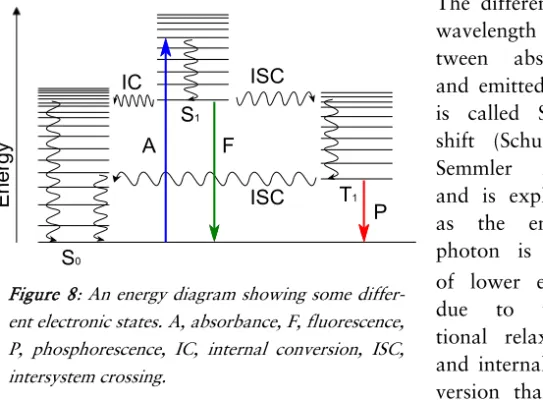
Fluorescence
Absorption and subsequent emission of light from a molecule is called photoluminescence and it can be further divided into fluo-rescence and phosphofluo-rescence depending on the time scale between absorption and emission. Fluorescence is the faster reaction and takes place typically within nanoseconds, whereas phosphorescence is delayed and generally takes place within microseconds (Licht-man & Conchello 2005).
The molecular orbitals of a substance describe the electron distri-bution, and in an electronic transition an electron changes molecu-lar orbital, causing increased or decreased energy. In addition to