DISSERTATION
ADVANCING POINT-OF-NEED BACTERIA DETECTION USING MICROFLUIDIC PAPER-BASED ANALYTICAL DEVICES
Submitted by
Katherine Elizabeth Boehle Department of Chemistry
In partial fulfillment of the requirements For the Degree of Doctor of Philosophy
Colorado State University Fort Collins, Colorado
Summer 2018
Doctoral Committee:
Advisor: Charles S. Henry Amber T. Krummel
Christopher J. Ackerson Brian J. Geiss
Copyright by Katherine Elizabeth Boehle 2018 All Rights Reserved
ii ABSTRACT
ADVANCING POINT-OF-NEED BACTERIA DETECTION USING MICROFLUIDIC PAPER-BASED ANALYTICAL DEVICES
Bacteria are responsible for more hospitalizations and deaths than any other foodborne contaminant, making the detection of these pathogens of utmost importance. To further complicate bacteria detection, the overuse of antibiotics and genetic plasticity of bacteria has caused antimicrobial resistant (AMR) bacteria to become a more
prevalent issue that threatens to be the number one cause of death worldwide by 2050 unless significant innovations are made. Although bacteria detection in the field is ideal, the current gold standards for detection require trained personnel and a central
laboratory. The primary work in this dissertation acts to improve upon current bacteria detection methods by designing, developing, and optimizing inexpensive user-friendly tests that detect bacteria at the point-of-need without trained personnel or expensive equipment. These goals are accomplished using microfluidic paper-based analytical devices (µPADs), a growing field for point-of-need detection that have been used for a variety of analytes and applications. Using paper as a platform has allowed for the simple development of user-friendly devices because of their easily designed and modifiable material that typically costs <$0.01 USD per device and allows for multiple tests to be completed from one sample addition.
Devices that will be described include colorimetric spot tests that detect common fecal indicator bacteria (FIB) species Escherichia coli and Enterococci spp. based on
iii
enzymes that are naturally produced by the bacteria. Utilizing these enzymes, a test was developed that turns from clear to yellow as an indication of live bacteria. These tests were successfully used in the detection of bacteria in food and water samples to demonstrate its efficacy in food safety applications. To improve specificity and
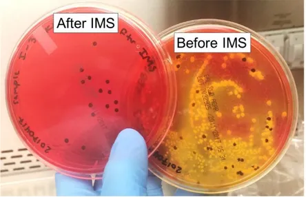
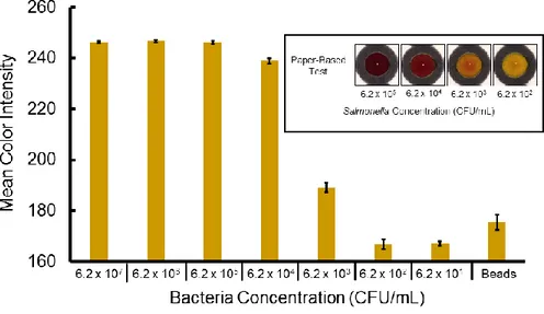
sensitivity of bacteria detection, a second spot test was developed that utilizes immunomagnetic separation (IMS) and an enzymatic sandwich immunoassay in the detection of another common foodborne pathogen, Salmonella typhimurium. This assay was developed specifically for detecting pathogens in complex matrices, such as one of the most common causes of pathogen contamination: animal feces.
Because AMR bacteria are becoming a more prevalent problem, devices were developed to specifically detect bacteria resistant to β-lactam antibiotics, the most common case of antimicrobial resistance observed in bacteria. The first generation of devices were developed to detect β-lactamase activity, an enzyme that facilitates resistance against β-lactam antibiotics. These devices were successful in detecting AMR in different species of bacteria isolated from environmental samples, and in the detection of AMR in sewage water. The second generation of devices enables detection of resistance against specific antibiotics through hydrolysis of the antibiotic and
detecting a change in pH. Although not yet demonstrated, these devices will eventually be used to determine if bacteria are resistant against specific classes of β-lactam antibiotics, including a commonly used class of last resort antibiotics, carbapenems.
Beyond bacteria detection, this dissertation also explores developing a field-ready device to identify falsified and substandard antibiotics. Because antibiotics are most commonly counterfeited in resource-limited settings, it is imperative to develop
iv
user-friendly point-of-need devices that can quantify the amount of active pharmaceutical ingredient in antibiotics. This was accomplished using enzyme competition, a method that had not been demonstrated paper-based devices.
Finally, all devices that have been developed and optimized in this dissertation utilized colorimetric detection. While a user-friendly and easily implemented method of detection, it does suffer from drawbacks such as sensitivity and user subjectivity when using the devices. To eliminate subjectivity, a portable system using a Raspberry Pi computer and 3D-printed light box and device holder have been optimized. Although the system has been demonstrated by automatically analyzing images and calculating Michaelis-Menten enzyme kinetic values, this system has limitless possibilities in
automatically analyzing colorimetric paper-based devices for truly objective colorimetric readouts and quantitative infield detection of pathogens or other analytes.
v
ACKNOWLEDGEMENTS
I would like to first and foremost thank my advisor, Dr. Chuck Henry, for all his mentoring and support throughout grad school. You are truly an inspiration in everything you do, all while having a family and being Chair of the Chemistry Department. Without your constant work, we would not keep breaking records in the number of papers we publish each year (we will achieve 40 publications in 2018!). Thank you for not only encouraging better science, but better communication. I have become a much better writer and presenter because of you, and I will always value your advice. Dr. Brian Geiss, thank you for teaching me everything I know about bacteria and biosafety. Without your knowledge, we would probably still be stuck on some bacteria detection projects. Thanks to you and Chuck, I am leaving CSU with a far more rounded
education than I could have asked for going into graduate school.
Also, a huge thank you goes to all Henry Group members and collaborators, past and present. Especially Dr. Jaclyn Adkins who was always there for my silly questions while I adjusted to doing lab work and taking over her bacteria detection projects.
Thanks for making the long drives to Laramie, Wyoming, and sometimes 12+ hour days, more entertaining with great conversation and audiobooks. Cody Carrell, thank you for all your help and second opinions with my projects. You are destined to do great things in and after graduate school. Dr. Robert Channon, although we never worked on
projects together, you have been a great friend, running partner, and volleyball team member. Thanks for being there for any science or non-science-related rants, and I will always value our great conversations. A few collaborators to thank include Sadie Henry,
vi
Erin Doan, Dr. Elizabeth Ryan, Jake Gilliland, and Amethyst Holder, who were all incremental in the work completed in this document, and would not have been completed without them.
Lastly, I would like to thank my supportive family and friends. My loving parents and sister for helping whenever I needed it, and my amazing fiancé for doing extra work around the house, and for our needy fur-child, when I worked 60-70 hours a week. I could not ask for a more supportive partner in life. I will always treasure all the fun lunches and outings with the amazing people I have met at Colorado State University. I will always treasure my time in graduate school and Fort Collins, and will miss everyone and the city greatly.
vii TABLE OF CONTENTS ABSTRACT ... ii ACKNOWLEDGEMENTS ... v Chapter 1: Introduction ... 1 Introduction ... 1 References ... 21
Chapter 2: Developing Paper-Based Spot Tests for Detecting Bacteria in Food Safety Applications ... 26
Introduction ... 28
Materials and Methods ... 34
Results and Discussion ... 46
Conclusions ... 66
References ... 69
Chapter 3: Utilizing Paper-Based Devices for β-lactam Resistant Bacteria Detection .. 72
Introduction ... 73
Materials and Methods ... 77
Results and Discussion ... 85
Conclusions ... 94
References ... 97
Chapter 4: Microfluidic Paper-Based Device for Detecting Antimicrobial Resistant Bacteria Through Antibiotic Hydrolysis ... 99
Introduction ... 100
Materials and Methods ... 103
Results and Discussion ... 108
Conclusions and Future Directions ... 120
viii
Chapter 5: Paper-Based Enzyme Competition Assay for Detecting Falsified Antibiotics
... 123
Introduction ... 124
Materials and Methods ... 129
Results and Discussion ... 135
Conclusions ... 153
References ... 154
Chapter 6: Developing a Raspberry Pi System to Quantify Color Change and Calculate Enzyme Kinetics on Paper-Based Devices ... 156
Introduction ... 157
Materials and Methods ... 161
Results and Discussion ... 168
Conclusions and Future Directions ... 180
References ... 183
Chapter 7: Conclusions and Future Directions ... 185
Conclusions and Future Directions ... 185
References ... 190
Appendix I: Diagnosing Fungal Infections Using a 3D-Printed Manifold and Paper-Based Microfluidics ... 191
Summary and Specific Aims ... 191
Significance ... 194 Innovation ... 197 Approach: Aim 1 ... 198 Approach: Aim 2 ... 204 Summary ... 211 References ... 212
1
CHAPTER 1. INTRODUCTION
Most bacteria present in the environment coexist with humans and animals without harm. However, certain strains of bacteria are pathogenic and result in serious illness or death, making the detection of bacteria of significant importance.1 While studying bacteria pathology and evolution is central, diagnostics is a underrepresented and important field. A recent study found that 2% of healthcare spending is used in diagnostics, yet account for up to 70% of clinical decisions.2 Bacterial diagnostics are especially underrepresented as demonstrated by the medical field still employing the same diagnostic procedure as 80 years ago: bacteria culture.3 While a reliable and accurate method, the assay is time-consuming, taking up to 2-3 days for results. Therefore, doctors are more likely to employ empirical diagnostics, where treatment is prescribed based on assumptions and previous experience.4 The primary pitfall of empirical diagnostics is the unnecessary prescription of antibiotics when the patient could have a viral or fungal infection instead of bacterial infection. It was recently
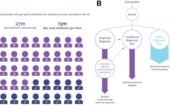
estimated that two-thirds of antibiotic prescriptions are unnecessary (Figure 1.1A).3 Due to unnecessary use of antibiotics and the genetic plasticity of bacteria, more pathogens have developed the ability to resist antimicrobials, giving rise to antimicrobial resistant (AMR) bacteria.5 At present, AMR infections cause 700,000 annual deaths worldwide and cost the United States upwards of $34 billion in additional healthcare costs.6 Unless significant innovations are made in the field, it is currently estimated that AMR infections will surpass cancer and heart disease as the global leading cause of death by 2050.3 AMR bacteria add an additional hurdle for diagnosing bacterial infections because even
2
if bacteria are causing the infection, the physician will not know what antibiotics to prescribe without additional diagnostic tests. Therefore, it is not only imperative that tests are developed to distinguish between bacteria and other pathogens, but to determine what antibiotics can treat a bacterial infection. In addition to new diagnostic methods being rapid, devices that are inexpensive, portable, and user-friendly also need to be developed for point-of-care (POC) diagnostics.7 Most of the world’s morbidity and mortality is occurring in resource-poor countries, where sending patient samples to a central laboratory is not an option.8 By developing and employing rapid diagnostic tests instead of empirical diagnosis, this can reduce unnecessary use of antibiotics, prescribing the correct treatment to patients as soon as possible, whether that patient is in a hospital or needing treatment at the POC (Figure 1.1).
Figure 1.1 │ Approximately two-thirds of antibiotic prescriptions for respiratory issues alone are unnecessary prescriptions (A), which can be prevented through the
development and use of rapid diagnostics tests instead traditional or empirical diagnostics (B).3
3
Foodborne Bacteria in the United States. Although bacteria detection needs
are most commonly associated with diagnosing bacterial infections in patients, a review found the most popular application was for food safety purposes, followed by clinical use, then environmental monitoring (Figure 1.2A).9 Bacteria detection is applicable in the United States food industry because foodborne illness outbreaks caused by bacteria results in 36,000 hospitalizations and over 800 deaths per year, more than any other foodborne contaminant.10 By implementing field-ready bacteria detection, outbreaks could be prevented by detecting bacteria before food is distributed, decreasing the approximate $36 billion USD lost to foodborne illnesses every year.11 Of these foodborne pathogens, Salmonella and Escherichia coli are estimated to cost over $4 billion USD per year in the US alone, contributing to their popularity as the most
reported species for bacteria detection (Figure 1.2B).9,12 Foodborne outbreaks are most commonly caused by animal fecal contamination of either the food directly or irrigation water.13-15 Therefore, it is not only important to detect bacteria in the food directly, but also in animal feces and water.16,17
Detecting Bacteria in the Field. Even though bacteria are most commonly
found in the environment, detecting bacteria currently requires samples to be transported to a central laboratory for laborious, time-consuming, and expensive diagnostic tests. Upon verbal correspondence with our funding agency, the National Wildlife Research Center, a division of the United States Department of Agriculture, around 200,000 wildlife and environmental samples are sent to their central laboratory for testing every year. Of these samples, approximately 90-95% are negative results. By developing inexpensive field-ready tests for bacteria detection, the number of samples
4
sent to the laboratory for comprehensive testing can be cut to 10,000-20,000 samples. At their reported $10 USD for the materials for each laboratory test, not including labor, this could result in at least $1.8 million USD in savings for this government agency alone. In addition to using field-ready bacteria tests for monitoring bacteria in wildlife samples, these developed portable biosensors would also be applicable to in-field testing of other environmental samples, food samples, and POC diagnostic testing for bacterial infections.
Figure 1.2 │ Trends in bacterial detection. (A) Academic papers over the last 20 years show food industry is the most popular application. (B) Salmonella and E. coli are the most commonly detected pathogen. (C) PCR is the most popular detection motif.9
5
Laboratory Methods for Bacteria Detection. Three of the most popular
methods for bacteria detection are culturing, polymerase chain reaction (PCR), and the enzyme-linked immunosorbent assay (ELISA). Although culturing has been the gold standard for bacteria detection for decades, PCR has surpassed the method in popularity in more recent scientific literature (Figure 1.2C).9 Traditional culturing is an ideal method because it detects viable bacteria, and selective culture can identify specific species, but the assay is time-consuming and can take up to several days to obtain a result.18 When detecting AMR bacteria, an additional step is required to test for susceptibility against certain antibiotics by growing the bacteria in the presence of the drugs.19 This method is still the gold standard for AMR bacteria diagnostics because it can quantify susceptibility against specific antibiotics, but the procedure can take several days. PCR is based on amplifying specific genes in the bacteria genome by denaturing the DNA and using a DNA polymerase to extend the genes using DNA primers and additional nucleotides in the solution (Figure 1.3A).20 The assay has gained traction because it is faster and more specific when compared to culturing. Depending on the assay, PCR can give results in as little as a few hours, and specific genes can be detected that are associated with pathogen strains or antimicrobial resistance.21,22
Although the results are fast and specific, PCR can suffer from inhibition effects and detects genes only, and it is therefore unknown whether the bacteria are dead or alive.23 ELISAs, which are based on specific antibodies adhering to the pathogen, have also been used in bacteria detection, but are less commonly used as compared to PCR and culturing.24 The assay is most commonly completed in a 96-well polystyrene plate and read with a plate reader. Although there are many different ELISA models, the sandwich
6
ELISA is one of the most popular motifs. The assay starts with a primary antibody, followed by the blocking agent (used to prevent nonspecific binding), bacteria sample, then completed with either biotinylated or enzyme-conjugated antibody (Figure 1.3B).25 Using a biotinylated antibody allows the user to implement any enzyme that has been conjugated to streptavidin, which forms a stable noncovalent bond with biotin.26
Figure 1.3 │ Two common detection motifs used in bacteria detection include (A) the
polymerase chain reaction (PCR) based on amplifying bacterial DNA associated with specific species or AMR mechanisms and (B) the enzyme-linked immunosorbent assay (ELISA) which is based on detection through antibodies specific to a certain bacteria species.
Between each step of the ELISA procedure, the sample is washed with buffer
containing Tween 20 to remove unbound substrates and prevent signal background. Antibodies are widely available biological substrates, making them easy to implement into new assays and optimize. However, aptamers and bacteriophages have also been suggested for specific detection of bacteria in a similar fashion to immunoassays.27,28 Although these biomolecules are not as widely available as antibodies, their ease to
7
mass produce and higher stability makes them a promising alternative for future applications. PCR and ELISA have also been combined for sensitive and specific
detection of various pathogens in food and clinical samples.29-32 All presented laboratory methods are reliable and accurate, but these procedures require trained personnel, expensive instrumentation, and are hence not favorable for field settings.
Developing Biosensors for Bacteria Detection. As seen in Figure 1.2C,
biosensors are a growing alternative to traditional bacteria detection and are predicted to surpass PCR and culturing in popularity.33 Biosensors can use different detection methods including optical and electrochemical measurements, and have incorporated three main classes of recognition elements: enzymes, antibodies, and nucleic acids. In addition to detection and diagnostics, biosensors have been developed for bacteria enumeration beyond the traditional and time-consuming culturing technique for more sensitive in-field detection.34 Nucleic acid amplification techniques have been applied to biosensors to decrease cost and increase user-friendliness to make the techniques more favorable for a point-of-care setting.35 For example, isothermal amplification techniques have been used as a replacement for PCR for amplifying genetic material in biosensors.36 PCR requires fluctuating temperatures throughout the assay,
necessitating an expensive thermocycler. Isothermal amplification, however, requires the solution to be heated to a set temperature, without fluctuation, to complete the assay. Another popular motif in biosensors for bacteria detection is electrochemical impedance spectroscopy (EIS), which is an electrochemical technique based on
electrical resistance. When the electrode surface becomes more crowded, for example, capturing more and more pathogens through antibodies covalently attached to the
8
electrode, decreasing electron transfer, and thereby increasing resistance. This method has been demonstrated in bacteria detection for a variety of applications and species including Salmonella, E. coli, and S. aureus, to name a few.37-41
Biosensors for specifically detecting AMR bacteria have also been developed. Card et al. introduced the use of expanded microarrays for accurately and
simultaneously testing gram-negative bacteria against 75 different antibiotics.42 Although this method tests for a breadth of antibiotic susceptibility, it requires an extensive procedure, including overnight culturing, cell lysing, and DNA extraction, which increases both detection time and assay costs. Microfluidic devices fabricated with polydimethysiloxane (PDMS) have also been developed for determining the
minimum inhibitory concentration (MIC) of different antibiotics against a specific bacteria isolate.43 One of these established PDMS devices can determine MIC within 3-4 hr by monitoring a single bacterium via microscopy.44 Another microfluidic device was recently developed for susceptibility testing that decreased total assay time to 1 hr by using a droplet generator and a fluorescent resazurin-based assay.45 Using a droplet generator effectively increased the concentration of bacteria without culture enrichment and fluorescence provided greater sensitivity compared to colorimetry. Many resazurin-based sensors have been optimized for detecting AMR bacteria as the assay is resazurin-based on detecting live vs. dead bacteria and can determine whether a certain antibiotic is effective against the bacteria.46-48 While these are all accurate and promising systems that provide alternatives to traditional methods, they require expensive equipment and trained personnel, making these assays more suitable for laboratory settings. To
9
portable, and inexpensive devices that do not require instrumentation or trained laboratory personnel for analysis still need to be developed.
Microfluidic Paper-Based Analytical Devices. While using paper in chemical
assays was established over 200 years ago,49 it was reintroduced as a microfluidic platform in 2007 for portable and inexpensive analytical assays.50 This reintroduction of paper was groundbreaking because Whitesides and coworkers incorporated
hydrophobic barriers into the paper to direct fluid flow to several channels and separate detection zones.50 Each zone was responsible for a different analytical test, enabling the detection of multiple analytes (glucose and protein in this device) from a single addition of sample (Figure 1.4A). Although this initial device had higher limits of detection and low sensitivity compared to conventional methods, this enabled the thousands of paper-based devices that followed this article and created the field known as microfluidic paper-based analytical devices (µPADs). Paper has gained significant popularity as a platform for analytical devices because of their inexpensive material (often <$0.01 per device), ability to store and stabilize chemical and biological reagents, natural fluid-wicking properties, and device disposability.51 Because of these properties, µPADs are specifically being developed as point-of-need analytical tests that enable the detection of analytes without trained personnel or a central laboratory.52 While the first device used qualitative detection based on color change, user-friendly quantitative detection has since been implemented using a “chemometer” design with readout similar to a common analog thermometer (Figure 1.4B).53 Instead of a simple yes or no answer, chemometers have been designed for the user to quantify an analyte by measuring the distance of color and relating that distance to a specific analyte concentration.
10
In addition to their ease of use, µPADs are also easy to design and fabricate and are practical for large-scale development and implementation.54 Paper-based devices are most often fabricated with Whatman chromatography paper, but almost any paper substrate can be used. To make µPADs, the user simply needs to develop a
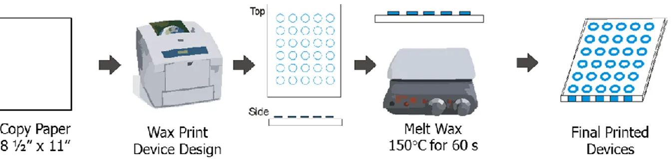
hydrophobic barrier that directs sample flow. The hydrophobic barrier can be many different materials including photo resist, or even scholar glue that is not vulnerable to potent chemicals like other barriers.55 One of the most popular hydrophobic barriers is wax, which can be deposited onto the paper using a common office wax printer56 or screen printing.57 Using a wax printer or screen printing allows for the user to create device designs using a common computer vector program such as Adobe Illustrator™ or Corel Draw™. Following wax application, the paper needs to be heated to melt the wax through the paper pores, creating a hydrophobic barrier, which can be completed using an oven or hotplates (Figure 1.4C). Once the hydrophobic barrier has been established, the user can modify the paper with chemicals to complete the desired analytical test. The back of the devices then need to be taped to prevent sample leakage.
Since the first device, µPADs have been developed for environmental and biological applications for the detection of metals, organic compounds, biomarkers, bacteria, and viruses, to name a few.58 While this first device used simple and user-friendly colorimetric detection, additional detection motifs have been established including electrochemical,59,60 fluorescence,61 and chemiluminescence,62 which were implemented into µPADs to lower limits-of-detection and increase sensitivity. As previously mentioned, paper has many different properties that are advantageous for
11
Figure 1.4 │ Microfluidic paper-based analytical devices (µPADs). (A) The first µPAD for the detection of glucose and protein using qualitative colorimetric detection.50 (B) Distance-based colorimetric detection of glucose for user-friendly quantification of analytes.53 (C) Fabricating paper-based devices by making a hydrophobic barrier with wax and taping the back of the device sheet.
point-of-need detection, but one of the most critical properties is its ability to store and stabilize chemical reagents. This allows for reactions to take place on the paper substrate without additional solutions beyond the sample, facilitating user-friendly detection of countless analytes in a field-setting, including bacteria.
Biological Assays on Paper-Based Devices. While PCR is the most popular
method in recent years for bacteria detection outside of µPADs, PCR has not been directly applied to paper-based devices. This is likely because PCR requires a
thermocycler, an expensive and complex piece of equipment that makes the assay not ideal for point-of-need settings. A thermocycler is necessary because PCR requires the
12
solution to shift between several different specific temperatures to complete the assay.20 Instead, isothermal amplification techniques including loop-mediated isothermal
amplification (LAMP) and recombinase polymerase amplification (RPA) has been applied to µPADs for sensitive and point-of-need detection of bacteria63 and viruses.64-66 While isothermal systems still require a heating element, LAMP only requires to be heated to a set temperature of 60 °C, and RPA requires a set temperature of 40 °C for amplification. To further establish isothermal amplification for field settings, an
inexpensive incubator was developed using a Styrofoam cup and chemical hand
warmers,67 and it has been reported that polyethersulfone is the best paper substrate to use for LAMP-based µPADs.68 While isothermal amplification has been demonstrated in paper-based devices as a conducive alternative to PCR for field settings, the underlying issue with nucleic-acid based techniques is the lack of live/dead verification.
Furthermore, in the application of AMR bacteria detection, it is unknown whether the AMR gene is expressed or simply present in the genome.
Enzymes are one of the most popular detection motifs associated with paper-based devices due to their catalytic abilities to accelerate a chemical reaction and amplify a detectable product. Enzymes can be used to detect an analyte of interest, including one of the most popular proof-of-concept reactions used in paper-based devices: glucose.69 Other enzymatic reactions that have been used in µPADs include the detection of other biomolecules such as uric acid, urine creatine, phenylalanine, and lactate.70-74 In addition to using enzymes to detect analytes, substrates can be used to detect enzyme activity in a sample. Detecting the activity of specific enzymes can indicate certain pathogens or health ailments. For example, assessing human health
13
based on enzyme detection has been established for many diseases that are
associated with the expression or lack of function of specific enzymes, including liver function,75 organ failure,76 male fertility,77 and even organophosphate poisoning.78 Using this concept, µPADs can be developed for bacteria detection based on unique enzymes the bacteria produce. Detecting bacteria based on enzymatic activity is an attractive platform because it detects healthy and viable bacteria, like traditional culturing or the resazurin assay. Additionally, it is not as specific as PCR and immunoassays, enabling pan-bacteria detection, which is advantageous for applications such as food safety monitoring. Using enzymes as a detection motif has been described previously for
colorimetric detection of E. coli, Listeria, and Salmonella in food and water samples.79-81 When the user does desire species-specific and sensitive detection,
immunoassays provide an attractive and robust platform that can easily be adapted to µPADs. Antibodies are naturally very specific to the analyte with little cross-reactivity, and can be manufactured to be specific to almost any analyte. One of the most
recognizable forms of paper-based devices is the pregnancy test, which is a lateral flow assay (LFA). Pregnancy tests are based on nanoparticle-conjugated antibodies specific to human chorionic gonadotropin (hCG), a protein women express when pregnant.82 A similar device was developed for detecting pathogenic bacteria where bacteria are captured by antibodies resulting in the nanoparticles aggregating resulting in the formation of colored line.83 Another popular application of antibodies in µPADs is through ELISAs where enzymes are covalently attached to antibodies instead of nanoparticles. Enzymes provide an advantage over nanoparticles because enzymes can continually amplify a substrate, providing a lower limit-of-detection. As previously
14
mentioned, ELISAs are traditionally completed in 96-well polystyrene plates, but ELISA was first introduced into paper-based devices in 2010.84 By moving the assay to paper, there was decreased reagent consumption, waste, and total assay time. Since its introduction to paper, ELISA has been demonstrated in the detection of
biomarkers,62,85,86 and pathogens including viruses87,88 and bacteria.89 To further
advance analyte detection in complex samples, antibodies are also used in the process of immunomagnetic separation (IMS), where antibodies are covalently attached to the surface of a magnetic bead.90 The immunomagnetic beads are added to a complex sample to adhere to the analyte before separating the beads from the sample using a magnet and reconstituting the beads in fresh buffer. Reconstituting the sample in fresh buffer also allows for the user to concentrate the sample by resuspending the beads in a smaller volume of buffer than the original sample. IMS has been demonstrated for efficiently separating target analytes and cells from complex mixtures such as blood,91 milk,92 meat,93 cheese and yogurt,94 and bovine feces,95,96 making the technique relevant to food safety applications.
Paper-Based Devices for Foodborne Pathogen Detection. The work
presented in this document revolve around improving bacteria detection at the point-of-need using paper-based devices, including both spot tests and microfluidic devices. The first set of devices that will be discussed are spot tests that detect common fecal
indicator bacteria, E. coli and Enterococci, and the most common foodborne pathogen,
Salmonella typhimurium, for the application of food safety monitoring. The first device
detects E. coli and Enterococci based on enzymes the bacteria produce: β-galactosidase and β-glucuronidase97 for E. coli detection and β-glucosidase for
15
Enterococci detection.98 This method was successfully demonstrated in detecting bacteria in irrigation water and alfalfa sprouts, which are a common source of food poisoning. While this method provided accurate and user-friendly results, the limits of detection were rather high at 108 CFU mL-1 of bacteria, necessitating a culture
enrichment step. By sampling the culture enrichment every four hours, we cut the analysis time to 8 hours for low bacteria concentrations compared to upwards of two days for traditional methods. However, the ideal bacterial detection system will have a low LOD without needing culture enrichment.
To meet these needs, another paper-based spot test was developed to
specifically detect S. typhimurium using antibodies. Implementing IMS as the first step of the procedure enabled us to isolate and concentrate the pathogen for detection in complex samples. Because IMS is a sample preparation step, not a detection method, an enzymatic sandwich immunoassay, similar to an ELISA, was added for sensitive and rapid analysis without expensive instrumentation. Using these two techniques for
isolation, concentration, and detection of the pathogen, our LOD was decreased to 102 CFU mL-1 in culture media. By using antibodies as our detection method, not only did we increase sensitivity of the assay, eliminating the need for culture enrichment, the assay is also very specific to S. typhimurium and showed no cross reactivity with E. coli bacteria. The final assay was demonstrated with bird feces and whole milk for food safety applications.
Detecting AMR Bacteria using µPADs. The next set of devices that will be
discussed are also based on enzymes the bacteria produce, but instead of identifying bacteria species, these devices detect enzymes that indicate AMR properties. There are
16
many different classes of antibiotics, therefore many different classes of antimicrobial resistance, and many of these resistance mechanisms are based off enzyme activity.99 Using this idea, we developed a paper-based spot test to detect β-lactamase, an enzyme that facilitates resistance against the most commonly prescribed antibiotics, penicillins and cephalosporins.100 lactamase enables resistance by hydrolyzing the β-lactam ring in β-β-lactam antibiotics, deactivating the compound. Taking advantage of this mechanism, we can use nitrocefin, a chromogenic cephalosporin, that turns from yellow to red upon enzyme hydrolysis (Figure 1.5).101 Using nitrocefin and paper-based
devices, a spot test was developed to detect the enzyme activity at a fraction of the cost and time as traditional methods. Contaminated water is a significant source of infection and outlet for the spread of AMR bacteria and is therefore a popular area of study for environmental scientists.102-105 To study AMR bacteria epidemiology, scientists currently must transport samples to a central laboratory for testing. Hence, it is not only important to use AMR tests in the application of point-of-care diagnostics, but also in
environmental monitoring. The test was demonstrated in environmental applications by detecting AMR bacteria in sewage water samples and 46 different environmental bacterial isolates. There was only one false result as verified by traditional methods, indicating 98% accuracy.
To further expand upon this idea of detecting AMR bacteria based on enzyme expression, another µPAD was developed to detect bacteria that produce
carbapenemase. This enzyme facilitates resistance against carbapenem antibiotics, a commonly used class of last resort antibiotics used in clinical cases.106 Carbapenem-resistant bacteria are becoming a prevalent problem and is recognized as one of the top
17
Figure 1.5 │ Using nitrocefin, a chromogenic substrate, to detect antimicrobial resistant bacteria based on bacterial enzymes that facilitate resistance against antibiotics through deactivation.
three most urgent threats of AMR in the US by the CDC.107,108 Although there is no chromogenic substrate for carbapenemase as there is for -lactamase, the hydrolysis the enzyme facilitates results in a decrease in pH. Using paper-based microfluidics, we developed a device that allows the bacteria sample to react with imipenem, a
carbapenem antibiotic, then the sample flows to another section of the device with pH indicators. If the antibiotic is hydrolyzed, the user will observe a corresponding decrease in pH as compared to a sample of bacteria that does not express carbapenemase. This device has not been demonstrated in detecting carbapenem-resistant bacteria
specifically, but has been developed and optimized for detecting specific penicillin antibiotics such as penicillin V and amoxicillin.
In-Field Devices for Counterfeit Antibiotic Screening. In addition to the use of
18
enzymes, analytes can be detected through enzyme inhibition, resulting in a lack of color change. This concept has been demonstrated in the detection of organophosphate pesticides that inhibit the reaction of acetylcholinesterase with a colorimetric substrate, resulting in less color change as the pesticide concentration increases.109,110 Using a similar concept, we have developed a µPAD to test for the authenticity of β-lactam antibiotics using β-lactamase and nitrocefin, but using enzyme competition, not direct inhibition. Counterfeit antibiotics is a prevalent problem in developing countries and it is estimated that up to 5% of global antibiotics are counterfeit.111,112 Of all counterfeit antibiotics, β-lactam antibiotics are the most counterfeited, accounting for over half of counterfeit antibiotics.113 To help combat this problem and monitor counterfeit antibiotics in the field, a µPAD was developed to detect the purity of β-lactam antibiotics using the same system that was used to detect β-lactam-resistant bacteria. The device operates through adding an antibiotic sample to the sample inlet where it travels down a channel, rehydrates nitrocefin, then travels to a detection zone where β-lactamase is stored. If the antibiotics are genuine, β-lactamase will statistically react more often with the concentrated β-lactam antibiotic compared to the dilute nitrocefin. If the antibiotics are counterfeit, β-lactamase will react with nitrocefin, turning the device red, indicating no or little active ingredient. Calibration curves for four different β-lactam antibiotics were generated and the device was tested with six common counterfeit ingredients, demonstrating its potential for in-field use of antibiotic screening.
Developing a Raspberry Pi for Automating Color Analysis. All devices
presented in this work use colorimetric readout for detection. While colorimetry is a user-friendly method that does not require external instrumentation, such as a
19
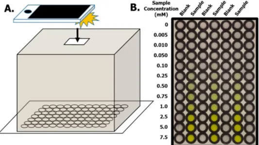
potentiostat in electrochemical detection, there is the underlying issue of subjectivity when reading the results. Currently, to quantify the color intensity in devices, an image is captured using either a Smart Phone camera and a light box, or a common desktop scanner. The image is then sent to a computer for analysis by an image software program such as NIH ImageJ or Adobe Photoshop. While this method works for laboratory research, it is not conducive to field settings. Consequently, smart phone applications have been developed to automatically analyze the device color following image capture.114,115 Although these applications are much more appropriate for field settings, the caveat of smart phone applications is the need to update the application for new and expensive phones and software. To enhance colorimetric detection capabilities in the field, we have chosen the Raspberry Pi format for developing a program where the user simply needs to input a command and the Raspberry Pi will automatically capture and analyze the images. A 3D-printed lightbox with a battery-powered light source was designed to hold the raspberry pi computer and camera and house the paper-based devices. The entire system costs <$100, which is cheaper than the average Smart Phone, and the system is portable and can be used in resource-limited settings. As a proof-of-concept, we have developed a program that automatically calculates Michaelis-Menten enzyme kinetics based on images captured by the Raspberry Pi™. This would enable the user to use the system for time-dependent reactions and calculate kinetics for enzyme-based detection methods.
Summary. The work in this dissertation advances in-field bacteria detection
through expanding biological assays to complex samples, such as food, milk, and animal feces. It also presents the first work on detecting antimicrobial resistant bacteria
20
using paper-based devices, both generally and to specific antibiotics. Although not directly related to bacteria detection, the first µPAD based on enzymatic substrate competition is used to develop the first quantitative µPAD for identifying substandard antibiotics. Finally, we describe the first Raspberry Pi system to use flood-fill in quantifying color change in the application of calculating Michaelis-Menten enzyme kinetic parameters.
21
REFERENCES
(1) Stephens, C.; Murray, W. Curr. Biol. 2001, 11, R53-R56.
(2) Rohr, U. P.; Binder, C.; Dieterle, T.; Giusti, F.; Messina, C. G.; Toerien, E.; Moch, H.; Schafer, H. H. PLoS One 2016, 11, e0149856.
(3) Tackling Drug-Resistant Infections Globally: Final Report and Recommendations Welcome Trust, UK Government, 2016.
(4) Morrell, M.; Fraser, V. J.; Kollef, M. H. Antimicrob. Agents Chemother. 2005, 49, 3640-3645.
(5) Levy, S. B.; Marshall, B. Nat. Med. 2004, 10, S122-129.
(6) Global Action Plan on Antimicrobial Resistance, World Health Organization, 2015. (7) Chin, C. D.; Linder, V.; Sia, S. K. Lab Chip 2012, 12, 2118-2134.
(8) Chin, C. D.; Laksanasopin, T.; Cheung, Y. K.; Steinmiller, D.; Linder, V.; Parsa, H.; Wang, J.; Moore, H.; Rouse, R.; Umviligihozo, G.; Karita, E.; Mwambarangwe, L.; Braunstein, S. L.; van de Wijgert, J.; Sahabo, R.; Justman, J. E.; El-Sadr, W.; Sia, S. K.
Nat. Med. 2011, 17, 1015-1019.
(9) Lazcka, O.; Del Campo, F. J.; Munoz, F. X. Biosens. Bioelectron. 2007, 22, 1205-1217.
(10) Painter, J. A.; Hoekstra, R. M.; Ayers, T.; Tauxe, R. V.; Braden, C. R.; Angulo, F. J.; Griffin, P. M. Emerg. Infect. Dis. 2013, 19, 407-415.
(11) Minor, T.; Lasher, A.; Klontz, K.; Brown, B.; Nardinelli, C.; Zorn, D. Risk Anal. 2015,
35, 1125-1139.
(12) Hoffman, S.; Batz, M.; Morris, J. G. J. Food Prot. 2012, 75, 1291-1302.
(13) Laidler, M. R.; Tourdjman, M.; Buser, G. L.; Hostetler, T.; Repp, K. K.; Leman, R.; Samadpour, M.; Keene, W. E. Clin. Infect. Dis. 2013, 57, 1129-1134.
(14) Steele, M.; Odumeru, J. J. Food Prot. 2004, 67, 2839-2849.
(15) Uyttendaele, M.; Jaykus, L.-A.; Amoah, P.; Chiodini, A.; Cunliffe, D.; Jacxsens, L.; Holvoet, K.; Korsten, L.; Lau, M.; McClure, P.; Medema, G.; Sampers, I.; Rao Jasti, P.
Compr. Rev. Food Sci. Food Saf. 2015, 14, 336-356.
(16) Sayah, R. S.; Kaneene, J. B.; Johnson, Y.; Miller, R. Appl. Environ. Microbiol. 2005,
71, 1394-1404.
(17) Renter, D. G.; Gnad, D. P.; Sargeant, J. M.; Hygnstrom, S. E. J. Wildl. Dis. 2006,
42, 699-703.
(18) Wang, Y.; Salazar, J. K. Compr. Rev. Food Sci. Food Saf. 2016, 15, 183-205. (19) Bauer, A. W.; Kirby, W. M.; Sherris, J. C.; Turck, M. Am. J. Clin. Pathol.1966, 45, 493-496.
(20) Schochetman, G.; Ou, C.; Jones, W. K. J. Infect. Dis. 1988, 158, 1154-1157. (21) Jamali, H.; Paydar, M.; Radmehr, B.; Ismail, S.; Dadrasnia, A. Food Control 2015,
54, 383-388.
(22) Clerc, O.; Prod'hom, G.; Senn, L.; Jaton, K.; Zanetti, G.; Calandra, T.; Greub, G.
Clin. Microbiol. Infect. 2014, 20, 355-360.
(23) Gonzalez, R. A.; Noble, R. T. Water Res. 2014, 48, 296-305.
(24) Magliulo, M.; Simoni, P.; Guardigli, M.; Michelini, E.; Luciani, M.; Lelli, R.; Roda, A.
22
(25) Drijvers, J. M.; Awan, I. M.; Perugino, C. A.; Rosenberg, I. M.; Pillai, S. 2017, In
Basic Science Methods for Clinical Researchers, Jalali, M.; Saldanha, F. Y. L.; Jalali,
M., Eds.; Elsevier, 2017, pp 119-133.
(26) Taninaka, A.; Takeuchi, O.; Shigekawa, H. Int. J. Mol. Sci. 2010, 11, 2134-2151. (27) Toh, S. Y.; Citartan, M.; Gopinath, S. C.; Tang, T. H. Biosens. Bioelectron. 2015,
64, 392-403.
(28) Singh, A.; Arutyunov, D.; Szymanski, C. M.; Evoy, S. Analyst 2012, 137, 3405-3421.
(29) Perelle, S.; Dilasser, F.; Malorny, B.; Grout, J.; Hoorfar, J.; Fach, P. Mol. Cell.
Probes 2004, 18, 409-420.
(30) Tamminen, M.; Joutsjoki, T.; Sjoblom, M.; Joutsen, M.; Palva, A.; Ryhanen, E. L.; Joutsjoki, V. Lett. Appl. Microbiol. 2004, 39, 439-444.
(31) Ge, B.; Zhao, S.; Hall, R.; Meng, J. Microbes Infect. 2002, 4, 285-290. (32) Daly, P.; Collier, T.; Doyle, S. Lett. Appl. Microbiol. 2002, 34, 222-226.
(33) Velusamy, V.; Arshak, K.; Korostynska, O.; Oliwa, K.; Adley, C. Biotechnol. Adv. 2010, 28, 232-254.
(34) Zhang, J. Y.; Do, J.; Premasiri, W. R.; Ziegler, L. D.; Klapperich, C. M. Lab Chip 2010, 10, 3265-3270.
(35) Niemz, A.; Ferguson, T. M.; Boyle, D. S. Trends Biotechnol. 2011, 29, 240-250. (36) Safavieh, M.; Ahmed, M. U.; Sokullu, E.; Ng, A.; Braescu, L.; Zourob, M. Analyst 2014, 139, 482-487.
(37) Muhammad-Tahir, Z.; Alocilja, E. C. Biosens. Bioelectron. 2003, 18, 813-819. (38) Yang, L.; Bashir, R. Biotechnol. Adv. 2008, 26, 135-150.
(39) Tan, F.; Leung, P. H. M.; Liu, Z.; Zhang, Y.; Xiao, L.; Ye, W.; Zhang, X.; Yi, L.; Yang, M. Sens. Actuators B Chem. 2011, 159, 328-335.
(40) Wang, Y.; Ping, J.; Ye, Z.; Wu, J.; Ying, Y. Biosens. Bioelectron. 2013, 49, 492-498.
(41) Wang, Y.; Ye, Z.; Ying, Y. Sensors 2012, 12, 3449-3471.
(42) Card, R.; Zhang, J.; Das, P.; Cook, C.; Woodford, N.; Anjum, M. F. Antimicrob.
Agents Chemother. 2013, 57, 458-465.
(43) Kim, K. P.; Kim, Y. G.; Choi, C. H.; Kim, H. E.; Lee, S. H.; Chang, W. S.; Lee, C. S.
Lab Chip 2010, 10, 3296-3299.
(44) Choi, J.; Jung, Y. G.; Kim, J.; Kim, S.; Jung, Y.; Na, H.; Kwon, S. Lab Chip 2013,
13, 280-287.
(45) Kaushik, A. M.; Hsieh, K.; Chen, L.; Shin, D. J.; Liao, J. C.; Wang, T. H. Biosens.
Bioelectron. 2017, 97, 260-266.
(46) Elavarasan, T.; Chhina, S. K.; Parameswaran, M.; Sankaran, K. Sens. Actuators B
Chem. 2013, 176, 174-180.
(47) Boedicker, J. Q.; Li, L.; Kline, T. R.; Ismagilov, R. F. Lab Chip 2008, 8, 1265-1272. (48) Deiss, F.; Funes-Huacca, M. E.; Bal, J.; Tjhung, K. F.; Derda, R. Lab Chip 2014,
14, 167-171.
(49) Davy, J. Phil. Trans. R. Soc. Lond. 1812, 102, 144-151.
(50) Martinez, A. W.; Phillips, S. T.; Butte, M. J.; Whitesides, G. M. Angew. Chem. Int.
Ed. Engl. 2007, 46, 1318-1320.
(51) Cate, D. M.; Adkins, J. A.; Mettakoonpitak, J.; Henry, C. S. Anal. Chem. 2015, 87, 19-41.
23
(52) Yetisen, A. K.; Akram, M. S.; Lowe, C. R. Lab Chip 2013, 13, 2210.
(53) Cate, D. M.; Dungchai, W.; Cunningham, J. C.; Volckens, J.; Henry, C. S. Lab Chip 2013, 13, 2397-2404.
(54) Akram, M. S.; Daly, R.; da Cruz Vasconcellos, F.; Yetisen, A. K.; Hutchings, I.; Hall, E. A. H. In Lab-on-a-Chip Devices and Micro-Total Analysis Systems, Castillo-Leon, J.; Svendson, W. E., Eds.; Springer International Publishing Switzerland, 2015, pp 161-195.
(55) Cardoso, T. M. G.; de Souza, F. R.; Garcia, P. T.; Rabelo, D.; Henry, C. S.; Coltro, W. K. T. Anal. Chim. Acta 2017, 974, 63-68.
(56) Carrilho, E.; Martinez, A. W.; Whitesides, G. M. Anal. Chem. 2009, 81, 7091-7095. (57) Dungchai, W.; Chailapakul, O.; Henry, C. S. Analyst 2011, 136, 77-82.
(58) Yang, Y.; Noviana, E.; Nguyen, M. P.; Geiss, B. J.; Dandy, D. S.; Henry, C. S. Anal.
Chem. 2017, 89, 71-91.
(59) Adkins, J.; Boehle, K.; Henry, C. Electrophoresis 2015, 36, 1811-1824.
(60) Mettakoonpitak, J.; Boehle, K.; Nantaphol, S.; Teengam, P.; Adkins, J. A.; Srisa-Art, M.; Henry, C. S. Electroanalysis 2016, 28, 1420-1436.
(61) Noor, M. O.; Krull, U. J. Anal. Chem. 2014, 86, 10331-10339.
(62) Wang, S.; Ge, L.; Song, X.; Yu, J.; Ge, S.; Huang, J.; Zeng, F. Biosens.
Bioelectron. 2012, 31, 212-218.
(63) Lafleur, L. K.; Bishop, J. D.; Heiniger, E. K.; Gallagher, R. P.; Wheeler, M. D.; Kauffman, P.; Zhang, X.; Kline, E. C.; Buser, J. R.; Kumar, S.; Byrnes, S. A.;
Vermeulen, N. M.; Scarr, N. K.; Belousov, Y.; Mahoney, W.; Toley, B. J.; Ladd, P. D.; Lutz, B. R.; Yager, P. Lab Chip 2016, 16, 3777-3787.
(64) Rodriguez, N. M.; Linnes, J. C.; Fan, A.; Ellenson, C. K.; Pollock, N. R.; Klapperich, C. M. Anal. Chem. 2015, 87, 7872-7879.
(65) Rodriguez, N. M.; Wong, W. S.; Liu, L.; Dewar, R.; Klapperich, C. M. Lab Chip 2016, 16, 753-763.
(66) Rohrman, B. A.; Richards-Kortum, R. R. Lab Chip 2012, 12, 3082-3088. (67) Huang, S.; Do, J.; Mahalanabis, M.; Fan, A.; Zhao, L.; Jepeal, L.; Singh, S. K.; Klapperich, C. M. PLoS One 2013, 8, e60059.
(68) Linnes, J. C.; Rodriguez, N. M.; Liu, L.; Klapperich, C. M. Biomed. Microdevices 2016, 18, 30.
(69) Liu, S.; Su, W.; Ding, X. Sensors 2016, 16.
(70) Gabriel, E. F.; Garcia, P. T.; Cardoso, T. M.; Lopes, F. M.; Martins, F. T.; Coltro, W. K. Analyst 2016, 141, 4749-4756.
(71) Chen, X.; Chen, J.; Wang, F.; Xiang, X.; Luo, M.; Ji, X.; He, Z. Biosens.
Bioelectron. 2012, 35, 363-368.
(72) Talalak, K.; Noiphung, J.; Songjaroen, T.; Chailapakul, O.; Laiwattanapaisal, W.
Talanta 2015, 144, 915-921.
(73) Robinson, R.; Wong, L.; Monnat, R.; Fu, E. Micromachines 2016, 7, 28.
(74) Dungchai, W.; Chailapakul, O.; Henry, C. S. Anal. Chim. Acta 2010, 674, 227-233. (75) Pollock, N. R.; Rolland, J. P.; Kumar, S.; Beattie, P. D.; Jain, S.; Noubary, F.;
Wong, V. L.; Pohlmann, R. A.; Ryan, U. S.; Whitesides, G. M. Sci. Transl. Med. 2012, 4. (76) Kannan, B.; Jahanshahi-Anbuhi, S.; Pelton, R. H.; Li, Y.; Filipe, C. D.; Brennan, J. D. Anal. Chem. 2015, 87, 9288-9293.
24
(77) Nosrati, R.; Gong, M. M.; San Gabriel, M. C.; Pedraza, C. E.; Zini, A.; Sinton, D.
Clin. Chem. 2016, 62, 458-465.
(78) Yen, T. H.; Chen, K. H.; Hsu, M. Y.; Fan, S. T.; Huang, Y. F.; Chang, C. L.; Wang, Y. P.; Cheng, C. M. Talanta 2015, 145, 66-72.
(79) Jokerst, J. C.; Adkins, J. A.; Bisha, B.; Mentele, M. M.; Goodridge, L. D.; Henry, C. S. Anal. Chem. 2012, 84, 2900-2907.
(80) Bisha, B.; Adkins, J. A.; Jokerst, J. C.; Chandler, J. C.; Perez-Mendez, A.;
Coleman, S. M.; Sbodio, A. O.; Suslow, T. V.; Danyluk, M. D.; Henry, C. S.; Goodridge, L. D. J. Vis. Exp. 2014, 88.
(81) Zakir Hossain, S. M.; Ozimok, C.; Sicard, C.; Aguirre, S. D.; Monsur Ali, M.; Li, Y.; Brennan, J. D. Anal. Bioanal. Chem. 2012, 403, 1567-1576.
(82) Posthuma-Trumpie, G. A.; Korf, J.; van Amerongen, A. Anal. Bioanal. Chem. 2009,
393, 569-582.
(83) Li, C.; Vandenberg, K.; Prabhulkar, S.; Zhu, X.; Schneper, L.; Methee, K.; Rosser, C. J.; Almeide, E. Biosens. Bioelect. 2011, 26, 4342-4348.
(84) Cheng, C. M.; Martinez, A. W.; Gong, J.; Mace, C. R.; Phillips, S. T.; Carrilho, E.; Mirica, K. A.; Whitesides, G. M. Angew. Chem. Int. Ed. Engl. 2010, 49, 4771-4774. (85) Murdock, R. C.; Shen, L.; Griffin, D. K.; Kelley-Loughnane, N.; Papautsky, I.; Hagen, J. A. Anal. Chem. 2013, 85, 11634-11642.
(86) Sun, X.; Li, B.; Tian, C.; Yu, F.; Zhou, N.; Zhan, Y.; Chen, L. Anal. Chim. Acta 2018, 1007, 33-39.
(87) Mu, X.; Zhang, L.; Chang, S.; Cui, W.; Zheng, Z. Anal Chem 2014, 86, 5338-5344. (88) Khan, M. S.; Pande, T.; van de Ven, T. G. Colloids Surf. B Biointerfaces 2015, 132, 264-270.
(89) Shih, C. M.; Chang, C. L.; Hsu, M. Y.; Lin, J. Y.; Kuan, C. M.; Wang, H. K.; Huang, C. T.; Chung, M. C.; Huang, K. C.; Hsu, C. E.; Wang, C. Y.; Shen, Y. C.; Cheng, C. M.
Talanta 2015, 145, 2-5.
(90) He, J.; Huang, M.; Wang, D.; Zhang, Z.; Li, G. J. Pharm. Biomed. Anal. 2014, 101, 84-101.
(91) Cohen, S. J.; Punt, C. J.; Iannotti, N.; Saidman, B. H.; Sabbath, K. D.; Gabrail, N. Y.; Picus, J.; Morse, M.; Mitchell, E.; Miller, M. C.; Doyle, G. V.; Tissing, H.; Terstappen, L. W.; Meropol, N. J. J. Clin. Oncol. 2008, 26, 3213-3221.
(92) Brandao, D.; Liebana, S.; Campoy, S.; Alegret, S.; Isabel Pividori, M. Talanta 2015,
143, 198-204.
(93) Skjerve, E.; Rorvik, L. M.; Olsvik, O. Appl. Environ. Microbiol. 1990, 56, 3478-3481. (94) Skjerve, E.; Olsvik, O. Int. J. Food Microbiol. 1991, 14, 11-18.
(95) Chapman, P. A.; Wright, D. J.; Siddons, C. A. J. Med. Microbiol. 1994, 40, 424-427. (96) LeJeune, J. T.; Hancock, D. D.; Besser, T. E. J. Clin. Microbiol. 2006, 44, 872-875. (97) Wu, J.; Stewart, J. R.; Sobsey, M. D.; Cormency, C.; Fisher, M. B.; Bartram, J. K.
Curr. Microbiol. 2018.
(98) Perry, J. D.; Morris, K. A.; James, A. L.; Oliver, M.; Gould, F. K. J. Appl. Microbiol. 2007, 102, 410-415.
(99) Poole, K. J. Pharm. Pharmacol. 2001, 53, 283-294.
(100) Murray, B. E.; Mederski-Samaroj, B. J. Clin. Invest. 1983, 72, 1168-1171. (101) O'Callaghan, C. H.; Morris, A.; Kirby, S. M.; Chingler, A. H. Antimicrob. Agents
25
(102) Lupo, A.; Coyne, S.; Berendonk, T. U. Front. Microbiol. 2012, 3, 116-128. (103) Jiang, L.; Hu, X.; Xu, T.; Zhang, H.; Sheng, D.; Yin, D. Sci.Total Environ. 2013,
458-460, 267-272.
(104) Holvoet, K.; Sampers, I.; Callens, B.; Dewulf, J.; Uyttendaele, M. Appl. Environ.
Microbiol. 2013, 79, 6677-6683.
(105) Baquero, F.; Martinez, J. L.; Canton, R. Curr. Opin. Biotechnol. 2008, 19, 260-265.
(106) Nordmann, P.; Poirel, L.; Dortet, L. Emerg. Infect. Dis. 2012, 18, 1503-1507. (107) Nordmann, P.; Naas, T.; Poirel, L. Emerg. Infect. Dis. 2011, 17, 1791-1798.
(108) Antibiotic Resistance Threats in the United States, Center for Disease Control and Prevention, 2013.
(109) Zakir Hossain, S. M.; Luckham, R. E.; McFadden, M. J.; Brennan, J. D. Anal.
Chem. 2009, 81, 9055-9064.
(110) Sicard, C.; Glen, C.; Aubie, B.; Wallace, D.; Jahanshahi-Anbuhi, S.; Pennings, K.; Daigger, G. T.; Pelton, R.; Brennan, J. D.; Filipe, C. D. Water. Res. 2015, 70, 360-369. (111) Delepierre, A.; Gayot, A.; Carpentier, A. Med. Mal. Infect. 2012, 42, 247-255. (112) Global Surveillance and Monitoring System for Substandard and Falsified Medical, World Health Organization, 2017.
(113) Kelesidis, T.; Falagas, M. E. Clin. Microbiol. Rev. 2015, 28, 443-464.
(114) Vashist, S. K.; Mudanyali, O.; Schneider, E. M.; Zengerle, R.; Ozcan, A. Anal.
Bioanal. Chem. 2014, 406, 3263-3277.
(115) Grushnikov, A.; Kikuchi, K.; Matsumoto, Y.; Kanade, T.; Yagi, Y. Adv. Biomed.
26
CHAPTER 2. DEVELOPING PAPER-BASED SPOT TESTS FOR DETECTING BACTERIA IN FOOD SAFETY APPLICATIONS
Foodborne illnesses caused by bacteria account for the highest number of hospitalizations and deaths compared to any other foodborne contaminant. Preventing foodborne illness outbreaks begs for faster and portable bacterial sensors that can be taken into the field to detect bacteria before food is distributed, cutting back on the approximate billions of dollars lost to outbreaks every year. Most commonly,
contamination occurs through fecal contamination of the food directly or the irrigation water. Hence, not only do these sensors necessitate the capability to detect bacteria on food, but also fecal and water samples. In this chapter, two different sets of bacteria devices are presented based on two different detection motifs.
The first set of devices are paper-based spot tests that detect Escherichia coli and Enterococcus species, as indicators of fecal contamination. These fecal indicator bacteria (FIB) were detected using substrates specific to enzymes produced by each species. β-galactosidase (β-gal) and β-glucuronidase (β-glucur) are both produced by
E. coli, while β-glucosidase (β-gluco) is produced by Enterococcus spp. Substrates
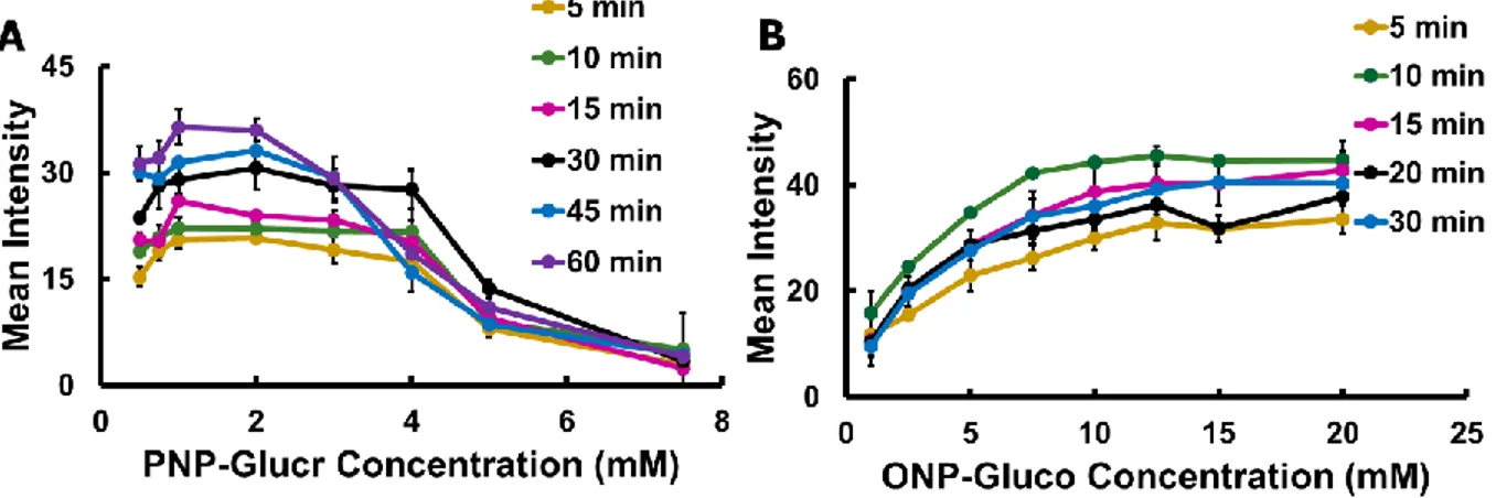
used produced either p-nitrophenol (PNP) or o-nitrophenol (ONP) as colorimetric products (from clear to yellow). Low concentrations (101 CFU mL-1) of pathogenic and nonpathogenic E. coli isolates and (100 CFU mL-1) E. faecalis and E. faecium strains were detected within 4 and 8 h of pre-enrichment.Alfalfa sprout and lagoon water
samples served as model food and water samples, and while water samples did not test positive, sprout samples did test positive within 4 h of pre-enrichment. Positive detection
27
of inoculated (2.3 × 10² and 3.1 × 101 CFU mL-1 or g-1 of E. coli and E. faecium,
respectively) sprout and water samples tested positive within 4 and 12 h, respectively. The second set of devices presents another colorimetric paper-based device that was combined with immunomagnetic separation (IMS) for detecting Salmonella
typhimurium. IMS was completed with anti-Salmonella coated magnetic beads that were
applied to capture and separate bacteria from the sample matrix and preconcentrate it into small volumes before testing on paper. To directly detect S. typhimurium after IMS, a sandwich immunoassay was used in the procedure with β-gal as the detection
enzyme. Using the antibody/enzyme complex, we performed a colorimetric assay with chlorophenol red-β-D-galactopyranoside (CPRG) for bacteria quantification, which has a noticeable color change from yellow to red.Using this system, the limit of detection of S.
typhimurium was found to be 102 CFU mL-1 in culturing solution without any
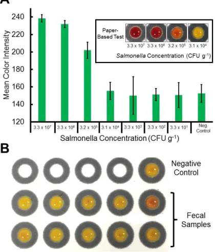
pre-enrichment or cross-reaction with other common bacteria species. Finally, the proposed platform was applied for detection of S. typhimurium in inoculated bird fecal samples and whole milk with detection limits of 105 CFU g-1 and 103 CFU mL-1, respectively, without any cultural enumeration.
Because each set of devices has their advantages and disadvantages, this chapter will conclude with comparing the two different methods and their applications. This chapter is a compilation of my personal contribution to two different projects which were both accepted for publication in Analytical Chemistry.1,2
Introduction
Of all contaminants found in food and water (bacterial, viral, chemical, etc.), bacterial contamination causes the highest number of hospitalizations and deaths within
28
the United States annually.3,4 Whereas drinking polluted water can lead to illness, the use of unsafe water for irrigation can also contaminate agricultural products causing foodborne illness.5,6 Leafy greens, for example, are responsible for 46% of foodborne outbreaks within the United States and, because alfalfa sprouts are cultivated in a moist humid growth environment that facilitates bacterial growth, they are one of the leading sources of multi-state foodborne outbreaks.7,8 Human and animal excreta (primarily feces) are major sources of food and waterborne diseases, but it is impossible to test for all possible transferable pathogens in a comprehensive manner.9 Instead, general indicators for bacterial contamination are commonly detected, and both E. coli and
Enterococcus spp. are used as standard fecal indicator bacteria (FIB).10-13 E. coli and enterococci are found in high concentrations, 109 and >104 colony forming units (CFU) per wet gram of stool respectively, predominantly in the gut of warm-blooded animals. Their presence is an indication of not only fecal contamination but also if conditions are amenable for the presence of other pathogens.13,14 FDA guidance and compliance regulations for both the agricultural production and industrial processing of food and beverages now call for the frequent testing of FIB species, necessitating portable, inexpensive, and user-friendly methods of testing.15
Of these other foodborne pathogens that can be present as indicated by FIB,
Salmonella is widely known as one of the most prevalent pathogens causing foodborne
illness outbreaks.16 Per the Center for Disease Control and Prevention (CDC),
Salmonella causes an estimated one million illnesses in the United States resulting in
19,000 hospitalizations and 380 deaths, more than any other pathogenic bacteria.17
29
milk18 also through animal fecal contamination.19 This is because, like E. coli and
enterococci, Salmonella live and replicate in the intestinal tracts of humans and animals, and therefore present in their feces.20 Studies have shown a strong correlation between skin and meat contamination of Salmonella, and prevalence in the animal’s feces, making feces an important sample matrix to detect the pathogen’s presence.21
Conventional methods are not practical for on-site detection of bacteria and the need for expensive equipment and trained lab personnel increases testing costs, making large scale studies of Salmonella and other pathogen epidemiology difficult.22
Due to the harmful role bacterial infections can play in human health, numerous bacteria detection methods have been developed. Common methods for bacterial detection include immunoassays, DNA amplification/detection methods such as
polymerase chain reaction (PCR), and traditional culture methods.23,24 While DNA and immunoassays have advantages such as selectivity and sensitivity, both can suffer from inhibition effects from sample components that lead to false positives or negatives as well as high instrumentation and/or test costs.25,26 As a result, the gold standard for bacterial detection has remained culture-based methods.27 Culturing microorganisms allows for sensitive isolation and confirmation of live target bacteria. Non-selective and selective media are used sequentially in conjunction with biochemical testing and microscopy, making this method time-consuming and material intensive. Accordingly, a rapid, user-friendly, cost-effective, and reliable approach for FIB and Salmonella
detection is required to overcome the drawbacks of conventional methods. The need for improved bacteria detection methods has led to the development of biosensors and analytical methods, including the use of paper-based analytical devices (PADs). PADs
30
provide a simple, easily modifiable and mass produced alternative platform, and can be incorporated with several different detection motifs.28,29
Other advantages of PADs include small sample and reagent consumption, rapid analysis, simple operation, disposability, and portability.30,31 PADs hold great promise for use as analytical tools in remote areas or areas where minimal instrumentation is available due to their natural fluid wicking properties and ability to store and stabilize reagents. This renders PADs to be attractive and simple platforms for analysis in fields such as environmental monitoring, medical diagnostics, point-of-care testing, and food safety control.28,30 However, there have been only a few reports on using PADs for rapid detection of bacteria, including Pseudomonas aeruginosa,32 Staphylococcus aureus,32,33
Escherichia coli,34-38 Salmonella typhimurium,35,39 and Listeria monocytogenes.35 Our lab has previously demonstrated bacteria detection in food and water samples based on bacterial enzyme expression.40,41 While detecting bacteria based on naturally expressed enzymes is a reliable method to detect viable bacteria, it does suffer from low limits-of-detection, necessitating a culture enrichment step. Although the entire process was still shorter than traditional methods, the ideal bacteria detection system will not necessitate culture enrichment.
Immunomagnetic separation (IMS) is an analytical method that was developed to separate targets of interest from complex sample matrices, and can also be used as an alternative to culture enumeration as a pre-enrichment step for pathogens. IMS is a procedure where antibodies specific to an analyte or cell are covalently attached to magnetic particles. These magnetic particles are added to the sample matrix to adhere to the target and are separated from the matrix with a magnet and re-suspended in
31
buffer. After separation from the sample matrix, many detection methods have been used including microscopy, broth enrichment, immunoassays, and PCR.42 IMS does not require bulky and expensive equipment to complete the procedure, making it ideal for in-field measurements. IMS has been demonstrated for efficiently separating target analytes and cells from complex mixtures such as blood,43 milk,44 meat,45 cheese and yogurt,46 and even bovine feces.47 With IMS, the antibodies attached to the beads can be specific to any analyte or cell of interest. Because of this, IMS has been
demonstrated for detecting many biomarkers,43,48 along with various bacteria45-47 and viruses.49-51 Combining IMS with paper-based devices has been previously described for the detection of E. coli in contaminated water.34 In this work, the authors describe the use of IMS to pre-concentrate samples from contaminated water before lysing the
bacteria and detecting bacterial enzymes β-galactosidase and β-glucuronidase. To the best of our knowledge, there has not been a paper-based device that is coupled with IMS for the detection of bacteria in more complicated sample matrices, such as animal feces and whole milk. Furthermore, despite the prevalence of Salmonella in bird feces, to the best of our knowledge, there has not been a proposed alternative detection method to traditional culture enrichment.
Herein, two different colorimetric paper-based spot tests for bacteria detection are reported. The first is to detect FIB bacteria, E. coli and Enterococcus spp., via their production of species-indicative enzymes. Both β-galactosidase and β-glucuronidase were used for E. coli detection and β-glucosidase for Enterococcus spp. detection. Due to their association with coliforms and FIB, these enzymatic reactions are also used as indicators of microbial safety.52 Substrates for each enzyme produced either ONP or
32
PNP, initiating a color change from clear to yellow. PNP and ONP can also be detected electrochemically (Figure 2.1), but this will not be discussed in depth. Pathogenic and non-pathogenic strains of E. coli, as well as Enterococcus faecalis and E. faecium were detected in pure culture as well as model surface irrigation water (uninoculated and inoculated lagoon water) and model food samples (uninoculated and inoculated alfalfa sprouts).
Figure 2.1 │ Reaction scheme showing the dual electrochemical and colorimetric
detection of formed PNP from reacting with bacterially produced β-glucr with PNP-glucr.
To improve the limit-of-detection and eliminate culturing that was used in the first set of devices, another method was developed by coupling PADs with IMS for specific colorimetric detection of S. typhimurium. IMS was applied to capture and separate target bacteria from the sample matrix, then preconcentrated into small volumes for further assays. By separating the pathogen from feces, this allowed us to complete a sandwich immunoassay to detect the presence of S. typhimurium in the sample (Figure
33
2.2A) without the concern of inhibition effects from the sample matrix. A second
anti-Salmonella antibody was conjugated with biotin, which was bound to streptavidin linked
to β-galactosidase (β-gal) to perform a colorimetric assay with chlorophenol red-β-D-galactopyranoside (CPRG) (Figure 2.2B). The PAD coupled with IMS demonstrated sensitive detection of S. typhimurium in media, and was also demonstrated in detecting
S. typhimurium in inoculated bird feces samples. To show the PAD’s promise for onsite
detection of contaminated food products, this method was also demonstrated in detecting S. typhimurium in inoculated whole milk.
Figure 2.2 │ System for detecting S. typhimurium. (A) Schematic of selected approach
for S. typhimurium. (B) S. typhimurium detection based on an enzymatic assay between β-gal and CPRG, resulting in chlorophenol red as a red-violet product.