This is the published version of a paper published in BMC Cardiovascular Disorders.
Citation for the original published paper (version of record):
Ahmeti, A., Henein, M Y., Ibrahimi, P., Elezi, S., Haliti, E. et al. (2017)
Quality of life questionnaire predicts poor exercise capacity only in HFpEF and not in HFrEF.
BMC Cardiovascular Disorders, 17: 268
https://doi.org/10.1186/s12872-017-0705-0
Access to the published version may require subscription.
N.B. When citing this work, cite the original published paper.
Permanent link to this version:
R E S E A R C H A R T I C L E
Open Access
Quality of life questionnaire predicts poor
exercise capacity only in HFpEF and not in
HFrEF
Artan Ahmeti
1,2, Michael Y. Henein
3,4, Pranvera Ibrahimi
1,3, Shpend Elezi
2, Edmond Haliti
1,2, Afrim Poniku
1,2,
Arlind Batalli
1,2and Gani Bajraktari
1,2,3*Abstract
Background: The Minnesota Living with Heart Failure Questionnaire (MLHFQ) is the most widely used measure of quality of life (QoL) in HF patients. This prospective study aimed to assess the relationship between QoL and exercise capacity in HF patients.
Methods: The study subjects were 118 consecutive patients with chronic HF (62 ± 10 years, 57 females, in NYHA I-III). Patients answered a MLHFQ questionnaire in the same day of complete clinical, biochemical and echocardiographic assessment. They also underwent a 5 min walk test (6-MWT), in the same day, which grouped them into; Group I:≤ 300 m and Group II: >300 m. In addition, left ventricular (LV) ejection fraction (EF), divided them into: Group A, with preserved EF (HFpEF) and Group B with reduced EF (HFrEF).
Results: The mean MLHFQ total scale score was 48 (±17). The total scale, and the physical and emotional functional MLHFQ scores did not differ between HFpEF and HFpEF. Group I patients were older (p = 0.003), had higher NYHA functional class (p = 0.002), faster baseline heart rate (p = 0.006), higher prevalence of smoking (p = 0.015), higher global, physical and emotional MLHFQ scores (p < 0.001, for all), larger left atrial (LA) diameter (p = 0.001), shorter LV filling time (p = 0.027), higher E/e’ ratio (0.02), shorter isovolumic relaxation time (p = 0.028), lower septal a’ (p = 0.019) and s’ (p = 0.023), compared to Group II.
Independent predictors of 6-MWT distance for the group as a whole were increased MLHFQ total score (p = 0.005), older age (p = 0.035), and diabetes (p = 0.045), in HFpEF were total MLHFQ (p = 0.007) and diabetes (p = 0.045) but in HFrEF were only LA enlargement (p = 0.005) and age (p = 0.013. A total MLHFQ score of 48.5 had a sensitivity of 67% and specificity of 63% (AUC on ROC analysis of 72%) for limited exercise performance in HF patients.
Conclusions: Quality of life, assessment by MLHFQ, is the best correlate of exercise capacity measured by 6-MWT, particularly in HFpEF patients. Despite worse ejection fraction in HFrEF, signs of raised LA pressure independently determine exercise capacity in these patients.
Keywords: Heart failure, The Minnesota Living with Heart Failure Questionnaire, Exercise capacity, 6 min walk test, Echocardiography, Quality of life
* Correspondence:gani.bajraktari@uni-pr.edu;gani.bajraktari@umu.se
1Clinic of Cardiology, University Clinical Centre of Kosova, Rrethi i Spitalit, P.N, 10000 Prishtina, Kosovo
2Medical Faculty, University of Prishtina, Prishtina, Kosovo Full list of author information is available at the end of the article
© The Author(s). 2017 Open Access This article is distributed under the terms of the Creative Commons Attribution 4.0 International License (http://creativecommons.org/licenses/by/4.0/), which permits unrestricted use, distribution, and reproduction in any medium, provided you give appropriate credit to the original author(s) and the source, provide a link to the Creative Commons license, and indicate if changes were made. The Creative Commons Public Domain Dedication waiver (http://creativecommons.org/publicdomain/zero/1.0/) applies to the data made available in this article, unless otherwise stated.
Ahmeti et al. BMC Cardiovascular Disorders (2017) 17:268 DOI 10.1186/s12872-017-0705-0
Background
Heart failure (HF) represents end stage heart disease ir-respective of the underlying etiology, and is acknowl-edged as a major cause of mortality and morbidity [1, 2]. Exercise intolerance and impaired quality of life (QoL) reflect poor prognosis in HF patients, and are considered the hallmark of disease severity, irrespective of left ven-tricular (LV) ejection fraction (EF) [3–7]. Treatment of HF aims at improving the clinical status, the functional capacity and QoL, as well as reducing mortality and hos-pitalizations [8]. Recently, QoL improvement has been shown as one of the most important treatment goals in HF, particularly with the documented increase in life pectancy [9, 10]. Moreover, bearing in mind the ex-pected short life expectancy in these patients, QoL seems to be an important objective that needs to always be addressed [11–13].
The QoL in HF is commonly assessed by the Minne-sota Living with Heart Failure questionnaire (MLHFQ) [14] and the Quality of Life with Heart Failure question-naire (QLHF) [15, 16]. We have translated these two questionnaires into Albanian language and used them in Kosovo Heart Failure Patients [17]. The relationship be-tween QoL and other demographic parameters proved controversial with some studies showing that older age is associated with lower QoL and others failing to show similar relationships [18–21]. Also, the relationship be-tween QoL and other parameters, such as gender and race, remain controversial [22, 23]. Few studies investi-gated the relationship of QoL with exercise capacity and breathlessness, which showed that QoL correlated with limited exercise and higher NYHA class [24, 25]. But, in those studies the relationship of QoL and LV EF remains not certain [25–27]. Therefore, the aim of this prospect-ive study was to assess the relationship between MLHFQ and exercise capacity in HF patients.
Methods
Study population
We studied 118 consecutive patients with a clinical diag-nosis of congestive HF (age 62 ± 10 years, 57 female) with ischemic or non-ischemic aetiology, who were in New York Heart Association (NYHA) functional class I-III, and were referred to the Clinic of Cardiology, University Clinical Centre of Kosovo, between December 2014 and September 2016. At the time of the study all patients were on full cardiac medications, optimized at least 2 weeks prior to enrollment. Patients with NYHA class IV, those with limited physical activity due to factors other than car-diac symptoms (e.g. arthritis), with more than mild renal or hepatic failure, with chronic obstructive pulmonary dis-ease, with recent acute coronary syndrome, stroke, psy-chological or psychiatric disorders, or those with severe anemia, were excluded from the study. All patients signed
a written informed consent to participate in the study, which was approved by the Ethics Committee of the Med-ical Faculty, University of Prishtina. This study was sup-ported and monitored by Kosovo Society of Cardiology [27], which is trying to implement European Society of Cardiology guidelines and other current diagnostic and therapeutic recommendations.
Data collection
A detailed history and clinical assessment were obtained in all patients. Routine biochemical tests, including hemoglobin, lipid profile, blood glucose level and kidney function, were also performed in all study patients. Esti-mated body mass index (BMI) was calculated from weight and height measurements. Waist and hip measurements were also made and waist/hip ratio was calculated.
Quality of life assessment
The MLHFQ contains 21 questions, whose aim is to determine how HF affects the physical, psychological and socioeconomic conditions of the patients (Additional file 1 Table S1). The questions refer to the signs and symptoms of HF, social relationships, physical and sexual activity, work and emotions [14] and assesses how HF affected the patient’s life during the previous month. The MLHFQ has a scoring range of 0 for no impairment to 105 for max-imum impairment. The questions cover symptoms and signs relevant to HF, physical activity, social interaction, sexual activity, work, and emotions. Three scores were de-termined: an overall score (21 items, 0–105), the physical dimension (8 items, 0–40), and the emotional dimension (5 items, 0–25), with the highest scores reflecting the worse QoL. The scale of answers to each question ranges from 0 (none) to 5 (very much), where 0 represented no limitation and 105 represented maximal limitation.
Echocardiographic examination
A single operator performed all echocardiographic ex-aminations using a Philips Intelligent E-33 system with a multi-frequency transducer, and harmonic imaging as appropriate. Using conventional landmarks and recom-mendations of the American Society of Echocardiog-raphy and European Association of EchocardiogEchocardiog-raphy [28, 29] we obtained all measurements including, inter-ventricular septal (IVS) thickness, posterior wall (PW) thickness, and LV dimensions, LV volumes and EF using the modified Simpson’s method and left ventricular mass (LVM) using Devereux formula [30].
Ventricular long axis motion was also studied using conventional methods previously described [31], from which the following measurements were obtained; total amplitude as the mitral annular plane systolic excursion (MAPSE) and the tricuspid plane systolic excursion (TAPSE), and long axis myocardial velocities in systole
(s’), early (e’) and late (a’) diastole. Mean value of the lat-eral and septal e’ velocities was also calculated. LV dia-stolic function was assessed from spectral Doppler recordings, from which LV early (E wave), late (A wave) diastolic velocities, E/A ratio and E/e’ (mean lateral and septal) ratio were all calculated. Finally, LV isovolumic relaxation time (IVRT) was measured. LV filling pattern was considered ‘restrictive’ when E/A ratio was >2.0, E wave deceleration time < 140 ms and the LA trasverse diameter was >40 mm [33]. LA diameter and volumes were measured, according to the guidelines of the American Society of Echocardiography and European Association of Echocardiography [29], maximal volume (LAV max) at the end systole and LA minimal volume (LAV min) at end diastole. LA total emptying fraction was calculated using the formula [32]:
LA total emptying fraction
¼ LAV max–LAV min=LAV max x 100
Measurements of LV dyssynchrony
Indirect assessment of LV dyssynchrony was obtained by measuring total isovolumic time (t-IVT), Tei Index and LV-RV pre-ejection time delay, a spreviously described [33] using total LV filling time and ejection times. Total isovolumic time (t-IVT) was calculated as 60 - (total ejec-tion time + total filling time) and was expressed in s/min [34]. Tei index was calculated as the ratio between t-IVT and ejection time [35].
Mitral and tricuspid regurgitation severity were assessed by colour and continuous wave Doppler and was graded as mild, moderate, or severe according to the relative jet area to that of the left atrium (LA) in line with the recom-mendations of the American Society of Echocardiography [36]. Retrograde trans-tricuspid pressure drop >35 mmHg was taken as an evidence for pulmonary hypertension [28]. All M-mode and Doppler recordings were made at a fast speed of 100 mm/s with a superimposed ECG (lead II). From the pulmonary artery flow recordings pulmonary artery acceleration time (PAAT) [37]. The LV outflow tract (LVOT) diameter and area were measured [38] in order to calculate the average velocity time integral (VTI) and the stroke volume (SV) [39].
NT-pro BNP measurement
Blood was taken from an antecubital vein in the morn-ing, sober and after staying extended for 20 min. Blood samples were collected into tubes containing potassium ethylenediaminetetraacetic acid (EDTA) (1 g/L plasma) and N-terminal proBNP were calculated with the Cobas Elecsys E411 analyzer (range 5–35,000 pg/mL) using chemiluminescent immunoassay kit (Roche Diagnostics, Grenach -Wyhlen, Germany).
Six minute walk test
Within 24 h of the echocardiographic examination a 6-MWT was performed on a level hallway surface and was administered by a specialized nurse blinded to the re-sults of the echocardiogram. According to the method of Gyatt et al. [40] patients were informed of the purpose and protocol of the 6-MWT, which was conducted in a standardized fashion without interrupting patient’s regu-lar medications [41]. A 15 m flat, obstacle-free corridor was used and patients were instructed to walk as far as they can, turning 180° after they had reached the end of the corridor, during the allocated time of 6 min. Patients walked unaccompanied so as not to influence walking speed. At the end of the 6 min the supervising nurse measured the total distance walked by the patient.
Statistical analysis
Data are presented as mean ± SD or proportions (% of patients). Continuous data was compared with two-tailed unpaired Student’s t test and discrete data with Chi-square test. Correlations were tested with Pearson coefficients. Predictors of 6-MWT distance were identi-fied with univariate analysis and multivariate logistic regression was performed using the step-wise method, a significant difference was defined as P < 0.05 (2-tailed). Patients were divided according to their ability to walk >300 m into good and limited exercise per-formance groups [42], and were compared using unpaired Studentt-test.
Results
The baseline characteristics of the study population are presented in Table 1. All 118 patients completed the MLHFQ. Patients mean age was 62 ± 9.8 years, and 48% were women. The most common comorbidities were hypertension (67%) and diabetes mellitus (27%) and 30% were smokers. Mean 6-MWT distance was 315 ± 115 m, and 47% were in NYHA class II. Table 2 presents base-line echocardiographic variables.
The score of total MLHFQ scale was 48 ± 17, whereas the physical and emotional MLHFQ subscales scores were 24 ± 9 and 9 ± 5, respectively (Table 1). The total physical and emotional MLHFQ subscale scores were not different in patients with HF and preserved EF (HFpEF) compared to those with HF and reduced EF (HFrEF) (Table 2).
Five of 59 (10%) patients with HFpEF had AF, com-pared to 13 of 59 (22%) patients with HFrEF (p = 0.07). LA diameter was significantly larger in AF patients com-pared to non-AF patients in HFrEF patients (p = 0.001), but not in HFpEF (p = 0.123). However, the 6-MWT dis-tance was not significant in both subgroups.
Patients with limited exercise vs. preserved exercise capacity (Tables 3 and 4)
Patients with limited exercise, who walked <300 m during 6-MWT, were older (p = 0.003), had higher NYHA func-tional class (p = 0.002), faster baseline heart rate (p = 0.006), higher prevalence of smoking (p = 0.015), and higher global, physical and emotional MLHFQ scores (p < 0.001, for all), compared to those with good exercise capacity. Patients with limited exercise also had larger LA
diameter (p = 0.001), shorter LV FT (p = 0.027), smaller septal MAPSE (p = 0.037), higher E/e’ ratio (0.020), shorter IVRT (p = 0.028), PAAT (p = 0.005), lower septal a’ (p = 0.019) and s’ (p = 0.023), compared to those with preserved exercise capacity. All other clinical and echocar-diographic parameters were not significantly different between two groups.
Relationship of total MLHFQ with clinical, biochemical and echocardiographic variables (Table 5)
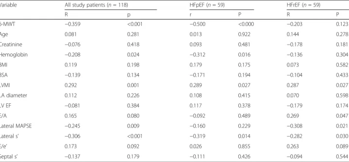
In the patients’ group as a whole, total MLHFQ score had strong correlation with 6-MWT distance, lateral s’ (p < 0.001 for both), good correlation with LVMI (p = 0.001) and with lateral MAPSE (p = 0.009), and weak correlation with hemoglobin level (p = 0.024). In HFpEF, total MLHFQ score had strong correlation with 6-MWT distance (p < 0.001, Fig. 1), and weak correl-ation with lateral s’ (p = 0.014), LVMI (p = 0.027) and with hemoglobin level (p = 0.016), whereas in HFrEF pa-tients it has only a weak correlation with lateral s’ (p = 0.03), LVMI (p = 0.027), lateral MAPSE (p = 0.027) and with E/A ratio (p = 0.047).
Table 1 Baseline characteristics of the study patients
Variable Means ± SD Age (years) 62 ± 9.8 BMI (kg/m2) 29 ± 3.8 Waist/hip ratio 0.96 ± 0.1 HR (beat/min) 83 ± 19 Diabetes mellitus (%) 27 Arterial hypertension (%) 67 Smoking (%) 30 LBBB (%) 14
NYHA class I, II, III (%) 30, 47, 23
Sinus rhythm (%) 80 B-blockers (%) 78 ACEi (%) 81 Diuretic (%) 76 Ca-blockers (%) 12 Aspirin (%) 77 Oral anticoagulants (%) 22 6MWT(m) 315 ± 115 Hemoglobin (g/dL) 12.6 ± 1.7 Creatinine (μmol/l) 96 ± 45 NT-ProBNP (pg/mL) 3630 ± 3742 MLHFQ - total score 48 ± 17 MLHFQ - physical score 24 ± 9 MLHFQ - emotional score 9 ± 5
BMI body mass index, BUN blood urea nitrogen, SBP systolic blood pressure, DBP diastolic blood pressure, HR heart rate, NYHA New York heart association, ACEi angiotensin converting enzyme inhibitors, Ca-blockers Calcium channel blockers, NT-ProBNP N-terminal pro b-type natriuretic peptide, LBBB left bundle branch block, WBC white blood cell, 6MWT 6 min walking test, MLHFQ minnesota living with heart failure questionnaire
Table 2 Comparison of quality of life between patients HFpEF and HFrEF
Variable HFpEF HFrEF P value
(n = 59) (n = 59)
MLHFQ total 47 ± 18 50 ± 16 0.328
MFHFQ physical 23 ± 9 26 ± 8 0.066
MFHFQ emotional 9 ± 5 9 ± 4 0.521
MLHFQ minnesota living with heart failure questioners
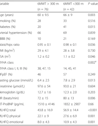
Table 3 Comparison of clinical and biochemical data between patients with limited exercise vs. preserved exercise capacity (6-min walk distance)
Variable 6MWT > 300 m 6MWT <300 m P value (n = 76) (n = 42) Age (years) 60 ± 9.5 66 ± 9 0.003 Smoking (%) 28 33 0.516 Diabetes (%) 20 41 0.015 Arterial hypertension (%) 66 69 0.839 LBBB (%) 10 21 0.169 Waist/hips ratio 0.95 ± 0.1 0.98 ± 0.1 0.036 BMI (kg/m2) 29 ± 4.1 28 ± 3.8 0.730 BSA (m2) 1.2 ± 0.2 1.1 ± 0.2 0.046 NYHA class 0.002a
NYHA class I, II, III (%) 38, 47, 15 14, 45, 41
HFpEF (%) 46 57 0.249
Fasting glucose (mmol/L) 6.4 ± 2.5 7.8 ± 2.9 0.013 Creatinine (μmol/L) 97.6 ± 54 93.0 ± 21 0.604 Hemoglobin (g/dL) 12.7 ± 1.6 12.3 ± 2.0 0.203 HR (beats/min) 72 ± 15 80 ± 13 0.006 NT-ProBNP (pg/mL 1510 ± 4146 1832 ± 2907 0.66 MLHFQ total 43.8 ± 16.9 56.9 ± 14.4 <0.001 MLHFQ physical 22.1 ± 9 27.6 ± 6.9 0.001 MLHFQ emotional 8.0 ± 4.3 10.9 ± 4.3 0.001 BMI body mass index, BSA body surface area, HR heart rate, NYHA New York heart association, LBBB left bundle branch block, NT-ProBNP N-terminal pro b-type natriuretic peptide, HFpEF heart failure with a preserved ejection fraction, MLHFQ minnesota living with heart failure questionnaire
a
Predictors of limited 6-MWT distance in HF patients (Table 6)
Predictors of limited 6 MWT distance in all HF patients
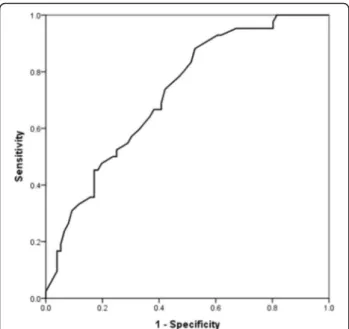
In univariate analysis, total MLHFQ (p < 0.001), physical MLHFQ (p = 0.002), emotional MLHFQ (p = 0.002), age (p = 0.005), diabetes (p = 0.017), atrial fibrillation (p = 0.006), LA diameter (p = 0.001), IVRT (p = 0.047), PAAT (p = 0.008), septal MAPSE (p = 0.04), E/e’ (p = 0.029), septal a’ (p = 0.033), and septal s’ (p = 0.041), predicted limited 6 MWT distance. In multivariate analysis, only total MLHFQ score (p = 0.005), age (p = 0.035) and the diabetes (p = 0.045) remained independent predictors of limited 6-MWT distance. A total MLHFQ score of 48.5 had a sensitiv-ity of 67% and specificsensitiv-ity of 63% (AUC on ROC analysis of 72%) for predicting limited exercise performance (Fig. 2).
Predictors of limited 6-MWT distance in HFpEF patients
Univariate analysis identified total MLHFQ (p = 0.001), physical MLHFQ (p = 0.026), emotional MLHFQ
(p = 0.007), BSA (p = 0.009), diabetes (p = 0.036), and NYHA class >1 (p = 0.012), hemoglobin level (p = 0.039), increased LVMI (p = 0.023), low lateral s’ (p = 0.013) and a’ (p = 0.032) as predictors of limited 6-MWT distance. In multivariate analysis, total MLHFQ (p = 0.007) and diabetes (p = 0.045) independently predicted the limited 6-MWT distance.
Predictors of limited 6 MWT distance in HFrEF patients
In univariate analysis, physical MLHFQ (p = 0.044), age (p = 0.015), NYHA class >1 (p = 0.036), LV mass (p = 0.036) and LA diameter (p = 0.008), predicted the 6-MWT limited exercise distance. In multivariate analysis, only LA enlarge-ment (p = 0.005) and age (p = 0.013) remained independent predictors of limited 6-MWT distance.
Discussion
Findings
The results of this study analysis can be summarized as follows: 1) the total scale, physical and emotional MLHFQ subscale scores were not different between HFpEF and HFrEF patients. 2) Patients with limited ex-ercise capacity were older, had higher NYHA functional class, faster baseline heart rate, higher prevalence of smoking and higher global, physical and emotional MLHFQ scores, compared to those with good exercise capacity. 3) Patients with limited exercise capacity, also had larger LA, shorter LV FT, worse longitudinal systolic function and raised LV filling pressures, compared to those with preserved exercise capacity. 4) Total MLHFQ score had strong correlation with 6-MWT distance in the patients group as a whole and in HFpEF subgroup, but not in HFrEF. 5) Total MLHFQ score, age and diabetes were the only independent predictors of limited 6-MWT distance in the whole group of patients and in HFpEF sub-group. It was LA enlargement and age which independ-ently predicted limited exercise capacity in HFrEF.
Results interpretation
MLHFQ irrespective of its components; physical or emo-tional seems to be a good measure of exercise capacity, since it correlated strongly with the 6-MWT distance in the HF group irrespective of EF. Thus, it could be used to reflect the overall cardiac status, when used to evaluate patients’ response to treatment. It however, does not re-flect the underlying cardiac structural or functional distur-bances, which contribute to the limited exercise capacity in individual patients, and which might need different treatments. Age seemed to be correlating with limited ex-ercise capacity but nothing can be done about it. On the other hand baseline heart rate proved to be an equally im-portant factor but can be managed by beta blockers [43] or other forms of heart rate controlling medications e.g. Ivabridine [44], or the combination of the two [45].
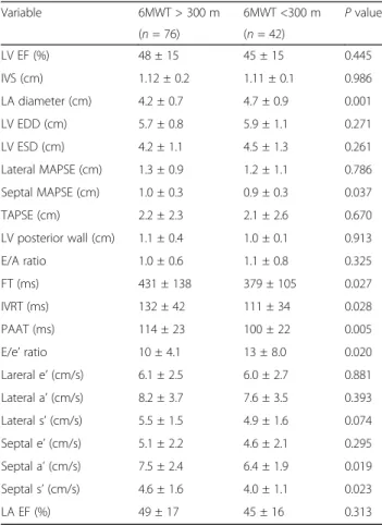
Table 4 Comparison of echocardiographic data between patients with limited exercise vs. preserved exercise capacity (6-min walk distance)
Variable 6MWT > 300 m 6MWT <300 m P value (n = 76) (n = 42) LV EF (%) 48 ± 15 45 ± 15 0.445 IVS (cm) 1.12 ± 0.2 1.11 ± 0.1 0.986 LA diameter (cm) 4.2 ± 0.7 4.7 ± 0.9 0.001 LV EDD (cm) 5.7 ± 0.8 5.9 ± 1.1 0.271 LV ESD (cm) 4.2 ± 1.1 4.5 ± 1.3 0.261 Lateral MAPSE (cm) 1.3 ± 0.9 1.2 ± 1.1 0.786 Septal MAPSE (cm) 1.0 ± 0.3 0.9 ± 0.3 0.037 TAPSE (cm) 2.2 ± 2.3 2.1 ± 2.6 0.670 LV posterior wall (cm) 1.1 ± 0.4 1.0 ± 0.1 0.913 E/A ratio 1.0 ± 0.6 1.1 ± 0.8 0.325 FT (ms) 431 ± 138 379 ± 105 0.027 IVRT (ms) 132 ± 42 111 ± 34 0.028 PAAT (ms) 114 ± 23 100 ± 22 0.005 E/e’ ratio 10 ± 4.1 13 ± 8.0 0.020 Lareral e’ (cm/s) 6.1 ± 2.5 6.0 ± 2.7 0.881 Lateral a’ (cm/s) 8.2 ± 3.7 7.6 ± 3.5 0.393 Lateral s’ (cm/s) 5.5 ± 1.5 4.9 ± 1.6 0.074 Septal e’ (cm/s) 5.1 ± 2.2 4.6 ± 2.1 0.295 Septal a’ (cm/s) 7.5 ± 2.4 6.4 ± 1.9 0.019 Septal s’ (cm/s) 4.6 ± 1.6 4.0 ± 1.1 0.023 LA EF (%) 49 ± 17 45 ± 16 0.313
LV left ventricle, EDD end-diastolic dimension, ESD end-systolic dimension, FT filling time, ET Ejection time, IVS interventricular septum, IVRT isovolemic relaxation time, e’ early diastolic myocardial velocity, s’ systolic myocardial velocity, LA left atrium, LA EF Left atrial emptying fraction, A atrial diastolic velocity, E early diastolic filling velocity, PAAT pulmonary artery acceleration time, MAPSE mitral annular plane systolic excursion, TAPSE tricuspid annular plane systolic excursion
Table 5 Correlation of MLHFQ total score in HF patients with clinical, biochemical and echocardiographic variables in study patients Variable All study patients (n = 118) HFpEF (n = 59) HFrEF (n = 59)
R p r P R P 6-MWT −0.359 <0.001 −0.500 <0.000 −0.203 0.123 Age 0.081 0.281 0.013 0.922 0.144 0.278 Creatinine −0.076 0.418 0.093 0.481 −0.178 0.181 Hemoglobin −0.208 0.024 −0.312 0.016 −0.136 0.304 BMI 0.119 0.198 0.179 0.175 0.073 0.582 BSA −0.139 0.134 −0.171 0.194 −0.104 0.433 LVMI 0.292 0.001 0.289 0.027 0.287 0.027 LA diameter 0.112 0.226 0.108 0.415 0.070 0.598 LV EF −0.081 0.384 0.117 0.378 −0.179 0.174 E/A 0.165 0.080 −0.092 0.489 0.269 0.047 Lateral MAPSE −0.245 0.009 −0.160 0.229 −0.308 0.021 Lateral s’ −0.306 <0.001 −0.319 0.014 −0.282 0.030 E/e’ 0.173 0.092 0.026 0.855 0.263 0.089 Septal s’ −0.137 0.179 −0.111 0.426 −0.094 0.544
MLHFQ minnesota living with heart failure questionnaire, 6-MWT 6 min walking test, BMI body mass index, BSA body surface area, EDD end-diastolic dimension, MAPSE mitral annular plane systolic excursion, A atrial diastolic velocity, E early diastolic filling, velocity, e’ early diastolic myocardial velocity, s’ systolic myocardial velocity, LVMI left ventricular mass index
Furthermore, patients with limited exercise capacity proved to have dilated LA [46, 47], the underlying patho-physiology of which is known to be complicated. It proved to be related to the high filling pressures in some [48] and poor LA emptying, as shown be short LV filling time, in others [49]. In addition to the variety of mechanisms of disturbed physiology, the matter is further complicated by the way patients differ in their response to treatment.
While the former group usually responds to LA pressure lowering medications i.e. ACE-inhibitors or A2 blockers [50], the latter respond better to cardiac resynchronization therapy [51]. Finally, it seems that predictors of the limited exercise capacity differed fundamentally according to the cardiac physiology. While specifically the causes of LA en-largement; pressure, mitral regurgitation, stiff LV, etc., that limited patients exercise in HFrEF, the respective reasons
Table 6 Predictors of limited exercise in HF patients
Variable Univariate predictors Multivariate predictors
OR CI 95% P value OR CI 95% P value
Whole HF study patients
MLHFQ - total score 1.053 (1.025–1.081) <0.001 1.080 (1.023–1.140) 0.005 MLHFQ - physical score 1.085 (1.031–1.141) 0.002 MLHFQ - emotional score 1.163 (1.060–1.277) 0.001 Age 1.069 (1.021–1.120) 0.005 1.101 (1.007–1.203) 0.035 Gender 0.576 (0.269–1.232) 0.155 Diabetes mellitus 2.765 (1.199–6.379) 0.017 4.876 (1.037–22.94) 0.045 LA diameter 2.500 (1.467–4.260) 0.001 FT 0.996 (0.993–1.000) 0.046 IVRT 0.984 (0.969–1.000) 0.047 PAAT 0.972 (0.952–0.993) 0.008 Septal MAPSE 0.235 (0.059–0.939) 0.040 E/e’ 1.090 (1.009–1.177) 0.029 Septal a’ 0.787 (0.632–0.981) 0.033 Septal s’ 0.663 (0.447–0.983) 0.041 HFpEF patients MLHFQ - total score 1.080 (1.032–1.131) <0.001 1.137 (1.036–1.249) 0.007 MLHFQ - physical score 1.088 (1.010–1.173) 0.026 MLHFQ - emotional score 1.213 (1.055–1.396) 0.007 Diabetes mellitus 3.556 (1.089–11.61) 0.036 26.88 (1.791–400.8) 0.017 Hemoglobin 0.711 (0.514–0.983) 0.039 NYHA class 3.038 (1.271–7.262) 0.012 BSA 0.003 (0.000–0.241) 0.009 LVMI 10.50 (1.007–1.096) 0.023 Lateral a’ 0.768 (0.604–0.978) 0.032 Lateral s’ 0.468 (0.258–0.850) 0.013 HFrEF patients MLHFQ - physical score 1.076 (1.002–1.156) 0.044 Age 1.071 (1.013–1.132) 0.015 1.113 (1.024–1.209) 0.012 NYHA class 2.501 (1.064–5.881) 0.036 LVM 1.010 (1.001–1.019) 0.036 1.015 (1.000–1.030) 0.047 LA diameter 3.183 (1.356–7.475) 0.008 7.401 (1.821–30.08) 0.005 LV EDD 1.070 (1.002–1.143) 0.044
MLHFQ Minnesota living with heart failure questionnaire, NYHA New York Heart Association, BSA body surface area, LV left ventricle, LVM left ventricular mass, LA left atrium, EDD end-diastolic dimension, FT filling time, IVRT isovolemic relaxation time, MAPSE mitral annular plane systolic excursion, E early diastolic filling, velocity, e’ early diastolic myocardial velocity, s’ systolic myocardial velocity, PAAT pulmonary artery acceleration time, LVMI left ventricular mass index, HFrEF heart failure with reduced ejection fraction
were multifactorial including age, diabetes, as well as emo-tional and physical scores that predicted exercise capacity in HFpEF. The latter finding adheres to what is known about HFpEF in terms of its etiology, comorbidities as well as limited benefit when using conventional guidelines-based treatment recommendations [52]. The lack of an ac-ceptable relationship between LA volume and exercise capacity in HFpEF could be explained by either strict early treatment with vasodilators which reduced cavity pressure and hence volume or less myocardial stiffness compared with HFrEF. Also, despite higher AF prevalence in HFrEF patients compared to HFpEF, our analysis suggest that AF was not necessarily a determinant factor for the difference in relationship between left atrial enlargement and 6-MWT. It seems therefore that more than one factor could contribute to the lack of direct relationship between the LA volume and exercise capacity in HFpEF. It was how-ever not feasible to run a number of permutations and combinations in order to identify the additive value of various individual variables in predicting exercise capacity.
Clinical implications
Our findings suggest that the MLHFQ correlates with 6MWT distance in heart failure patients as a whole and is able, fairly accurately, to predict those with limited ex-ercise capacity. These findings apply better to patients with HFpEF much more than those with HFrEF in whom clearly signs of raised LA pressures are those which independently determine their limited exercise capacity. These differences support the need for continu-ing the use of detailed Doppler echocardiographic follow up of heart failure patients in order to better understand
the pattern of disturbances that explain symptoms as well as the most accurate treatment option.
Limitations
Obvious limitations can easily be seen in this study. The small number of patients included in this study limits general application of the findings before results are revalidated in a larger cohort. We consider that further prospective cohort studies with a larger sample size, are undoubtedly needed to strengthen or refute our findings. Speckle tracking ultrasonography to measure the global longitudinal strain, which might be associated with re-duced functional capacity in HF patients was not used. However, assessing longitudinal LV function with con-ventional tools, provided an estimate of other overall longitudinal LV function. We cannot ignore the emo-tional element in conducting the 6-MWT and patient encouragement to walk faster, although unassisted. We did not assess the reproducibility of the results of the MLHFQ neither the 6-MWT distance, which could have shown significant differences.
Conclusion
Although the conventionally used MLHFQ, irrespective of its components, correlates closely with the 6-MWT dis-tance in HF patients particularly HFpEF. Raised filling pressures seem to be the strongest independent predictor of limited exercise capacity in HFrEF. These differences might impact treatment options in the two conditions.
Additional file
Additional file 1: Table S1. The Minnesota Living with Heart Failure Questionnaire. (DOC 29 kb)
Abbreviations
6-MWT:Minute walk test; A: Atrial diastolic velovity; a’: Atrial myocardial velocity; BMI: Body mass index; BSA: Body surface area; DT: Deceleration time; E: Early diastolic velocity; e’: Early diastolic myocardial velocity; EF: Ejection fraction; FT: Filling time; HF: Heart failure; HFpEF: Heart failure with preserved ejection fraction; HFrEF: Heart failure with reduced ejection fraction; LA EF: Left atrial emptying fraction; LA: Left atrium; LAV max: Left atrial maximal volume; LAV min: Left atrial minimal volume; LV: Left ventricle; LVM: Left ventricular mass; LVMI: Left ventricular mass index; LVPWd: Left ventricular posterior wall in diastole; MAPSE: Mitral annular plane systolic excursion; MLHFQ: The Minnesota Living with Heart Failure Questionnaire; NYHA: New York Heart Association; PA: Pulmonary artery; QoL: Quality of life; RV: Right ventricle; s’: Systolic myocardial velocity; TAPSE: Tricuspidal annular plane systolic excursion
Acknowledgements Not applicable. Funding Not applicable.
Availability of data and materials
The datasets used and analyzed during the current study are available from the corresponding author on reasonable request.
Fig. 2 ROC-curve of MLHFQ - total score in predicting poor exercise performance on 6-min walk test in patients with heart failure
Authors’ contributions
GB, SE and MH designed the manuscript; Data acquisition was made by AB, AP and EH; AA, PI and GB analyzed and interpreted the data; AB, AA AP and EH drafted the manuscript; MH, PI, GB and SE revised critically. All authors read, approved the manuscript for publication and agreed to be authors of the manuscript.
Ethics approval and consent to participate
All patients signed a written informed consent to participate in the study, which was approved by the Ethics Committee of the Medical Faculty, University of Prishtina (Statement Nr.3729, date 22.10.2010). This study was supported and monitored by Kosovo Society of Cardiology [27], which is trying to implement European Society of Cardiology guidelines and other current diagnostic and therapeutic recommendations.
Consent for publication Not applicable. Competing interests
The authors declare that they have no competing interests.
Publisher’s Note
Springer Nature remains neutral with regard to jurisdictional claims in published maps and institutional affiliations.
Author details
1
Clinic of Cardiology, University Clinical Centre of Kosova, Rrethi i Spitalit, P.N, 10000 Prishtina, Kosovo.2Medical Faculty, University of Prishtina, Prishtina, Kosovo.3Department of Public Health and Clinical Medicine, Umeå University and Heart Centre, Umeå, Sweden.4Molecular & Clinical Sciences Research Institute, St George University London, London, UK.
Received: 21 August 2017 Accepted: 13 October 2017
References
1. Jessup M, Marwick TH, Ponikowski P, Voors AA, Yancy CW. 2016 ESC and ACC/AHA/HFSA heart failure guideline update - what is new and why is it important? Nat Rev Cardiol. 2016;13(10):623–8.
2. Working Group on Cardiac Rehabilitation & Excercise Physiology and Working Group on Heart Failure of the European Society of Cardiology. Recommendations for exercise testing in chronic heart failure patients. Eur Heart J. 2001;22(1):37–45.
3. Wilson JR, Hanamanthu S, Chomsky DB, Davis SF. Relationship between exertional symptoms and functional capacity in patients with heart failure. J Am Coll Cardiol. 1999;33(7):1943–7.
4. Bittner V, Weiner DH, Yusuf S, Rogers WJ, McIntyre KM, Bangdiwala SI, et al. Prediction of mortality and morbidity with a 6-min walk test in patients with left ventricular dysfunction. SOLVD investigators. JAMA. 1993;270(14):1702–7.
5. Alahdab MT, Mansour IN, Napan S, Stamos TD. Six minute walk test predicts long-term all-cause mortality and heart failure Rehospitalization in African-American patients hospitalized with acute Decompensated heart failure. J Card Fail. 2009;15(2):130–5.
6. Morcillo C, Aguado O, Delás J, Rosell F. Utilidad del Minnesota Living With Heart Failure Questionnaire en la evaluación de la calidad de vida en enfermos con insuficiencia cardiaca. Rev Esp Cardiol. 2007;60(10):1093–6. 7. Bussoni MF, Guirado GN, Roscani MG, Polegato BF, Matsubara LS, Bazan
SGZ, et al. Diastolic function is associated with quality of life and exercise capacity in stable heart failure patients with reduced ejection fraction. Braz J Med Biol Res Rev Bras Pesqui Médicas E Biológicas Soc Bras Biofísica Al. 2013;46(9):803–8.
8. Ponikowski P, Voors AA, Anker SD, Bueno H, Cleland JGF, Coats AJS, et al. 2016 ESC guidelines for the diagnosis and treatment of acute and chronic heart failure: the task force for the diagnosis and treatment of acute and chronic heart failure of the European Society of Cardiology (ESC) developed with the special contribution of the heart failure association (HFA) of the ESC. Eur Heart J. 2016;37(27):2129–200.
9. Wasywich CA, Gamble GD, Whalley GA, Doughty RN. Understanding changing patterns of survival and hospitalization for heart failure over two
decades in New Zealand: utility of“days alive and out of hospital” from epidemiological data. Eur J Heart Fail. 2010;12(5):462–8.
10. Joffe SW, Webster K, McManus DD, Kiernan MS, Lessard D, Yarzebski J, et al. Improved survival after heart failure: a community-based perspective. J Am Heart Assoc. 2013;2(3):e000053.
11. Stevenson LW. Design of therapy for advanced heart failure. Eur J Heart Fail. 2005;7(3):323–31.
12. Jaarsma T, Beattie JM, Ryder M, Rutten FH, McDonagh T, Mohacsi P, et al. Palliative care in heart failure: a position statement from the palliative care workshop of the heart failure Association of the European Society of cardiology. Eur J Heart Fail. 2009;11(5):433–43.
13. Hoekstra T, Lesman-Leegte I, van Veldhuisen DJ, Sanderman R, Jaarsma T. Quality of life is impaired similarly in heart failure patients with preserved and reduced ejection fraction. Eur J Heart Fail. 2011;13(9):1013–8. 14. Rector TS, Cohn JN. Assessment of patient outcome with the Minnesota
living with heart failure questionnaire: reliability and validity during a randomized, double-blind, placebo-controlled trial of pimobendan. Pimobendan multicenter research group. Am Heart J. 1992;124(4):1017–25. 15. Dobre D, de Jongste MJL, Haaijer-Ruskamp FM, Sanderman R, van
Veldhuisen DJ, Ranchor AV. The enigma of quality of life in patients with heart failure. Int J Cardiol. 2008;125(3):407–9.
16. Guyatt GH. Measurement of health-related quality of life in heart failure. J Am Coll Cardiol. 1993;22(4):A185–91.
17. Ahmeti A, Bajraktari G. Use of the“Minnesota living with heart failure questionnaire” quality of life questionnaire in Kosovo’s heart failure patients. Int Cardiovasc Forum J. 2016;9(1):36–40.
18. Clark DO, Tu W, Weiner M, Murray MD. Correlates of health-related quality of life among lower-income, urban adults with congestive heart failure. Heart Lung J Acute Crit Care. 2003;32(6):391–401.
19. Gott M. Predictors of the quality of life of older people with heart failure recruited from primary care. Age Ageing. 2006;35(2):172–7.
20. Pascale JM, Sosa CE, Orn A. Evaluation of interleukin-2 soluble receptor in patients with trypanosomiasis americana in Panama. Rev Med Panama. 1991;16(3):184–8.
21. Adebayo S, Olunuga T, Durodola A, Ogah O. Quality of life in heart failure: a review. Niger. J Cardiol. 2017;14(1):1.
22. Hoekstra T, Jaarsma T, van Veldhuisen DJ, Hillege HL, Sanderman R, Lesman-Leegte I. Quality of life and survival in patients with heart failure. Eur J Heart Fail. 2013;15(1):94–102.
23. Mbakwem AC, Aina FO, Amadi CE, Akinbode AA, Mokwunyei J. Comparative analysis of the quality of life of heart failure patients in south western Nigeria. World J Cardiovasc Dis. 2013;3(1):146–53.
24. Juenger J, Schellberg D, Kraemer S, Haunstetter A, Zugck C, Herzog W, et al. Health related quality of life in patients with congestive heart failure: comparison with other chronic diseases and relation to functional variables. Heart Br Card Soc. 2002;87(3):235–41.
25. dos SJJA, JEA P, PRS B. Quality of life and clinical indicators in heart failure: a multivariate analysis. Arq Bras Cardiol. 2009;93(2):159–66.
26. Bajraktari G, Batalli A, Poniku A, Ahmeti A, Olloni R, Hyseni V, et al. Left ventricular markers of global dyssynchrony predict limited exercise capacity in heart failure, but not in patients with preserved ejection fraction. Cardiovasc Ultrasound. 2012;10(1):36.
27. Bajraktari G. The Kosovo Society of Cardiology. Int Cardiovasc Forum J [Internet]. 2015 12 [cited 2016 Sep 18];2(1). Available from: http://icfjournal. org/icfj/index.php/icfj/article/view/93.
28. Gardin JM, Adams DB, Douglas PS, Feigenbaum H, Forst DH, Fraser AG, et al. Recommendations for a standardized report for adult transthoracic echocardiography: a report from the American Society of
Echocardiography’s nomenclature and standards committee and task force for a standardized echocardiography report. J Am Soc Echocardiogr Off Publ Am Soc Echocardiogr. 2002;15(3):275–90.
29. Lang RM, Bierig M, Devereux RB, Flachskampf FA, Foster E, Pellikka PA, et al. Recommendations for chamber quantification. Eur J Echocardiogr J Work Group Echocardiogr Eur Soc Cardiol. 2006;7(2):79–108.
30. Devereux RB, Alonso DR, Lutas EM, Gottlieb GJ, Campo E, Sachs I, et al. Echocardiographic assessment of left ventricular hypertrophy: comparison to necropsy findings. Am J Cardiol. 1986;57(6):450–8.
31. Höglund C, Alam M, Thorstrand C. Atrioventricular valve plane displacement in healthy persons. An echocardiographic study. Acta Med Scand. 1988;224(6):557–62. 32. Wakatsuki Y, Funabashi N, Mikami Y, Shiina Y, Kawakubo M, Takahashi M, et
al. Left atrial compensatory function in subjects with early stage primary
hypertension assessed by using left atrial volumetric emptying fraction acquired by transthoracic echocardiography. Int J Cardiol. 2009;136(3):363–7. 33. Henein MY, Das SK, O’Sullivan C, Kakkar VV, Gillbe CE, Gibson DG. Effect of
acute alterations in afterload on left ventricular function in patients with combined coronary artery and peripheral vascular disease. Heart Br Card Soc. 1996;75(2):151–8.
34. Duncan AM, Francis DP, Henein MY, Gibson DG. Importance of left ventricular activation in determining myocardial performance (Tei) index: comparison with total isovolumic time. Int J Cardiol. 2004;95(2–3):211–7. 35. Tei C, Ling LH, Hodge DO, Bailey KR, JK O, Rodeheffer RJ, et al. New index of
combined systolic and diastolic myocardial performance: a simple and reproducible measure of cardiac function–a study in normals and dilated cardiomyopathy. J Cardiol. 1995;26(6):357–66.
36. Zoghbi WA, Enriquez-Sarano M, Foster E, Grayburn PA, Kraft CD, Levine RA, et al. Recommendations for evaluation of the severity of native valvular regurgitation with two-dimensional and Doppler echocardiography. J Am Soc Echocardiogr Off Publ Am Soc Echocardiogr. 2003;16(7):777–802. 37. Olariu A, Wellnhofer E, Gräfe M, Fleck E. Non-invasive estimation of left
ventricular end-diastolic pressure by pulmonary venous flow deceleration time. Eur J Echocardiogr J Work Group Echocardiogr Eur Soc Cardiol. 2003;4(3):162–8. 38. Mowat DH, Haites NE, Rawles JM. Aortic blood velocity measurement in
healthy adults using a simple ultrasound technique. Cardiovasc Res. 1983; 17(2):75–80.
39. Finegold JA, Manisty CH, Cecaro F, Sutaria N, Mayet J, Francis DP. Choosing between velocity-time-integral ratio and peak velocity ratio for calculation of the dimensionless index (or aortic valve area) in serial follow-up of aortic stenosis. Int J Cardiol. 2013;167(4):1524–31.
40. Guyatt GH, Sullivan MJ, Thompson PJ, Fallen EL, Pugsley SO, Taylor DW, et al. The 6-min walk: a new measure of exercise capacity in patients with chronic heart failure. Can Med Assoc J. 1985;132(8):919–23.
41. Guyatt GH, Thompson PJ, Berman LB, Sullivan MJ, Townsend M, Jones NL, et al. How should we measure function in patients with chronic heart and lung disease? J Chronic Dis. 1985;38(6):517–24.
42. Ingle L, Rigby AS, Nabb S, Jones PK, Clark AL, Cleland JGF. Clinical determinants of poor 6-min walk test performance in patients with left ventricular systolic dysfunction and no major structural heart disease. Eur J Heart Fail. 2006;8(3):321–5.
43. Kotecha D, Flather MD, Altman DG, Holmes J, Rosano G, Wikstrand J, et al. Heart rate and rhythm and the benefit of Beta-blockers in patients with heart failure. J Am Coll Cardiol. 2017;69(24):2885–96.
44. Roth S, Fernando C, Azeem S, Moe GWI. There a role for Ivabradine in the contemporary Management of Patients with chronic heart failure in academic and community heart failure clinics in Canada? Adv Ther. 2017; 34(6):1340–8.
45. Hidalgo FJ, Anguita M, Castillo JC, Rodríguez S, Pardo L, Durán E, et al. Effect of early treatment with ivabradine combined with beta-blockers versus beta-blockers alone in patients hospitalised with heart failure and reduced left ventricular ejection fraction (ETHIC-AHF): a randomised study. Int J Cardiol. 2016 Aug 15;217:7–11.
46. Acarturk E, Koc M, Bozkurt A, Unal I. Left atrial size may predict exercise capacity and cardiovascular events in patients with heart failure. Tex Heart Inst J. 2008;35:136–43.
47. Batalli A, Ibrahimi P, Bytyi? I, Ahmeti A, Haliti E, Elezi S, et al. Different determinants of exercise capacity in HFpEF compared to HFrEF. Cardiovasc Ultrasound [Internet]. 2017 Dec [cited 2017 Jun 15];15(1). Available from: http:// cardiovascularultrasound.biomedcentral.com/articles/10.1186/s12947-017-0103-x. 48. Bajraktari G, Dini FL, Fontanive P, Elezi S, Berisha V, Napoli AM, et al.
Independent and incremental prognostic value of Doppler-derived left ventricular total isovolumic time in patients with systolic heart failure. Int J Cardiol. 2011;148(3):271–5.
49. Bytyçi I, Bajraktari G, Ibrahimi P, Berisha G, Rexhepaj N, Henein MY. Left atrial emptying fraction predicts limited exercise performance in heart failure patients. IJC. Heart Vessel. 2014;4:203–7.
50. Henein MY, O’Sullivan CA, Coats AJ, Gibson DG. Angiotensin-converting enzyme (ACE) inhibitors revert abnormal right ventricular filling in patients with restrictive left ventricular disease. J Am Coll Cardiol. 1998;32(5):1187–93.
51. Salukhe TV, Francis DP, Morgan M, Clague JR, Sutton R, Poole-Wilson P, et al. Mechanism of cardiac output gain from cardiac resynchronization therapy in patients with coronary artery disease or idiopathic dilated Cardiomyopathy. Am J Cardiol. 2006;97(9):1358–64.
52. Oktay A, Shah S. Diagnosis and Management of Heart Failure with preserved ejection fraction: 10 key lessons. Curr Cardiol Rev. 2014;11(1):42–52.
• We accept pre-submission inquiries
• Our selector tool helps you to find the most relevant journal • We provide round the clock customer support
• Convenient online submission • Thorough peer review
• Inclusion in PubMed and all major indexing services • Maximum visibility for your research
Submit your manuscript at www.biomedcentral.com/submit