Molecular Biology- Master
DegreeProject (30 ECTS)
Spring/2013
Student;
Omar AL-Bayati. MS.
a11omaab@student.his.se
Superviser;
Dr. Anna Karin Pernestig, PhD.
Examiner;
School of Life Sciences
Högskolan i Skövde
BOX 408 SE-541 28
Skövde, Sweden.
Quick detection of disease causing pathogens
in sepsis by Fluorescence in situ Hybridization
utilizing Peptide Nucleic Acid Probes
Optimizing the Fluorescence In situ
hybridization technique for a more rapid
inspection of Sepsis causative pathogens by
Abstract
Sepsis is a serious clinical condition that is characterized by a systemic inflammatory response syndrome resulting from a known or suspected infection. The major clinical symptoms involve an abnormal WBC count, elevated body temperature, respiration and pulse rate. Reported cases with high mortality rate range between 13 - 20 million. In order to treat Sepsis, the detection of bacteria in blood culture is extremely crucial. Treating patients with broad spectrum antibiotics is usually related to adverse effects, drug resistance, increased mortality, and high cost. In the past decades, researches had detected that E. coli and S. aureus are the major role players that cause sepsis. These microbes are molecularly tested by methods like MALDI TOF, FISH and Microarrays.
In this analysis, DNA fluorescence in situ hybridization (FISH) assessment for the identification of S. aureus, one of the most frequent blood pathogens, was optimized in the labs of Högskolan i Skövde. As a result, the growth of S.
aureus was observed very carefully, optimizing the FISH procedure for gram
positive bacteria was done and the sensitivity, stability and specificity of the DNA probe were examined under variant conditions like the continuous decrease in the bacteria cells number and utilizing a mixture of different types of bacteria cells.
.
Contents
1. Introduction ...…..……….. 1
2. Materials and Methods ……….……….4
2.1. Culturing of S.aureus ……….4
2.2. Growth of bacteria ………. 4
2.3. Fluorescence in situ hybridization ………. 4
2.3.1. Permeabilization ………. 4
3. Results and Discussion ………..………… 6
3.1. Growth of bacteria ……… 6
3.2. Fluorescence in situ hybridization ……… 7
3.2.1. Permeabilization ………. 7
3.2.2. Optimizing of probe binding ……….. 9
3.2.3. Sensitivity ………..10 3.2.4. Specificity ………..11 4. Conclusion ………...………. 14 5. Future prospect ………..……….. 14 6. Acknowledgment …... 14 7. References …... 16 8. Appendices ………...……… 20
Abbreviations
BC Blood culture
CFU/mL Colony forming unit/milliliter DAPI 4',6-diamidino-2-phenylindole
E. coli Escherichia coli
FICH fluorescein filter
FISH Fluorescent in situ hybridization HiS Högskolan I Skövde
KSS Kärnsjukhuset i Skövde LB Luria Broth
MALDI TOF Matrix- assisted laser desorption ionization-
. time of flight mass spectrometry min minute
MOD multiple organ dysfunction O.D Optical Density
PBS Phosphate Buffer Saline PCR Polymerase chain reaction
SIRS Systemic inflammatory response syndrome
S. aureus Staphylococcus aureus S. pneumoniae Streptococcus pneumoniae
1
Introduction
Sepsis is a serious clinical condition that is characterized by a systemic inflammatory response syndrome (SIRS) resulting from a known or suspected infection (Levy et al., 2003). Each year, approximately 13-20 million patients are diagnosed with severe sepsis worldwide (Tissari et al., 2010; Laakso et al., 2011). Almost 135000 people in Europe die every year due to severe sepsis (Tissari et al., 2010). Sepsis is demonstrating a high mortality and a high occurrence rate, among 25% - 50% of the diagnosed patients die every year (Laakso et al., 2011; Rangel-Frausto, 2005). Sepsis represented by acute organ dysfunction, the presence of infection, and inflammatory response (Walter et
al., 2004). Most common clinical symptoms in patients with sepsis may
include white blood cells (WBC) <4000cells/mm³ or>12000 cells/mm³, fever>38°Cor<36°C, respiration rate>20 breaths /min, and pulse >90/min (Nguyen et al., 2006). If the infection and inflammation in an early stage are not treated at the right time, they could lead to septic shock, severe sepsis, and multiple organ dysfunction (MOD) then eventually the death of the patient (Mancini et al., 2010). In magnitude, septic shock and sepsis possess the same severe lethal effect as in other severe life threatening states i.e. stroke, trauma, and acute myocardial infarction (Nguyen et al., 2006). When pathogens invade, the body reacts with a hyper-immune response. The neutrophils and macrophages in the blood release large amounts of Cytokines during intense attacks causing the activation of the underlying coagulation pathway and complement system resulting finally in the damage of the organ (Stearns-Kurosawa, et al., 2011).
Leading microbes that contribute to septicemia and sepsis syndrome belong to
Streptococcus spp., Enterococcus spp., Staphylococcus spp., Candida spp., and Escherichia coli. (Weinstein et al., 1995; Wischnewski et al., 1998).
The identification of the causative agent in a clinical laboratory takes minimally 48 hours, standard laboratory detection of bacteremia is usually done a computed blood culture bottle, and that will take at least one day. In case the blood culture bottle is detected positive, gram staining and subculturing on agar plates is performed to identify the exact species of the
2
bacteria, and that will take one or two days (Kempf et al.,2000; Jansen et al, 2000). In the mean while physicians treat the patients with broad-spectrum antibiotics against pathogenic microorganisms, and that will lead to an additional cost because the patients need to stay longer in the hospital.
According to several studies, there is a relation between the early detection of the causative pathogen, the early treatment with effective antibiotics, and the increased survival of the patients (Laakso et al., 2011; Wolk and Fiorello, 2010). Since different bacteria show different susceptibility against antibiotics, a rapid identification of the bacteria is of high importance (Wellinghausen et
al., 2004). This is important to decrease the risk of antibiotic resistance, which has become a serious problem in recent years (Wellinghausen et al., 2004).
The modern techniques for quicker identification and detection of bacteria in positive blood cultures are real-time PCR, Matrix- assisted laser desorption ionization- time of flight mass spectrometry (MALDI TOF), fluorescence in
situ hybridization (FISH) and Microarrays (Mancini et al., 2010).
FISH is a method that’s rapid, accurate, and simple to operate. It’s used for the direct identification of pathogens from positive blood culture bottles. FISH bypasses nucleic acid amplification step resembles that of PCR, and thus the odds of having contamination are less (Volkhard et al., 2000). DNA probes with fluorescent dyes are utilized In order to identify target sequences, and are supplement to the target DNA sequence. The DNA probes utilized in FISH adopt Watson Crick base pairing (Moter & Goebel et al., 2000). The most frequently target molecule utilized for the identification of bacteria in FISH is 16S rRNA.16S rRNA is a feature of the small subunits of the bacterial ribosome. It’s genetically very stable, and its domain structure involves variable and conserved regions that may be utilized for probes targeting various taxonomic levels of bacteria. In addition to that, 16Sr RNA has a high copy number which makes it an appropriate target (Clarridge, 2004; Möter & Göbel, 2000).
3
This treatise project is a part of a highly ranked Project entitled “Community acquired severe sepsis and septic shock in adults in Skaraborg, Sweden” that’s being fulfilled through the association of Högskolan I Skövde (His) and Unilabs AB Sweden and Skaraborg hospital (SkaS).The treatise projects is a launching point for approving FISH as one of the molecular methods to be employed in Unilabs AB, to attain a more precise and preceding identification of microbes the positive blood culture sample of patients who had sepsis. The major objective of this project is to establish molecular methods in the direction of a quicker and more precise identification of the etiologic agents in human samples that are leading to Sepsis and affirm that the new methods are more financially adaptable considering available methods i.e. The blood culture system and MALDI TOF. The fluorescence in situ hybridization (FISH) method will be utilized to localize and distinguish the absence or presence of S. aureus by utilizing a DNA probe. The optimizing of the FISH technique in gram positive bacteria demands the Permeabilization of the cell wall that is so thick in gram positive bacteria in order to make it possible for the probe to get inside the bacteria and bind to the target DNA. Then examine the particularity of the probe to distinguish only S. aurues in a mixture of various types of bacteria which S. aureus is one of them.
4
Materials and Methods
2.1 Culturing of S. aureus
S. aureus strain CCUG 43764 (Unilabs AB) was used in this study, and
cultured on Blood agar. The plates were then incubated overnight at 37ºC. The
S. aureus worked as a pet in the entire lab experimentation.
2.2 Growth of bacteria
One S. aureus colony strain CCUG 43764 was extracted and transferred into 10ml of LB broth (5g yeast, 10g tryptone, and 10g NaCl per liter), and kept overnight in a shaking incubator at 37ºC and 120 rpm. Next day 1ml from overnight broth culture was inoculated in 100ml LB broth at 37ºC and 120 rpm, every hour 1 ml was taken for a total of 8 hours until the stationary phase of the bacterial growth was reached. Measuring the optical density (OD) at a wavelength of 600nm was done utilizing a WPA CO8000 cell density meter (Saveen & Werner) to determine the growth of the bacteria every hour. When the OD600 reading was >1, dilutions were made to adjust for high cell density. In order to determine the Colony forming unit /milliliter (CFU/ml) and count the colonies, the same samples were diluted using 1x phosphate buffer saline PBS. The samples were similar to those that were used for the measurement of the OD in order to assess the growth curve. Following an overnight incubation, colonies were counted at 37ºC in order to estimate the CFU/ml corresponding to every hour of S. aureus growth.
2.3 Fluorescence in situ hybridization 2.3.1 Permeabilization
The DNA probe Saureus2FISHTX (Table 1) was used for the detection of S.
aureus strain CCUG 43764. The target molecule for the Saureus2FISHTX
probe was 16S rRNA. The 16S rRNA is most commonly used as target molecule for FISH in the identification of bacteria. It is a component of the bacterial ribosome small subunit. It possesses a high genetic stability and its
5
domain structure includes conserved and variable regions that can be used for probes targeting different taxonomic levels of bacteria. Furthermore, 16S rRNA is a suitable target because of its high copy number (Clarridge, 2004; Möter & Göbel, 2000). In each experiment the probe EU338flour (Table 1) was used as a positive control because it can bind to all bacteria.
Table 1. List of probes that used during the experiment.
Probe name Chemistry Mods5' Tm DNA sequence
Saureus2FISHTXa DNA Oligos Tex red via
Amine C6
60 GAAGCAAGCTTCTCGTCC
G
SaureusFISHTX1b DNA Oligos Tex red via
Amine C6
48 GCT TCT CGT CCG TTC
EUB338 flour c DNA Oligos FICH
fluorescein
60 GCTGCCTCCCGTAGGAGT
a
(Trebesius et al., 2000) (BioNordika).
b
(Kempf et al.,2000; Jansen et al, 2000) (BioNordika).
c
(Amann et al., 1990) (BioNordika).
One S. aureus colony was chosen and diluted using 100µl PBS followed by taking 20µl from the sample diluted for streaking onto a glass slide and left to dry at 80ºC. Sample was fixed using a few absolute methanol drops, then for permeabilization step, two assays were chosen and tested. One Step Permeabilization Assay (Poppert et al., 2012), using lysis Reagent (15 mg/ml lysozyme (Sigma), 0.1 mg/ml lysostaphin (Sigma), 20mM Tris–HCl at pH 7.0), and Two Steps Permeabilization Assay (Yan et al., 2008), using first lysozyme (15 mg/ml lysozyme (Sigma), in 20mM Tris–HCl at pH 7.0) then Lysostaphine (0.1 mg/ml lysostaphin (Sigma), 20mM Tris–HCl diluted in PBS). Then the slides were placed in a Hybridization buffer (30% formamide (BioRad), 0.9M NaCl, 0.01% SDS, a 1µM probe, 20mM Tris–HCl at pH 8.0), and incubated in PNA FISH workstation (AdvanDX) at 47ºC. Then the slides were incubated with washing buffer (0.9M NaCl, 0.01% SDS, 10mM EDTA, 20mM Tris–HCl pH 8.0) in a water bath at 47ºC for 10 min. The negative control included the slides that went through all the pervious steps but without
6
adding the S. aureus bacteria, In order to make sure that the slides were not contaminated and the probe did not bind to any component that have been use during the experiment. The slides where mounted with VectaShield Hard Set Mounting Medium with 4',6-diamidino-2-phenylindole (DAPI) (Vector Laboratories) and examined using a Leica DMRA fluorescence microscope.
Results and Discussion
Sepsis is a critical clinical state and it is extremely crucial to identify the causative type of bacteria as quick as possible. A usual diagnosis could take three days before giving any identification. Therefore, there is an urgent need to come up with more advanced methods that provide a quicker identification and examination of the bacteria in the blood samples. In this report, a protocol was established in order to detect one of the most frequent blood pathogens
S.aureus utilizing FISH.
3.1 Growth of bacteria
To detect the sensitivity of the DNA probes utilized in the FISH, it is important to parallelly estimate the growth curve of S. aureus and the CFU/ml. The S.
aureus growth in LB medium was initially tested by calculating the
OD600 value and counting the CFU/ml of a thriving culture of bacteria. Two of the performed experiments results are depicted (Figure 1). The S. aureus grew slowly in the lag phase during the first hour. The lag phase is characterized by a very small increase in cell number, that is due to the bacteria’s adaptation to the new medium and preparation for cell division, but later on the cultures, the bacteria portrayed an integral growth until the sixth hour during the log phase. During the seventh hour the growth went slow. At the eighth hour, the growth practically stopped according to the results of the OD, but the cells number was still increasing. This result was contrary to what was expected, because the OD results should be directly proportional to the number of cells.
7
Figure 1: Portrays the growth of S. aureus in LB media calculated OD600 and CFU/ml. The experiment and growth were performed two times and the outputs were nearly the same, the figure shows the average of both.
3.2 Fluorescent in situ hybridization
3.2.1 Permeabilization
The Permeabilization step includes two enzymes, Lysozyme and Lysostaphin. The Lysozyme is a small enzyme that attacks the protective cell walls of bacteria. Bacteria build a tough skin of carbohydrate chains, interlocked by short peptide strands, lysozyme functions by attacking peptidoglycans found in the cell walls of bacteria, especially Gram-positive bacteria and hydrolyzes the glycosidic bond that connects N-acetylmuramic acid with the fourth carbon atom of N-acetylglucosamine (McKenzie et al., 1991). Lysostaphin is capable of cleaving the crosslinking pentaglycin bridges in the cell wall of the Staphylococci (Schindler et al., 1964).
This report shows that the One Step Permeabilization Assay (Poppert et
al., 2012) has shown a high effectiveness in permeabilizing the S. aureus cells.
All bacterial cells on the slides are permeabilized and the probe Saureus2FISHTX penetrated the cells and bound to the complementary target DNA (Figure 2). This indicated that the Permeabilization step was successful
8
in breaking the thick cell wall found in the S. aureus , which is also one of the project's aims. The Two Step Permeabilization Assay (Yan et al.,
2008) showed less effectiveness in permeabilizing the bacterial cells (Figure 3), for this reason the One Step Permeabilization assay (Poppert et al., 2012) was utilized throughout the rest of the lab work during the project.
Figure 2: FISH of S. aureus (One Step Permeabilization)
(A): DAPI binds to any bacterial DNA and produces a light, hence indicating a presence of bacteria. The green is considered a FICH fluorescein filter, the overlay shows the two images, for both the DAPI and the FICH fluorescein, both in one image. The negative control is a slide without bacteria, the positive is the utilized probe EU338flour which binds to any bacterial DNA but after the process of permeabilizing the bacteria.
(B): DAPI binds to any bacteria DNA and produces a light, hence indicating a presence of bacteria. Tex is the Tex Red filter. The negative control is a slide without bacteria, sample signifies the combination of bacteria with the Saureus2FISHTX probe that utilizes Tex Red as fluorescent, and binds particularly to the S. aureus bacteria. The overlay shows the two images ,for both the DAPI and the Tex Red, both in one image. In both Groups. DAPI, 4’,6-diamidino-2-phenylindole. FISH, Florescence in situ hybridization.
9
Figure 3: FISH of S. aureus (Two Step Permeabilization)
(A): DAPI binds to any bacterial DNA and produces a light, hence indicating a presence of bacteria. The green is considered a FICH fluorescein filter, the overlay shows the two images, for both the DAPI and the FICH fluorescein, both in one image. The negative control is a slide without bacteria, the positive is the utilized probe EU338flour which binds to any bacterial DNA but after the process of permeabilizing the bacteria.
(B): DAPI binds to any bacteria DNA and produces a light, hence indicating a presence of bacteria.Tex is the Tex Red filter. The negative control is a slide without bacteria, sample signifies the combination of bacteria with the Saureus2FISHTX probe that utilizes Tex Red as fluorescent, and binds particularly to the S. aureus bacteria. The overlay shows the two images ,for both the DAPI and the Tex Red, both in one image. In both Groups. DAPI, 4’,6-diamidino-2-phenylindole. FISH, Florescence in situ hybridization.
3.2.2 Optimizing of probe binding
While observing the bacteria microscopically, it was noticed that the level of brightness in the sample when using the Saureus2FISHTX probe was less than the level seen in earlier experiments but the brightness level in the positive
10
control when using the EU338flour probe was normal. In order to increase the brightness, adjustments of some parameters had to be done. One parameter was changed every time and any difference in the brightness level was monitored. First, the formamide concentrations used in the experiment were changed. Formamide is used to lower the melting point and annealing temperature of the nucleic acid strands (McConaughy et al., 1969) and the examined concentrations were (10%, 20% and 40%). This change did not increase the level of brightness (not shown). Then the time of the hybridization was increased the tested times were (15 min, 20 min and 25 min). Changing the times did not increase the level of brightness (not shown). A different probe known as SaureusFISHTX1 (Table1) was introduced. The new probe did show more brightness, but it was still considered less compared to that of the pictures from previous studies. According to the manufacturer of the mounting media, the DAPI substance found in the Mounting Medium could be reducing the brightness, therefore it was replaced with the Mounting Medium without DAPI (Vector Laboratories). This modified procedure was used, and the One Step Permeabilization Assay (Poppert et al., 2012) was followed for preparing the slides, the slides that were mounted using a mounting media with no DAPI showed great brightness(Figure 4).
Figure 4: FISH of S. aureus. Both slides mounted with mounting media without DAPI, in positive control (A) the EU338flour probe have used, in sample(B) the SaureusFISHTX1 was used. DAPI, 4’,6-diamidino-2-phenylindole. FISH, Florescence
in situ hybridization.
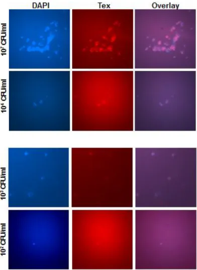
3.2.3 Sensitivity
In order to test and verify the sensitivity of the DNA probe, S. aureus was collected from exponential phase of growth and diluted up to 101 CFU/ml. From each dilution 10µl was taken on each slide and smears were made. DNA
11
Probe SaureusFISHTX1 worked well and detected S. aureus done to 100 bacterial cells per ml (Figure 5). Similar results were obtained in a previous study, where the used probe was a PNA probe (Humayun Iqbal, 2012, thesis work).
Figure 5: Sensitivity of the SaureusFISHTX1 probe. Various dilutions of FISH stained
S.aureus. For such large amount of bacteria the staining is clear, but it's highly
unspecific for lower amounts. DAPI, 4’,6-diamidino-2-phenylindole, Tex, Texas Red.
3.2.4 Specificity
Experiments were conducted in order to verify the specific binding of the SaureusFISHTX1 probe to the DNA of S. aureus and not to other types of bacteria. In the first set of experiments, the probe was set to react with one type of bacteria E. coli or S. pneumonia. The One Step Permeabilization Assay (Poppert et al., 2012) was followed for preparing the slides that contain
12
coli used because it’s gram negative bacteria and have thin a cell wall. The
slides where mounted with VectaShield Hard Set Mounting Medium without DAPI (Vector Laboratories) and examined using a Leica DMRA fluorescence microscope. The bacteria was present when using the FICH filter, but when it was replaced by the Tex Red filter, no light was observed, which means that the probe didn’t bind to the bacteria (figure 6,7).
Figure 6: FISH of E. coli. Both pictures for the same section but the blue one using DAPI filter and the red one using Tex Red filter. DAPI, 4’,6-diamidino-2-phenylindole. FISH, Florescence in situ hybridization.
Figure 7: FISH of S. pneumonia Both pictures for the same section but the blue one using DAPI filter and the red one using Tex Red filter. DAPI, 4’,6-diamidino-2-phenylindole. FISH, Florescence in situ hybridization.
The second set of experiments involved the SaureusFISHTX1 reacting with a mixture of bacteria (E. coli, S. aureus, S. pneumoniae). The One Step Permeabilization Assay (Poppert et al., 2012) was followed for preparing the slides. The slides where mounted with VectaShield Mounting Medium with DAPI (Vector Laboratories) and examined using a Leica DMRA fluorescence microscope. The E. coli could not be observed using the DAPI filter (not shown), this could be because that the permeabilization destroyed the bacterial cells, since the cell wall is very thin in the gram negative bacteria. While comparing the images when using a DAPI filter then when replaced with the
13
Tex Red filter for the same section, it was observed that the S. aureus, and S.
pneumoniae types of bacteria appeared under DAPI yet only S. aureus showed
under Tex Red which means the probe is specific to the S. aureus and it didn’t bind to the S. pneumonia (figure 8). It is easy to differentiate between them because the S. aureus appears more clearly under the DAPI filter while the S.
pneumoniae appears less clear and in most cases the S. pneumoniae appears
like two adjacent cells.
Figure 8: FISH of mix of bacteria E. coli, S. aureus, S. pneumonia. Both pictures for the same section but the blue one using DAPI filter and the red one using Tex Red filter, 4’,6-diamidino-2-phenylindole. FISH, Florescence in situ hybridization.
Another study (Juliane Strietz, 2012, Project work) has established the usefulness of using a DNA probe in the detection of E. coli. The probe works specifically, and its sensitivity is down to the concentration of 1000 cells per ml.
Another study (Humayun Iqbal, 2012, thesis work) has established that using a PNA probe is useful for the detection of S. aureus, the probe works pecifically, and its sensitivity is down to the concentration of 100 cells per ml. The PNA probes are more stable, enter the cell more easily and have a stronger and more rapid ability to hybridize than the DNA probes, but they cost twice as much which makes them more expensive than the DNA probes (Stender et al., 2002). The total cost for a PNA FISH equipment is about $6111 USD , and the probe kit costs approximately $1044 USD/50 tests (AdvanDX MA USA., 2012). If the usage of a DNA probe in FISH method was optimized carefully, taking into account the stability of the probe, then it will be economically more beneficial than using the PNA probe. MALDI-TOF is the method with the lowest cost of
14
the molecular techniques, costing only $2USD /test, and it differentiates among several pathogens through the aid of large data bases, yet it is not sensitive enough to accurately perform clinical diagnosis (Gorton et
al., 2011).
Conclusion
This study reports that One Step Permeabilization Assay has shown high effectiveness in permeabilizing the S. aureus cells and helping the DNA Probes to enter the bacterial cells and bind to its target DNA. The FISH method can diagnose the bacterial infection in patients with suspected sepsis in a shorter time. These results show that the SaureusFISHTX1 probe had high sensitivity as it could detect the S. aureus up to 102 CFU/ml and bound specifically to S. aureus only.
Future prospects
It's beneficial to start testing this method on clinical specimens for individuals who had sepsis in their blood. This will help to compare the obtained resulted with the original results in order to know the matching rate among them. If the matching rate is high then it's possible to start thinking about utilizing the FISH technique in the laboratory in order to get rapid results at a low cost.
Acknowledgement
I would like to start by thanking my supervisor Anna-Karin Pernestig PhD, Senior Lecturer Department of Life Sciences at the University of Skövde, for her supervision, teaching, and developing the motivation and confidence in me. Furthermore, I heartily thank Mikael Ejdebäck PhD, course responsible for Master’s program molecular biology for his help and support for us students. I also like to thank Heléne and kajsa for their help during the project work. I dedicate this thesis to my beloved parents who had faith in me, and without whom I wouldn't be where I am today. I also dedicate it to my precious little
15
nephews Hassan and Yasir, and to my dear friend Dhaif Alabady for his continuous support and help.
16
References
AdvanDx (2012) S. aureus PNA FISH® Staphylococcus aureus culture
identification kit. (Cat. No. KT001., 2012) AdvanDx MA, USA.
Amann RI., Binder., BJ and Olson RJ. (1990) Combination of 16S rRNA targeted oligonucleotide probes with flow ytometry for analyzing mixed microbial populations. Appl Environ Microbiol. 56, (p 1919-1925).
Clarridge J. E. (2004) Impact of 16S rRNA Gene Sequence Analysis for Identification of Bacteria on Clinical Microbiology and Infectious Diseases.
Clin. Microbiol. Rev. 17(4), (p. 840-862).
Gorton, RL., Stone, NARH., Ramnarain, P., Barker, K ., Mchugh, TD., Kibbler, CC (2011). A comparison of three rapid identification techniques for the identification of yeasts from positive blood cultures: Gram's stain, PNA-FISH and MALDI-TOF. Mycosis, 54. (p. 78-80).
Jansen J.G., Mooibroek M., Idema J., Harmsen H.J.M., Welling G.W., Degener J.E. (2000) Rapid identification of bacteria in blood cultures by using
fluorescently labeled oligonucleotide probes. J. Clin. Microbiol. 38(2), (p. 814-817).
Kempf V. A. J., Trebesius K. and Autenrieth I. B. (2000) Fluorescent In Situ Hybridization Allows Rapid Identification of Microorganisms in Blood Cultures. J. Clin. Microbiol. 38(2), (p. 830-838).
Laakso S., Kirveskari J., Tissari P. and Mäki M.(2011) Evaluation of High-Throughput PCR and Microarray-Based Assay in Conjunction with Automated DNA Extraction Instruments for Diagnosis of Sepsis. PLoS ONE 6: e26655 (p. 1-8).
Levy M. M., Fink M. P., Marshall J. C., Abraham E., Angus D., Cook D., Cohen J., Opal S. M., Vincent J. L. and Ramsay G. (2003) 2001
17
SCCM/ESICM/ACCP/ATS/SIS International Sepsis Definitions Conference.
Crit. Care Med. 31(4), (p. 1250-1256).
Mancini N., Carletti S., Ghidoli N., Cichero P., Burioni R. and Clementi M. (2010) The era of molecular and other non-culture-based methods in diagnosis of sepsis. Clin. Microbiol. Rev. 23(1) (p. 235-251).
McConaughy BL., Laird CD. and McCarthy BJ (1969) Nucleic acid reassociation in formamide. Biochemistry, 8, (p3289-3295).
lactalbumin: structure,
-Lysozyme and alpha HA, White FH (1991). McKenzie . ) 315 -173 41: (p.
Adv. Protein Chem.
function, and interrelationships.
Moter, A. & Goebel U. B. (2000) Fluorescence in situ hybridization (FISH) for direct visualization of microorganisms. J. Microbiol Methods, 41, (p85–112). Nguyen H. B. and Rivers, E. P. (2006) severe sepsis and septic shock: review of the literature and emergency department management guidelines. Ann.
Emerg. Med. 48, (p. 28-54).
Poppert S, Riecker M, Wellinghausen N. (2010) Accelerated identification of
S. aureus from blood cultures by a modified fluorescence in situ hybridization
procedure. J Med Microbiol, 59. (p. 65–68).
Rangel-Frausto M.S. (2005) Sepsis: Still going strong. Archives of medical
research. 36, (p. 672-681).
Schindler, C. A.; V.T. Schuhardt (1964). Lysostaphin: A New Bacteriolytic Agent for theStaphylococcus. Proceedings of the National Academy of Sciences 51 (3). (p. 414–421).
Stearns-Kurosawa D. J., Osuchowski M. F., Valentine C., Kurosawa S. and Remick D. G. (2011). The Pathogenesis of Sepsis. Annu. Rev. Pathol. Mech.
18
Stender H., Fiandaca, M., Hyldig-Nielsen, J. J. and Coull J. (2002) PNA for rapid microbiology. J. Microbiol. Met. 48, (p. 1-17).
Tissari P., Alimuddin Z., Tarkka E., Mero S., Savolanien L., Vaara M., Aittakorpi A., Laakso S., Lindfors M., Piiparinen H., Mäki M., Carder C., Huggett J. and Gant V. (2010) Accurate and rapid identification of bacterial species from positive blood cultures with a DNA-based microarray platform: an observational study. Lancet 375 (p. 224-230).
Trebesius K. Leitritz L and Adler K. (2000). Culture independent and rapid identification of bacterial pathogens in 18ybridizati fasciitis and streptococcal toxic shock syndrome by fluorescence in situ 18ybridization. Med Microbiol
Immunol.188, (p. 169-175).
Volkhard A., Kempf J., Trebesius K and Autenrieth I. B.(2000). Blood Cultures Rapid Identification of Microorganisms in Fluorescent In Situ Hybridization Allows. J. Clin. Microbiol.38 (2), (p. 830-838).
Walter T., Zwirble L and Angus D. (2004) severe sepsis epidemiology: sampling, selection, and society. Critical Care.8, (p. 222- 226).
Weinstein, M. P., Mirrett S, Reimer G., Wilson M., Smith E. S.,Chuard C. R., Joho K. L, and Reller L. B. (1995) Controlled evaluation ofBacT/Alert standard aerobic and FAN aerobic blood culture bottles for detection of bacteremia and fungemia. J. Clin. Microbiol.33, (p. 978-981).
Wellinghausen N., Wirths B., Franz A.R., Karolyi L., Marre R. and Reischl U. (2004) Algorithm for the identification of bacterial pathogens in positive blood cultures by real-time LightCycler polymerase chain reaction (PCR) with sequence-specific probes. Diagnostic microbiology and infectious disease. 48, (p. 229 -241).
Wischnewski N., Kampf G.,Gastmeier P., Schlingmann J., Daschner F., Schumacher M., and Ruden. H (1998) Prevalence of primary bloodstream infectionsin representative German hospitals and their association with centraland peripheral vascular catheters. Bakteriol. 287, (p. 93-103).
19
Wolk. D.M.and Fiorello A.B. (2010) Code Sepsis: Rapid methods to diagnose Sepsis and detect hematopathogenes – Part II: Challenges to the laboratory diagnosis of sepsis. Clinical Microbiology Newsletter. 32, (p. 41-49).
Yan C., Ding B. and Lan X. (2008) The toxicity study on marine lowtemperature lysozyme. Food Chem. Toxicol. 46, (p. 604-609).
20
Appendix 1
and S. aureus 16s rRNA of to bind to was utilized SaureusFISHTX1 The probe DNA. S. aureus probe against by blasting the confirmed this is21
Appendix 2
S. 16s rRNA of to bind to was utilized 1 Saureus2FISHTX The probe . DNA S. aureus probe against by blasting the confirmed and this is aureus22
Appendix 3