School of Sustainable Development of Society and Technology
Genetic and phenotypic characterization of trypanosomas
Linda Bech
Degree Project, ECTS 30.0
Supervicer at Mälardalens Högskola Prof. Carl Påhlason
Examiner at Mälardalens Högskola Dr. Magnus Neumüller
Abstract
Trypanosoma theileri, of the subgenus Megatrypanum, a non-pathogenic cosmopolitan blood
dwelling parasite of bovine. T. theileri can be cultured at room temperature in several culture media.
Blood samples were collected from deer’s. To see if the blood was infected with
trypanosomes it was cultivated in 2 ml sheep blood or cell cultivation medium DMEM with antibiotics.
Growth was detected by microscopy to see if there were any trypanosomes.
To determine the species of trypanosomes that was in the deer blood a DNA-preparation was done before a Polymerase Chain Reaction (PCR) could be done. With sequencing the
trypanosomes where determined to be Trypanosoma theileri.
Different tests were made to see in what way the trypanosomes best were caught to the objective slides.
Forty samples of borrelia positive serum from forty different patients were tested with the fluorescent microscopy. Forty different samples from blood donors were tested the same way. Blood samples from 16 different fissiped were taken and to see if they were infected with trypanosomes. Three different PCR’s were done on the 16 blood samples.
A small test on human blood was also performed.
Protein identification by immunoblot with western blot and silver staining was done. With the electron microscopy tests were done in the ordinary way and Critical Dry Point to see if both of the techniques worked.
Enzyme-Linked Immuno Sorbent Assay (ELISA) test were accomplished on two 96 well plates. The wells on the plates were diluted in different ways before they were processed.
Acknowledgment
I would like to take the opportunity to thank Professor Carl Påhlsson, at The Academy for HST at Mälardalens Högskola, for his knowledge and for his time to help me with this degree project.
Thank you! Linda Bech
Contents
2. Introduction 6
2.1 History 6
2.2 What’s a Trypanosoma? 6
2.3 Detection of Trypanosoma 7
2.4 The looks of Trypanosoma 7
2.5 Trypanosoma theileri 7
2.6 The Purpose 8
3. Materials and methods 8
3.1 Cultivation and detection 8
3.2 DNA-preparation and Polymerase Chain Reaction (PCR) 8 3.2.1 PCR program for first PCR and nested 9
3.3 Gel electrophoresis 9
3.4 Cloning 9
3.4.1 LB-agar plates preparation 9
3.4.2 Cloning PCR 10 3.5 Sequencing 10 3.5.1 Sequence PCR preparation 10 3.5.2 Sequence PCR 10 3.5.3 Gel preparation 11 3.6 Fluorescent microscopy 11 3.6.1 Acridinorange 11 3.6.2 Serology test 11 3.6.3 Adherence optimisation 12 3.7 16 Direct detection by PCR 12
3.8 Mammalian vs. sheep blood 13
3.9 Protein identification by immunoblot 13
3.9.1 Preparation of the sample 13
3.9.2 Preparation of the gel 13
3.9.3 Western blot 14
3.9.4 Blocking of the membrane 15
3.9.5 Silver staining 15
3.10 Electron microscopy 15
3.10.1 The ordinary way 15
3.10.2 Critical Dry Point 15
4. Results 17 5. Discussion 21 Referents 22 Appendix 1 24 Appendix 2 25 Appendix 3 26 Appendix 4 27
2. Introduction
2.1 History
The first man to see a trypanosome was probably Antony van Leeuwenhoek. In 1680 he wrote to Robert Hooke at the Royal Society saying that he had seen numerous small
micro-organisms in the gut of a horsefly through his microscope. “They lay mingled with the thin matter that was in the fly's guts and moved forward very quickly”. He almost certainly described the epimastigotes of Trypanosoma theileri in their insect vector (Goodwin, L. G).
Trypanosomes a parasitic protozoon of the family Trypanosomidae, were during the mid-nineteenth century seen in fish, frogs, moles and mice and it was soon recognized in all vertebrates and invertebrates and even some plants (Goodwin, L. G).
At the beginning of 1900, the importance of trypanosomes as pathogens was recognized by Timothy Richards Lewis and Griffith Evans in India and by Sir David Bruce in East Africa all of whom gave their names to species of trypanosomes (Goodwin, L. G). Almost
simultaneously tryponosomes were recorded by Laveran in South Africa (Rodrigues, A.C et
al.). African sleeping sickness is transmitted from antelopes to man and his domestic stock by tsetse flies and Changas disease from the wild animals of South America by reduviid bugs that rapidly became adapted to breed and feed in human dwellings. Both forms of
trypanosomiasis are a great deal more pathogenic in their new hosts (Goodwin, L. G).
2.2 What’s a Trypanosoma?
Trypanosoma spp. is a universal protozoan parasite with representative species infecting
nearly all vertebrate species that can cause devastating human and animal diseases, for which there is currently no adequate treatment (Lefebvre, M. F. et al., Lira, and C.B.B. et al.).
There is a large amount of different kind of trypanosomosis around the world that have different effects. For example Africa and South America suffer great economic importance affecting agriculture development in large areas due to high mortality or severe production losses in cattle associated with chronic trypanosomosis (Geysen, D. et al., Lefebvre, M. F
et.al.).
The disease is caused by trypanosomes which are transmitted by the bite of a blood sucking arthropod (Lefebvre, M. F et al.).
Trypanosomes are divided into two different kinds’ stercorarian and salivarian. In the
stercorarian (T. cruzi, T. theileri) kind the trypanosomes are taken up in the blood meal and grow and divide in the hindgut of the insect vector. Infection of the vertebrate host is by contamination, when the insect feeds it defecates and the trypanosomes gain entry through the feeding site or by the skin being scratched. The salivarian (T. vivax, T. brucei brucei, T.
evansi and T. congolense) trypanosomes develop in the mid gut of the vector, which is a fly
and are injected via the salivary glands when the fly feeds (Lefebvre, M. F et.al., van Hellemond, J. J et al., www.aber.ac.uk)
2.3 Detection of Trypanosoma
The parasites count are often so low that it is only detectable by cultivation of blood samples and a way to detect the parasites is by microscope (Geysen, D. et al., Goossens, B et al., Townsend, J et al, Verloo, D et al.). A way to identify the parasites at levels far below the detection limit is to use polymerase chain reaction (PCR). Species specific
DNA-hybridisation probes have been developed for trypanosome detection (Geysen, D. et al.).
2.4 The looks of Trypanosoma
Trypanosome grows thinner and more or less narrow at the end of the body protoplasm (Laveran, A.). Two masses of chromatin can be seen, when stained. The first one is a large mass towards the middle of the body, the nucleus. The other one is smaller and usually located near the posterior end, the centrosome. The trypanosoma have a flagella which starts from the centrosome and bounds the undulating membrane usually ends at the anterior extremity (Laveran, A.). The DNA-strands are inside the kinetoplastid which is circular and the organelles that are in the cytoplasm are for example the endoplasmic reticulum, Golgi, lysosomes, glycosomes and more (Marquardt et al.).
2.5 Trypanosoma theileri
Trypanosoma theileri, of the subgenus Megatrypanum, has been described as a
non-pathogenic cosmopolitan blood dwelling parasite of bovine with high incidence on every continent except Antarctica (Cross, R. F et al., Goossens, B et al., Lefebvre, M. F et al., Rodrigues, A.C et al., Townsend, J et al., van Hellemond, J. J et al.).
In Western Europe, T. theileri is the only trypanosome species occurring in cattle (van Hellemond, J. J et al., Verloo, D et al.).
T. theileri is a relatively large, trypanosomal cell that is between 30 and 80µm and it is longer than those of other trypanosomes parasite of bovine (Townsend, J et al., van Hellemond, J. J
et al.).
A T. theileri infection in bovid may persist for many years with no recognized ill effects. T.
theileri seem to become pathogenic when stress situation occur or when infections with other
pathogens are present e.g. East Coast Fever (Goossens, B et al., Rodrigues, A.C et al., Verloo, D et al.). There is also evidence that T. theileri crosses the bovine placenta, sometime causing abortions (Lefebvre, M. F et al., Hussain, K. et al.).
T. theileri can be cultured at room temperature in several culture media such as veal infusion or blood agar (Verloo, D et al.). Attempts to culture T. theileri in defined medium, several non-defined and semi-defined media have failed. In fact, several media permit a quick growth of T. theileri for a short time, but few media permit multiplication for more than 20–30 days (Rodrigues, A.C et al.).
2.6 The Purpose
The purpose of the work was to try to optimize the cultivation what regarded the temperatures, moisture and the composure of different culture medium, to optimize the growth conditions for the trypanosomes.
Different methods and cycling programs for Polymerase Chain Reaction (PCR) had to be optimized that included finding specific primers for the trypanosomes.
3. Materials and methods
Blood samples were collected from deer’s, one female and two youngsters during hunting season. One of the youngsters were smaller than the other, it turned out that that youngster was infected with trypanosomes.
To cultivate the trypanosomes 8.5 g Tryptic Soya broth, 3 g agar-agar and 200 ml MQ-H2O
was autoclaved for 20 min in 120°C with 1 atm pressure. After the autoclave the agar was cooled down to about 49°C before 11 ml of sheep blood (at room temperature) was added. 5 ml of the blood agar was pipetted to 15 ml of centrifuge tubes. The agar had to cool and set before they were stored in the refrigerator.
3.1 Cultivation and detection
The trypanosomes were cultivated in 2 ml sheep blood or cell cultivation medium, DMEM with addition of Ampicillin 200 µl/ml final concentration. 30 µl from the deer blood was added to the cultivation tubes and incubated at 28°C. The tubes were inspected weekly for 3 weeks. For cultivation 5 µl samples from the tubes were taken and put on objective slides with cower slides on top. The slides were examined by microscopy to see if the trypanosomes had started to grow, in that case 10 µl were transferred to new tubes.
3.2 DNA-preparation and Polymerase Chain Reaction (PCR)
DNA-preparation was made from the tubes with cell culture medium by following the QIAGEN DNeasy® Blood & Tissue kit (250). The DNA- prep was then used to make a PCR with different primers, depending on which PCR was to be done. The primers that were used were Tryp3f, Tryp3nR and Tryp3R (Geysen et al). There was also a theileri specific primer Tryp-thelR that had been located with the help from NCBI´s gene data bank (Salmijärvi).With “ready to go” tubes, a tube that the masters mix already is in. All that have to be added to the tubes are template, primers and dH2O. The total volume of the reaction mix was
25µl/tubes.
After the first PCR was done a nested PCR was done to confirm that there were trypanosomes in the samples.
Chart 1. Annealing position for the primers and the sequence that have been used to the rRNA 18s gene in T. theileri.
Primer Position Sequence Length
Tryp3f 1272-1297 AAACGATGACACCCATGAATTGGGGA 26 Tryp3nR 1868-1892 CACTACAATGTCAGTGAGAACAAGACAC 28
Tryp3R 1969-1991 GTATTGCAATTATTGGTCGCGCA 23
Tryp-thelR 2196-2212 TAATCTCATCGGAAAATGATCCAG 24
Chart 2. The primer name and product length.
PCR Primer pars Product length Notes
1st Tryp3f/Tryp-thelR 940bp
Nested Tryp3nR/Tryp-thelR 344bp Theileri specific product
Nested Tryp3R/Tryp-thelR 243bp Trypanosomes general product
3.2.1 PCR program for first PCR and nested
94°C 4min
94°C 1min 4sec (Denaturating)
55°C 30sec (Annealing) X40 72°C 2min (Elongation)
72°C 10min 4°C 99h
3.3 Gel electrophoresis
To confirm that the right product was gained in the PCR a 1.5% agarose gel electrophoresis was performed in TBE X1 and 3µl Ethidium bromide. The gel was loaded and then run on 100V, for about 45 min.
The PCR gave, as expected, a product at 940 bp, for the first PCR. The nested PCR also gave the expected result 243 bp.
3.4 Cloning
A cloning was done with TOPO TA cloning® Kit for sequencing version J. The cloning was done to separate the trypanosoma DNA from the sheep/deer DNA.
3.4.1 LB-agar plates preparation
30g Tryptic Soya broth 15g Agar
1L dH2O
5ml Ampicillin (conc. 10mg/ml)
The Tryptic, agar and dH2O were mixed and autoclaved for 20 min in 120°C with 1 atm
pressure then it was cooled to about 50°C before the ampicillin was added. The LB-agar medium was pored in Petri-dishes and left to set.
The cloning was only done on samples from the first PCR. After the cloning preparation the samples were spread with a glass-policeman on to three ampicillin plates with the
concentrations 50µl, 80µl and the rest. The three amp-plates were then put in a 37°C incubator over night.
3.4.2 Cloning PCR
A cloning-PCR was done with primers T7-forward and M13 reverse on seven clones.
94°C 2min
94°C 1min 4sec (Denaturating)
55°C 1min (Annealing) 40X 72°C 1min (Elongation)
72°C 7min 4°C 99h
A gel electrophoresis was done to verify that the amplification had accrued.
3.5 Sequencing
3.5.1 Sequence PCR preparation
A sequence PCR was prepared with two of the verified clones. Master Mix
2µl template 1µl primer 12µl dH2O
The Master Mix was made twice, once with Cy5 T7 primer and once with Cy5 M13 primer. For each sample four empty tubes (not ready to go tubes) was used. There were 2 cloned that turned out to be god, so eight tubes were used. The tubes were marked with A, C, G and T, one tube for each nucleotide, one for the nucleotide A, one for nucleotide C and so on. 3µl of each nucleotide was used. In one set of four tubes with one of the clones contained Cy5 T7 forward and from the same clone but a new set of four tubes contained M13 reverse. The other clone was prepared in the same way. Then a sequence PCR was done.
3.5.2 Sequence PCR
94°C 2min 94°C 30sec (Denaturating) 55°C 30sec (Annealing) X25 72°C 1min (Elongation) 72°C 10min 4°C 99hWhen the sequence PCR was done 6µl formamid loading dye was added. The formamid denaturises the DNA strands so that they got single stranded. The formamid also contains glycerol that has the effect of making the sample sink to the bottom of the well. It also is a dye, so that the sample is visible. To do so that the strands denaturises a sequence boiling was done, in the PCR apparatus at 72°C for 3 min.
3.5.3 Gel preparation
To see what nucleotides there were in the samples a 0.5 acrylamid “long read repro gel TM” was made. The gel was made between two special slides that were set to polymerase under a UV-light. When the gel was set in the sequencing apparatus 2L TBE buffer X0.5 was added. The wells were washed with the buffer before they were loaded with 7µl samples/well. The sequence apparatus were connected to a computer that received the results of the nucleotides that were in the samples. The lines of nucleotides were then blasted with NCBI´s database to see what the samples contained.
3.6 Fluorescent microscopy
3.6.1 Acridinorange
A 5µl sample were pipetted to a slide and left to air dry. 5µl 95% Ethanol were then added on to the sample and left dry for 2min. 5µl acridinorange were added and left to work for 2min before it was rinsed with tap water. The result was examined in the fluorescence microscopy (see fig. 1)
Fig. 1 Acredinorange trypanosomes
showed in the fluorescence microscopy.
3.6.2 Serology test
1ml trypanosome medium was pipetted in to a tube. The tube was centrifuge and the supernatant was removed. The pellet was then resolved in 100µl 1/10 glutaraldehyd. The sample was left for 1h. The tube was then centrifuge again and the glutaraldehyd was removed. The pellet was then resolved in PBS.
A diluting series, 1/20, 1/80, 1/320, 1/640 and 1/1280, was done on the glutaraldehyd washed trypanosomes.
Forty samples of borrelia positive serum from forty different patients were tested with the fluorescent microscopy. 5µl of trypanosome were pipetted to slides and let to air dry. 95% ethanol was added and let to dry. When the ethanol had dried 20µl of a borrelia positive sample, diluted 1/25, was added to the samples on the slides. The slides were put in a moist camber and incubated at 40°C for about 30min. The slides were then washed with PBS 2x5min. After the wash 20µl Rabbit anti- human IgG FITC, diluted 1/40, were added. The slides were then incubated again for 1h. The slides where then washed again in PBS 2x5min. When the slides had dried a drop of DakoCytomation Fluorescent Mounting Medium were
added and then a coverslip on top. The result could then be seen in the fluorescent microscopy (see fig. 5).
The same test was then done in the same way but with serum from forty blood donors.
3.6.3 Adherence optimisation
Different ways to get the trypanosomes to stick to the slides was carried out. When the different solutions had been added the slides were treated as in the antibody test.
1. A gelatine mixture was added to an objective glass and left to set before the trypanosomes were added.
2. An agar mixture was left to set after it had been poured on to the objective glass. 3. Trypanosomes were pipetted on to the objective glass and left to set. When they were
set 95% ethanol were pipetted on top of the trypanosomes and left to dry.
4. The last test was to compare egg yolk, 95% ethanol and natural (only trypanosomes on the glass) and to see if there was any difference in the trypanosomas ability to stick to the objective slides.
3.7 Direct detection by PCR
Blood samples from 16 different fissiped (roe deer, elk and red deer) were collected. DNA preparations were done with Nucleo Spin, MACERY-NAGEL. With the DNA-preparation, 16 different PCR’s were set up. The first PCR was made in 16 different ready to go tubes market rigorous. First PCR Mix 5µl Template 1µl Tryp3f 1µl Tryp-thelR 18µl dH2O
Then a nested PCR was done, were 1µl from the first PCR were taken. It was important to not contaminate the nested PCR. It was also important to take the µl from the tube with the same marking. There was also done a second nested PCR, the same way as the first nested PCR, but with different primers.
First nested PCR 5µl Template 1µl Tryp3nR 1µl Tryp-thelR
1µl from the first PCR with the same marking 17µl dH2O
Second nested PCR 5µl Template 1µl Tryp3R 1µl Tryp-thelR
1µl from the first PCR with the same marking 17µl dH2O
3.8 Mammalian vs. sheep blood
One test was carried out to see if the T. theileri also could grow in mammalian blood. The test was set up as in ordinary cultivation 5ml blood agar, 2ml of human blood in two test tubes and 2ml of sheep blood in two different tubes. The same amount of trypanosomas was added to the test tubes. The tubes were incubated and the growth was watched in the microscopy almost every day.
3.9 Protein identification by immunoblot
3.9.1 Preparation of the sample
The lyses of the trypanosomes were made by adding 45µl lysis buffer and 5µl 10% SDS.
Lysis buffer 25% sucrose 50mM Tris
1mM EDTA (pH 8)
The tube was then boiled at 95°C in a heating block for 10 min to denaturise the protein. After the heating 10µl bromothymol blue and glycerol were added and mixed vigorously so the sample would be clear. If it wasn’t clear urea could have been added.
3.9.2 Preparation of the gel
The test was started by assembling the apparatus according to the manual by the technician. The preparation of two 12% Tris/Glycine SDS-polyacrylamide gel electrophoresis was then done by the making the first gel, for the two gels. The TEMED was added at the end just before it was to be poured between the slides, it made the gels to set. The first gel was poured to the half of the slides and left to set for about 15min.
15ml solution 5ml H2O 6ml 30% Acrylamid mix 3.8ml 1.5M Tris (pH 8.8) 0.15ml 10% SDS 0.15ml 10% APS 0.006ml TEMED
Meanwhile the first gels was setting the second gel (STACK) was prepared. When the first gels had set the STACK was poured on top of the first gels. The TEMED was added just before it was poured between the slides. When the STACK had been added the combs was adjusted to get wells, so the samples could be loaded.
5ml STACK 3.4ml H2O 0.83ml 30% Acrylamid mix 0.63ml 1.0M Tris (pH 6.8) 0.05ml 10% SDS 0.05ml 10% APS 0.005ml TEMED
When the both gels had set they were assembled in to the apparatus and running buffer x1 was filled up in the inner and the outer reservoir. The combs were removed and the samples were loaded, 1µl, 3µl, 5µl, 10µl and a 5µl marker. The two gels were loaded the same way. The gels were then run at 100V for about 1h.
1L Running buffer x10 30.3g (0.25M) Tris base 144.2g (1.92M) Glycine 10g (1.0%) SDS
dH2O to 1000ml
This was a stem solution so it was diluted ten times with dH2O before use.
The gels were run in duplicate of which one was used for western blot and one was stained with silver staining.
3.9.3 Western blot
When the gels were done, one of the gels was set up as followed. The set up were made in the Towbin buffer.
• Black grid (bottom) • Sponge
• 2 Whatman paper • Gel
• Hybond-P polyvinyl membrane (Amersham) Pretreated by incubation:
10sec in methanol 5min in H2O
>10min in Towbin buffer • 2 Whatman paper
• Withe grid (top)
Inserted the black grid towards the black of the container = negative pool. The set up was run at 50V in a cooling room overnight.
x10 Towbin stem solution 29g Tris base
145g glycine dH2O to 1000ml
The mix of the x1 Towbin solution 100ml x10 stem solution
200ml methanol 700ml dH2O
3.9.4 Blocking of the membrane
The next day the membrane was put in a plastic bag with 2% skim milk powder diluted in PBS to 10ml and left overnight on a shaking table. The next day positive Borrelia antibodies diluted 1/200 were added and again left overnight in the refrigerator. The membrane was then washed x2. After the wash peroxidase anti-human were added and left overnight. Then the membrane was washed x2, before it was processed with diaminobenzidine (DAB).
3.9.5 Silver staining
The other gel was stained with Silver staining kit for protein following the manufactures instructions (GE Healthcare).
3.10 Electron microscopy
3.10.1 The ordinary way
5µl of glutaraldehyd washed trypanosomes was left to dry on a filter before it was plated with a thin layer of gold under high pressure. Then the trypanosomes were visible in the electron microscopy.
3.10.2 Critical Dry Point
The start was the same, 5µl of glutaraldehyd washed trypanosomes was left to dry on a filter. The filter was then put in a steel cylinder with bolts on both sides. Dry ice (solid carbon dioxide) was put in the cylinder and left overnight. The pressure that has been built up over the night, which happens when solid carbon dioxide change from solid phase to liquid, was let out gently. Then the filter was plated with a thin layer of gold and then watched at in the electron microscopy.
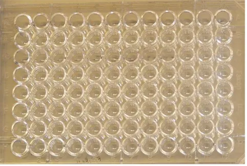
3.11 Enzyme-Linked ImmunoSorbent Assay (ELISA)
The ELISA test were accomplished on two 96 well plates. The trypanosomes were first diluted in carbonate buffer (pH 9.8). The trypanosomes were then diluted, column wise and left so that the trypanosomes could fixate in the plate.
One plate was diluted as shown in fig. 2 and the other as in fig. 3.
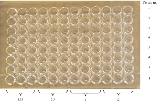
Fig. 3 Different serum in dilution 1/100 tested against dilutions of antigen (in triplets).
After the fixation 200µl 1% serum albumin were added as a blocking solution. The wells were then washed two to three times with a washing solution.
The washing solution 13.5g NaCl
1.5ml Tween 20 1.5L dH2O
After the wash, 50µl Serum Borrelia (1/100) was added row wise and incubated for one hour. The plates was then washed again two, three times.
4µl of Polyclonal Rabbit Anti-human IgG/HRP was added to 12ml of PBS. 50µl of the solution was added to the plates and left to incubate in the refrigerator over night.
The next day the plates was washed two, three, before 100µl of Dako Cytomation OPD tabl, 2mg were added. When a tawny colour appeared the reaction was terminated with Sulfuric acid (H2SO4).
4. Results
PCR and Sequencing
The PCR products that were obtained, gave the expected product length. A nested PCR was done to confirm that there were trypanosomes in the samples.
The cloning was done to separate the trypanosomes DNA from any other DNA. The cloning PCR with primers T7-forward and M13 reverse also gave the expected length (see fig. 4).
neg,1,2,3,4,5,6,7,ladder
Fig. 4 The cloning PCR gave the result that was expected ~1100bp.
The sequencing was done from the two strongest bands (3 and 4), from the cloning. The result that was given was compared with sequences of Trypanosomes theileri in the NCBI database (see appendix1-4).
Fluorescent microscopy
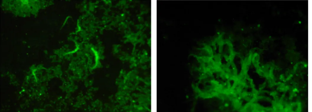
Some of the Borrelia serums were possible to use as primary antibodies in the fluorescent microscopy (see fig. 5).
Fig. 5 Trypanosomes in the fluorescence microscopy from the antibody test.
The assay to optimize adherents to slides both gelatine and the agar mixtures worked, and the use with 95% ethanol worked best. Also in the test with egg yolk, 95% ethanol worked best.
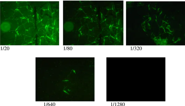
The serology test showed that the conjugate don’t give fluoresce; and patient serum have primary anti-Trypanosoma antibody to a titer of 1/640 (see fig. 6).
1/20 1/80 1/320
1/640 1/1280
Fig. 6 Serology test showed in the fluorescence microscopy.
Direct detection by PCR
The test was done to see if it was possible to screen blood samples for Trypanosoma theileri by using PCR. The result was that it did, the three different PCR’s revealed the same results. Three of the 16 samples were positive for T. theileri.
Mammalian vs. sheep blood
Trypanosoma theileri did not grow in the mammalian blood at all.
Protein identification by immunoblot
Unfortunately no antibody reaction was found by western blot. (See fig.7)
1µl laddar 10µl 5µl 3µl 1µl
Electron microscopy
The vacuole on the trypanosome exploded in the ordinary way in electron microscopy due to the high pressure in the gold plating camber (see fig. 8).
Fig. 8 The vacuole is broken due to the high pressure in the gold plating camber.
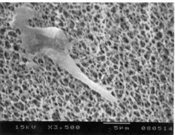
The Critical Dry Point experiment worked very fine (see fig. 9).
Fig. 9 Trypanosoma theileri in the Critical Dry Point experiment.
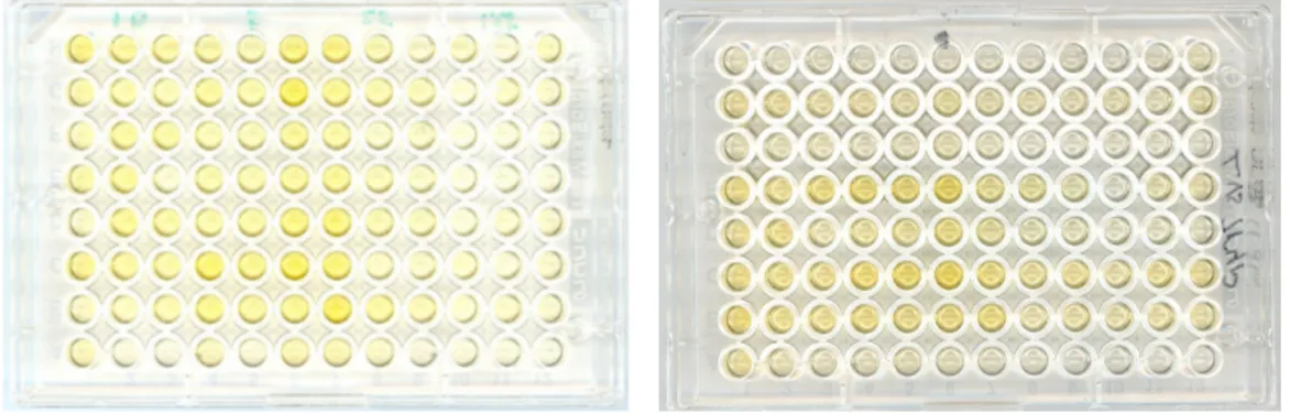
ELISA
Since the trypanosomes are a relatively large parasite it was very difficult to fixate them in the wells of the ELISA plates. Many of them were washed away in the different washing steps. There was a little difference in the colour on the plates. The Borrelia serum gave results for the trypanosomes (see fig. 10).
Fig. 10 The left ELISA plate, is dilution of trypanosomes 10, 5, 2.5 and
1.25µl/well respectively, tested in triplets. Optimal concentration is 2.5-5ul/well.
In the plate on the right eight different serums was diluted from undiluted to 1/1024 column wise and added row wise, in row 2,4,6,7 shows reaction.
5. Discussion
The fact that it was more common with antibodies in Borrelia positive serum indicate that it is plausible that T. theileri are transmitted, even if it is impossible to culture Trypanosoma in human blood.
Some blood donors have positive serology for T. theileri. That could be explained with that it is difficult to find out if the donors have been bitten by any arthropods that are carrier of the trypanosomes.
During the work it was discovered that the trypanosomes easily could be cultivated on the bench top as in the incubator, indicating that they are not so sensitive to temperatures for growth.
At some point the trypanosomes stopped to grow and replicate in the sheep blood. It wasn’t known at that time what caused the death. After a search on the internet some articles was found about the subject. Lira, C. B. B et al. and Dreesen, O., and Cross, G. A. M. wrote about
T. brucei having 20 telomeres. It seemed to be the case of T. theileri as well; a few very weak trypanosomes from a tube with sheep blood were pipetted in to a tube with bovine blood and incubated. After a few days the trypanosomes had started to grow and replicate and they were more motile than they ever had been in the sheep blood.
The test on human blood that was done in this report may not be significant. There was only one set of human blood. Maybe there was something in that blood that the trypanosomes didn’t like.
To see if the trypanosomes really don’t growth in human blood there should be more tests done with more human blood from different persons.
Referents
Cross, R. F., Smith, C. K., Redman, D. R. (1971) Observations on Trypanosoma theileri Infection in Cattle. Canadian Journal of Comparative Medicine (1971) Vol. 35 12-17
Dreesen, O., Cross, G. A. M. (2007)
Telomere length in Trypanosoma brucei.Experimental Parasitology 118 (2008) 103-110
Geysen, D., Delespaux, V., Geerts, S. (2002)
PCR–RFLP using Ssu-rDNA amplificationas an easy method for species-specific diagnosis of Trypanosoma species in cattle.
Veterinary Parasitology 110 (2003) 171–180
Goodwin, L. G. (1985) Trypanosomiasis: Introduction. British Medical Bulletin (1985) Vol.
41, No. 2, 103-104
Goossens, B., Mbwambo, H., Msangi, A., Geysen, D., Vreysen, M. (2006) Trypanosomosis
prevalence in cattle on Mafia Island (Tanzania). Veterinary Parasitology 139 (2006) 74–83
Hussain, K., Brodie, B., Ott, R.S., Montealegre, F. (1985) Prevalence of Trypanosoma
theileri in cows and fetuses. American journal of Veterinary Research (1985) jun; 46(6):1256-8
Laveran, A. (1907) Protozoa as causes of diseases. Nobel Lecture Dec, 11 1907
Lefebvre, M. F., Semalulu, S. S., Oatway, A. A., Nolan, J. W. (1997) Trypanosomiasis in
woodland caribou of northern Alberta. Journal of wildlife Diseases 33(2) (1997) 271-277
Lira, C. B. B., Giardini, M.A., Siqueira Neto, J. L., Conte, F. F., Cano, M. I. N. (2007)
Telomere biology of trypanosomatids: beginning to answer some questions. Trends in
Parasitology (2007) vol.23 No.8
Marquardt, W.C., Demaree, R.S., Grieve, R. B. (2000) Parasitology and vector biology.
Academic press. IAP Harcourt; ISBN: 0-12-473275-5
Rodrigues, A.C., Campanera, M., Takata, C.S.A., Dell’ Porto, A., Milder, R.V., Takeda, G.F., Teixeira, M.M.G. (2003) Brazilian isolates of Trypanosoma (Megatrypanum)
theileri: diagnosis and differentiation of isolates from cattle and water buffalo based on biological characteristics and randomly amplified DNA sequences. Veterinary Parasitology 116 (2003) 185–207
Salmijärvi, S. (2007) Genetisk art bestämmning och karakterisering av Trypanosoma
Theileri. (2008) Mälardalens Högskola, Eskilstuna, Sverige.
Townsend, J., Duffus, W. P. H., Glauert, A. M. (1982) An Ultrastructural Study of the
interaction in Vitro between Trypanosoma theileri and bovine Leucocytes. Journal of Cell
van Hellemond, J. J., Hoek, A., Wichgers Schreur, P., Chupin, V., Özdirekcan, S., Geysen, D., van Grinsven, K. W. A., Koets, A. P., Van den Bossche, P., Geerts, S., Tielens, A. G. M (2007) Energy metabolism of bloodstream from Trypanosoma theileri.
Eukaryotic cell (2007) 1693-1696
Verloo, D., Brandt, J., van Meirvenne, N., Büscher, P. (1999) Comparative in vitro
isolation of Trypanosoma theileri from cattle in Belgium. Veterinary Parasitology 89 (2000) 129-132