Department of Biology and Chemical Engineering
Coordination Chemistry of Novel Drug Candidates
for the Treatment of Iron Overload
Anneli Norin
Degree Project, ECTS 30.0 At the University of Queensland Brisbane, Australia 2006 Supervisor at the University of Queensland Prof. Paul V. Bernhardt Examiner at Mälardalen University Ass. Prof. Simon Dunne
ABSTRACT
Iron overload is a serious clinical condition caused by excessive iron in the body, which can be largely prevented by the use of iron-specific chelating agents. At the moment there are only a few chelators in clinical use for the treatment of Fe overload. One of them, and so far the best working one, is desferrioxamine (DFO). This iron chelator has a major disadvantage of being orally inactive and is given by long and frequent subcutaneous infusions (12-24 hours/5-6 days/week) to patients. 1
Consequently, the design of an orally active, nontoxic, selective iron chelator has become a high priority. To design an iron chelator for clinical use, the important factors to consider are metal selectivity and affinity, ligand-metal complex stability,
bioavailability and toxicity. The best iron chelator should be highly selective for iron(III) in order to minimize chelation of other biological essential metal ions which could lead to deficiency with prolonged usage. Favouring the Fe (III) oxidation state avoids Fenton chemistry and the production of toxic free radicals.
This report deals with complexes of aroylpicolylhydrazines. Chemical and biological testing has established that these molecules function extremely well as iron chelators in vivo. The most effective candidates are more efficient at promoting iron efflux (release of iron) from cells than the existing drug DFO. Although the ligands discussed in this report, are selective for iron, there is no record of how they interact with other essential metal ions (Mn, Co, Ni, Cu and Zn) in our body and it is this that is the topic of this report. It was found that the complexes with H2PPH (N,N’-bis(α-picolinoyl)hydrazine)
probably built polymeric complexes and therefore they were almost totally insoluble in all common solvents. The complexes of H2BPH (N-(benzoyl)-N’-(picolinoyl)hydrazine))
were also problematic in terms of insolubility. Another problem was decomposition of the complexes in solution, with resultant precipitation of the ligand and that happened with all of the ligands. The ligand H2TPH formed complexes with Ni and Co, [MII(HL)2]
and [MIII(L)2]-, and crystals were grown suitable for x-ray structure studies. The cobalt
complex is the first one reported of its kind. There was also crystals grown suitable for x-ray work of a protonated picolylhydrazide and that is also the first crystal structure reported.
TABLE OF CONTENTS
INTRODUCTION 5
Chelation therapy... 8
EXPERIMENTAL 13 Instrumentation & physical methods ... 13
Materials ... 13 Commercial Materials... 13 Precursors... 14 Methyl picolinate, C7H7NO2... 14 Picolyl hydrazide, C6H8N3O ... 14 Methyl benzoate, C8H9O2... 14 Benzoyl hydrazide, C7H9N2O ... 14 SYNTHESES 15 The N-(isonicotinoyl)-N’-(Picolinoyl) Hydrazine Ligands ... 15
N-(benzoyl)-N’-(picolinoyl)hydrazine), H2BPH... 15
N,N’-bis(α-picolinoyl)hydrazine, H2PPH. ... 15
N-(2-Thiophenecarbonyl)-N’-(Picolinoyl)hydrazine, H2TPH ... 16
Bis-Ligand Complexes of N-(isonicotinoyl)-N’-(Picolinoyl) Hydrazine Ligands ... 16
[Cu3(BPH)4](OH)2·4 H2O (AN23)... 16
[Cu(HBPH)2]·1.5 H2O (AN24) ... 17
[Zn3(HBPH)4](OH)2·0.5H2O (AN29) ... 17
[Ni(HBPH)2]·1.5 H2O (AN34)... 17
[Co(BPH)2]·H2O (AN40)... 18
Attempted synthesis “[Mn(BPH)2]” (AN41) ... 18
[Cu(PPH)2]·3.5 H2O (AN13) ... 19
[Zn(PPH)2]·0.5H2O (AN51)... 19
Attempted synthesis “[Zn(PPH)2]” (AN35)... 19
Attempted synthesis “[Zn(PPH)2]” (AN17)... 20
Attempted synthesis “[Ni(PPH)2]” (AN30) ... 20
Attempted synthesis “[Ni(PPH)2]” (AN103) ... 20
Attempted synthesis “[Ni(PPH)2]” (AN69) ... 21
[Co(PPH)2]·1.75 H2O (AN39) ... 21
[Co(PPH)2]·4.25H2O (AN54) ... 21
Attempted synthesis [Co(PPH)2] (AN79)... 22
Attempted synthesis “[Mn(PPH)2]” (AN55) ... 22
[H4PPH]Cl2 (AN79B) ... 22
Attempted synthesis “[Cu(TPH)2]” (AN59) ... 23
Attempted synthesis “[Zn(TPH)2]” (AN63) ... 23
[Ni(TPH)2]·0.5H2O (AN73)... 23 [Ni(TPH)2]·1.75H2O (AN92)... 24 [Ni(TPH)2]·1.5H2O (AN97)... 24 [Co(TPH)2]·1.25 H2O (AN100B) ... 25 NH4[Co(TPH)2]·H2O (AN89A) ... 25 [Co(TPH)2]·0.5H2O (AN98:1) ... 26
Attempted synthesis “[Mn(HTPH)2]” (AN88) ... 26
Attempted synthesis “[Mn(HTPH)2]” (AN78) ... 27
RESULTS/DISCUSSION 28 Syntheses and physical properties ... 28
Spectroscopic properties ... 32
Description of crystal structures ... 40
Electrochemistry ... 50
CONCLUSIONS/FUTURE DIRECTIONS 53
REFERENCES 54
INTRODUCTION
Iron is essential for all life with the exception of two bacterial species. Almost all cells and organisms require iron to perform basic cellular processes. Oxygen transport, energy production and DNA synthesis depend on iron-containing proteins.
Iron exists in two valency states in biological systems: ferrous (FeII) and ferric (FeIII),
but almost exclusively it exists in the less soluble oxidized state (FeIII). The chemistry with iron is complex, because of its dual valency and reactivity with oxygen. Ionic iron is toxic to cells and is an active promoter of free radical reaction, a process called the
Fenton reaction. In the body, iron is bound in a safe form. Serum iron is bound to proteins called transferrin, and most bodyiron is present as iron porphyrin complexes,
(haemoglobin, myoglobin and haem-containing enzymes) but iron can also be stored as ferritin and haemosiderin.
Iron has a central role in the energy metabolism of cells. It is a transition metal and can take part in redox processeses where it converts between its Fe(II) and Fe(III) redox states. The most well known example of this redox reaction is the iron containing proteins haemoglobin and myoglobin, which are involved in oxygen transport and
storage. We also have important forms of iron in cytochromes, iron-sulphur proteins, iron enzymes and lactoferrin.
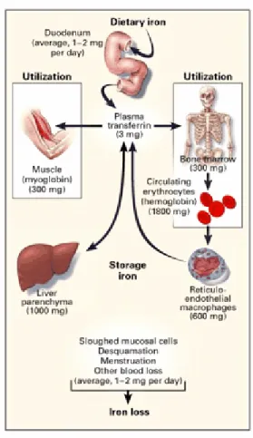
About two thirds of bodyiron is present in haemoglobin in red blood cells. Since humans do not have a physiological mechanism for eliminating iron, the absorption process plays a major role in the maintenance of iron levels in the body. The distribution of iron in tissue is shown in figure 1.
Figure 1. Distribution of iron in adults. Reproduced with permission from review article
by Andrews.29
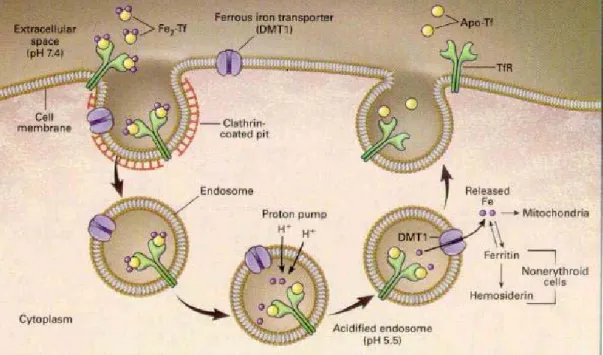
Absorption of iron consists of three steps: uptake into the intestinal mucosal cell, movement through the intestinal cell and release from the cell to the circulation. The uptake of iron is performed by a specific carrier protein called transferrin. When ferric iron is bound to transferrin, it is nonreactive. Transferrin bound to iron is recognized by receptors on the outer membrane of cells, which make a vesicle and transport the transferrins to a lysosome where the metal is released due to the lower pH of the lysosomal environment. The transferrin receptor is present on the surface of only those cells that need iron, because cells regulate how much transferring receptors they make according to their needs. The pathway of iron within the mucosal cell is not fully understood. There is no way that ionic iron can exist in the cell, so when the carrier protein releases the ion it is immediately bound to protein. There are many suggestions that Fe bound to Fe chelators like ATP and amino acids, and iron could also be bound to chaperon molecules or mediated via interactions of organelles such as mitochondria.30.31 Dietary iron is absorbed by the barrier cells of the duodenum and released into blood and is circulated to tissues throughout the body where it is used or put into storage by the protein ferritin (See Figure 2).
Figure 2. The Transferrin Cycle. Reproduced with permission from review article by
Andrews.29
Lack of iron can cause anaemia but excessive iron is toxic to humans. Iron toxicity for humans is above 20 milligrams of iron for every kilogram of weight, and 60 milligrams per kilogram is a lethal dose. 2,3,4
Iron overload is a collective name given for diseases causing excessive iron in the body. Iron overload may be caused by increased absorption of iron from the intestine even when the body does not require it. The most common genetic disorders classified as Iron overload disease are hereditary hemochromatosis, Thalassemia and Friedreich’s Ataxia. The elevated levels of iron lead to free radical-mediated tissue/organ damage and eventual death. 5
Hereditary haemochromatosis is an inherited disease. It is thought to be mainly caused by a mutation of a gene called HFE, which probably allows excess iron to be absorbed from the diet. This mutation is known as C282Y and to develop haemochromatosis you usually need two genes (one from each parent) to be C282Y. Not everyone who is borne with two copies of the mutated HFE gene develops the disease. Hereditary
haemochromatosis usually occurs between the ages of 30 and 60, as the build up of iron takes years. If left untreated excess iron builds up in the organs especially the liver, heart and pancreas. This may cause heart or liver failure, which can be fatal.
Haemochromatosis is more common in Caucasian or white populations, having a prevalence of approximately 1 in 200.
Many people now days are being diagnosed with hereditary haemochromatosis without showing any symptoms. Early symptoms of iron overload or hemochromatosis include:
fatigue, weakness, weight loss, joint pain and abdominal pain. The first method used for treatment is therapeutic phlebotomy. 5
There are two forms of thalassemia, α- and β-thalassemia. The disease leads to low production, and excessive destruction, of red blood cells. β-Thalassemia patients have a mutation in the beta globin chain. In the major form of treatment is blood transfusions, but iron overload from the transfusions may cause damage to the heart, liver and
endocrine systems. The mild form of β-thalassemia produces small red blood cells, with no symptoms. α-Thalassemia is caused by deletion of a gene or genes from the α-globin chain. The treatment is regular blood transfusions and folate supplementation. The regular blood transfusions causes iron overload and patients who receive a lot of blood require chelation therapy to remove iron from the body. This iron chelating agent binds to iron and causes it to be excreted in the urine. Untreated, thalassemia major leads to heart failure as well as liver dysfunction. 6
Friedreich’s Ataxia (FA) is a hereditary neurodegenerative condition. FA is an
autosomal recessive disease, which means that two defective gene copies, one from each parent, must be inherited to develop symptoms. The gene for FA codes for a protein called frataxin. Normal frataxin is found in the mitochondria, where it is thought to be involved in regulating the transport of iron. The disease is due to a noncoding sequence of DNA within the FRDA gene. The result is a decrease in gene expression, leading to a deficiency of frataxin, which leads to excess of iron within the mitochondria. 7
There are two ways of removing iron and the methods are: therapeutic phlebotomy and chelation therapy.
If production of red blood cells is normal but absorption of iron from the diet is too high, as in hereditary hemochromatosis, therapeutic phlebotomy is the method of
treatment. Therapeutic phlebotomy is simply removing blood to lower the amount of iron in the body. The best outcomes are achieved if therapy is begun before symptoms start. Men usually need to have 3 to 4 pints of blood taken each year (about once every 3 months) and women may need to have 1 to 2 pints of blood taken each year (about once every 6 months). 8
Chelation therapy
Patients who receive multiple blood transfusions for one reason or another may
eventually end up with iron overload. Although transfusion therapy prevents many of the complications of severe anemia, the body is unable to eliminate the excess iron contained in the transfused blood. The excess iron deposits in tissues and organs, resulting in
damage and organ failure. Chelation therapy is used to help the body eliminate the excess iron and prevent the iron overload.
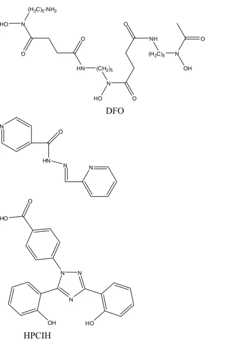
The most widely used iron chelator in haematology over the past 30 years is Desferrioxamine-B (DFO) (Fig. 3), also known as DesferalTM and manufactured by Novartis. It is a bacterial siderophore, produced to sequester iron from the environment.9 This drug has a major disadvantage being orally inactive and also very expensive. The administration of the drug is through long and frequent subcutaneous infusions, 12-24 hours a day, for 5-6 days a week. Therefore, there is an urgent need for an orally active iron chelating agent. Also, many patients are in the third world, so treatment needs to be cheap to reach maximum number of people. 1
To design an iron chelator for clinical use, the important factors to consider are metal selectivity and affinity, ligand-metal complex stability, bioavailability and toxicity. There have been many attempts to develop effective chelators, because of the problems with DFO. 10,32
One of the most promising ones has been deferiprone (Fig. 3), known as Ferriprox® and manufactured by Apotex Inc. Ferriprox is the only orally active Fe chelator approved for use in treating Fe overload. It has been shown that the oral iron chelator Ferriprox helps patients with Thalassemia. Unfortunately, when taken orally it is not as effective as subcutaneously given DFO. 11,12
ICL 670 or Deferasirox (Fig. 3) has recently been approved by the FDA (November 2005). Also known as ExjadeTM and manufactured by Novartis. This is an orally effective
drug that promotes removal of excess iron from the body. 13ICL 670 combines with the iron and both are removed by the kidneys.
Studies have been made of the 2-pyridylcarbaldehyde isonicotinoyl hydrazone class (HPCIH) (Fig. 3) showing that they have potential as orally active iron (Fe) chelators.14,15 The studies showed that some of the chelators of this class are more effective than DFO at preventing Fe uptake from Fe-transferrin and mobilizing Fe from cells. The same group has also been studying the H2IPH analogs and reported their high efficacy at
mobilizing Fe from cells and inhibiting Fe uptake from the serum Fe transport protein, transferrin.15 Three of those H2IPH analogs, namely H2BPH
(N-(benzoyl)-N’-(picolinoyl)hydrazine)), H2PPH (N,N’-bis(α-picolinoyl)hydrazine) and H2TPH
(N-(2-thiophenecarbonyl)-N’-(Picolinoyl)hydrazine) are the ones discussed in this report (Fig. 4).
N O HO (H2C)5-NH2 HN N HO O O NH (H2C)5 N OH O O (CH2)5 DFO N HN O N N N N N OH HO HO O HPCIH ICL670
Figure 3. Known Fe chelators and relevant biologically active compounds discussed in
N H N H O O N N N H O O N N H H2BPH H2PPH N N H NH O O S H2TPH
Figure 4. The H2IPH analogs: H2BPH (N-(benzoyl)-N’-(picolinoyl)hydrazine)), H2PPH
(N,N’-bis(α-picolinoyl)hydrazine) and H2TPH
(N-(2-Thiophenecarbonyl)-N’-(Picolinoyl)hydrazine).
Earlier studies have shown that these ligands displays great affinity for FeIII and the question is whether it is the same with other metal ions that are biological important. Several of the metal ions in the transition series (Mn, Co, Ni, Cu and Zn) are essential when it comes to the body but iron is by far the most abundant.
Iron is the most common metal in membranes, organelles and primitive cells. Zinc is the most common trace metal ion in the cytoplasm of advanced aerobic cells (much less common than some group 1 and 2 metal ions). Manganese is common in vesicles or organelles and copper is as common outside the cytoplasm and outside cells. Nickel and cobalt are not common anywhere in eukaryotes. 16
Nickel and cobalt will not be a problem since the concentration of both in human serum is below 1 µg/L compared with 100 µg/L for zinc, iron and copper. The activity of nickel in humans is confined to the enzyme urease where it acts as an acid catalyst for the decomposition of urea to generate ammonia. 16
The activity of cobalt is almost totally due to the functions of vitamin B12. There are
other cobalt enzymes though, that do not use B12, one is in methionine amino peptidase in
the cytoplasm. 16
The concentration of free manganese in cell cytoplasm is less than 10-7M. The biochemistry of Mn in all oxidation states is dominated by transfer to selected
in mitochondria and chloroplasts in such enzymes as superoxide dismutase as well as in lysosomes in acid phosphatases. 16
Free CuII is toxic to cells and it can damage biomolecules by radical formation. The free CuII concentration is less then 10-20 M (<1 atom per cell!!!!). Copper in cells is extremely tightly bound to proteins. Copper is exchanged between intracellular proteins known as copper chaperones. The chaperones have very high affinity for copper and their function is to transfer the ions to specific sites within cellular compartments or enzymes. This makes it very difficult for a Fe chelator to compete with the chaperone. 16
The biggest potential problem would be zinc. Zinc is the most common trace metal ion in the cytoplasm. The concentration of free zinc ions within biological cells varies from about 10-11M in the cytoplasm to about 10-3M in some vesicles. Zinc basically has three roles: acid catalyst, control ion and structural ion. On the other hand Zn depletion can be easily overcome by supplementation if necessary. 16
EXPERIMENTAL
Instrumentation & physical methods
Solution UV / visible spectra were measured on an Analytikjena Specord 210
spectrophotometer. Infrared spectra were measured on a Perkin-Elmer Model 1600 FT-IR spectrophotometer using an ATR accessory, diamond window. 1H NMR and 13C spectra were obtained with Bruker AV400 (400 MHz) instrument with TMS as internal standard. EPR spectra were recorded at 77 K on a Bruker ER200D spectrometer at X-band
frequency (9.303 GHz). Samples were prepared as 1 Mm solutions in 1:2 DMF:water. Spectra were simulated with the program EPR50F. 17
Cyclic voltammetry was performed with a BAS100B/W analyzer employing a platinum working electrode. For voltammetry in H2O an aqueous Ag/AgCl reference electrode was
employed (E° = 196 mV vs. NHE). For non-aqueous electrochemical experiments in DMF, a Ag/Ag+(DMF) reference electrode was used and referenced using ferrocene. Concentrations were 1 mM and 0.1 M phosphate (pKa2=7.2) buffer was used as the
supporting electrolyte for aqueous media and 0.1 M Et4NClO4 for non-aqueous solvent
systems.
MS (ESI) were run on a Finnigan MAT 900 XL-Trap instrument with a Finnigan APIIII electrospray source, using methanol as solvent. The measurements were done by
magnetic scan at a resolving power of 1,000.
X-ray crystallography. Cell constants at 293 K were determined by least-squares fit to the setting parameters of 25 independent reflections measured on an Enraf-Nonius CAD4 four-circle diffractometer employing graphite-monochromated Mo-Kα radiation
(0.71073 A) and operating in the ω-2θ scan mode within the range 2 < 2θ < 50 Å. Data reduction and empirical absorption corrections (ψ-scans) were performed with the WINGX suite of programs. Structures of metal complexes were solved by Patterson methods with SHELX or by SIR92, SIR 97 or SIR 2002. Crystal data appears in table 4 and selected bond lengths and angles are presented in table 5. The atomic nomenclature is defined in Figs. 11, 12,13 and 14 drawn with the program ORTEP3. 18
Materials
Commercial Materials
All commercially available chemicals and solvents used in this work were of analytical grade and were used without any further purification.
Precursors
Methyl Picolinate, C7H7NO2
2-Picolinic acid (20.0g, 6.1545 mmoles) was dissolved in 200 mL MeOH and the
solution was cooled to -10 ºC in a CaCl2/EtOH/icebath. SOCl2 (12 mL, 0.1645 mol) was
added dropwise, keeping the temperature below 0 ºC. The mixture was then stirred at room temperature for 1 hour and refluxed for 2 hours. MeOH was then removed by rotary evaporation. 25 mL MeOH was added and the solution taken to dryness again. 200 mL saturated Na2CO3 was added to the residue. The solution was extracted with 3x100 mL
CHCl3. The aqueous solution was dried with MgSO4 and filtered. Evaporation again
resulted in a clear colourless liquid. Yield 9 g (40 %)
Picolyl-hydrazide, C6H8N3O
Methyl picolinate (9 g, 65.6 mmoles) was dissolved in 40 mL MeOH. Hydrazine hydrate (7 mL, 0.144 mol) was added dropwise to the stirred solution. The mixture was refluxed for 12 hours. MeOH was removed on rotary evaporater. Diethyl ether was added to the residue and the product was obtained as a precipitate. The precipitate was recovered by filtration and washed twice with diethyl ether. The white solid was dried in a vaccum desiccator over night. Yield 9.59 g (107 %).
Methyl benzoate, C8H9O2
A MeOH solution of benzoic acid was cooled to -10 ºC in a CaCl2/EtOH/ice bath. SOCl2
(12 mL, 0.163 mol) was added dropwise, keeping the temperature below 0 ºC. The reaction was stirred at room temperature for one hour and then refluxed for two hours. MeOH was removed on a rotary evaporator. MeOH (25 mL) was added and the solution taken to dryness again. Saturated Na2CO3 solution (200 mL) was added to the residue.
The aqueous solution was extracted with CHCl3 (3x100 mL). The solution was dried with
MgSO4 then filtered. Evaporation resulted in a clear colourless liquid. Yield 21 g (95 %)
Benzoyl hydrazide, C7H9N2O
Methyl-benzoate (21g, 0.154 mol) was dissolved in 40 mL MeOH. To the stirred solution, hydrazine hydrate (15 mL, 0.308 mol) was added dropwise. The mixture was refluxed for 12 hours. MeOH was removed on the rotary evaporator. Diethyl ether was added to the residue and the product precipitated. The solid was washed twice with diethyl ether. The solid was dried in a vacuum desiccator overnight. Yield 15.54 g (75 %)
SYNTHESES
The N-(Isonicotinoyl)-N’-(picolinoyl) hydrazine Ligands
N-(Benzoyl)-N’-(picolinoyl)hydrazine), H2BPH
Benzoic acid (8.01 g, 0.0656 mol) and 1-methylimidazole (16.16 g, (3eqv.) 0.1968 mol) were placed in a flask with 125 mL DMF. The solution was cooled to 0 ºC. A mixture of p-toluenesulfonyl chloride (15.01 g, (1.2 eqv) 0.0787 mol) dissolved in 125 mL DMF was added and the reaction was stirred on ice for 30 minutes. Picolinic hydrazide (9g, 0.0656 mol) was dissolved in 75 mL DMF and added in small portions to the above solution still on ice, with the reaction temperature kept below 0 ºC. Reaction allowed to stir over night at 0 ºC initially, but slowly warming to RT. DMF (bp 153 ºC) was removed using a high vacuum pump. A yellow oil was obtained. 25 mL of diethyl ether was added to the oil and a precipitate was collected by filtration. The oil that remained was taken to dryness on the rotary evaporator. 100 mL H2O was added to the residue and
the product was extracted with 3x100 mL ethyl acetate. Ethyl acetate was removed on the rotary evaporator. The white solid was washed well with water. The product precipitated as a white mass and was filtered and washed twice with water, then dried in desiccator overnight. The product was recrystallized in 300 mL hot EtOH. Yield 11.83 g (75%). Melting point: 214-216 ºC.
IR: 3157.55(w, N-H str), 1679.23 (w), 1634.44 (s), 1579.02 (w, pyridine), 1487.44 (s, amide I), 1461.26 (m, amide I), 1433.87 (w, amide II), 1281.55 (s), 682.75 (s,
monosub.benzene) cm-1. 1H NMR (DMSO-d 6): δ=7.51 (m, 3H), 7.58 (m, 1H), 7.65 (m, 1H) 7.91 (d, 1H), 8.03 (m, 2H), 8.70 (d 1H), 10.53 (s, 1H), 10.62 (s, 1H) 13C NMR (DMSO-d 6): δ=122.39, 127.02, 127.5, 128.5, 131.86, 132.58, 137.86, 148.69,
149.28, 163.28, 165.46, 165.87 ppm. Extrapeaks δ=137.86 due to 1-methylimidazole and δ=165.87 due to formic acid, byproducts from decomposed DMF.
N,N’-Bis(α-picolinoyl)hydrazine, H2PPH.
Picolinic acid (10 g, 0.0812 mol) and 1-methylimidazole (20.03 g, 0.244 mol) were placed in a flask with 125 mL DMF and cooled to 0 ºC. A mixture of p-toluenesulfonyl chloride (18.6 g, (2.4 eqv) 0.0975 mol) dissolved in 125 mL DMF was added and the reaction was stirred on ice for 30 minutes. Hydrazine hydrate (2.03 g, 0.0406 mol) was added to the above solution, still on ice. The reaction was allowed to stir overnight at 0 ºC initially, but slowly warming to RT. A white precipitate began to form after ca. 20
minutes. This was collected by filtration and washed with two 5 mL portions of diethyl ether. The majority of the product remained in the filtrate. DMF was removed from the filtrate under high vacuum. Diethyl ether (100 mL) was added to the residue and the mixture was placed in the freezer overnight to induce the product to precipitate. The
product was obtained as a white powder after filtration and dried over vacuum. Total yield 7.37 g (75%). The product was recrystallized in 650 mL hot EtOH and cooled to RT overnight. The product was obtained as white crystals.
IR: 3317.50(s, N-H str), 1707.23 (m), 1669.15 (s), 1586.85 (w, pyridine), 1566.27 (w, pyridine), 1478.04 (s, amide I), 1459.91 (s, amide I), 1418.48 (m, amide II) cm-1.
1H NMR (DMSO-d
6): δ=7.66 (m, 2H), 8.01 (m, 4H), 8.69 (dt, 2H), 10.61 (s, 2H). 13C NMR (DMSO-d
6): δ=122.40, 127.02, 137.86, 148.67, 149.24, 163.00 ppm.
N-(2-Thiophenecarbonyl)-N’-(picolinoyl)hydrazine, H2TPH
2-Thiophenecarboxylic acid (11.35 g, 0.0886 mol) and 1-methylimidazole (21.80 g, 0.266 mol) were placed in a flask with 125 mL DMF and cooled to 0 ºC. A mixture of p-toluenesulfonyl chloride (20.3 g, (1.2eqv) 0.1063 mol) dissolved in 125 mL DMF was added and stirred on ice for 30 minutes. Picolyl-hydrazide (12.15 g, 0.0886 mol) was added to the above solution, still on ice. The reaction was stirred overnight at 0 ºC initially, but slowly warming to RT. DMF was removed using a high vacuum pump. Water (150 mL) was added to precipitate the product. The product was collected by filtration and washed with ice-cold water. The product was obtained as a white powder. The wet product was recrystallized in 1000 mL hot EtOH/water (9:1). After cooling to RT overnight, the product was obtained as a white powder. The solid was vacuum-dried. Yield 9.14 g (41.5%) IR: 3158.00 (s, N-H str), 3000.49 (s), 1620.76 (s), 1545.67 (m), 1502.72 (s), 1462.00 (w), 1435.87 (w), 1415.50 (m), 1356.48 (s), 1284.08 (s) cm-1. 1H NMR (DMSO-d 6): δ= 7.21 (t, 1H), 7.66 (m, 1H), 7.86 (m, ), 8.04 (dd, ), 8.70 (d, 1H), 10.52 (s, 1H), 10.64 (s, 1H). 13C NMR (DMSO-d 6), major conformer: δ=122.41, 127.04, 128.18, 129.05, 131.74,
137.16, 137.86, 148.68, 149.00, 160.86, 163.36 ppm. Extra peaks due to minor conformer δ=128.13, 128.96, 131.57, 137.45 ppm.
Bis-Ligand Complexes of N-(isonicotinoyl)-N’-(Picolinoyl) Hydrazine
Ligands
[Cu3(BPH)4](OH)2·4 H2O (AN23)
H2BPH (0.89 g, 0.0037 mol) was dissolved in 75 mL hot EtOH in a conical flask.
Cu(OAc)2·H2O (0.37 g, 0.0018 mol) was dissolved in 10 mL water and added to the
ethanolic H2BPH solution. The mixture was warmed gently for 10 minutes with stirring,
then cooled to RT. The product was filtered and obtained as a light green powder. It was dried in a vacuum dessicator overnight. Yield 0.36 g (35.9 %). The precipitate was
insoluble in all common solvents and only sparingly soluble in DMF/water (1:1).
Microanalysis found C, 49.41; H, 3.35; N, 13.04 %; calculated for C52H38Cu3N12O8(OH)2
· 4H2O gives C, 49.74; H, 3.85; N, 13.39 %.
IR: 1621.87 (w), 1598.98 (s), 1503.79 (w,), 1478.93 (s, amide I), 1434.76 (w, amide II), 1404.74 (s, amide I), 683.50 (s, benzene) cm-1
[Cu(HBPH)2]·1.5 H2O (AN24)
H2BPH (0.89 g, 0.0037 mol) was dissolved in 75 mL hot EtOH in a conical flask.
Cu(ClO4)2·6H2O (0.68 g, 0.00184 mol) was dissolved in 10 mL EtOH and added to the
H2BPH/ EtOH solution. The mixture was allowed to warm gently for 10 minutes under
stirring. Cooled to RT. The product 1a was filtered and obtained as a light green powder. Placed in dessicator over night to dry. Yield 0.21 g (20.9 %). A satisfactory microanalysis of 1a could not be obtained. The precipitate was insoluble in all common solvents. The filtrate was left for slow evaporation.This gave a second precipitate 1b, with a different composition to 1a. Yield 0.31g (39 %).
IR for 1a: 1624.21 (w), 1601.26 (w), 1582.76 (w), 1523.41 (s), 1505.55 (w), 1480.29 (s), 1436.07 (w), 1402.90 (s), 712.84 (s, benzene) cm-1.
Microanalysis found for 1b: C, 54.16; H, 3.74; N, 14.36 %; calculated for C26H20CuN6O4·1.5H2O gives C, 54.69; H, 4.06; N, 14.72 %.
IR for 1b: 1586.82 (w), 1480.77 (s), 1435.67 (w), 1399.20 (s), 685.04 (s, benzene) cm-1.
[Zn3(HBPH)4](OH)2·0.5H2O (AN29)
H2BPH (0.88 g, 0.0037 mol) was dissolved in 50 mL hot MeOH in a conical flask.
Zn(OAc)2·H2O (0.40 g, 0.00183 mol) was dissolved in 10 mL water and added to the
H2BPH/ MeOH solution. The mixture was allowed to warm gently for 10 minutes under
stirring. Cooled to RT. The product was filtered and obtained as a bright yellow powder. Placed in dessicator overnight to dry. Yield 0.77 g (77.0 %).
Microanalysis found C, 51.54; H, 3.55; N, 14.59 %; calculated for C52H38Zn3N12O8(OH2)·0.5H2O gives C, 52.13; H, 3.45; N, 14.03 %.
IR: 3057.11(w, N-H str), 1651.05 (w), 1589.45 (w), 1529.83 (s), 1439.53 (w), 1396.90 (s), 688.61 (s, benzene) cm-1.
UV/VIS (CHCl3/MeOH): λmax = 329 nm. ε329 = 24350 M-1cm-1.
[Ni(HBPH)2]·1.5 H2O (AN34)
H2BPH (0.90 g, 0.0037 mol) was dissolved in 50 mL hot MeOH in a conical flask.
H2BPH/ MeOH solution. The mixture was allowed to warm gently for 10 minutes with
stirring. A bright green solution formed, although no precipitation was observed. The solution was cooled to room temperature and left to slowly evaporate. After 3 days, a green precipitate was observed. This was collected by filtration and dried in a desiccator over night. Yield 0.92 g (92.0 %).
Microanalysis found C, 54.79; H, 3.46; N, 15.66 %; calculated for C26H20NiN6O4·1.5H2O
gives C, 55.15; H, 4.09; N, 14.84 %.
IR: 3055.73(w, N-H str), 1656.20 (w), 1588.31 (w), 1523.39 (s), 1489.38 (w), 1439.27 (w), 1395.19 (s), 684.62 (s, benzene) cm-1.
UV/VIS (CHCl3): λmax = 353 nm. ε353 = 11070 M-1cm-1.
[Co(BPH)2]·H2O (AN40)
H2BPH (0.89 g, 0.0037 mol) was dissolved in 40 mL hot MeOH in a conical flask.
Co(OAc)2·4H2O (0.46 g, 0.00185 mol) was dissolved in 20 mL water and added to the
H2BPH/ MeOH solution. A brownish solution formed and a light green precipitate was
observed. The mixture was allowed to warm gently for 30 minutes under stirring. Cooled to RT. The product was filtered and obtained as a light green powder. Placed in
dessicator overnight to dry. Yield 0.36 g (36 %).
Microanalysis found C, 55.81; H, 3.68; N, 15.05 %; calculated for C26H20CoN6O4·H2O
gives C, 56.02; H, 3.98; N, 15.08 %.
IR: 1648.93 (w, benzene), 1587.62 (w, pyridine), 1524.52 (s), 1489.37 (w), 1439.85 (w), 1393.45 (s), 686.96 (s, benzene) cm-1.
UV/VIS (Dioxane): λmax = 580 nm, ε580 = 253 M-1cm-1. Attempted synthesis “[Mn(BPH)2]” (AN41)
H2BPH (0.90 g, 0.00375 mol) was dissolved in 40 mL hot MeOH in a conical flask.
Triethylamine was added (1 eq). MnCl2·4 H2O (0.37 g, 0.0019 mol) was dissolved in 10
mL water and added to the H2BPH/ MeOH solution. A bright orange solution formed and
a orange precipitate was observed. The mixture was allowed to warm gently for 30 minutes under stirring, then cooled to RT. The product was filtered and obtained as a bright orange powder. Placed in desiccator overnight to dry. Yield 0.26 g (26 %). A satisfactory microanalysis could not be obtained and the precipitate was insoluble in all common solvents.
IR: 3435.03 (w), 3065.23 (w), 1641.85 (w), 1591.79 (w), 1524.39 (s), 1469.13 (w), 1442.22 (w), 1393.14 (s), 689.39 (s, benzene) cm-1.
[Cu(PPH)2]·3.5 H2O (AN13)
H2PPH (0.89 g, 0.0037 mol) was dissolved in 70 mL hot EtOH in a conical flask.
Cu(OAc)2·H2O (0.36 g, 0.0018 mol) was dissolved in 10 mL water and added to the
H2PPH/ EtOH solution. A bright green solution formed and a light green precipitate was
observed. The mixture was allowed to warm gently for 10 minutes under stirring, then cooled to RT. The product was filtered and obtained as a light green powder, then placed in desiccator overnight to dry. Yield 0.91 g (45.5%).
Microanalysis found C, 47.57; H, 3.67; N, 18.16 %; calculated for C24H18CuN8O4·3.5
H2O gives C, 47.33; H, 4.14; N, 18.40 %.
IR: 3449.68(w, N-H str), 1627.33 (w), 1601.80 (s), 1571.56 (w, pyridine), 1510.73 (s, amide I), 1434.71 (w, amide II), 1406.12 (s, amide I) cm-1.
Absorbs at 370 nm, shoulder (DMF)
[Zn(PPH)2]·0.5H2O (AN51)
H2PPH (0.44 g, 0.0018 mol) was dissolved in 50 mL hot CHCl3 in a conical flask.
Piperazine (0.18 g, 0.0009 mol) was added. Zn(NO3)2· 6H2O (0.27 g, 0.0009 mol) was
dissolved in 10 mL water and added to the H2PPH/ CHCl3 solution. The mixture was
allowed to warm gently for 30 minutes under stirring, then cooled to RT. The product precipitated but the powder was too fine so the solution was evaporated to dryness. Two 20 mL potions of EtOH were added and the mixture taken to dryness each time. The product was suspended in diethyl ether and then filtered. The product was obtained as a bright yellow powder and placed in desiccator with potassium hydroxide as a drying agent overnight. The precipitate was insoluble in all common solvents. Yield 0.44 g (89 %).
Microanalysis found C, 51.44; H, 3.43; N, 20.72 %; calculated for C24H18ZnN8O4·0.5
H2O gives C, 51.77; H, 3.44; N, 20.12 %.
IR: 3319.41(w, N-H str), 1670.15 (w), 1530.55 (s), 1474.63 (w), 1409.30 (m) cm-1.
Attempted synthesis “[Zn(PPH)2]” (AN35)
H2PPH (0.44 g, 0.0018 mol) was dissolved in 50 mL hot MeOH in a conical flask. LiOH
(0.15 g, 0.0036 mol) was added. Zn(NO3)2·6 H2O (0.54 g, 0.0018 mol) was dissolved in
10 mL MeOH and added to the H2PPH/ MeOH solution. The mixture was allowed to
warm gently for 30 minutes under stirring, then cooled to RT. The product was obtained as a bright yellow powder and placed in desiccator with potassium hydroxide as a drying agent overnight. Yield 0.72 g (72 %).
A satisfactory microanalysis could not be obtained. The precipitate was insoluble in all common solvents.
IR: 3404.08 (w, N-H str), 1657.29 (w), 1605.44 (w), 1573.60 (w), 1523.67 (s), 1472.80 (w), 1435.90 (w), 1407.01 (s), 1395.57 (s) cm-1.
Attempted synthesis “[Zn(PPH)2]” (AN17)
H2PPH (0.88 g, 0.00365 mol) was dissolved in 75 mL hot EtOH in a conical flask.
H2PPH did not dissolve so triethylamine was added dropwise until the ligand dissolved.
Zn(OAc)2·H2O (0.40 g, 0.0018 mol) was dissolved in 10 mL water and added to the
H2PPH/ EtOH solution. The mixture was allowed to warm gently for 10 minutes with
stirring, then cooled to RT. The product precipitated but the powder passed through the filter. The reaction was evaporated to dryness. 2x20 mL EtOH was added and then taken to dryness again. The product was washed in diethylether and filtered. The product was obtained as a bright yellow powder and placed in desiccator with potassium hydroxide as a drying agent over night. Yield 1.16 g (58 %).
A satisfactory microanalysis could not be obtained. The precipitate was insoluble in all common solvents.
IR: 2978.11(w, N-H str), 2602.66 (w, N-H str), 2496.46 (w, N-H str), 1671.34 (w), 1530.78 (s, amide I), 1475.83 (m, amide I), 1440.58 (w, amide II), 1408.94 (m, pyridine), 1396.59 (m, pyridine) cm-1.
Attempted synthesis “[Ni(PPH)2]” (AN30)
H2PPH (0.89 g, 0.0037 mol) was dissolved in 200 mL hot acetone in a conical flask.
Triethylamine was added (0.00185 mol). Ni(OAc)2·4H2O (0.46 g, 0.00185 mol) was
dissolved in 10 mL water and added to the H2PPH/ acetone solution. The mixture was
allowed to warm gently for 10 minutes with stirring, then cooled to RT. The reaction was then allowed to stand overnight. The yellow precipitate was collected by filtration. Yield 0.35 g (35 %).
A satisfactory microanalysis could not be obtained. IR: 1531.57 (s), 1401.11 (s) cm-1.
UV/VIS (CHCl3/MeOH): λmax = 377 nm, ε377 = 15245 M-1cm-1.
Attempted synthesis “[Ni(PPH)2]” (AN103)
H2PPH (0.20 g, 0.0008 mol) was dissolved in 15 mL hot CH2Cl2/ 15 mL hot MeOH in a
conical flask. Triethylamine (0.0005 mol) was added. NiCl2·6H2O (0.1 g, 0.0005 mol)
was dissolved in 5 mL MeOH and added to the H2PPH/ CH2Cl2/MeOH solution. The
reaction was stirred with heating for 20 minutes. The solution turned bright green immediately upon addition of NiCl2, though no precipitation occured. The mixture was
cooled to RT, then allowed to slowly evaporate from the beaker. Crystals of H2PPH were
Attempted synthesis “[Ni(PPH)2]” (AN69)
H2PPH (0.22 g, 0.0009 mol) was dissolved in 25 mL hot CHCl3 in a conical flask.
Triethylamine (0.091 g, 0.0009 mol) was added. NiCl2·6H2O (0.22 g, 0.0009 mol) was
dissolved in 5 mL EtOH and added to the H2PPH/CHCl3 solution. The mixture was
allowed to warm gently for 20 minutes under stirring, then cooled to RT. A cloudy apple green solution was observed with a mint green precipitate. The product was collected by filtration. Yield 0.37 g (76 %).
A satisfactory microanalysis could not be obtained.
IR: 3215.78 (w), 1657.10 (m), 1640.39 (s), 1592.83 (s), 1568.22 (m), 1471.18 (w), 710.45 (s), 683.83 (s) cm-1.
UV/VIS (H2O): λmax = 351 nm, ε351 = 4227 M-1cm-1. λmax = 267 nm, ε267 = 5865 M-1cm-1.
[Co(PPH)2]·1.75 H2O (AN39)
H2PPH (0.89 g, 0.0037 mol) was dissolved in 50 mL hot MeOH in a conical flask.
Co(OAc)2·4H2O (0.46 g, 0.0018 mol) was dissolved in 10 mL MeOH and added to the
H2PPH/ MeOH solution. The mixture was allowed to warm gently for 10 minutes under
stirring, then cooled to RT. After leaving the solution for several hours, an orange brown oily solid formed. This was collected by filtration and dried in a desiccator. Yield 0.3 g (30 %).
The precipitate was insoluble in all common solvents.
Microanalysis found C, 50.10; H, 3.17; N, 19.45 %; calculated for C24H18CoN8O4·1.75
H2O gives C, 50.32; H, 3.78; N, 19.56 %.
IR: 3319.95 (w), 1709.52 (w), 1650.62 (w), 1603.61 (w), 1524,94 (s), 1462.76 (m), 1435.67 (w), 1404.48 (m) cm-1.
[Co(PPH)2]·4.25H2O (AN54)
H2PPH (0.45 g, 0.0018 mol) was dissolved in 50 mL hot CHCl3 in a conical flask.
CoCl2·6H2O (0.22 g, 0.0009 mol) was dissolved in 15 mL EtOH and added to the
H2PPH/ CHCl3 solution. Triethylamine (0.091 g, 0.0009 mol) was added. The mixture
was allowed to warm gently for 30 minutes under stirring, then cooled to RT. No
precipitation was observed, so the mixture was left for slow evaporation. After 7 days of slow evaporation there was a gum in the beaker. Attempts to re-dissolve the gum in hot MeCN resulted in a light brown precipitate, which was filtered to obtain the product. Yield 0.27 g, (54 %).
Microanalysis found C, 46.85; H, 4.88; N, 17.41 %; calculated for C24H18CoN8O4·4.25
H2O gives C, 46.65; H, 4.32; N, 18.13 %.
IR: 3386.55 (w), 1626.37 (s), 1603.47 (s), 1566.19 (m), 1513.10 (s), 1473.91 (m), 1393.79 (m) cm-1.
UV/VIS (H2O): λmax = 422 nm, ε422 = 12102 M-1cm-1. λmax = 376 nm, ε376 = 22162 M -1cm-1.
Attempted synthesis [Co(PPH)2] (AN79)
H2PPH (0.45 g, 0.0018 mol) was dissolved in 50 mL hot MeCN in a conical flask.
CoCl2·6H2O (0.22 g, 0.0009 mol) was dissolved in 10 mL MeOH and added to the
H2PPH/ MeCN solution. Triethylamine (0.091 g, 0.0009 mol) was added. The mixture
was allowed to warm gently for 20 minutes with stirring, then cooled to RT. A dark green solution was noted at first (smell of acetic acid) then the solution turned into milky olive green solution containing an olive green precipitate. After standing overnight, the product was collected by filtration. Yield 0.31 g, (62 %).
A satisfactory microanalysis could not be obtained.
IR: 3082.23 (w), 1625.26 (w), 1563.31 (s), 1505.07 (m), 1471.64 (w), 1450.46 (w), 1381.32 (m) cm-1.
UV/VIS (H2O): λmax = 375 nm, ε375 = 4187 M-1cm-1. λmax = 340 nm, ε340 = 5753 M-1cm-1.
λmax = 261 nm, ε261 = 17972 M-1cm-1.
Attempted synthesis “[Mn(PPH)2]” (AN55)
H2PPH (0.45 g, 0.0019 mol) was dissolved in 50 mL hot CHCl3 in a conical flask.
MnCl2·4H2O (0.185 g, 0.0009 mol) was dissolved in 10 mL EtOH and added to the
H2PPH/ CHCl3 solution. Triethylamine (0.091 g, 0.0009 mol) was added. The mixture
was allowed to warm gently for 30 minutes under stirring, then cooled to RT. The orange solution was allowed to slowly evaporate. After 7 days the solution left for slow
evaporation was dry. The residue was tried to redissolved in CHCl3 and the product
precipitated as an orange-brown powder. Yield 0.21 g, (42 %).
A satisfactory microanalysis could not be obtained. The precipitate was insoluble in all common solvents.
IR: 1648.43 (w), 1525.21 (s), 1469.45 (w), 1434.33 (w), 1392.04 (s), cm-1.
[H4PPH]Cl2 (AN79B)
H2PPH (0.45 g, 0.0018 mol) was dissolved in 50 mL hot MeCNin a conical flask.
CoCl2·6H2O (0.22 g, 0.0009 mol) was dissolved in 10 mL MeOH and added to the
H2PPH/ MeCN solution. Triethylamine (0.091 g, 0.0009 mol) was added. During the
addition of the dissolved CoCl2 salt there was a strong smell of acetic acid. The solution
became dark green immediately, but after ~5 min it turned milky olive green. The
olive green powder was obtained by filtration. Yield 0.31 g (62 %). The pale green filtrate was left for slow evaporation. After 2 days, light brown crystals had formed suitable for X-ray work. According to the IR it was not PPH.
Microanalysis found C, 47.06; H, 3.70; N, 18.17 %; calculated for C12H12Cl2N4O2 gives
C, 45.73; H, 3.84; N, 17.78 %.
IR: 3319.92 (w), 3078.20 (m), 2722.29 (m), 2071.82 (w), 1996.29 (w), 1659.17 (s), 1597.73 (s), 1520.76 (w), 1467.84 (s), 1440.44 (s) cm-1.
Attempted synthesis “[Cu(TPH)2]” (AN59)
H2TPH (0.44 g, 0.0018 mol) was dissolved in a mixture of 30 mL hot CHCl3 and 30 mL
hot MeOH in a conical flask. Triethylamine (0.091 g, 0.0009 mol) was added.
CuCl2·2H2O (0.15 g, 0.0009 mol) was dissolved in 5 mL EtOH and added to the H2TPH/
CHCl3/MeOH solution. The mixture was allowed to warm gently for 20 minutes under
stirring, then cooled to RT. A green precipitate was formed immediately. The product was filtered and put in desiccator to dry. Yield 0.32 g, (64 %).
A satisfactory microanalysis could not be obtained. The precipitate was insoluble in all common solvents.
IR: 3087.67 (w), 1669.15 (s), 1565.50 (m), 1523.64 (s), 1486.80 (m), 1421.04 (m), 1403.23 (m) cm-1.
Attempted synthesis “[Zn(TPH)2]” (AN63)
H2TPH (0.44 g, 0.0018 mol) was dissolved in 30 mL hot CHCl3 and 30 mL hot MeOH in
a conical flask. Zn(OAc)2·2 H2O (0.2 g, 0.0009 mol) was dissolved in 5 mL MeOH and
added to the H2TPH/ CHCl3/MeOH solution. The mixture was allowed to warm gently
for 20 minutes with stirring, then cooled to RT. A pale yellow cloudy precipitate was formed immediately upon mixing. The precipitate was put in a desiccator to dry. Yield 0.27 g, (54 %).
A satisfactory microanalysis could not be obtained. The precipitate was insoluble in all common solvents.
IR: 1592.63 (w), 1563.58 (w), 1530.76 (m), 1500.42 (s), 1470.06 (m), 1433.10 (s), 1400.71 (m), 1373.04 (m) cm-1.
[Ni(TPH)2]·0.5H2O (AN73)
H2TPH (0.45 g, 0.0018 mol) was dissolved in 30 mL hot CHCl3 and 30 mL hot MeOH in
0.0009 mol) was dissolved in 5 mL MeOH and added to the H2TPH/ CHCl3/MeOH
solution. The mixture was allowed to warm gently for 20 minutes with stirring, then cooled to RT. A bright green solution was formed, but no precipitate. After 5 days x-ray quality crystals were obtained by vapour diffusion of acetone into this solution. Yield 0.02 g (9 %).
Microanalysis found C, 46.41; H, 2.99; N, 14.47 %; calculated for C22H16NiN6O4S2·0.5H2O gives C, 46.42; H, 3.19; N, 14.76 %.
IR: 3066.25 (w), 1651.15 (s), 1602.33 (w), 1501.45 (s), 1419.21 (m), 1393.90 (s), 1351.70 (m), 1302.57 (m), 710.45 (s), 683.83 (s) cm-1.
UV/VIS (DMF): λmax = 368 nm, ε368 = 10428 M-1cm-1.
[Ni(TPH)2]·1.75H2O (AN92)
H2TPH (0.22 g, 0.0009 mol) was dissolved in 15 mL hot CHCl3 and 15 mL hot MeOH in
a conical flask. Triethylamine (0.091 g, 0.0005 mol) was added. NiCl2·6H2O (0.11 g,
0.0005 mol) was dissolved in 5 mL MeOH and added to the H2TPH/ CHCl3/MeOH
solution. The mixture was allowed to warm gently for 20 minutes under stirring, then cooled to RT. From the bright green solution, precipitation occured after five minutes and was filtered. Yield 0.04 g (16 %). X-ray quality crystals were obtained after 5 days by vapour diffusion of acetone into the filtrate solution.
Microanalysis found C, 45.13; H, 2.96; N, 14.3 %; calculated for C22H16NiN6O4S2·1.75
H2O gives C, 45.34; H, 3.37; N, 14.42 %.
IR: 3065.11 (w), 2947.72 (w), 1650.91 (s), 1602.11 (w), 1500.24 (s), 1419.66 (m), 1393.38 (s), 1351.11 (m), 1303.40 (m), 700.60 (s), 684.22 (s) cm-1.
UV/VIS (1:2, MeOH/MeCN): λmax = 353 nm, ε353 = 3633 M-1cm-1.
[Ni(TPH)2]·1.5H2O (AN97)
H2TPH (0.22 g, 0.0009 mol) was dissolved in 30 mL hot CHCl3 and 30 mL hot MeOH in
a conical flask. Triethylamine (0.091 g, 0.0005 mol) was added. NiCl2·6H2O (0.11 g,
0.0005 mol) was dissolved in 5 mL MeOH and added to the H2TPH/ CHCl3/MeOH
solution. The mixture was allowed to warm gently for 20 minutes under stirring. The bright green solution was cooled to RT. The solution was adsorbed onto an aluminia column (30x1 cm Merck, Al2O3 90 active neutral, Activity stage 1), containing MeOH.
Two bands were eluted, using MeOH containing 1 % aq. NH3 as the eluting agent. The
first band was a dark yellow colour. A precipitate formed after two days slow
evaporation. The second band was lighter yellow colour. Band 1: yield 0.04 g (16%). The precipitate was insoluble in all common solvents.
Microanalysis found C, 46.02; H, 2.8; N, 14.2 %; calculated for C22H16NiN6O4S2·1.5 H2O
gives C, 45.7; H, 3.31; N, 14.53 %.
IR: 3064.95 (w), 2802.24 (w), 1651.46 (m), 1518.32.11 (s), 1418.73 (m), 1394.85 (s), 1369.16 (w), 1351.30 (m), 711.58 (s), 685.13 (s) cm-1.
[Co(TPH)2]·1.25 H2O (AN100B)
H2TPH (0.22 g, 0.0009 mol) was dissolved in 15 mL hot CH2Cl2 and 15 mL hot MeOH
in a conical flask. Triethylamine (0.00045 mol) was added. CoCl2·6H2O (0.11 g, 0.00045
mol) was dissolved in 5 mL MeOH and added to the H2TPH/ CHCl3/MeOH solution. The
mixture was allowed to warm gently for 20 minutes with stirring, then cooled to RT. An olive green solution was obtained but no precipitate. A green precipitate in beaker began to form after 10 minutes. This was collected by filtration. Yield 0.03 g (12 %).
Microanalysis found C, 46.17; H, 2.74; N, 14.49 %; calculated for C22H16CoN6O4S2·1.25H2O gives C, 46.04; H, 3.25; N, 14.64 %.
IR: 3067.56 (w), 2988.38 (w), 1644.09 (m), 1601.04 (w), 1523.23 (s), 1420.31 (m), 1393.79 (m), 712.52 (s), 688.88 (s) cm-1.
UV/VIS (CHCl3/MeOH ): λmax = 332 nm, ε332 = 24356 M-1cm-1.
NH4[Co(TPH)2]·H2O (AN89A)
H2TPH (0.22 g, 0.0009 mol) was dissolved in 30 mL hot CHCl3 and 30 mL hot MeOH in
a conical flask. Triethylamine (0.091 g, 0.00045 mol) was added. CoCl2·6H2O (0.11 g,
0.00045 mol) was dissolved in 5 mL MeOH and added to the H2TPH/ CHCl3/MeOH
solution. The solution was refluxed for 20 minutes with stirring. An olive green solution formed. This was cooled at 4 °C overnight. A precipite of TPH formed. The TPH was removed by filtering the mixture. The filtrate was adsorbed onto an alumina column (30x1 cm Merck, Al2O3 90 active neutral, Activity stage 1), containing MeOH, to
separate any remaining free ligand from the complex. Five bands were eluted, using MeOH containing 1 % aq. NH3 as the eluting agent. The first band was a yellow colour.
A green precipitate formed immediately. The second band was lighter yellow colour. Band 3 was dark yellow, band 4 lighter yellow and band 5 paler yellow than band 4. The 5 fractions were left for slow evaporation. After one day there were dark brown crystals suitable for x-ray work found in fraction 4. Band 4: yield 0.01 g. After 7 days there were dark brown crystals suitable for x-ray work found in fraction 1. Band 1: yield 0.04g. Microanalysis found C, 41.76; H, 3.88; N, 16.72 %; calculated for C22H16CoN6O4S2·NH4
gives C, 41.83; H, 3.51; N, 16.63 %.
IR: 3067.65 (w), 2958.71 (w), 1643.03 (m), 1600.60 (w), 1523.61 (s), 1500.75 (s), 1419.52 (m), 1392.73 (m), 711.25 (s), 687.67 (s) cm-1.
UV/VIS (H2O): λmax = 404 nm, ε375 = 13322 M-1cm-1. λmax = 302 nm, ε340 = 18581 M -1cm-1. λ
max = 245 nm, ε261 = 30850 M-1cm-1.
[Co(TPH)2]·0.5H2O (AN98:1)
H2TPH (0.22 g, 0.0009 mol) was dissolved in 15 mL hot CHCl3 and 15 mL hot MeOH in
a conical flask. Triethylamine (0.091 g, 0.0009 mol) was added. CoCl2·6H2O (0.21 g,
0.0009 mol) was dissolved in 5 mL MeOH and added to the H2TPH/ CHCl3/MeOH
solution. The mixture was allowed to warm gently for 20 minutes with stirring, then cooled to RT. After five minutes the dark green solution turned orange brown and was allowed to slowly evaporate. After three days several crystals had formed suitable for X-ray work. Filter. Yield 0.015 g
Microanalysis found C, 46.64; H, 2.92; N, 14.33 %; calculated for C22H16CoN6O4S2·0.5
H2O gives C, 46.77; H, 3.12; N, 14.88 %.
IR: 3667.51 (w), 3081.29 (w), 2987.35 (w), 1643.43 (m), 1600.51 (w), 1524.46 (s), 1418.85 (m), 1392.44 (m), 709.61 (s), 686.48 (s) cm-1.
UV/VIS (DMF): λmax = 339 nm, ε339 = 47881 M-1cm-1
ESI-MS (m/z): 597 (4 %) [CoII(HTPH)2 + 2 Na]
596 (12 %) [CoIII(HTPH)(TPH) + 2 Na] 595 (60 %) [CoIII(TPH)2 + 2 Na]
613 (19 %) [[CoII(HTPH)
2 + 2 Na + O]
612 (15 %) [CoIII(HTPH)(TPH) + 2 Na + O] 611 (100 %) [CoIII(TPH)2 + 2 Na + O]
629 (12 %) [CoII(HTPH)2 + 2 Na + 2O]
628 (4 %) [CoIII(HTPH)(TPH) + 2 Na + 2O] 627 (52%) [CoIII(TPH)2 + 2 Na + 2O]
Attempted synthesis “[Mn(HTPH)2]” (AN88)
H2TPH (0.11 g, 0.0005 mol) was dissolved in 15 mL hot CHCl3 and 15 mL hot MeOH in
a conical flask. Triethylamine (0.091 g, 0.0005 mol) was added. MnCl2·4H2O (0.18 g,
0.0009 mol) was dissolved in 5 mL MeOH and added to the H2TPH/ CHCl3/MeOH
solution. The mixture was allowed to warm gently for 20 minutes under stirring, then cooled to RT. Precipitation occured immediately and the precipitate collected by filtration.Yield 0.11 g (22 %).
A satisfactory microanalysis could not be obtained. The precipitate was insoluble in all common solvents, only sparingly in DMF.
IR: 2030.82 (w), 1590.00 (m), 1563.30 (m), 1532.63 (m), 1486.47 (s), 1426.44 (m) cm-1. UV/VIS (DMF): λmax = 351 nm, ε351 = 18059 M-1cm-1
Attempted synthesis “[Mn(HTPH)2]” (AN78)
H2TPH (0.45 g, 0.0018 mol) was dissolved in 65 mL hot MeCNin a conical flask.
Triethylamine (0.091 g, 0.0009 mol) was added. MnCl2·4H2O (0.18 g, 0.0009 mol) was
dissolved in 5 mL MeOH and added to the H2TPH/ MeCN solution. The mixture was
allowed to warm gently for 20 minutes under stirring, then cooled to RT. A yellow solution was obtained, but no precipitate. The mixture was left to slowly evaporate and one vial for vapor diffusion in acetone.
RESULTS/DISCUSSION
Syntheses and physical properties
The aim of this project was to investigate the coordination chemistry of the H2TPH,
H2BPH and H2PPH ligands with metals of biological importance, other than iron. These
ligands have great potential as orally active iron chelators, but to be suitable for in vivo use they must bind selectively to iron, despite the presence of other metals within the cell. For this reason, the coordination behaviour of these ligands with the biologically
important metals manganese, cobalt, nickel, copper and zinc was investigated. The coordination chemistry with biological important metals and these ligands is important for the understanding in the affinity towards metals and if metals other than iron can act as competitors for Fe.
There are several other methods for making the ligands. For example, H2PPH can be
made by reacting picolyl chloride with hydrazine in aqueous solution.19 The previous methods of preparing H2TPH, H2BPH has been to react a pentafluorophenolate ester with
an aromatic hydrazide (see reaction scheme 1). These reagents are expensive and PFP, in particular, is toxic. 20 N O O + OH F F F F F + N O O F F DCC F F F + DCU (1) (2) N O O F F F F F + NH NH O Ar O Ar NHNH2 O
Scheme 1. Reaction scheme for making H2TPH, H2BPH and H2PPH.
An alternative method have been used herein to synthesize the ligands. The ligands H2TPH, H2BPH and H2PPH were synthesized through in situ, coupling picolinic
hydrazide with the corresponding acid: picolinic acid for H2PPH, benzoic acid for
reactive and the reagents cheap. Yields for the reaction were good, in the range 50-75 % after work-up (see reaction in scheme 2).
O O N N + i)TsCl/DMF 0oC, 30 min N N O N N O + H 2NNH N O i) DMF 0oC, 2 h NH O NH O N
Scheme 2. Reaction scheme for making H2BPH.
From the H2TPH, H2BPH and H2PPH ligands, bis-ligand [MII(HL)(L)] and
[MIII(HL)(L)] complexes were synthesized and isolated, even when the metal and ligand ratio were reacted in a 1:1 ratio. This in contrast to the behaviour observed with iron, where complexes of formula [M(HL)(L)] and [M(HL)Cl2] have been isolated.
Isolating the complexes was not straight forward and the same method could not be used for the different ligands, mostly due to insolubility in different solvents. With the different ligands, the choice of solvent, base and metal salt was a critical step in isolating the complexes and most importantly getting the complexes to crystallize. The added base helps to deprotonate the ligand. The pKa of the first hydrazine proton falls in the range
2.45 – 2.74. Most often one equivalent of metal and two equivalents of ligand were allowed to react in the solvent and the complex precipitated almost immediately. The individual methods described in the experimental section were found to be the optimum methods for making the complexes for each ligand and metal.
The synthesis of complexes with the H2BPH ligand was problematic, since most of the
obtained products were insoluble in nearly all solvents and simply did not give any crystals, due to rapid precipitation from solution. The complexes that were formed precipitated immediately from the reaction mixture and recrystallization or further investigation of the product was often impossible, due to their insolubility in all common solvents. Another problem was decomposition of the complexes in solution, with
[M(HL)2] + 2H+ M2++ 2H2L
The equilibrium is drawn to the right by precipitation of the ligand from solution. This is also in contrast to the behaviour observed for iron, where the complexes were extremely stable, once formed.
While this work was in progress, a report was found of the crystal structures of the ligand BPH with Ni, Mn and V.21 The reported experimental procedure did not, in this case, give the results reported. There is also another group reporting the crystal structure of nickel and the H2BPH ligand, using approximately the same method as herein.22
Some interesting results were obtained from the reactions between the ligand BPH and the acetate salts of Zn and Cu. The microanalysis suggested a formulation of 3 metals bound to 4 ligands, [Zn3(BPH)4](OH2)·4H2O and [Cu3(BPH)4](OH2)·0.5H2O
respectively. Copper and zinc can both form complexes with donor numbers of 5 or 6 and ligands having oxygen or nitrogen as donor atoms are able to form complexes with these metals. H2BPH can act as a tridentate ligand, using three donor atoms (N, N, O) in
bonding to a single metal ion. It could also act as a bridging ligand, potentially binding to two metal centres. Therefore, there are different possibilities of how the structure, [M3L4]
could look. NMR was performed on the [Zn3(BPH)4]·3H2O complex in the aim of
eliminating some of the possibilities by examining the symmetry of the complex, but unfortunately it did not give a result due to low solubility.
The synthesis of complexes with the H2PPH ligand was also problematic, with the same
problems as with the H2BPH ligand. The complex, that was formed, precipitated
immediately from the reaction mixture and recrystallization or further investigation of the product was hampered by insolubility in all common solvents. When the best synthetic method finally was found, the next problem was keeping the complex in solution long enough to get crystals, because the ligand often precipitated. Crystals were isolated but these were only of H2PPH itself. This may indicate that the metal only binds to one
ligand and therefore reactions were tried with a 1:1 M:L ratio, but with no better result. Some problems were encountered with different zinc salts. The Zn ions seemed too be bound too hard to the counterions in the salts and the ligand was unable to compete for binding. For example, ZnCl2 and the ligand H2PPH did not react at all. In one attempt, 10
equivalents of triethylamine were added to the solution to totally deprotonate the ligand, but again there was no reaction. Different bases and different salts were tried to get a better result, but with zinc the result was either totally insoluble compounds or
compounds that had an uncertain formulation. Similar problems were seen with nickel. EPR of [Cu(HPPH)2]·3.5H2O showed a broad spectrum which is consistent with
suspended solid or polymerization. The insolubility in all common solvents points to the likelihood of H2PPH forming polymeric compounds with other metals than iron.
Other groups working with the same ligand have reported syntheses with the ligand in its deprotonated form (L2-) forming complexes with Au and Ru.23,19 These metals are much less labile than the first row transition metals, which would explain why discrete metal complexes were obtained. Another group has described a related ring opening of a reduced tetrazine. The hydrolysis of this tetrazine, favoured the formation of monomeric
complexes. They did not observe the hydrazido(2-) formation.24 None of the groups observed anything pointing to polymerization. Bernhardt et al. found that H2PPH resisted
all attempts to form a 1:2 Fe:L complex and proposed that it is possible that more complex oligomeric structures are being formed in solution.19
An attempt to synthesise [Co(HPPH)(PPH)] led to an unexpected result. Light brown crystals were found in the filtrate left for slow evaporation. IR spectroscopy was used to exclude the crystals being H2PPH. One crystal was mounted for X-ray crystallography
and it was subsequently revealed as the doubly protonated ligand; [H4PPH]Cl2. No acid
was added so the question is where the protons came from. Indeed, triethylamine was added to the reaction. The protonated form of H2PPH has been studied before in
connection with its anti-HIV and antitumor potential.25
The synthesis of complexes with the H2TPH ligand was more straight forward since the
work with the other two ligands prepared this way. The only thing that changed was the solvent, because H2TPH was not soluble in pure MeOH. A 1:1 mixture of CHCl3/MeOH
was used. Problems were seen here also with [Cu(TPH)2] being insoluble in all common
solvents. Again the reactions between the ligand and different Zn salts did not result in complex formation. The only Zn salt that gave a reaction was Zn(OAc)2, but the product
from the reaction was insoluble in most common solvents. It was sparingly soluble in DMSO, but not soluble enough to obtain a NMR spectrum. With MnCl2 no satisfactory
result was obtained. There was a reaction as evidenced by a colour change, but the complex was unstable in solution and H2TPH kept precipitating. The reactions with NiCl2
and CoCl2 and the ligand gave a small amount of crystals suitable for x-ray
crystallography after several days. Unfortunately, the solutions also contained
precipitated H2TPH. The crystals were found after precipitated ligand had been filtered
off several times. The ratio of L:M was changed to 1:1, and then to 1:2, but no better results were observed. An unexpected result was that the crystal of [Co(HTPH)2]·0.5H2O
revealed a CoII complex, which is surprising, as the reaction was performed in a flask open to the air and because CoII is readily oxidized to CoIII in solution under open air. In an attempt to separate the Co complex from the free ligand, the reaction mixture was passed over an alumina column. MeOH was used as an initial eluent, with the last fraction being eluted with MeOH containing a few drops of aqueous ammonia. This last fraction was left in a beaker in the fumehood and dark brown crystals formed overnight. These crystals had a different colour compared to the other [CoII(TPH)2] crystals, which
were green. Assuming that adding NH3 must have forced the CoII to oxidize, this new
compound must be the CoIII complex (see scheme 3).
[CoII(HTPH) 2] + NH3 [CoII(HTPH)(TPH)]-+ NH4+ O2+ 4H+ 2 H2O [CoIII(HTPH)(TPH)] + NH3 [CoIII(TPH) 2]-+ NH4+
Scheme 3. Oxidation of CoII complex to CoIII.
The microanalysis results could be fitted to [CoIII(TPH)2]·1.5NH4Cl but the crystal
structure was off insufficient resolution to locate the ions (R = 12 %) (The structure is discussed under description of crystal structures).
Spectroscopic properties
The IR spectra for the ligand H2BPH shows two bands at 1679 cm-1 and 1634 due to the
two different carbonyl groups. Only one strong band at 1669 cm-1 is seen for the symmetrical H2PPH (Figures 5 and 6). For H2TPH (Figure 7) only one strong (νc = 0)
band at 1620 cm-1 could be seen, when two bands were expected. This may be due to the carbonyl bands laying on top of each other. The H2TPH spectrum also shows the amide II
band at 1500 cm-1 and a much weaker amide III band at 1280 cm-1. Two bands around 680-620 cm-1 are due to the thiophene ring.
4000.0 3000 2000 1500 1000 550.0 70.9 74 76 78 80 82 84 86 88 90 92 94 96 98 100 102 103.7 cm-1 %T 3008.32 1686.40 1639.98 1580.19 1510.15 1490.98 1461.71 1434.69 1300.87 1282.53 1245.34 1121.50 1040.95 997.77 916.93 879.87 820.57 743.78 682.85 619.84 Figure 5. IR spectrum of H2BPH.
4000.0 3000 2000 1500 1000 550.0 51.0 55 60 65 70 75 80 85 90 95 100.0 cm-1 %T 3314.15 3062.91 1707.31 1670.66 1588.47 1566.69 1476.43 1459.12 1419.81 1294.70 1278.05 1237.68 1141.29 1087.22 1040.85 995.36 974.04 927.28 888.44 842.58 814.36 767.41 746.66 725.00 694.25 647.33 616.81 591.47
4000.0 3000 2000 1500 1000 550.0 52.0 55 60 65 70 75 80 85 90 95 99.4 cm-1 %T 3158.00 3000.49 1620.76 1589.13 1545.67 1502.72 1462.00 1435.87 1415.50 1356.48 1329.24 1284.08 1247.51 1114.61 1098.10 1039.69 997.96 907.94 841.29 711.58 688.07 608.64 Figure 7. IR spectrum of H2TPH.
The IR spectra of the complexes with H2BPH show bands at 1650 cm-1, which may be
assigned to the non-coordinated C=O group, while bands at 1590 cm-1 may be assigned to the coordinated C=O group. This is a shift of 30-60 cm-1 from the free ligand, which shows a weakening of the C=O bond upon complexation. The bands around 1525-1530 and 1395 cm-1 are assigned to the amide I and amide II bands of the singly-deprotonated ligand.
The IR spectra of the complexes with H2PPH show bands at 1627 or 1670 cm-1 which
may be assigned to the non-coordinated C=O group, while bands at 1603 cm-1 may be
assigned to the coordinated C=O group. The exception here is the complex between Zn and H2PPH which only shows one band at 1670 cm-1 and indicating that the C=O groups
are both non-coordinated (Figure 8). The peaks around 1510-1530 and 1400 cm-1 are assigned to the amide I and amide II bands of the singly-deprotonated ligand. The zinc complex only has one band at 1524 cm-1 and that may be assigned to the enol -O-C=N group of the fully deprotonated ligand (Figure 8).
4000.0 3000 2000 1500 1000 550.0 50.3 55 60 65 70 75 80 85 90 95 99.3 cm-1 %T 3319.41 1670.15 1530.55 1474.63 1409.30 1394.53 1293.49 1156.83 1088.74 1041.13 996.80 896.88 813.54 749.06 728.27 690.17 637.89 617.19 Figure 8. IR spectrum of [Zn(PPH)2].
The IR spectra of the complexes with H2TPH show bands at 1645 cm-1 which are
assigned to the non-coordinated C=O group, while bands at 1602 cm-1 are assigned to the coordinated C=O group. The exception here is the complex between Cu and H2TPH
which only shows one band at ca. 1669 cm-1 and that may again indicate that the C=O groups are both non-coordinated (Figure 9). The absorption maxima around 1520 and 1395 cm-1 are assigned to the amide I and amide II bands of the singly-deprotonated ligand. The copper complex has only one absorption maxima at. 1524 cm-1 and that may be assigned to the enol -O-C=N group of the fully deprotonated ligand.
4000.0 3000 2000 1500 1000 550.0 71.6 74 76 78 80 82 84 86 88 90 92 94 96 98 100 101.9 cm-1 %T 3161.04 3087.67 1669.15 1604.88 1565.50 1523.64 1486.80 1421.04 1403.23 1358.67 1313.78 1271.891131.17 1094.51 1024.23 861.28 831.76 798.75 751.83 711.26 673.10 647.96 562.30
Figure 9. IR spectrum of [Cu(TPH)2].
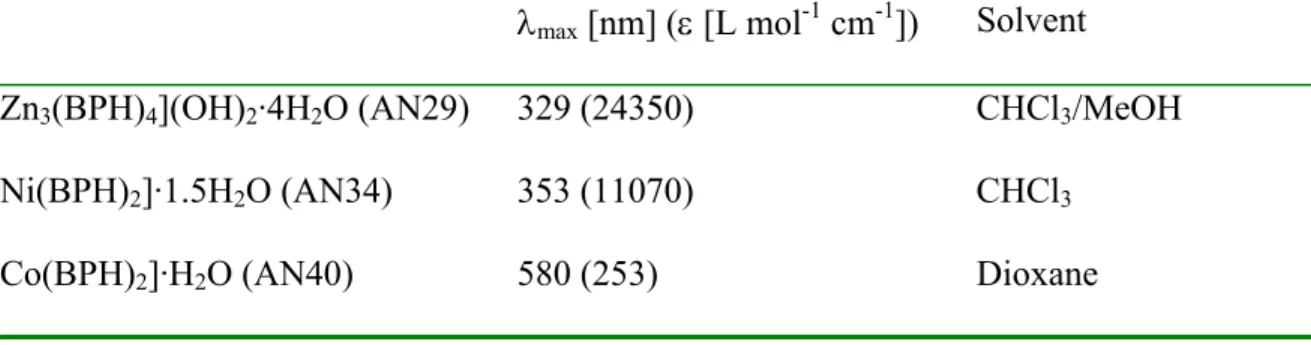
The electronic spectra of bis-ligand metal complexes of the ligands are listed in Tables 1, 2 and 3. Because of solubility issues, different solvents were used. The LMCT bands for the different complexes appears between 320 and 380 nm. These energy absorptions are most probably due to ligand-to-metal charge transfer and intraligand transitions. This is supported by the fact that the transition is seen for ZnII, which having a 3d10
configuration, has no d-d transitions. The extinction co-efficients are also higher than the value expected for d-d transitions (10 to 100 M-1cm-1).
Table 1. Electronic spectral data (different solvents, 298 K) of the metal complexes with
H2BPH.
λmax [nm] (ε [L mol-1 cm-1]) Solvent
[Zn3(BPH)4](OH)2·4H2O (AN29) 329 (24350) CHCl3/MeOH
[Ni(BPH)2]·1.5H2O (AN34) 353 (11070) CHCl3
[Co(BPH)2]·H2O (AN40) 580 (253) Dioxane
Table 2. Electronic spectral data (different solvents, 298 K) of the metal complexes of
H2PPH.
λmax [nm] (ε [L mol-1 cm-1]) Solvent
[Co(PPH)2]·4.25H2O (AN54) 422 (12100), shoulder
376 (22160) H2O
“[Co(PPH)2]” (AN79) 375 (4190), shoulder
340 (5750) 261 (17970)
H2O
“[Ni(HPPH)2]” (AN30) 377 (15240) CHCl3/MeOH
“[Ni(HPPH)2]” (AN69) 351 (4230)
267 (5870)
H2O
]Table 3. Electronic spectral data (different solvents, 298 K) of the metal complexes of
H2TPH.
λmax [nm] (ε [L mol-1 cm-1]) Solvent
[Co(TPH)2]·1.25H2O (AN100B) 332 (24360) CHCl3/MeOH
NH4 [Co(TPH)2]·H2O (AN89A) 404 (13320)
302 (18580) 245 (30850)
H2O
[Co(TPH)2]·0.5H2O (AN98;1) 339 (47880) DMF
[Ni(TPH)2]·1.75H2O (AN92) 353 (3630) MeCN/MeOH
[Ni(TPH)2]·0.5H2O (AN73) 368 (10430) DMF
“[Mn(TPH)2]” (AN88) 351 (18060) DMF
The metal complexes of H2TPH, H2BPH and H2PPH ligands are all, except for the
complexes with ZnII and CoIII, paramagnetic and hence give rise to paramagnetic shifts and line-broadening in their NMR spectra. The problem is that NMR active compounds must be extremely pure to produce an NMR spectrum. Mixtures of paramagnetic metal ions nullify all signals. Due to these problems no NMR spectra of NH4 [Co(TPH)2]·H2O
were obtained. Attempts to obtain a 1H NMR spectrum was thwarted by the presence of
CoII paramagnetic impurities.
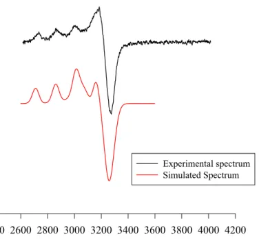
Since copper (II) complexes are paramagnetic, EPR spectra of 1 mM DMF:water (1:2) solutions (saturated) of [Cu3(BPH)4](OH)2·4H2O and [Cu(PPH)2]·3.5 H2O at 77 K
(Figure 10) were collected. The spectrum of [Cu(PPH)2]·3.5 H2O showed a broad EPR
spectrum which is consistant with suspended solid. That is understandable since the complex only was sparingly soluble in DMF.
Experimental spectra arising from copper complexes normally contain a mixture of the two copper isotopes (63Cu and 65Cu). Cu2+ typically yields an axial EPR spectrum. The two isotopes both have nuclear spins of 3/2 so that the Zeeman line will split into four lines. The hyperfine coupling along g// for Cu2+ is always much greater than that along g⊥,
resulting in a large splitting of the g// line with only minor splitting of g⊥. The EPR
spectrum of [Cu3(BPH)4](OH)2·4H2O were simulated with axially symmetric spin
Hamiltonian parameters (g// = 1.980, g⊥ = 2.157, A// = 50.94 G and A⊥ = 19.21 G). The
Graph of experimental and simulated epr spectra of [Cu3(BPH)4](OH)2.4H2O
Gauss
2400 2600 2800 3000 3200 3400 3600 3800 4000 4200
Experimental spectrum Simulated Spectrum
Figure 10. Experimental X-band (9.303 GHz) EPR spectrum of
[Cu3(BPH)4](OH)2·4H2O. Simulated spectrum (red line) is shown on the bottom.
The epr spectrum is consistent with a mononuclear complex in solution. If the complex was multinuclear, the copper centres would interact, affecting the epr spectrum.
ESI-MS was performed on the some of the BPH complexes but the complexes did not ionize in the beams and that means that the complexes are neutral. They have to be positively charged to be detected.
ESI-MS was also performed on the CoII(TPH)2·0.5H2O complex. MS showed 3 groups of
triplets. Each group was separated by 16 mass units, the members of each triplet were separated by one mass unit. The lowest mass group has a molecular weight corresponding to [CoII(HTPH)2 + 2 Na]. The presence of a triplet pattern is explained by sequential loss
of a proton and oxidation of the metal centre.


![Figure 9. IR spectrum of [Cu(TPH) 2 ].](https://thumb-eu.123doks.com/thumbv2/5dokorg/4841736.131010/36.918.156.821.135.612/figure-ir-spectrum-cu-tph.webp)
![Figure 11. View of the [Ni II (HTPH) 2 ]·0.5H 2 O structure (30% probability ellipsoids)](https://thumb-eu.123doks.com/thumbv2/5dokorg/4841736.131010/41.918.331.675.367.774/figure-view-ni-ii-htph-structure-probability-ellipsoids.webp)

