Academy of
Sustainable Development and Technology.
Comparison of Mesenchymal Stem Cells derived from
Amniotic Fluid and Umbilical Cord
Master of Science Thesis (30hp)
Flemmingsberg, Sweden 2011
Åsa Ekblad
Supervisor
Cecilia Götherström, PhD
Assistant Research Professor
Department of Clinical Sciences, Intervention and Technology
Karolinska Institutet
Examiner
Sven Hamp
Sustainable Development and Technology
Mälardalens University
Abstract
Background
Mesenchymal stem cells (MSC) are non-hematopoietic multipotent stromal cells that can differentiate into lineages such as adipocytes, osteocytes and chondrocytes. MSC are immune privileged and also possess immunosuppressive properties, which in combination with their differentiative properties makes them excellent candidates for tissue engineering, an
alternative treatment solution for of congenital malformations. This study will investigate mesenchymal stem cells from amniotic fluid and umbilical cords to evaluate which tissue that is superior for tissue engineering.
Methods and Results
Mesenchymal stem cells were isolated from amniotic fluid (afMSC) obtained at routinely performed amniocenteses and from umbilical cords (ucMSC) collected at births from elective caesarean sections. afMSC were cultured in Dulbecco’s modified Eagle medium-low glucose (DMEM) and ucMSC isolated with 3 different protocols were cultured in two different media; modified Eagle medium Alpha (MEMα) and DMEM.
After expansion, the cell populations were characterized in regards to theirphenotype, immunological properties, proliferative capacity and in vitro differentiation abilities. Mixed lymphocytes culture (MLC) showed that the af and ucMSC were immune privileged and also possessed immunosuppressant properties. Furthermore, the cells cultured in MEMα suppressed the immune response to a greater extent than cells cultured in DMEM.
MSC from both sources showed varied differentiation potential towards the osteogenic and adipogenic lineages, but overall with low efficiency. All cells were positive for CD105, CD44, CD73 and HLA I and negative for CD45, CD31, CD14, CD80, HLA-DR and HLA II. MSC from uc were negative for CD34 and positive for CD90, whereas afMSC were
intermediately positive for CD34 and CD90. The cells were cultured for 10 passages to investigate their proliferative capacity and their population doubling times were calculated. The average doubling time for afMSC was 49.3 ± 12,7 hours at passages 1 to 5, 42.3 ± 7.5 hours for ucMSC.
Conclusions
Based on this data we will in the forthcoming studies isolate cells from amitotic fluid using positive selection for various markers and culture the cells in MEMα. With these changes we hope to obtain a potent homogenous cell population that can be used in the treatment of congenital malformations.
Table of Contents
Introduction ... 1
Background ... 1
Aims of the study ... 1
Materials and methods ... 2
Isolation and culture of MSC from amniotic fluid ... 2
Isolation and culture of MSC from umbilical cord ... 2
Proliferation and population doubling time ... 3
Phenotype of fetal MSC ... 3
Differentiation ability of MSC ... 4
Mixed lymphocyte culturing (MLC) ... 4
Results ... 5
MSC cultures from amniotic fluid ... 5
Isolation and cultures of MSC from umbilical cords ... 5
Phenotype of afMSC and ucMSC ... 6
In vitro differentiation of afMSC and ucMSC ... 7
Mixed lymphocyte culturing ... 8
Discussion ... 9
1
Introduction
Background
This thesis is a part of a larger study at Karolinska Institutet concerning treatment of congenital malformations with severe soft tissue defects, such as gastroschisis or spina bifida. Approximately 3 % of all the newborns are affected of major congenital malformations1, and these defects are responsible for more than a third of all pediatric hospital admissions and for up to 50 % of the total cost of pediatric hospital treatment2. These defects are also the leading cause of deaths during the neonatal period3.
The treatment of many congenial malformations involves surgical interventions and grafts, but the treatment is often hindered by insufficient availability of suitable tissues or organs. Autologous tissue engineering could be an alternative solution, and this study will use fetal and term mesenchymal stem cells (MSC) as a cellular source for tissue engineering and transplantation. This thesis consists of characterization and comparison of MSC derived from amniotic fluid (afMSC) and umbilical cord (ucMSC) to investigate which source of MSC that is superior for autologous tissue engineering in the future.
Mesenchymal stem cells are non-hematopoietic multipotent stromal cells that can differentiate into lineages such as adipocytes, osteocytes and chondrocytes4. MSC have been isolated from various tissues, such as adipose tissue, liver, amniotic fluid and umbilical cord blood but the main source is adult bone marrow5. Amniotic fluid and umbilical cord are two sources from where cells can be obtained relatively easy; amniotic fluid can be acquired during routine amniocentesis without any additional risks for the mother or the fetus and the umbilical cord can be obtained at birth.
Aims of the study
To isolate and culture mesenchymal stem cells derived from 2nd trimester amniotic fluid and term umbilical cords.
To develop and refine methods for isolation of MSC from umbilical cord tissue.
To characterize the MSC in regards to theirphenotype, immunological properties, proliferative capacity and in vitro differentiation abilities.
To evaluate which source of stem cells that is superior for transplantation and tissue engineering, in regards to their potential and also the isolation procedure.
2
Materials and methods
The study is approved by the Regional Ethical Review Board in Stockholm. Written consent was obtained from all the patients.
To isolate human mesenchymal stem cells, two term (38th - 40th gestational week) umbilical cords were collected at births from planned caesarean sections where patients had volunteered to donate the umbilical cords. Amniotic fluid was previously obtained from healthy donors by routine amniocentesis performed at mean week 15th +3 (range 14th +6 to 16th+0) of gestation for routine fetal karyotyping.
Isolation and culture of MSC from amniotic fluid
Amniotic fluid (approximately 8 ml) was centrifuged at 400-500 g for 15 minutes at room temperature. The cell pellet was resuspended in Dulbecco’s modified Eagle medium-low glucose (DMEM) (HyClone Laboratories Inc., Utah, USA) supplemented with 10 % FSC (PAA Laboratories GmbH, Austria) that had been specially selected for the growth and differentiation of MSC, and 1/100 antibiotic-antimycotic solution (Invitrogen, Paisley,
Scotland). The cells were plated in one well in a 24-well culture plate and maintained at 37˚ C in a humidified environment containing 5 % CO2. After three days the non-adherent cells
were removed and the medium was replaced. The medium was replaced every 3-4 days thereafter. At approximately 70 % confluence, the cells were detached by treating them with 0.05 % trypsin and 0.53 mM EDTA (Invitrogen) and replated at a density of 4000 cells/cm2 in culture flasks.
Isolation and culture of MSC from umbilical cord
Umbilical cord MSC was isolated by three different methods as described below. The cells and the tissues were split equally and cultured in two different media for comparison; modified Eagle medium Alpha (MEMα) (Invitrogen) supplemented with 15 % FCS (PAA Laboratories) and 1/100 antibiotic-antimycotic solution (Invitrogen), and DMEM (HyClone Laboratories Inc) supplemented with 15 % FCS (PAA Laboratories) and 1/100 antibiotic-antimycotic solution (Invitrogen).
1.) Approximately 15 cm of the umbilical cords were cut into 2 cm pieces, the arteries were removed and immersed into 300 units/ml collagenase solution (Sigma-Aldrich, St Louis, MO., USA) and incubated for 3,5 or 16 h at room temperature. Thereafter the enzymatic reaction was quenched by dilution with PBS (Substrate department, Karolinska Institutet, Flemmingsberg Sweden). To obtain single cells, the cell suspension was passed through a 70 µm cell strainer and centrifuged at 500 g for 10 minutes. The cell pellets were collected and resuspended in medium and plated into two 25 cm2 culture flasks (one for DMEM and one for MEMα) per each umbilical cord, and incubated at 37˚ C in a humidified environment containing 5 % CO2. After
2 days the non-adherent cells were removed and the medium was replaced. The medium was replaced every 3-4 days thereafter. At approximately 70 % confluence, the cells were detached by treating them with 0.05 % trypsin and 0.53 mM EDTA (Invitrogen) and replated at a density of 4000 cells/cm2 in culture flasks.
3
2.) After the vessels had been removed some of the Wharton´s Jelly tissue were excised and cut into approximately 4 mm3 pieces, which were placed on four 10 cm culture plates and left for 5-10 minutes at room temperature to allow tissue adherence to the plastic. The tissue pieces were thereafter covered with culture media and incubated at 37˚ C in a humidified environment containing 5 % CO2. The medium was
replaced after 3 days and thereafter every 3-4 days. After 8 days the tissue pieces were removed, and when the cells reached approximately 70 % confluence the cells were detached by treating them with 0.05 % trypsin and 0.53 mM EDTA
(Invitrogen) and replated at a density of 4000 cells/cm2 in culture flasks.
3.) The remaining tissue containing both arteries and Wharton’s jelly was cut into small pieces and placed into a sterile 50 ml tube and incubated in collagenase type 1 (300 units/ml, Sigma) and hyaluronidase (1 mg/ml, Sigma-Aldrich) in DMEM (HyClone Laboratories Inc) for 1 h in a 37˚ C water bath. The tissue pieces were transferred to a tube containing trypsin-EDTA (0.1%, Invitrogen) solution and incubated for 30 minutes in a 37˚ C water bath. The remaining supernatant was kept. The tissue was removed and the supernatant centrifuged at 500 g for 10 minutes. The cells were resuspended in 3 ml medium and the cells from the two enzymatic digestion steps were combined and plated in two 25 cm2 culture flasks. After 2 days the non-adherent cells were removed and the medium was replaced. The medium was replaced every 3-4 days thereafter. At approximately 70 % confluence, the cells were detached by treating them with 0.05 % trypsin and 0.53 mM EDTA (Invitrogen) and replated at a density of 4000 cells/cm2 in culture flasks.
Proliferation and population doubling time
The different cell populations were cultured for 10 passages or until they became senescent, by plating 0,1×10⁶ cells into a 25 cm2 culture flask. At approximately 70 % confluence they were detached using trypsin as described above. The viable cells were counted in a
hemocytometer, and replated at the same density. The cell doubling time was calculated using the equation
𝐷𝑇 = 𝑙𝑜𝑔 𝑡
2 𝑦 𝑥
y = number of cells when counted x = initial number of cells
t = time from plating to counting the cells
Phenotype of fetal MSC
For flow cytometry, cultured af and ucMSC were harvested by treatment with trypsin and EDTA as previously described. The cells were washed two times with MSC medium and resuspended in PBS (Substrate department, Karolinska University Hospital) in aliquots of 0.2×10⁶ cells. To identify surface epitopes, the cells were incubated with fluorescein-conjugated antibody against different antigens; CD45, CD14, CD31, CD34, CD80, CD73 (Becton-Dickinson, San Jose, CA, USA), CD44 (Immunotech, Marseille, France), CD90, CD105 (Ancell, Bayport, MN, USA), HLA-I, HLA-II (Dako), HLA-DR (Becton-Dickinson)
4
for 30 minutes in the dark at 4˚ C. The cells were washed and resuspended in PBS (Substrate department). Nonspecific fluorescence was determined by using equal aliquots of cells incubated with mouse monoclonal antibodies (γ1/γ2a), (Becton-Dickinson). The cells were analyzed in a flow cytometer (FACScan, Becton Dickinson), and the data was analyzed using the software Flow-Jo (Tree Star, Inc., Ashland, OR USA). The antibodies were conjugated to two different fluorescent compounds; FITC (fluorescein) and PE (phycoerythrin).
Differentiation ability of MSC
The ability of MSC to differentiate into adipogenic and osteogenic lineages was assayed. For adipogenic differentiation, MSC were cultured in two different adipogenic media; induction media: Dulbecco´s modified Eagles medium-High glucose (DMEM-HG) (Invitrogen), 0.05 U/ml penicillin, 0.05 μg/ml streptomycin (Invitrogen), 10 % FCS (PAA Laboratories), 1,0 μM dexamethasone (Sigma), 0.2 mM indomethacin (Sigma), 0.5 mM
3-isobutyl-1-methylxanthine (Sigma) and 0.01 M insulin (Sigma) or supportive medium: DMEM-HG (Invitrogen), 0.05 U/ml penicillin, 0.05 μg/ml streptomycin (Invitrogen), 10 % FCS (PAA Laboratories ) and 0.01 M insulin (Sigma). The cells were plated at a density of 2.1×10⁴ cells/cm2, the medium was replaced every 1-3 days for three cycles alternating between induction and supportive media.
Oil Red O staining for lipid vacuoles; the cells were washed 2× with PBS and fixed in 10 % formalin (Merck KGaA Darmstadt, Germany) for 30-60 minutes. Thereafter the cells were rinsed with 60 % isopropanol (Merck KGaA) and thereafter stained with Oil Red O solution (Sigma) for 10 min. to visualize the accumulation of neutral lipids in fat vacuoles.
For osteogenic differentiation, MSC were cultured in DMEM (HyClone Laboratories Inc), 0.05 U/ml penicillin, 0,05 μg/ml streptomycin (Invitrogen), 10 % FCS, 10 nM dexamethasone (Sigma), 0.05 mM ascorbic acid-2-phosphate (Sigma) and 10 mM β-glycerophosphate
(Sigma). The cells were plated at a density of 3,2×10³ cells/cm2, the medium was replaced every 3-4 days for 21 days.
Staining for presence of calcium and phosphates; the cells were washed 2× with PBS (Substrate department) and fixed in 10 % formalin (Merck KGaA). Von Kossa staining for phosphates; after formalin fixation, the cells were stained with freshly made 2 % silver nitrate solution (Scharlau Chemie S.A, Spain) for 10 min in the dark, and thereafter the cells were exposed to bright light for 15 min. Alizarin Red staining for calcium; after the cells were fixed in 10 % formalin the cells were stained with freshly made 40 mM Alizarin Red solution (Sigma-Aldrich), pH 4.2, for 10 min with rotation at room temperature.
Mixed lymphocyte culturing (MLC)
Adult peripheral blood lymphocytes (PBL) were prepared by centrifuging peripheral blood on a Ficoll gradient (Lymphoprep, Nycomed Pharma, Oslo Norway) at 500 × g for 20 min at room temperature. Three samples of PBLs were prepared, two from two different donors and one pooled from six donors that were used as stimulator cells. The stimulator cells (100,000) were irradiated with 20 Gy and co-cultured with 100,000 non irradiated responder cells. Triplicate samples of 1×10⁵ cells were cultured in 0,2 ml RPMI medium (RPMI 1640,
5
Invitrogen) supplemented with 10 % pooled human AB serum, 100 U/ml Penicillin, 100 μg/ml Streptomycin (Invitrogen) and 20 mM L-Glutamine (Invitrogen) in 96 wells plates. To study how ucMSC and afMSC affect lymphocyte alloreactivity 1000 (1 %), 10,000 (10 %) or 50,000 (50 %) irradiated MSC was added to the lymphocytes at the beginning of the
experiment. The cultures were incubated at 37˚C in humidified 5 % CO2 for 6 days. At the
fifth day, 1 μCi/ml 3H-thymidine (Amersham Pharmacia Biotech, Little Chalfont, UK) was added to the cultures for incorporation in DNA synthesis. After 24 h the cells were harvested on a glass fibre filter (Wallac, Turku, Finland) using a semiautomatic harvesting machine (Harvester 96, Tomec, Orange CT, USA). Radioactivity was determined as counts per minute (cpm) with an Intertechnique beta-counter (Wallac, Turku, Finland). Background cpm for each type of PBL was determined using the same technique on cells co-cultured with irradiated cells from the same donor, with and without the various number of MSC.
Results
MSC cultures from amniotic fluid
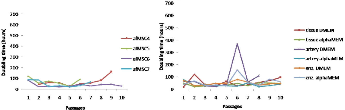
The amniotic fluid cells were a heterogeneous population with predominance of fibroblastic morphology (Figure 1) The cells were cultured for up to 10 passages with an average doubling time of 49.3 ± 12,7 hours at passages 1 to 5. At later passages the doubling time varied among the different cultures (Figure 2A). One culture became senescent at the 7th passage, the average doubling time was 60,5 ± 38,5 hours at passages 6 to 10.
Figure 1. Cells derived from amniotic fluid, at the 2nd passage.
Isolation and cultures of MSC from umbilical cords
Isolation of MSC from the first umbilical cord was successful except with the method for the arteries. The method was improved and isolation of MSC from the second umbilical cord was successful using all the methods. The cell populations appeared homogenous; colonies
appeared after 4-6 days (Fig 3A). These cell populations were also cultured for up to 10 passages with an average doubling time of 42.3 ± 7.5 hours at passages 1 to 5 and at passages after that doubling times differed among the cultures (Figure 2B). Cells derived from arteries,
6
cultured in DMEM became senescent at the 9th passage, the average doubling time was 66,2 ± 70,1 hours at passage 6 to 10.
Figure 2 Cell doubling time afMSC (A) and ucMSC (B). The cell cultures shows analogous proliferation rate
throughout the 5 first passages and thereafter they diverge, afMSC5 became senescent at passage 7 average doubling time was 59,1 ±76,9 hours at passages 1 to 10, cell culture derived from uc arteries, cultured in DMEM became senescent at passage 9 the average doubling time was 62,1 ± 42,3 hours at passage 1 to 10.
Phenotype of afMSC and ucMSC
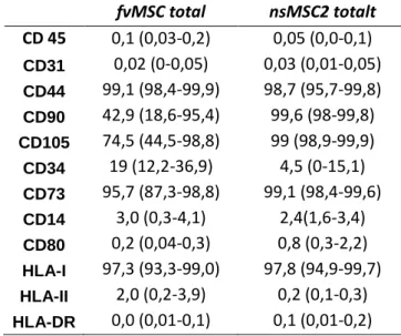
The cells were positive for CD105, CD44, CD73 and HLA I and negative for CD45, CD31, CD14, CD80, HLA-DR and HLA II. MSC from uc were negative for CD34 and positive for CD90, whereas afMSC were intermediately positive for CD34 and CD90 (Figure 4 and table 1).
Figure 4 Representative flow cytometry analysis of cultured MSC with monoclonal antibodies against CD45,
CD14, CD34 CD80, CD90, HLA-II, HLA-DR, HLA-I, CD105, CD73, CD44 and CD31 (bold lines). Dashed line indicates isotype-matched mouse IgG antibody control staining.
7
Table 1 The average expression of different surface epitopes by af and ucMSC in percentage from the flow
cytometry analysis. The range of expression is in brackets.
fvMSC total nsMSC2 totalt CD 45 0,1 (0,03-0,2) 0,05 (0,0-0,1) CD31 0,02 (0-0,05) 0,03 (0,01-0,05) CD44 99,1 (98,4-99,9) 98,7 (95,7-99,8) CD90 42,9 (18,6-95,4) 99,6 (98-99,8) CD105 74,5 (44,5-98,8) 99 (98,9-99,9) CD34 19 (12,2-36,9) 4,5 (0-15,1) CD73 95,7 (87,3-98,8) 99,1 (98,4-99,6) CD14 3,0 (0,3-4,1) 2,4(1,6-3,4) CD80 0,2 (0,04-0,3) 0,8 (0,3-2,2) HLA-I 97,3 (93,3-99,0) 97,8 (94,9-99,7) HLA-II 2,0 (0,2-3,9) 0,2 (0,1-0,3) HLA-DR 0,0 (0,01-0,1) 0,1 (0,01-0,2)
In vitro differentiation of afMSC and ucMSC
The success in differentiation of af and ucMSC to osteogenic and adipogenic cells was low. Most cell cultures in the experiment detached totally or partly from the plastic surface in the culturing wells.
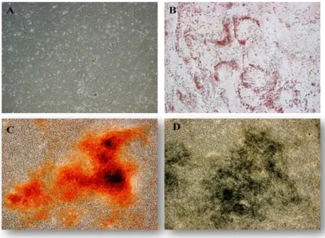
afMSC derived from 2 out of 4 different samples differentiated to osteogenic cells (see figure 3C and D) and one afMSC population differentiated to adipogenic cells
ucMSC derived from arteries, both cultured in DMEM and in MEMα differentiated to
adipogenic cells. ucMSC derived from tissue pieces, cultured in MEMα differentiated also to adipogenic cells (see figure 3B). No ucMSC from the different treatment differentiated to osteogenic cells.
8
Figure 3. A) Primary culture day 6 of ucMSC derived from arteries cultured in MEMα showing distinct spindle-shaped fibroblastic morphology. B) Adipogenic differentiation of ucMSC, indicated by accumulation of neutral lipid vacuoles stained with Oil Red O (red staining indicates lipid fat droplet)s. Osteogenic differentiation of afMSC indicated by calcium depositions stained with C) Alizarin Red, (indicated by red staining) and D) von Kossa method (indicated by black staining)
Mixed lymphocyte culturing
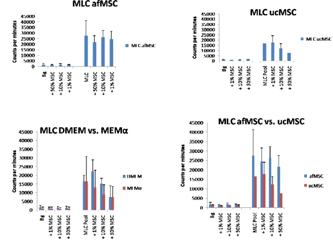
Mixed lymphocytes cultures showed that afMSC and ucMSC are immune privileged cells and furthermore, possess immunosuppressant properties (Figure 5). ucMSC suppressed at average 22,5 % the lymphocyte alloreactivity, afMSC 7,3 % (data not shown). Cells cultured in MEMα suppressed the immunoresponse to a greater extent than cells cultured in DMEM, at average 14,9 % DMEM vs. 32,9 % MEMα (data not shown).
9
Figure 5. Summary of the results from mixed lymphocytes cultures. MLC afMSC is an average of 4 different experiments, MLC ucMSC is an average of 6 experiments. MLC DMEM vs. MEMα compares ucMSC cultured in the different mediums, 3 experiments each. MLC afMSC vs. ucMSC compares the results from MLC af and ucMSC described above. The standard deviations are shown as the black error bars in the graphs.
Discussion
The cell populations from the amniotic fluids were quite heterogeneous and the cells displayed various morphology and some cultures became senescent at early passages. The advantage of using MSC from amniotic fluid is the time, one would have approximately half of a pregnancy (20 weeks) to isolate, culture, characterize and validate the cells before being used in autologous transplantation. But to be able to use the cells for autologous tissue engineering the protocol must be further optimized since the cells we isolated are not potent enough. S. Da Sacco et al.6 showed that Chang medium allowed expansion of cells from amniotic fluid for up to 50 passages with maintained original cell morphology, and that expansion in DMEM only was possible for 10 passages, thereafter the cells ceased to grow. That shows the importance of choosing a proper medium for the cultures. D. Schmidt et al7 isolated cells from amniotic fluid using positive isolation of CD133 cells, and obtained a homogenous cell population that could be used for fabricating human living heart valves. The isolation of cells from the umbilical cord involves many different procedures and in some protocols different enzymatic treatments. There are numerous opportunities for inadvertent contaminations in the protocols we used, and the enzymatic treatments of the cells may have
10
an impact to the original cell properties. If the purpose with the cell isolation and expansion is, as in this case, autologous tissue engineering, both the present methods and the time scale are inappropriate. Major congenital malformations often need to be surgically corrected shortly after birth and at that time there would be insufficient number of cells available for autologous transplantation using these protocols.
One of the criteria to identify MSC is that the population must express CD105, CD73 and CD90. They must also lack expression of CD45, CD34, CD14 and HLA class II4. In the flow cytometry analysis the cells from af were intermediately positive for CD34 and CD90, which confirms that the populations were heterogeneous. In two samples only approximately 50 % of the cells were viable and therefore there could have been unselective binding of the monoclonal antibodies which leads to deceptive results. This experiment must be repeated. Phermthai et al also received weaker expression of CD90 in flow cytometry analysis of MSC derived from amniotic fluid8, although the reason for this is unknown. The heterogeneity of the cells analyzed may be one explanation.
The differentiation assays are well established in our laboratories, and have been proven successful9-12. In this study the cells in most experiments detached from the plastic surface, so in the forthcoming experiments we will consider coating the culture wells with appropriate proteins e.g. FCS13 or Collagen before starting a differentiation assay.
In the mixed lymphocyte culture assays some of the MSC populations rather stimulated than inhibited lymphocyte proliferation, which also can confirm that the populations were
heterogeneous, although K. Le Blanc et al14 showed that MSC at low concentrations can stimulate or mildly inhibit lymphocyte proliferation.
To conclude, af and ucMSC were successfully isolated and cultured for several passages. The cells had characteristics similar to MSC isolated from other sources, although the populations were heterogeneous, which was confirmed by the experiments.
Based on this data we will in the forthcoming studies isolate cells from amitotic fluid using positive selection for various markers and culture the cells in MEMα or other culture medium. With these changes we hope to obtain a potent homogenous cell population that can be used in the treatment of congenital malformations.
11
References
1. Whiteman, Valerie E.; Reece, E. Albert; Prenatal diagnosis of major congenital malformations. Current Opinion in Obstetric and Gynecology,1994, 6(5):459-467
2. McCandless SE, Brunger JW, Cassidy SB; The burden of genetic disease on inpatient care in a children's hospital. Am J Hum Genet. 2004;74:121-127
3. Amulya K. Saxena, M.D.; Congenital Anomalies of Soft Tissues: Birth Defects Depending on Tissue Engineering Solutions and Present Advances in Regenerative Medicine. Tissue Engineering Part B: Reviews, 2010, 16(5): 455-466.
4. M. Dominici et al; Minimal criteria for defining multipotent mesenchymal stromal cells, The
International Society for Cellular Therapy position statement. Cytotherapy 2006 Vol. 8, No 4, 315-317
5. Husein K. Salem, Chris Thiemermann; Mesenchymal Stromal Cells: Current Understanding And Clinical Status. Stem Cells 2010;28:585-596
6. S. Da Sacco et al.; Human Amniotic Fluid as a Potential New Source of Organ Specific Precursor Cells for Future Regenerative Medicine Applications, The Journal of Urology, 2010, Vol. 183, 1193-1200
7. D. Schmidt et al.; Prenatally Fabricated Autologous Human Living Heart Valves Based on Amniotic Fluid-derived Progenitor Cells as Single Cell Source, Circulation, 2007; 116:I-64 – I-70
8. Phermthai et al.; A novel method to derive amniotic fluid stem cells for therapeutic purposes,BMC Cell Biology 2010, 11:79
9. C. Götherström et al.; Immunomodulatory effects of human foetal liver-derived mesenchymal stem cells, Bone Marrow transplantation, 2003, Vol. 32, 265-272
10. C. Götherström et al.; Immunologic properties of human fetal mesenchymal stem cells, American Journal of Obstetrics and Gynecology, 2004, Vol. 190, 239-245
11. Mikael Rydén et al.; Functional characterization of human mesenchymal stem cell-derived adipocytes, Biochemical and Biophysical Research Communications, 2003, Vol. 311, 391-397
12. K. Le Blanc et al.; Fetal Mesenchymal Stem-Cell Engraftment in Bone after In Utero Transplantation in a Patient with Severe Osteogenesis Imperfecta, Transplantation, 2005, Vol. 79, 1607-1614
13. K. Bieback et al.; Critical Parameters for the Isolation of Mesenchymal Stem Cells from Umbilical Cord Blood, Stem Cells, Vol. 22, 625-634
14. K. Le Blanc et al.; Mesenchymal Stem Cells Inhibit and Stimulate Mixed Lymphocyte Cultures and Mitogenic Responses Independently of the Major Histocompability Complex, Scandinavian Journal of Immunology 57 (2003), 11-20