DISSERTATION
AFFINITY MATURATION AND CHARACTERIZATION OF NOVEL BINDERS TO THE HIV-1 TAR ELEMENT BASED ON THE U1A RNA RECOGNITION MOTIF
Submitted by David W Crawford Department of Chemistry
In partial fulfillment of the requirements For the Degree of Doctor of Philosophy
Colorado State University Fort Collins, Colorado
Fall 2018
Doctoral Committee:
Advisor: Brian McNaughton Christopher Ackerson Eric Ross
Copyright by David W Crawford 2018 All Rights Reserved
ABSTRACT
AFFINITY MATURATION AND CHARACTERIZATION OF NOVEL BINDERS TO THE HIV-1 TAR ELEMENT BASED ON THE U1A RNA RECOGNITION MOTIF
The increased understanding of the importance of RNA, both as a carrier of information and as a functional molecule, has led to a greater demand for the ability to target specific RNAs, but the limited chemical diversity of RNA makes this challenging. This thesis documents the use of yeast display to perform affinity maturation for the ability of a protein to bind the TAR element of HIV-1, which is a desirable therapeutic target due to its prominent role in the HIV-1 infection cycle. To accomplish this, we used a “semi-design” strategy—repurposing a natural RNA binding protein to bind a different target—by creating a library based on important binding regions (es-pecially the β2β3 loop) of the U1A RRM. Following selection for TAR binding, a strong consensus sequence in the β2β3 loop emerged. The affinity of certain library members for TAR was mea-sured by ELISA and SPR, and it was determined that the best binder (TBP 6.7) had remarkable affinity (KD= ~500 pM). This TAR binding protein also proved capable of disrupting the Tat–TAR interaction (necessary for HIV-1 replication) both in vitro and in the context of extracellular tran-scription. Through collaboration, we were able to obtain a co-crystal structure of TBP 6.7 and TAR. This crystal structure showed that the overall structure of TBP 6.7 was largely unchanged from that of U1A, thereby validating our semi-design strategy. We also found that the β2β3 loop played a disproportionately large role in the binding interaction (~⅔ of the buried surface area). The prominence of this region’s role in the interaction inspired the creation and characterization of peptide derivatives of the TBP 6.7 β2β3 loop. These β2β3 loop derived peptides maintain affin-ity for TAR RNA (KD= ~1.8 μM), and can disrupt Tat/TAR-dependent transcription. Ultimately, the project yielded a novel platform of TAR binding peptides and a crystal structure which will inform future RNA targeting efforts in addition to generating the tightest known binder of TAR.
ACKNOWLEDGEMENTS
I would formally like to acknowledge everyone who provided financial support for the re-search in this thesis, including the NIH, Novartis, and Colorado State University.
I would also like to thank the faculty of the CSU Chemistry Department, for their classroom instruction and generous open door policies, especially Prof. James Neilson for instilling in me a deep-seated love of the perovskite structure (which is always the answer).
I’d like to thank John Anderson and the entire Wilusz2Lab for treating a demanding guest as a dear friend. It enabled my most challenging experimental work, and I will never forget it.
I would like to thank my colleagues in the McNaughton Lab for the camaraderie we shared through the highs and lows of graduate school. I’d liketo particularly thank AngelineTa, without whose friendship and scientific support I never would have completed a PhD.
I’d like to acknowledge my mentor, Dr. Brett Blakeley, for teaching me how to hold a pipette, and my mentees, Patrick Beardslee and Zachary Fleishhacker, for being receptive and creative.
I would next like to thank a group of people I I have never met, but with whom I am honored to share an author line: my collaborators. Most especially, I’d like to thank Dr. Ivan Belashov and Prof. Joseph Wedekind for the PDB file that made me cry with delight.
I am grateful for the long-term support from my committee members: Prof. Pat Bedinger who guided me through my first practical foray into molecular biology so many years ago; Prof. Chris Ackerson who mentored me through my first publishable work; and Prof. Eric Ross who treated a one-time rotation student as his very own during all the time I spent at CSU.
Most especially, I’d like to thank my advisor, Dr. Brian McNaughton for always being open to a scientific discussion, giving me sound research advice, and for opening so many doors for me. Brian helped me do excellent work which speaks for itself, and it speaks even more eloquently and convincingly because he also found the collaborators it needed to become truly great.
Finally, I’d like to express my appreciation for the $1 americano refills at Mugs Coffee Lounge which fueled the creation of this thesis, and Maryann Crawford for proofreading it.
DEDICATION
For all the times that I felt my struggles and failures as a scientist were all that defined me, and all the times you showed me they were not: I dedicate this thesis to my mother, my father, and my sisters. Your
TABLE OF CONTENTS
ABSTRACT . . . ii
ACKNOWLEDGEMENTS . . . iii
DEDICATION . . . iv
LIST OF TABLES . . . xii
LIST OF FIGURES . . . xiii
Chapter 1 Introduction . . . 1
1.1 Biological Macromolecule Background . . . 1
1.1.1 Proto-Biochemistry was Focused on Proteins . . . 2
1.2 The Central Dogma . . . 3
1.2.1 Structure and Function of DNA . . . 3
1.2.2 Formulation of the Central Dogma . . . 4
1.3 Bioinformatics Era . . . 5
1.3.1 Gene Editing . . . 6
1.3.2 Limits . . . 6
1.4 The Centrality of RNA to Life . . . 7
1.4.1 The RNA World Hypothesis . . . 9
1.5 Roles of RNA in Living cells . . . 9
1.5.1 Dogmatic Roles of RNA . . . 9
1.5.2 RNA Interference (RNAi) . . . 10
1.5.3 CRISPR-Cas . . . 14
1.5.4 Viral RNAs . . . 14
1.6 HIV I TAR RNA . . . 15
1.6.1 TAR Induces a Transcription Cascade . . . 16
1.6.2 TAR as pre-miRNA . . . 16
1.6.3 Relevance of TAR RNA . . . 18
1.7 Basis of Nucleic Acid Behavior . . . 18
1.7.1 Nucleotides . . . 18
1.7.2 Nucleic acid as a Polymer String . . . 20
1.7.3 Nucleic Acid as Ribbon . . . 21
1.7.4 Nucleic Acid Base-Pairing . . . 23
1.8 Nucleic Acid 3D-Structures . . . 25
1.8.1 DNA Structure . . . 25
1.8.2 RNA Structure . . . 26
1.8.3 RNA Tertiary Structure . . . 29
1.8.4 Structure Conclusion . . . 29
1.9 Nucleic Acid as Sequence . . . 31
1.10 Dictates of Binding Nucleic Acids . . . 32
1.10.1 Small Molecules . . . 32
1.10.2 Nucleic Acids . . . 33
1.11 Thesis Goal . . . 35
1.11.1 Develop a Protein-Based Binder for TAR RNA . . . 35
1.11.2 Advance Understanding of Protein–RNA Binding . . . 35
1.12 Development of a Modular Binder for dsDNA . . . 35
1.12.1 Modular DNA Binding Proteins . . . 35
1.13 TALENs . . . 36
1.13.1 TAL Domain Natural Origin . . . 37
1.13.2 TAL Informatics . . . 37
1.13.3 Structure of TAL Domain . . . 38
1.13.4 TAL Domain Engineering . . . 40
1.13.5 TAL Domain Lessons . . . 40
1.13.6 Binding DNA vs. Binding RNA . . . 40
1.14 ssRNA binding . . . 41
1.14.1 Introduction to ssRNA . . . 41
1.15 Pumilio Repeat Proteins . . . 42
1.15.1 Natural Origins of Pumilio Repeat Proteins . . . 42
1.15.2 Structure of Pumilio repeats . . . 42
1.15.3 Mechanism of Pumilio Repeat Binding . . . 43
1.15.4 Engineering Pumilio Repeats . . . 45
1.15.5 Cytosine Binder Evolution . . . 45
1.15.6 Pumilio Repeat Domain Utilization Arc . . . 48
1.15.7 Other ssRNA binders . . . 50
1.16 Structured RNA Binding Proteins . . . 51
1.16.1 Background . . . 51
1.16.2 RNA Recognition Motifs (RRMs) . . . 52
1.16.3 U1A–U1hpII Mechanism of Binding . . . 55
1.16.4 U1A E19S . . . 57
Chapter 2 Affinity Maturation of U1A E19S for TAR RNA Binding . . . 59
2.1 Chapter 2 Introduction . . . 59
2.1.1 Chapter 2 Summary . . . 59
2.2 Chapter 2 Attribution . . . 60
2.2.1 Chapter 2 Background . . . 60
2.3 Design of Screening Strategy . . . 62
2.3.1 Screening Method . . . 62
2.3.2 Screening strategy . . . 64
2.4 Yeast Display Confirmation . . . 65
2.4.1 Cloning U1A Variants into Yeast . . . 65
2.4.2 Positive Controls . . . 66
2.5 Library Preparation . . . 68
2.5.1 Cloning the β2β3 Loop Library . . . 68
2.5.2 Library Transformation . . . 70
2.6 Library Screening . . . 71
2.6.1 General Screening Conditions . . . 71
2.6.3 Sorting Conditions . . . 72
2.6.4 Diversification . . . 74
2.6.5 Screening of Rounds 4–6 . . . 77
2.6.6 Sequence Analysis of Round 4 . . . 78
2.6.7 C-Helix Rationale . . . 78
2.7 Properties of Sixth Generation TAR Library . . . 80
2.7.1 β2β3 Loop Homology . . . 80
2.7.2 C-Helix Homology . . . 83
2.8 Initial Characterization Attempts . . . 84
2.8.1 Fluorescence Polarization . . . 84
2.8.2 Characterization via Qualitative Yeast Display . . . 85
2.8.3 Yeast Display KD . . . 88
2.9 Conclusions . . . 89
Chapter 3 Characterization of TAR Binding Proteins . . . 90
3.1 Chapter 3 Introduction . . . 90 3.1.1 Chapter 3 Summary . . . 90 3.1.2 Chapter 3 Attribution . . . 90 3.1.3 Chapter 3 Background . . . 91 3.2 ELISA Preparation . . . 91 3.2.1 Assay preliminaries . . . 91 3.2.2 Cloning . . . 92 3.2.3 Protein Purification . . . 93 3.3 ELISA Assay . . . 95
3.3.1 General ELISA Protocol . . . 95
3.3.2 ELISA results . . . 98 3.3.3 Quantitative ELISAs . . . 101 3.4 SPR Analysis . . . 104 3.4.1 Protein Preparation . . . 104 3.4.2 SPR Experiment . . . 104 3.4.3 SPR Analysis . . . 105
3.5 Characterization of TAR Binding Selectivity . . . 108
3.6 SHAPE Analysis . . . 111
3.6.1 SHAPE Context . . . 112
3.6.2 SHAPE RNA Prep . . . 112
3.6.3 SHAPE Method . . . 113
3.6.4 SHAPE Results . . . 114
3.6.5 Qualitative Binding . . . 115
3.6.6 SHAPE Data . . . 115
3.7 Disrupting the Tat–TAR Interaction . . . 118
3.7.1 ELISA . . . 119
3.7.2 ITC . . . 119
3.8 Suppression of Tat–Dependent Transcription by a Synthetic TAR-Binding Protein . . . 121
Chapter 4 Crystallization of TBP 6.7 and TAR . . . 125 4.1 Chapter 4 Introduction . . . 125 4.1.1 Chapter 4 Summary . . . 125 4.1.2 Chapter 4 Attribution . . . 125 4.1.3 Chapter 4 Background . . . 126 4.2 Preliminary Work . . . 126 4.3 Crystallization . . . 127 4.3.1 Protein Purification . . . 128
4.3.2 Crystallization and X-Ray Data Collection . . . 129
4.3.3 Phase Determination, Refinement and Analysis . . . 129
4.3.4 Molecular Dynamics (MD) Simulations . . . 131
4.4 Structural Analysis of the HIV TAR-TBP 6.7 Complex . . . 133
4.4.1 Comparison to Previous Structures . . . 133
4.4.2 TBP 6.7 Uses the RNP Motif to Recognize Double-Stranded RNA . . . 136
4.5 Thermodynamic Analysis . . . 138
4.5.1 Contributions of R52 . . . 138
4.5.2 Vital Contributions of R47A . . . 139
4.6 Conclusions . . . 144
4.6.1 General Conclusions . . . 144
4.6.2 Relationship to Prior Work . . . 145
Chapter 5 Peptide Derivatives of TBP 6.7 . . . 147
5.1 Chapter 5 Introduction . . . 147
5.1.1 Chapter 5 Summary . . . 147
5.1.2 Attribution . . . 147
5.1.3 Background . . . 148
5.2 Synthesis of Constrained Peptide . . . 148
5.2.1 General reagent information for synthesis of constrained peptide 1 and peptide 1s . . . 149
5.2.2 Synthesis of Constrained peptide 1 and peptide 1s . . . 150
5.2.3 LC-MS Analysis of Constrained peptides . . . 151
5.3 Preparation of TBP 6.7, SUMO, and SUMO-β2β3 Fusions for ELISA or Transcription . . . 151
5.4 Fluorescence Emission Analysis of TAR binding to peptide 1 . . . 152
5.4.1 Fluorescence Emission Assay . . . 152
5.4.2 Fluorescence Emission Results . . . 152
5.5 ITC Inhibition Assays . . . 155
5.5.1 ITC Inhibition Assay Methods . . . 155
5.6 Transcription Assay . . . 158
5.6.1 Transcription Assay Methods . . . 158
5.6.2 Statistical Analysis . . . 159
5.6.3 Results . . . 159
5.6.4 Transcription Assay Summary . . . 162
5.7 SUMO Fusions of the TBP 6.7 β2β3 Loop . . . 162
5.7.2 ELISA Results . . . 163
5.8 Surface Display Assays . . . 164
5.8.1 Bacterial Display . . . 165
5.8.2 Yeast Display . . . 165
5.8.3 Display Assay Results . . . 166
5.9 Conclusions . . . 167
Chapter 6 Conclusions and Future Directions . . . 169
6.1 Project Background and Goals . . . 169
6.2 Achievement of Project Goals . . . 170
6.2.1 Develop a Protein-Based Binder of TAR RNA . . . 170
6.2.2 Advance Understanding of Protein–RNA Binding . . . 171
6.2.3 Develop a Peptide Based Binder of TAR RNA . . . 171
6.3 Future Directions . . . 172
6.3.1 Optimization of Peptide Derivatives of TBP 6.7 . . . 172
6.3.2 Optimized Surface Display . . . 173
6.3.3 Optimized Recombinant Expression . . . 174
6.4 Progress Toward a Binding Code . . . 174
6.4.1 Structural Considerations . . . 175
6.4.2 Base Interactions . . . 176
6.4.3 Conclusions . . . 177
Bibliography . . . 178
Appendix A Helical Grafting of E6AP . . . 198
A.1 Background . . . 198
A.1.1 Significance . . . 198
A.1.2 The E6/E6AP/p53 Ternary Complex . . . 198
A.1.3 The E6/E6AP Binding Interaction . . . 200
A.1.4 Research Goals . . . 200
A.2 Helical Grafting Strategy . . . 201
A.2.1 Helical Stabilization . . . 201
A.2.2 Screening System Design . . . 202
A.2.3 Purification of sfGFP-E6 . . . 203
A.3 Grafting Strategy . . . 205
A.4 Materials Preparation and General Methods . . . 206
A.4.1 Cloning . . . 206
A.4.2 Protein Purification . . . 207
A.4.3 Protein Purification Assay Consequences . . . 207
A.4.4 Yeast Preparation . . . 208
A.5 Yeast Display Assays . . . 210
A.5.1 Sac7d-E6AP and E6AP Display on Yeast . . . 210
A.5.2 Initial Tests of sfGFP-E6 binding to Displayed E6AP . . . 210
A.5.3 sfGFP-E6/E6AP Binding with Longer Incubation Times . . . 212
A.6.1 Protocol . . . 214
A.6.2 Results . . . 214
A.7 “Helical Grafting of E6AP” Conclusions . . . 217
A.8 Future Directions . . . 217
A.8.1 Replication . . . 217
A.8.2 Yeast Display . . . 217
A.8.3 ITC . . . 218
A.8.4 Other Assays . . . 218
A.8.5 Introducing p53 to E6/E6AP Interaction . . . 218
Appendix B Other Experiments of Possible Interest . . . 220
B.1 Binding of TBP 6.7 to ΔC25 TAR RNA . . . 220
B.1.1 Introduction . . . 220
B.1.2 Methods . . . 220
B.1.3 Results . . . 222
B.1.4 Conclusions . . . 222
B.2 Alternate β2β3 Loop Display Strategies . . . 223
B.2.1 Introduction . . . 223
B.2.2 TEV-cleavage displayed β2β3 loop peptides . . . 225
B.2.3 Maleimide FITC Conjugation of TEV-Cys-β2β3-Cys-TEV . . . 227
B.2.4 Conclusions and Future Directions . . . 228
B.3 TBP 6.7 Expresses in Mammalian Cells . . . 231
B.3.1 Introduction . . . 231
B.3.2 Materials Preparation . . . 231
B.3.3 Conclusions . . . 233
B.4 Sac7d Based Binders of CUG10RNA . . . 233
B.4.1 Introduction . . . 233
B.4.2 Library Creation and Screening . . . 233
B.4.3 Yeast Display Methods . . . 234
B.4.4 Analysis of the Best Binder: CUG10Binding Protein 5.21 . . . 234
B.4.5 Conclusions . . . 235
B.5 Others . . . 237
B.5.1 Enzymatic Creation of Inorganic Nanoparticles . . . 237
B.5.2 Alternate Library Selection Method . . . 239
B.5.3 Small Molecule Induced Dimerization . . . 239
B.5.4 Library Screening for Other RNAs . . . 239
Appendix C Protein and DNA Sequences . . . 240
C.1 Sequences from Chapter 2, “Affinity Maturation of U1A E19S for TAR RNA Binding” . . . 240
C.1.1 Selected Primers from Chapter 2 . . . 240
C.1.2 wtU1A in pCTcon2 . . . 241
C.1.3 U1A E19S in pCTcon2 . . . 242
C.1.4 1st Gen Library Receiving Plasmid/BsaI U1A . . . 242
C.1.6 2nd Gen Library Receiving Plasmid . . . 243
C.1.7 2nd Gen Library Amplicon . . . 243
C.2 Sequences from Chapter 3, “Characterization of TAR Binding Proteins” . 244 C.2.1 Selected Primers from Chapter 3 . . . 244
C.2.2 Generic TAR Binding Protein with C-terminal His6and FLAG Tags . . 245
C.2.3 TBP 6.7 . . . 245
C.2.4 TBP 6.6 . . . 246
C.2.5 RNAs . . . 247
C.2.6 Tat Sequences . . . 247
C.2.7 PLAI-BS Transcript Sequence . . . 248
C.3 Sequences from Chapter 4, “Crystallization of TBP 6.7 and TAR” . . . 249
C.3.1 TBP 6.7 Variants . . . 249
C.4 Sequences from Chapter 5, “Peptide Derivatives of TBP 6.7” . . . 250
C.4.1 TBP 6.7 Used in ITC Assays . . . 250
C.4.2 SUMO Fusions . . . 250
C.4.3 Aga2 Control . . . 251
C.4.4 TBP 6.7 β2β3 Loop for Yeast Display . . . 252
C.4.5 TBP 6.7 β2β3 Loop–eCPX for Bacterial Display . . . 252
C.5 Sequences from Appendix A, “Helical Grafting of E6AP” . . . 253
C.5.1 Sac7d for Yeast Display . . . 253
C.5.2 Sac7d-E6AP for Yeast Display . . . 254
C.5.3 E6AP Peptide for Yeast Display . . . 254
C.5.4 sfGFP-E6 . . . 254
C.5.5 sfGFP . . . 256
C.5.6 Sac7d for Expression . . . 257
C.5.7 Sac7d-E6AP for Expression . . . 258
C.5.8 p53 Core . . . 258
C.6 Sequences from Appendix B, “Other Experiments of Possible Interest” . 259 C.6.1 ΔC25 TAR . . . 259
C.6.2 TEV-Cys-β2β3-Cys-TEV . . . 259
C.6.3 Cys-β2β3-Cys . . . 260
C.6.4 Z-Peptide . . . 260
C.6.5 Gblock sequence from Section B.3, “TBP 6.7 Expresses in Mammalian Cells” . . . 261
C.6.6 Primers from Section B.3, “TBP 6.7 Expresses in Mammalian Cells” . . 261
C.6.7 TBP 6.7-* . . . 262
C.6.8 TBP 6.7-FLAG-* . . . 262
C.6.9 TBP 6.7-NLS-* . . . 263
C.6.10 Sac7d Library . . . 264
C.6.11 Sac7d with N-terminal myc . . . 264
LIST OF TABLES
1.1 Composition of a Cell . . . 3
2.1 Sequences of library members in first three rounds of sorting . . . 73
2.2 Yeast Display Round Conditions . . . 74
2.3 Yeast sorted in fourth round, both from the C-Helix and β1α1 libraries . . . 79
2.4 Sequences from TAR 6G Library . . . 81
3.1 Statistical Values of SPR for TBP 6.6 and TBP 6.7 and wtU1A for TAR and U1hpII RNAs 106 3.2 Kinetic and Equilibrium Values of SPR for TBP 6.6, TBP 6.7, and wtU1A for TAR RNA and U1hpII RNA . . . 108
3.3 Folding Energies of RNA Hairpins Used in Selective Binding Studies . . . 110
3.4 Reagent concentrations for TAT/TAR Dependent Transcription Assay . . . 121
4.1 X-ray Diffraction and Refinement Statistics of TBP 6.7–TAR Co-crystal . . . 130
4.2 Thermodynamic Parameters for TAR-TBP 6.7 Binding at 20 °C . . . 139
5.1 Thermodynamic Parameters for ITC, at 25 °C, of Tat Peptide Titrated into TAR RNA, with and without pre-complexing of β2β3 SUMO and peptide 1s . . . 156
LIST OF FIGURES
1.1 The Central Dogma as Formulated by Francis Crick . . . 5
1.2 RNA Roles in the Central Dogma . . . 8
1.3 Overview of RNA Interference Pathways . . . 11
1.4 micro-RNA Processing Overview . . . 13
1.5 CRISPR-Cas9 with RNA Modifications . . . 14
1.6 Two Examples of Functional Viral RNAs . . . 15
1.7 Tat–TAR Induced Transcription Cascade . . . 17
1.8 The HIV I TAR Element in the Context of the HIV Genome . . . 17
1.9 Polynucleotide (RNA) vs. DNA vs. Polypeptide . . . 19
1.10 Polynucleotide as a Two-Sided Chemical Ribbon . . . 22
1.11 Nucleic Acid Base Pairing . . . 24
1.12 RNA Secondary Structure Overview . . . 27
1.13 DNA vs. RNA Tertiary Structure Comparison . . . 30
1.14 Polyamide Scaffold for Binding DNA . . . 33
1.15 TAL Recognition Code . . . 38
1.16 Structure of a TAL Domain Bound to dsDNA . . . 39
1.17 Structure of a Pumilio Repeat Domain . . . 44
1.18 Yeast-3-Hybrid for Finding Cytosine-Binding Pumilio Repeat . . . 47
1.19 Modular “Pumby” Domain Code . . . 49
1.20 Example Classes of RNA Binding Proteins . . . 53
1.21 Examples RNA Recognition Motif Proteins . . . 54
1.22 The HIV I TAR Element . . . 57
2.1 Wild-type U1A Crystal Structure . . . 62
2.2 Overview of the Yeast Display Technique . . . 64
2.3 Functional Display of U1A Variants . . . 68
2.4 Cloning Diagram for Diversification of the β2β3 Loop . . . 71
2.5 Yeast Sorts . . . 75
2.6 Diversification Strategy . . . 77
2.7 Yeast Display Rounds 4 and 5 . . . 78
2.8 TAR 6G Sequence Logo . . . 83
2.9 TAR Binding Protein 3.1 and 6.2 Fluorescence Polarization . . . 86
2.10 Qualitative Yeast Display for Characterizing Generation 6 TAR Binding Proteins . . 87
2.11 Quantitative Yeast Display Assays . . . 88
3.1 PAGE Gel of TBP 6.6 and TBP 6.7 . . . 94
3.2 General ELISA Scheme . . . 96
3.3 Results of a Single Plate of ELISA Assays using 50 nM U1A Variant . . . 99
3.4 ELISA Survey of 6th Generation TAR Binding Proteins . . . 100
3.5 ELISA Signal of TBP 6.7 vs. U1A E19S for TAR Binding . . . 101
3.7 Finalized Quantitative ELISA-based Binding Curves . . . 103
3.8 SPR Binding Curves of TBP 6.6, TBP 6.7 and wtU1A against TAR and U1hpII . . . 107
3.9 Affinity of TBP 6.6 and TBP 6.7 for Modified TAR . . . 109
3.10 mFold Calculations of Hairpins used to analyze selectivity . . . 111
3.11 RNA Folding Conditions and Baseline Reactivities for SHAPE . . . 112
3.12 SHAPE Data Using 4:1 Protein:RNA . . . 115
3.13 TBP 6.7 Binding TAR via Gel Shift Assay . . . 116
3.14 SHAPE Data Using 8:1 Protein:RNA . . . 117
3.15 Disruption of Tat–TAR interaction Measured by ITC . . . 120
3.16 Biochemical Overview of Transription Assay . . . 122
3.17 TBP 6.7 inhibition of TAT/TAR based Transcription . . . 123
4.1 ELISA Assays to Analyze Variation Between U1A Scaffolds . . . 127
4.2 Electron Density Map of TBP 6.7–TAR Crystal Structure . . . 132
4.3 Fractional Occupancy and Molecular Dynamic Simulations for TAR-TBP 6.7 Complex 133 4.4 Overview of TAR Binding Protein 6.7–TAR complex . . . 135
4.5 The RNP Motif in the TBP 6.7–TAR Complex . . . 137
4.6 Detailed View of the β2β3 loop in the TBP 6.7–TAR Complex . . . 138
4.7 ITC Plots of TBP 6.7 Mutants Titrated into TAR . . . 140
4.8 Schematic diagram of cation-π contacts between HIV-1 TAR bases and guanidinium groups contributed by the TBP 6.7 β2-β3 loop . . . 141
4.9 Fractional Occupancy . . . 143
5.1 Structures and LC-MS Analysis of Constrained Peptides . . . 149
5.2 Fluorescence Assay Measuring Binding of peptide 1 to TAR . . . 153
5.3 Fluorescence Assay Measuring Binding of TBP 6.7 to (2AP)-TAR . . . 154
5.4 Inhibition of Tat–TAR Complex Formation by SUMO β2β3 and peptide 1s as mea-sured by ITC . . . 157
5.5 Transcription Assay Full Gels . . . 160
5.6 TAT/TAR Transcription Assay with peptide 1s . . . 161
5.7 ELISA Data Showing Binding of SUMO Fusions of the TBP 6.7 β2β3 Loop, and asso-ciated Arg→Ala mutant . . . 163
5.8 Flow Cytometry Analysis of Bacterial Displaying a β2β3 loop . . . 166
5.9 Flow Cytometry Analysis of Yeast Displaying a β2β3 loop . . . 167
6.1 Project Summary . . . 172
A.1 Crystal Structure and Cartoon of E6/E6AP/p53 Complex . . . 199
A.2 Detail of the E6/E6AP Binding Interaction . . . 200
A.3 Proposed Yeast Display System for Measuring Binding of E6 to E6AP . . . 204
A.4 E6/E6AP complex vs. E6/Sac7d-E6AP Complex . . . 205
A.5 Initial Purifications of Sac7d(-E6AP) and sfGPF(-E6) . . . 208
A.6 Final sfGFP-E6 Purification . . . 209
A.7 Initial Confirmation of Sac7d-E6AP and E6AP Display on Yeast . . . 211
A.8 E6 Binding by Displayed E6AP, 45 min. Incubation . . . 212
A.10 ITC Titrations of Sac7d-E6AP into sfGFP-E6 . . . 215
A.11 ITC of Sac7d into Buffer Compared to Sac7d-E6AP into sfGFP-E6 . . . 216
B.1 Dinucleotide Bulge TAR . . . 221
B.2 Binding of TBP 6.7 to ΔC25 TAR . . . 222
B.3 Schemes for Detection of Cyclization of Displayed β2β3 Loop Peptide . . . 224
B.4 Display of β2β3 Peptide Variants Before and After TEV Cleavage . . . 226
B.5 Mass Spectrum of Supernatant Following TEV Cleavage . . . 227
B.6 Results of Incubating Cys-β2β3-Cys with Maleimide-FITC . . . 228
B.7 Proposed use of TEV Cleavage on a Bacterial Surface to Detect Binding Events . . . 230
B.8 Western Blot Demonstrating TBP 6.7 Expression in HEK 293T Cells . . . 232
B.9 CBP 5.21 Binding to Cy-5 Labelled CUG10RNA or FITC conjugated anti-myc Antibody 235 B.10 Concentration Series of Displayed CBP 5.21 Incubated with 1–10 μM Cy5-CUG10RNA 236 B.11 NADPH-based Monitoring of Enzymatic Selenite Reduction . . . 238
Chapter 1
Introduction
1.1 Biological Macromolecule Background
The intertwined fields of Biochemistry, Molecular Biology, and Chemical Biology can be imperfectly generalized as the study of the properties and interactions of four major classes of biological polymers- lipids, carbohydrates, proteins, and nucleic acids. Nearly every genetic sur-vey, every pharmaceutical, every in vitro cellular study, and every assay in all these fields are focused on some aspect of chemically classifying one of these four macromolecules and their interactions with each other and small-molecule adjuncts to form pathways. These four classes of molecules have not, however, had equal time and resources devoted to them. The degree to which our understanding of biochemistry is based on giving importance to what we could study, and that it is based on what we already know is not always appreciated. Practically, this means that proteins are the most studied molecule, nucleic acids a comfortable second, and carbohy-drates and lipids far behind
To be fair, proteins account for a vast array of cellular function ranging from catalysis to structure, but the original conception of the other macromolecule classes as mere facilitators of protein chemistry was reductive; Wide-ranging their function may be, but proteins do not tell the whole story of cellular chemistry. Of the non-protein macromolecules, Ribonucleic acid (RNA) especially has emerged as a material responsible for functional entities ill-defined by the old Central Dogma. New perspectives are needed to conceptualize the remarkable scope and dynamism of RNA’s cellular roles, and new tools will need to be developed to study it.
The work described in this thesis advances two goals. The first goal, which sees signifi-cant progress represented here, is the development and characterization of possible effector molecules for a single functional RNA of biochemical interest—the Trans-activating Response element of HIV-1 (TAR). The second, more abstract goal, for which this work represents a small
but important step, is progress toward the eventual ability to develop binders for arbitrary and specific functional RNAs.
In the service of understanding these two aims, and why they are only recently coming into focus, let us takea look back at how our understanding of biochemistry has evolved, how the path this understanding took has informed our assumptions, and how this has necessarily resulted in limitations. By understanding this history, we can recalibrate our goals and build new tools to achieve them.
1.1.1 Proto-Biochemistry was Focused on Proteins
In the 19th century Dutch chemist Gerardus Johannes Mulder came to realize that a vast amount of biological substances could be well-described by a single empirical formula— C400H620N100O120P1S1. Mulder’s correspondent Jöns Jacob Berzelius suggested that this massive class of substances deserved the name “Protein,” which roughly means, from its Greek roots, “of first importance” [1, 2]. This lofty designation certainly reflects the priorities of the nascent field of biochemistry. The focus on proteins as the primary enablers of cellular chemistry grew when James Sumner crystallized Urease in 1926 [3], proving what most enzymologists already guessed—that enzymes were proteins (Nobel Prize 1946) [4]. Early structural biology was almost entirely focused on proteins—most notably Linus Pauling’s hydrogen-bond based justification for the structure of the protein α-helix and β-sheet secondary structure elements [5] (Nobel Prize 1954).
This focus on proteins was not unwarranted, as Table 1.1A shows that this class of molecules makes up a majority of the dry mass of the cell, and this massive percentage does, in fact, correlate to a wide array of function.
The common catchphrases of the day demonstrate this explicit focus on protein. One fa-mous example, “One Gene, One Enzyme” [8] (soon amended to “One Gene, One Polypeptide) highlights this focus. The “gene” had been formulated purely as an abstraction, and the first, and
Table 1.1: Composition of a Cell A: Breakdown of mammalian cellular dry mass by molecule type [6] B:
Breakdown of RNA mass by RNA type [7]
A % of Dry Mass B % of RNA
Protein 59.31% rRNA 80%
DNA 0.82% tRNA 15%
RNA 3.62% mRNA 5%
Polysaccharides 6.59% Other Functional RNAs 0.1%
Lipids 16.47%
Small molecules 13.18%
for decades primary, concrete conceptual connection between the abstract idea of the heritable gene and the gene’s effect on observed reality was as an enabler of protein chemistry.
In the early 20th century, the hypothesis that Proteins were somehow the basis for genetic transfer was taken as a fait acccompli. How could a molecule as chemically simple as DNA, with only four bases, possibly conduct the complicated business of genetic transfer? However, the definitive proof that the nucleic acids were the physical basis of the gene was discovered through observation of the heritability of infectivity in Streptococcus pneumoniae by Oswald Avery, Colin MacLeod, and Maclyn McCarty in 1944 [9]. The fact that Avery did not win the Nobel Prize before his death in 1955 is testament to the unwillingness of his contemporaries to accept his (correct) conclusions, and not until an experiment by Hershey using bacteriophage was published in 1952 did the conclusion that nucleic acids were the basis for genetics become inescapable [10,11].
1.2 The Central Dogma
1.2.1 Structure and Function of DNA
The scope and promise of the new field of Molecular Biology snapped into focus in 1953 when James Watson and Francis Crick proposed a structure for the Deoxyribonucleic acid (DNA) poly-mer [12]. A final phrase in this seminal paper, “It has not escaped our notice that the specific pairing we have postulated immediately suggests a possible copying mechanism for the genetic
material” uses the newly discovered structure of DNA to bridge the gap between the function it had already been determined to have, and a mechanism for fulfilling that function.
The chemical limitations of the nucleic acids was no longer puzzling, it was a feature. A sam-ple of DNA was no longer a hodgepodge of functional groups, it was a strand, a linear sequence which underpins life itself. There is a reason that the enduring public symbol of Biochemistry and Molecular Biology is the elegant DNA double-helix. From the moment the structure was known, the broad strokes of how the information that is life had propagated itself from the misty past, and would continue doing so for the foreseeable future, was obvious. The abstract idea of the “gene” was now “information [13].”
Life itself was as readable as this sentence, once the code was learned.
1.2.2 Formulation of the Central Dogma
The code correlating DNA and protein sequence was indeed cracked in a very few years, and Francis Crick formulated the famous Central Dogma of Molecular Biology in 1958:
“[O]nce ‘information’ has passed into protein it cannot get out again. In more detail, the transfer of information from nucleic acid to nucleic acid, or from nucleic acid to protein may be possible, but transfer from protein to protein, or from protein to nucleic acid is impossible. Information means here the precise determination of sequence, either of bases in the nucleic acid or of amino acid residues in the protein [14,15].”
This simplifies well to the “DNA→RNA→Protein,” approximation taught in high school bi-ology, but all the possibilities that Crick foresaw are shown in Figure 1.1, and Crick’s primary assertion—that sequence-based information only flows from nucleic acid to protein—has been remarkably robust. However, the initial role that RNA was given—uninteresting messenger— was quickly found to be too reductive. RNA was soon known to be responsible for catalyzing peptide bond formation in the ribosome, and for acting as an adapter between mRNA and pro-tein primary sequence. These two roles actually encompass ~95% of the mass of RNA in the
cell (Table 1.1) while the sequences and structures of the RNAs involved in these roles are highly conserved across all phyla of life [6].
Figure 1.1: The Central Dogma as Formulated by Francis Crick In Francis Crick’s own words: “A tentative
classification for the present day [1970]. Solid arrows show general transfers; dotted arrows show special transfers. Again, the absent arrows are the undetected transfers specified by the central dogma.” Adapted from [15]
1.3 Bioinformatics Era
Counter-intuitive as it may seem, this new focus on DNA actually increased the fixation on the fact of proteins as being of “first importance.” Once the code was cracked, and it seemed clear that DNA didn’t (directly) code for anything other than protein, everything else became second-class. If heritable life was “information,” and all the chemistry that was coded for was contained in proteins, it follows that the rest of the vast array of chemistry that takes place in the cell can be extrapolated in total if the information in the DNA is sufficiently understood.
The fundamental promise and hope of this formulation was that Molecular Biology is know-able. If all function in a cell ultimately derives from DNA, and this DNA is both finite and read-able, maybe we could understand, and control everything, in a cell. In the 1990s, as the human genomeprojectwasnearingcompletion, thisoptimismwasatitsheight. HumanGenomeProject luminary Eric Lander penned a roadmap in 1996 that states “[o]nce all proteins are known, it should be possible to assemble comprehensive ‘interaction maps’ of genomes. [16]” This
assump-tion that “gene” and “protein” are essentially synonymous, and that a “comprehensive interacassump-tion map” could be assembled using only the genome are hallmarks of the era.
1.3.1 Gene Editing
The therapeutic hope of the bioinformatics era—-that all cell disease states can ultimately be traced back to the genome and corrected there—has been partially borne out. Our increasing knowledge of the genome has indeed allowed us map disorders back to the gene variant which causes them. For instance, Huntington’s Disease is understood to be caused by a mutated version of the “Huntingtin” protein which has too many glutamine residues near the N-terminus. We know this because we can read the extra “CAG” repeats in the gene coding for this protein in Huntington’s sufferers [17]. We also know that in cellulo, correcting the gene fixes the problem [18]. As examples like this demonstrate, there is a great deal of well-placed interest in Gene Therapy [19–21].
1.3.2 Limits
But the rapid DNA-based increase in our knowledge has, until recently, obscured the limits of our current approaches and models. The importance of DNA as the source of the master tem-plates for the proteins in the cell is obvious, but it to call it the “template” or “blueprint” is an over-statement. In hindsight, the belief that gaining a complete understanding of DNA would grant anything like total understanding of cellular processes was extraordinarily naïve. The genome is, more-or-less a parts list, not a manual. Assuming this parts list would be enough to fully un-derstand the dynamic processes that make up up cellular biology is, at a basic level, comparable to assuming that the best way to assemble IKEA furniture is to look at a photograph of a com-pleted item and draw from disorderly piles of every component used by IKEA. Life is not the inevitable result of recorded information, but instead the result of the careful, ordered, limited, and precise expression of this information within the pre-existing context of the cell, and within the derived context of a multi-cellular organism.
An example of the power, but also the fundamental limitation, of a DNA informatics ap-proach is the successful synthesis of an artificial genome by Craig Venter’s team. This group of researchers synthesized a full genome of Mycoplasma mycoides and subsequently successfully transplanting it into a cell, which then grew normally. The achievement is obvious, but so is the limitation. This genome doesn’t spontaneously create an organism around it, and would not do so even if transcription/translation machinery and biopolymer building blocks were available. The genome contained the “complete” information for replication, but only if the necessity of an emphM. capriculum cellular environment can be considered free of information [22].
Cellular environment may not follow the clear rules of the DNA code, but it is as important a form of “information” as the easily readable DNA sequences. To take the next steps in biochem-ical understanding, we need to understand the interaction of every piece of the cell. Arguably the most pervasive and dynamic aspect of that cellular milieu is the seemingly boring RNA.
1.4 The Centrality of RNA to Life
In Molecular Biology as described by the Central Dogma, DNA is the master composer cre-ating a timeless work of genius, Protein is the orchestra expressing that genius, while RNA is relegated to the role of simple amanuensis, making sure the conductor and players have their scores. In Crick’s original formulation, RNA dutifully copies down the information contained in the DNA, transmitting it from the nucleus to the parts of the cell that can bring the coded-for proteins into existence, and then degrades without a trace. This is an important job to be sure, but it isn’t an apparently complicated one.
However, ever since that boring relegation, RNA continually gets caught doing something interesting. Soon after the formulation of the central dogma, RNA was found to be the adapter between the genetic code and protein synthesis, and to catalyze the synthesis of polypeptides from amino acids within the ribosome. In fact, the ribosomal RNA (rRNA) and transport RNA (tRNA) account for ~95% of the RNA in a mammalian cell (Table 1.1). More recently, it was dis-covered that RNA doesn’t get transcribed from the genome as a perfect copy of a coded gene,
DNA
Transcription Translation
RNA Protein
lncRNAs Splicing RNAi tRNA rRNA
Polypeptide Transport RNA
(tRNA)
Ribosomal RNA (rRNA)
Peptide Bond Formation
micro RNA (miRNA) small-interfering RNA (siRNA)
RNA interference (RNAi)
X
Splicing
Long non-coding RNAs (lncRNAs)
A
A B C D
B
C
D
Figure 1.2: RNA Roles in the Central Dogma RNA plays a role in every process described in the Central
Dogma, from the A long non-coding RNAs that regulate transcription to the B post-transcriptional splic-ing needed to make readable mRNA, to the C RNA interference pathways which regulate RNA levels, to the D transport and ribosomal RNAs which catalyze polypeptide synthesis.
but needs to be spliced post-transcriptionally into a properly readable form , and that it can even perform this splicing without the aid of protein [23]. More recently still it was discovered that long non-coding RNAs (lncRNAs) regulate gene expression prior to transcription [24,25], mean-ing that RNA plays a role in every step of the Central Dogma (illustrated in Figure 1.2).
1.4.1 The RNA World Hypothesis
More recent decades have seen the discovery of RNAs that could catalyze reactions, now known as Ribozymes [26], and that the RNA in an RNA/protein complex can act in a catalytic role outside of the Ribosome [27]. This realization that RNA can both carry and replicate infor-mation and led to the widespread acceptance of the “RNA World” hypothesis, which posits that life as we understand it emerged from self-replicating RNA molecules [28].
Though the RNA World hypothesis is not directly germane to the challenge of targeting any specific RNA, it is always worth considering as a universal effector of life. If the RNA World hypothesis is correct, then the chemical trappings of life in all their complexity were pulled into the dance of replication and descent with variation that defines “life” due to their relationship with RNA. It is unsurprising that hardly any processes in a cell that are unaffected by RNA.
1.5 Roles of RNA in Living cells
1.5.1 Dogmatic Roles of RNA
rRNA and tRNA
The vast majority of RNA in the cell (~95%) [6] is either rRNA (~80%) or tRNA (~15%). This is important to consider when designing an RNA binding protein, because these abundant RNAs are not viable therapeutic targets. The structures of rRNA and tRNA are complex, varied, and highly conserved among species [7]. This means that any possible therapeutic which has spe-cific activity toward rRNA or tRNA, general activity toward RNA structural elements, or general activity toward RNA, is simply going to be drawn to the ribosome or the tRNA, which every cell needs to survive. Any such therapeutic would be too generally cytotoxic to deserve the name.
RNA as Messenger (mRNA)
The most familiar role of RNA is that of messenger RNA (mRNA), which makes up ~5% of cel-lular RNA Table 1.1. Messenger RNA is responsible for carrying genetic information (canonically, the primary sequence of proteins) between the DNA in the nucleus which stores this information and the ribosomes in the cytoplasm which express it. Even if this were the only variable class of RNA, mRNA would still be a tempting therapeutic target. The genome is more “parts list” than “blueprint”, and is identical among all cells in an organism. A human kidney cell and a human brain cell accomplish very different tasks, but the fundamental genetic difference between them is not the information that resides within the nucleus (this is identical), but the information that gets sent out.
Messenger RNA is transcribed from the DNA genome. While ~75% of the DNA genome is transcribed into RNA, but only about 2% directly codes for protein. Initial transcripts are known as pre-mRNA [24], and the protein and RNA mediated process known as “splicing” occurs before the eventual mRNA is exported from the nucleus to the cytoplasm. Indeed, without this splicing process, it would not be ribosome-readable. Mis-splicing causes many diseases and disorders, notably certain Muscular Dystrophies (caused by expanded (CUG) repeats), as well as neuron disorders [29]. Specifically targeting such RNAs is an active area of research [29,30].
1.5.2 RNA Interference (RNAi)
RNAi is the general term for an extensive set of pathways which regulate cellular RNA levels. Thestudyofthesepathways, andtheRNAsandproteinsassociatedwiththem, isafieldofscience in and of itself. Briefly, there are two types of RNAi: microRNA (miRNA) and small interfering RNA (siRNA). Figure 1.3 summarizes the pathways these RNAs participate in.
siRNA
siRNA involves short, single stranded RNAs which are perfectly complementary to an mRNA target. The dsRNA complex formed by the siRNA and the target mRNA activates the protein complex known as Argonaute, which catalytically cleaves any mRNAs complementary to the
5' Dicer Argonaute dsRBP mRNA Dicing Strand selection RISC loading Passenger strand ejection Silencing Deadenylation Translational repression GW Ribosome PABP RISC-loading complex RISC Cropping siRNA miRNA A(n) Slicing Slicing ? AUG 5' 5' 3' 3' 5' 3' 3' 5' Microprocessor dsRNA 3' 5' Cytoplasm Nucleus Exterior 3' RNase dsRBD PAZ Platform Helicase N terminal MID Domains
Figure 1.3: Overview of RNA Interference Pathways This figure illustrates the pathways that siRNA and
miRNA utilize to regulate gene expression. Though they have different results, both work, generally speaking, through their interaction with the RNA binding protein/nuclease Argonaute. Adapted from [31]
one held in the RISC complex. This is presumably a remnant cellular immune system against dsRNA, since the most reliable source of dsRNA is an infecting virus.
Though the siRNA knockdown pathway is not as robust in mammals as the analogous path-way in plants, when it does work, the mechanism of action is catalytic, leading to the targeted mRNA not being translated [31].
miRNA
miRNA uses much of the same machinery, but importantly, they are not perfectly comple-mentary to their targets. They adopt a common conformation involving stretches of complemen-tarity followed by stretches of mismatched “internal loops” (see Section 1.8.2 for a discussion of RNA structural elements). Transcribed miRNAs are not functional, and need processing (shown in Figure 1.3) in order to become active. The active form of an miRNAs is a double stranded stem-loop, and the RNA loaded onto the Argonaute protein which facilitates miRNA regulation is single-stranded. In general, miRNAs have a distinct stem-loop-bulge shape, and a two nu-cleotide overhang on their 3′end. A general survey of miRNAs, covering their processing from the transcribed pri-miRNA into functional miRNAs can be seen in Figure 1.4.
These RNAs are involved in regulation, rather than the binary knockdown of the vestigial siRNA cellular immune systems, and are enmeshed into the cellular network, with 92% of cellu-lar RNA binding proteins likely involved in miRNA binding [33].
miRNAs are a perfect example of the types of RNAs this thesis hopes to outline general strate-gies for targeting. They are functional based on both sequence and three-dimensional structure, and many are disease-relevant. For instance, miR-21 is responsible for the regulation of many tu-mor supressor genes, and its upregulation is associated with many cancers [34]. They are struc-tured, but not distinct enough to be individually targeted by their structure alone. In short, they are the perfect molecular recognition challenge.
Dicer cleavage
Argonaute A(n) Drosha Two nucleotide 3′ overhangpri-miRNA
pre-miRNA
miRNA
Figure 1.4: micro-RNA Processing Overview The processing steps required between transcription of a
pri-miRNA and active regulation. The precise placement of structural elements is notable. Adapted from [32]
1.5.3 CRISPR-Cas
Conceptually similar is the famous CRISPR-Cas system, which evolved to enable a prokary-ote to store the sequences of viral RNAs into permanent DNA storage is shown in Figure 1.5. The more groundbreaking application has been the adaptation of this bacterial immune response system to write arbitrary sequences into DNA storage, and is now most associated with genome editing. Though genome editing is, of course, a DNA modification, it is worth noting that the mechanism of action is predicated upon the Cas9 nuclease binding a guide RNA. These RNAs are frequently engineered to be able to bind effector proteins such as fusions to the MS2-coat protein [35]. This use of RNA modifications to CRISPR gRNA is llustrated in Figure 1.5. See Sec-tion 1.15.5 for more detail on the MS2 system.
Engineered
sgRNA
Ef
fe
cto
rs
Cas9
Figure 1.5: CRISPR-Cas9 with RNA Modifications This figure illustrates the CRISPR-Cas9 system, which
utilizes a guide RNA (gRNA) as a means of providing a template for editing the genome, and also illustrates a method of manipulating the CRISPR-Cas9 system with a well-understood Protein–RNA interaction— the MS2-MS2 Binding protein pair. Novel synthetic RNA/Protein binding partners would enable further modifications. Adapted from [35]
1.5.4 Viral RNAs
Viruses, minimal as they are, frequently use RNA in a functional manner, and essentially all functional RNAs from viruses that infect human cells can be considered disease-relevant.
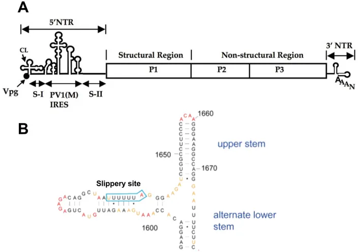
Though this thesis focuses nearly exclusively on the HIV TAR element, two additional ex-amples of viral RNAs and their functions are shown in Figure 1.6.
Slippery site
A
B
Figure 1.6: Two Examples of Functional Viral RNAs A. The poliovirus Internal Ribosome Entry Site (IRES) Element is required for bypassing the ribosomal requirement for 5′caps, so the poliovirus ORF
can be translated. The structured cloverleaf and Vpg elements (which do not code for protein) make up fully ~5% of the poliovirus genome. Adapted from [36]. B The HIV I Frameshift Element allows the simultaneous transcription of two overlapping, but frameshifted, open reading frames (Gag and Pol) into a single polypeptide. Remarkably the ratio of Gag to combined Gag-Pol transcription is conserved across retroviruses [37]. Adapted from [38]
1.6 HIV I TAR RNA
The specific viral RNA target this thesis is most concerned with is the trans-activation response element of HIV I (TAR). The TAR region of HIV canonically occupies the first 45 nucleotides of the ~9000 nucleotide HIV I mRNA genome, and is part of the 5′Untranslated region (5′UTR)
[39]. TAR plays two important roles in HIV replication (Figure 1.22) which will be discussed in detail shortly, but important to note is that the key facilitator of both roles is a small, structured RNA element occupying positions 17–43 on the HIV genome.
1.6.1 TAR Induces a Transcription Cascade
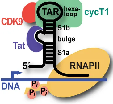
The first, and most important role TAR plays in the HIV life-cycle, is that of a pseudo-promoter facilitating the replication of HIV mRNA, which in the case of HIV is both template for protein production and packaged genome in the virion. Leaky transcription of the HIV I DNA genome leads to the mRNA transcript of the 5′UTR of HIV, which base-pairs with the DNA genome. The trans-activator of transcription (Tat) protein from HIV I [40–42], binds the TAR element on the mRNA and recruits the elements from the host cell necessary to transcribe the genome into mRNA with a positive feedback loop [43,44]. This transcription cascade marks the shift from latent to active HIV infection (illustrated in Figure 1.7).
1.6.2 TAR as pre-miRNA
The second key feature of the TAR element is its ability to act as an anti-apoptotic miRNA, keeping the host T-cell “alive” and manufacturing HIV virions [46–48]. The entire canonical TAR element, as well as a further ~10 nucleotides, are a pre-miRNA, while the mature miRNA is derived from the base-paired region (with mismatches) immediately before and after the struc-tured TAR element (see Figure 1.4 and Figure 1.3 for further details). The TAR element can be seen in the context of the HIV I genome in Figure 1.8.
A key point to note about the two roles of TAR (which are necessary for successful HIV I pro-liferation), is that they are likely less susceptible to mutational evasion as other HIV I targets, such as reverse transcriptase [49–51]. This is due to the fact that, in theory, any mutation which allowed TAR to evade a therapeutic would also require a compensatory mutant to the Tat gene so Tat/TAR-dependent transcription would occur, or would require any mutation to leave the pre-miRNA properties of the TAR element intact.
Tat
cycT1
P
i
P
i
P
i
RNAPII
DNA
TAR
5´
CDK9
S1a
S1b
hexa-loop
bulge
Figure 1.7: Tat–TAR Induced Transcription Cascade The viral transactivation response (TAR) element
RNA comprises lower (S1a) and upper (S1b) stems. The positive transcription elongation factor b (p-TEFb) comprising cyclin T1 (green) and CDK9 (red) is recruited to TAR by the HIV-1 protein Tat (purple), which binds the central RNA bulge allowing cyclin T1 to interact with the apical loop. The bound complex stim-ulates host RNA polymerase II (yellow) by phosphorylation to produce full-length viral transcripts from proviral DNA From: [45]
5′ 3′ G C G C CU AU A C G U G C G U A G U G C U A U A A U AG CC G C G A U G C U A U CU G C A U G C C G CUG GGA 30 10 20 40 40 47 5'
Tat binding site
A
B
mature miRNA
pre-miRNA
Figure 1.8: The HIV I TAR Element in the Context of the HIV Genome The ~5000 bases on the 5′end of
the HIV I genome, and the secondary structure of same, can be seen in A, with the TAR element indicated by a box. B shows the regions of the TAR element which engage in the two main functions of TAR: Tat recruitment and miRNA activity.
1.6.3 Relevance of TAR RNA
HIV afflicts over 36 million people worldwide, and no cure (or vaccine) yet exists. Current treatments are effective, but evolving resistance is always a concern, especially since there are only four current drug targets (entry, reverse transcriptase, integrase, and protease) [52,53].
For reasons outlined above, much effort has gone into generating binders of TAR RNA. These efforts have resulted in TAR-binding molecules ranging in size from small molecules to cyclic peptides [54–58]. These efforts have had various degrees of success, but none has resulted in a useful therapeutic.
1.7 Basis of Nucleic Acid Behavior
Since the importance of generating specific binders for arbitrary DNAs and RNAs should be clear, as should the worth of a binder for the HIV I TAR element, let’s look at the challenges associated with the goal of targeting an RNA.
As a general statement, the first step to targeting anything is understanding how the target behaves. To understand an RNA target, let’s consider what a typical DNA or RNA macromolecule is built from and the general properties of a DNA or RNA macromolecule. while using the former to understand the latter. Finally, we will examine how these emergent properties inform the challenge of creating a binder for an arbitrary polynucleic acid vs. accomplishing the same for a protein of similar size.
1.7.1 Nucleotides
The nucleotide is the monomeric building block of an extended DNA or RNA molecule. A fundamental difference in engineering a protein binder for a nucleic acid vs. another protein emerges from the differences between their monomers: nucleotides and amino acids respec-tively. Proteinaceous amino acids account for 20 different side-chains with fairly diverse chemi-cal functionality including carboxylic acids, primary amines, amides, guanidino, thiol, thioether, as well as various degrees of hydrophobicity. There are, however, only five canonical
nucle-obases, and only four can occupy a given position on a DNA or RNA polymer. In addition to the limited number of possibilities at each position, these nucleobases are limited in chemical diversity (and are of similar hydrophobicity). All nucleobases can be reductively described as small, minimally modified heterocycles, with either purine or pyrimidine as the foundational heterocycle. Figure 1.9 shows the various nucleobases.
RNA polynucleotide polypeptide DNA nucleotide Nucleobase ribose phosphate deoxyribose No 2′ hydroxy
Adenine Guanine Uracil Cytosine Thymine
Figure 1.9: Polynucleotide (RNA) vs. DNA vs. Polypeptide) vs. Polypeptide A polynucleotide is made
up of nucleotides while a polypeptide is made up of amino acids. The key differences between the two classes of nucleotide are the substitution of thymine for uracil as a nucleobase in DNA and the lack of a hydroxy group on the 2′carbon in the backbone sugar of DNA (deoxyribose vs. ribose). The primary
implications of a nucleotide monomer vs. an amino acid monomer are the minimal chemical diversity of the nucleotides vs. the proteinogenic amino acids, and the largely exposed backbone of a polynucleic acid vs. the internalized backbone of a polypeptide.
These nucleobases are connected to a cyclic pentose, with the glycosidic bond attached to carbon 1 on the pentose. RNA (ribonucleic acid) nucleotides have ribose as the backbone pen-tose, while DNA (deoxyribonucleic acid) has deoxyribose, which lacks a hydroxy group on the 2′
carbon. This small difference leads to drastically different chemical properties, and affects the way that RNA and DNA interact with the cellular environment and pathways.
The pentose is attached to a phosphate group through the 5′oxygen. Since these backbone elements are identical on any given DNA or RNA they cannot be used to target a specific DNA or RNA. The nucleobase, sugar, and phosphate together are referred to as a nucleotide (or deoxynu-cleotide) monophosphate in this form. Importantly, at physiological conditions, the phosphate groups carries a negative charge in both the monomer form and as part of a polymer. This fact is critically important to the emergent properties of a polynucleic acid chain.
Since the sugar and phosphate groups are identical scaffolding connecting the different DNA or RNA bases, they are usually referred together as the sugar/phosphate backbone. Worth noting is that nucleotide monomers with different numbers of phosphate groups (especially adenine mono- di- and triphosphate) are also important signaling molecules, energy sources for reac-tion catalysis, and enzyme co-factors within a cell in addireac-tion to their role as building blocks of complicated polynucleic acids. Therefore, anything which targets an individual nucleotide too specifically will wreak havoc with almost all cellular processes.
1.7.2 Nucleic acid as a Polymer String
Though the nucleotide monomers are important, DNA and RNA functionality is predi-cated upon the polymerization of these nucleotides. This polymerization occurs strictly on the sugar/phosphate backbone, with a phosphate forming a bridge between the 5′-OH group on one nucleotide and the 3′-OH group on the next, in what is referred to as a phosphodiester linkage. Nucleic acid sequences are conventionally written in the 5′→3′direction, since this is both the direction in which they are synthesized by a polymerase, and read by the ribosome [7].
Importantly, if one draws a line through one end of the glycosidic bond, the variable nucle-obases will all be on the one side of the line, and the sugar/phosphate backbone will be on the other (as can be seen in Figure 1.9). In contrast, if one draws a line down the peptide backbone of a protein the functional side chains of the amino acids will be found on either side of the
backbone. Thus the non-variable backbone of a polynucleic acid cordons off the variable bases, while in a polypeptide the variable side chains cordon off the non-variable backbone.
This is a gross oversimplification of the actual three-dimensional, structured, reality of both macromolecule classes, but it is a valuable approximation to build on as we move to the next model.
1.7.3 Nucleic Acid as Ribbon
Given the nature of a polynucleic acid as segregated anionic and hydrophobic surfaces, a single-stranded polynucleic acid can be roughly understood as a two-surfaced chemical ribbon with the same essential properties from end to end, but with drastically different properties on either side (illustrated roughly in Figure 1.10).
One side of this two-sided ribbon is the sugar phosphate backbone (Figure 1.10A); the salient characteristic of this side of the ribbon are the regular and identical phosphate groups which give it a uniform negative charge. Due to this negative charge, a polynucleic acid will have gen-eral affinity for cations and cationic moieties (notably the side-chains of Lys and Arg residues in proteins) will have fairly strong non-specific interactions with a DNA or RNA chain.
The other side of the ribbon can be approximated as a regular series of aromatic loops with the π-systems perpendicular to each other. The variations in this pattern are minor, since any chemical diversity emerges from a limited set of chemically similar nucleobases. The salient characteristics of this side of the ribbon is its hydrophobic character (and therefore its tendency to be buried while in aqueous solution), and the ability of external aromatic π-systems (such as those found on the side-chains of Tyr, Trp, and Phe) to participate in π-π interactions with the nucleotides on this chain Figure 1.10B.
Both sides of the ribbon, of course, participate in hydrogen bonding interactions, but the backbone side has repeating, identical units of sugar hydroxy groups, while the nucleobase sur-face has heteroatoms on the purine and pyrmidine rings which participate in hydrogen bond-ing interaction. It is only on this side of the ribbon that there is any variability in the hydrogen
–
–
–
–
–
–
–
π π
π
π π
π π π
π π π
nucleobase surface
backbone surface
hybridized ribbon
B
C
A
Figure 1.10: Polynucleotide as a Two-Sided Chemical Ribbon Considering a polynucleic acid as a
two-sided ribbon is an enlightening mental model. The key point of this model is that each surface has dif-ferent overwhelming tendencies. The nucleobase surface tends toward interaction with hydrophobic ar-eas, while the backbone side tends toward interaction with cations. A secondary, related, point is that hybridization tends to bury the nucleobase surface, meaning that the majority of exposed surface in a double-stranded DNA or RNA is the charged, hydrophobic, constant backbone side of the ribbon.
bonding pattern. Indeed, minor variations in hydrogen bond and space-filling pattern are the only chemical difference between polynucleic acids of different sequence.
Considering a nucleic acid in this manner further clarifies the challenge of targeting a polynucleic acid vs. targeting a protein of similar size. When a nucleic acid hybridizes in this model, it does so by two nucleobase surfaces coming together, therefore the nucleobase surface (which is, after all, where specific recognition usually occurs) will expose relatively less surface, and the more exposed anionic backbone forms a wall around it.
In contrast, while the peptide backbone is obviously the wellspring of many properties of proteins, it does need not usually need to be considered when targeting a specific protein to the degree that the negatively charged backbone of a nucleic acid does. Any specific binder of an RNA walks a tightrope of anionic interaction. Such a binder needs to either interact with or tolerate the drastic chemical property of a persistent and constant negative charge, but it can’t interact too strongly with this charge or it will bind all nucleic acids rather than a specific nucleic acid.
1.7.4 Nucleic Acid Base-Pairing
This “ribbon” model of a nucleic acid also informs an essential emergent chemical prop-erty of nucleic acids which is the basis for nearly all of the three-dimensional structure of these molecules—the antiparallel binding of two strands of nucleic acid with complementary se-quences. On a conceptual level, this can be approximated by thinking of the hydrophobic sides of two nucleic “ribbons” coming together, burying the hydrophobic bases and exposing the hy-drophilic sugar-phosphate backbone.
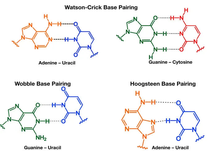
In the nucleotide level view of a double-stranded chain, each nucleobase participates in spe-cific hydrogen-bonding interactions with a partner nucleobase with the opposite heterocycle foundation, this is known as “Watson-Crick” base pairing (the two hydrogen-bond mediated A·T pair, and the three hydrogen-bond mediated G·C pair). There are also alternative base pairings, such as the “wobble” and “Hoogsteen” base-pairings. These are less favorable than standard
Watson-Crick base, but can still stabilize the structure of polynucleic acids. The different types of base pairing are illustrated in Figure 1.11.
Watson-Crick Base Pairing
Wobble Base Pairing Hoogsteen Base Pairing
Adenine – Uracil Guanine – Cytosine
Adenine – Uracil Guanine – Uracil
Figure 1.11: Examples of the primary modes of nucleic acid base pairing The most important base
pair-ing interactions are the Watson-Crick A·T and G·C, but Wobble base pairs can pair non-canonical part-ners, and Hoogsteen pairs can stabilize alternate conformations (as can be seen in Figure 4.4
The phenomenon of hybridization is often thought of as being driven by these hydrogen bond interactions, but it is more accurate to say that it is enabled by them. This becomes clear when considering the Gibb’s free energy equation (ΔG = ΔH - TΔS). The spatially specific binding of two massive biopolymer ribbons is most assuredly entropically costly, but so are the ordered, clathrate-like water formations around the nucleobases of a single-stranded nucleic acid. The hydrogen bonds between purine and pyrimidine bases allow this ordered water to disperse into disorder, making the ΔS value of complex formation less negative than it would otherwise be.
The driving force for hybridization—the ΔH—comes from the hydrophobic effect. When a nucleic acid is base-paired, it minimizes the surface area exposure of the hydrophobic nucle-obase side of the ribbon, making the charged backbone the site of the primary interactions. Though it is, importantly, a more predictable and specific structure formation than protein-folding, it is driven by the same forces [59].
1.8 Nucleic Acid 3D-Structures
This tendency of a polynucleic acid to base-pair to a complementary strand leads to the for-mation of complex three-dimensional structures. Unlike the specifics of protein folding—which remain difficult to predict from a primary sequence—the structure formed by a polynucleic acid stems from specific and predictable interactions between bases. It is here that the properties and tendencies of DNA and RNA (at least in a biological context) begin to diverge.
1.8.1 DNA Structure
For DNA, discussion of biologically relevant structure begins with the familiar double-helix. The double-helix of popular imagination is known as B-DNA. B-DNA is a right-handed double helix, with constant, uneven spacing between the two helices, creating a major groove (22 Å in width) and a minor groove (12 Å in width). The helix undergoes a full rotation every ~10.5 base-pairs. The spatial relationship between bases is repeated constantly with minimal variation [7].
Realistically, this is also where a discussion of the biologically relevant structural variation of DNA ends. Single-stranded DNA (ssDNA) is certainly a chemical possibility, but is seldom seen in a cellular context outside of the context of DNA replication. Additionally, other forms of double-stranded DNA exist, sometimes even in a cellular context, but the proportion of these other forms can be generously described as “trace” [60]. Ultimately, this lack of structural variation makes targeting biologically relevant DNA a simpler, more systematic exercise than targeting complex and amorphous structured RNAs [61]. Though simpler than targeting RNA, the considerations
involved in targeting DNA inform the challenge of targeting structured RNA, and as such one such systematic solution will be described at length in Section 1.13 on page 36.
1.8.2 RNA Structure
RNA, in contrast, demonstrates a more expansive palette of structural variation within the cell. Double-stranded RNA is important to a cell, but unless a cell is infected with a Baltimore Class III viruses with a dsRNA genome, extensive swaths of dsRNA are not of particular concern. Longer strands of RNA in a cellular environment are all-but entirely single-stranded mRNA or essential and conserved tRNAs or rRNA (Table 1.1). And while three-dimensional structural el-ements are vital in the functional RNAs (such as miRNA or viral RNAs) discussed in Section 1.5, these structured regions are non-extensive enough that they are best thought of as elements. Prac-tically, this means that even though a given structured RNA may be locally similar to a B-DNA double helix, these areas are generally small enough to bend and breathe in a way that chro-mosomal DNA does not, and structured RNA molecules cannot be assumed to have the same absolute spatial regularity as a B-DNA helix.
The basic types of RNA structural elements are discussed here and illustrated in Figure 1.12.
Stem-Loops
Stem loops are the fundamental secondary structural element in an RNA. A stem-loop oc-curs when a series of RNA bases is able to form Watson-Crick base-pairs with a complementary sequence of RNA on the same strand, which is nearby but not contiguous. The paired bases form the stem, while the bases between form a loop. Generally speaking, the loop is between 4–8 bases, though this is sometimes as many as 10 bases (as in U1hpII, an important RNA for this work [62]). A loop at the end of a strand of paired RNA is also called a hairpin loop. A related structural element is the “multi-branch loop,” which occurs when stem loops are separated by unpaired RNA strands.
internal loop
hairipin loops/stem loops
pseudoknot
multi-branch loop
Figure 1.12: RNA Secondary Structure Overview Watson-Crick base-pairing results in a variety of
loop-based secondary structure, examples of which are shown here. Important to note is that the base-paired stem regions are largely helical, while the structure elements represent a departure from helicity. Adapted from [63]
![Table 1.1: Composition of a Cell A: Breakdown of mammalian cellular dry mass by molecule type [6] B:](https://thumb-eu.123doks.com/thumbv2/5dokorg/4336627.98466/19.918.167.749.164.335/table-composition-cell-breakdown-mammalian-cellular-mass-molecule.webp)