Dental implants in patients with Sjögren’s syndrome: a case series and a systematic review Running title: Sjögren’s syndrome and dental implants
Bruno Ramos Chrcanovic 1, Jenö Kisch 2, Ann Wennerberg 3
1
Department of Prosthodontics, Faculty of Odontology, Malmö University, Malmö, Sweden. bruno.chrcanovic@mau.se; brunochrcanovic@hotmail.com
2
Department of Prosthodontics, Faculty of Odontology, Malmö University, Malmö, Sweden. jeno.kisch@gmail.com
3 Department of Prosthodontics, Sahlgrenska Academy, University of Gothenburg, Göteborg, Sweden ann.wennerberg@odontologi.gu.se
DEPARTMENT OF PROSTHODONTICS, FACULTY OF ODONTOLOGY, MALMÖ UNIVERSITY, MALMÖ, SWEDEN; DEPARTMENT OF PROSTHODONTICS, SAHLGRENSKA ACADEMY, UNIVERSITY OF GOTHENBURG, GÖTEBORG, SWEDEN
* Corresponding author:
Bruno Ramos Chrcanovic. Department of Prosthodontics, Faculty of Odontology, Malmö University, Carl Gustafs väg 34, SE-214 21, Malmö, Sweden. bruno.chrcanovic@mau.se; brunochrcanovic@hotmail.com Mobile: +46 725 541 545 Fax: +46 40 6658503
Funding/grant support: Scientific Research Council of Sweden (Vetenskapsrådet), Folktandvården Skåne AB, Sweden
KEYWORDS
ABSTRACT
The purpose of the present study was to assess the clinical outcome of dental implants in a series of patients with Sjögren’s syndrome (SS), as well as review of the literature. The study consisted of two parts: report of a case series and a systematic review. Results of the clinical series: 19 patients received 107 implants, followed for a mean of 125 months. Two patients lost 3 implants (failure rate 2.80%, 3/107). At the last follow-up, there was a mean±SD marginal bone loss (MBL) of -2.190±1.384 mm. Estimation of 4.39 mm of MBL after 30 years. Results of the review: 19 studies, including the present clinical series, with 712 implants in 186 patients (failure rate 4.11%, 29/705; failed at a mean time of 12.9±31.7 months), followed up for a mean of 72.5 months. The probability of failure was 2.8% (95% CI 1.6%, 4.1%). Primary SS had lower implant failure rate (2.54%, 3/118) than secondary SS patients (6.52%, 12/184). As a conclusion, dental implants should be considered by dentists as a viable treatment option in patients with SS, as the failure rate is fairly low. The SS patients may, however, present a higher MBL around implants than in the general population.
KEYWORDS
INTRODUCTION
Primary and secondary Sjogren’s syndromes (SS) are systemic disorders characterized by lymphocyte infiltration and progressive destruction of exocrine glands leading to mucosal dryness, particularly of the eyes and mouth.1 SS have a significant impact to the oral health, and its oral implications include xerostomia due to reduced salivary flow; rampant caries; chronically inflamed and irritated oral mucosa; inflamed, enlarged, and hardening salivary glands; an increased incidence of chronic candidiasis;2, 3 angular cheilitis; increased plaque retention, changes in taste perception; difficulty swallowing; chronic tissue discomfort, recurrent denture sores, difficulty masticating, and mandibular denture instability.4 Because of the great susceptibility for caries, patients with SS often loose many teeth during their lives and at an earlier age compared to the general population. Due to the difficulties or the inability to wear dentures because of the dry and sensitive oral mucosa, many SS patients and dentists have opted for an oral rehabilitation with dental implants instead.
There is a great demand for dental implants in SS patients, but much is conjectured on how successful this treatment may be in these patients.5 According to the notes of SS patients from one study,6 a considerable number of dentists and rheumatologists advised them not to have dental implants, as the professionals were concerned about initial osseointegration or a higher risk of implant failure. The present study aimed to assess the clinical outcome of dental implants in a series of patients with SS, as well as to review the cases of SS patients receiving dental implants described in the literature.
MATERIALS AND METHODS
The present study consists of two parts: (a) report of a case series and (b) a systematic review.
a) Case series
Materials. This retrospective study included patients treated with dental implants during the period 1980-2014 at one specialist clinic (Clinic for Prosthodontics, Centre of Dental Specialist Care, Malmö, Sweden). The study was approved by the regional Ethical Committee, Lund, Sweden (Dnr 2014/598; Dnr 2015/72).
Definitions. An implant was considered a failure if presenting signs and symptoms that led to implant removal, i.e. a lost implant.
Inclusion and exclusion criteria. Implants installed in patients being diagnosed with SS were included. The patients could have had primary (another underlying rheumatic disorder does not occur) or secondary SS (associated with another underlying rheumatic disease, such as systemic lupus erythematosus, rheumatoid arthritis, or systemic sclerosis), and the syndrome needed to have been diagnosed having performed tests to do so, usually a combination of some of (but not limited to) the following tests: blood test for the autoantibody SSA, ANA (Anti-Nuclear Antibody) test, lip biopsy (salivary gland biopsy), Schirmer tear test, examination of the surface of the eye with rose bengal and lissamine green, sialogram, salivary flow, and salivary scintigraphy.
Modern threaded cylindrical- or conical-design implants were included, and zygomatic implants were excluded. Only implants with first radiographs taken within 12 months after implant installation were considered for MBL evaluation. Negative values of MBL were considered as bone loss.
Data collection. The data were directly entered into a SPSS file (SPSS software, version 25, SPSS Inc., Chicago, IL, USA) as the dental records of the patients were being read, and it consisted of several implant-, site-, and patient-related factors.
The patients were periodically followed up by a dental hygienist at the clinic, with attendance based on individual needs.
Marginal bone level evaluation. Reproducible intra-oral radiographs were used. When there were no available digital radiographies from the baseline appointment, the analogue periapical radiographies were scanned at 1200 dpi (Epson Perfection V800 Photo Color Scanner; Nagano, Japan). Marginal bone level (MBL) was measured after calibration based on the inter-thread (pitch) distance of each implant type, the Nobel implant being the most common (0.60 mm). Measurements were taken from the implant-abutment junction to the marginal bone level, at both mesial and distal sides of each implant, and then the mean value of these two measurements was considered. MBL was calculated by comparing bone-to-implant contact levels to the radiographic baseline examination. The Image J software (National Institute of Health, Bethesda, USA) was used for all measurements.
b) Systematic review
The PRISMA Statement guidelines7 were followed.
Objective. The focused question was elaborated by using the PICO format (participants, interventions, comparisons and outcomes): What are the clinical outcomes, i.e. implant survival and MBL, of dental implants installed in patients with SS?
Search strategies. A search without time restrictions was undertaken in July 2018 in the five electronic databases: PubMed/Medline, Web of Science, Science Direct, J-Stage, and Lilacs. The following terms were used:
Google Scholar was also checked. A manual search of dental implants-related journals was performed.
Inclusion and Exclusion Criteria. Publications reporting cases of patients diagnosed with Sjögren’s syndrome rehabilitated with implant-retained and/or implant-supported oral prosthetic rehabilitation were included. Publications reporting clinical cases of prosthetic rehabilitation not using dental implants were not included.
Study selection. The titles and abstracts of all reports identified through the electronic searches were read independently by the authors. For studies appearing to meet the inclusion criteria, or for which there were insufficient data in the title and abstract to make a clear decision, the full report was obtained. Disagreements were resolved by discussion between the authors.
Data extraction. The following data were extracted on a standard form: year of publication, number of patients, patient’s sex, age, implant type, implants placed and lost in maxilla and mandible, implant healing period, period between implant placement and loss, type of prosthetic reconstruction, MBL, and follow-up period. Contact with authors for possible missing data was performed.
c) Statistical analyses
The mean, standard deviation (SD), and percentage were calculated for several variables. The test perfomed were the following: Kolmogorov–Smirnov (to evaluate the normal distribution), Levene’s test (to evaluate homoscedasticity), Student’s t-test or Mann-Whitney (for two independent groups, continuous variables), Pearson’s chi-squared or Fisher’s exact test (for categorical variables), correlation and linear regression (to check the relationship between MBL and time of follow-up), and untransformed proportion random-effects DerSimonian-Laird method8 (for implant failure). The degree of statistical significance was considered p < 0.05. All data were statistically analyzed using the SPSS version 25 software (SPSS Inc., Chicago, IL, USA) and the software OpenMeta[Analyst].9
RESULTS a) Case series
A number of 19 subjects among the 2,670 patients treated with implants in this clinic were diagnosed with SS. These patients received a total of 107 oral implants – 56 implants in maxillae, 51 in mandibles, 11 in 1 male, 96 in 18 females. The mean±SD patients’ age was 63.3±8.1 (min-max, 50.4-80.3). A total of 107 threaded implants with a cylindrical design were installed - 43 Nobel turned, 38 Nobel MK III TiUnite, 13 Astra TiOblast, 10 Astra Osseospeed, 2 Bego Semados, and 1 Nobel Active. All implants were inserted with open flapped surgery in healed sites with delayed loading. The abutment connection was performed after a mean±SD healing time of 163±46 days (min-max, 74-233). The mean length of the implants was 12.8±1.8 mm (min-max, 7-15) and the mean diameter 3.74±0.17 mm (min-max, 3.3-4.3).
According to the Lekholm and Zarbclassification, and concerning bone quantity, 50 implants were placed in type B, 41 in type C, and 8 in type D bone. Concerning bone quality, 9 implants were placed in type 1, 35 in type 2, 49 in type 3, and 6 in type 4. This information was available for 99 implants.
Three implants were used for single-crown restorations, 22 implants for 8 fixed partial prostheses of 2-6 prosthetic elements, 6 implants for a fixed partial prosthesis of 7-10 prosthetic elements, 74 implants for 14 full-arch fixed prostheses, and 2 implants to support an overdenture.
The patients were followed-up for a mean±SD of 125.5±82.5 months (min-max, 5.6-341.2). Two patients lost 3 implants, all due to loss/lack of osseointegration, and no implant fracture was identified. Failures were recorded for 3 Nobel turned implants only in mandible, and they happened 36 days, 8.5 months, and 159.6 months after implant installation. One of the lost implants was part of a full-arch prosthesis, and 2 implants were lost before the abutment connection and were planned for a fixed partial prosthesis of 7-10 prosthetic elements. These two lost implants were later replaced by another two implants. The three lost implants were installed in bone B2 (n=1) and C4 (n=2).
Marginal bone level evaluation. A total of 35 implants were excluded from the analysis of MBL for the following reasons: no radiograms were found in the dental records (n=13), the first radiogram was taken after one year of implant installation surgery (n=15), very short follow-up (n=5), and early implant failure (n=2). Of the remaining 72 implants, 293 MBL measurements were performed, considering the several radiological follow-ups for each implant - a mean of 4.1 MBL measurements per implant. The baseline radiograms were taken at a mean±SD of 4.4±3.2 months (min-max, 0-9.6) after implant placement. The implants were radiologically followed up for mean±SD of 125.2±80.5 months (minmax, 0.5329.9). At the last followup, there was a mean±SD MBL of -2.190±1.384 mm (min, max; -7.571, 0.000) for all implants (n=72). Table 1 shows that, at the last radiological follow-up, 25% of the implants presented 3 or more millimeters of MBL. There was a steady increase in the mean MBL with time of follow-up (Table 2). There was an estimated trend to loss bone with time (Figure 1), reaching an estimated 4.39 mm of bone loss 30 years after implant installation. According to the linear regression equation (y = -0.79 – 0.01x), there was an estimated loss of 0.01 mm of bone for every additional month of follow-up. There was a moderate correlation between MBL and follow-up time (R = 0.563, R2 = 0.317, p < 0.001, Pearson correlation). In the last radiological follow-up, the mean±SD MBL in primary SS patients (-2.371±1.451; n=53; mean follow-up 133.6±92.3 months) was higher than in secondary SS patients (-1.686±1.053; n=19; mean follow-up 101.8±13.0 months), even though with no statistically significance (p = 0.093, Mann-Whitney test).
b) Systematic review
Literature search
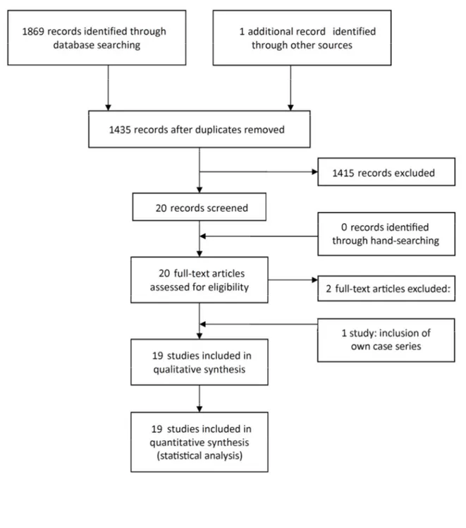
The study selection process is summarized in Figure 2. At the end of the process, the full-text reports of the remaining 20 articles led to the exclusion of 2 publications.10, 11 In one of them the clinical case was later reported in another publication,4 but with follow-up information. In the second excluded article, the same cases were reported in another published study by the same research
group.12 Thus, a total of 18 publications were included in the review. As the patients of the present case series were also included in the analyses, 19 studies were included in the review.
Description of the Studies and Analyses
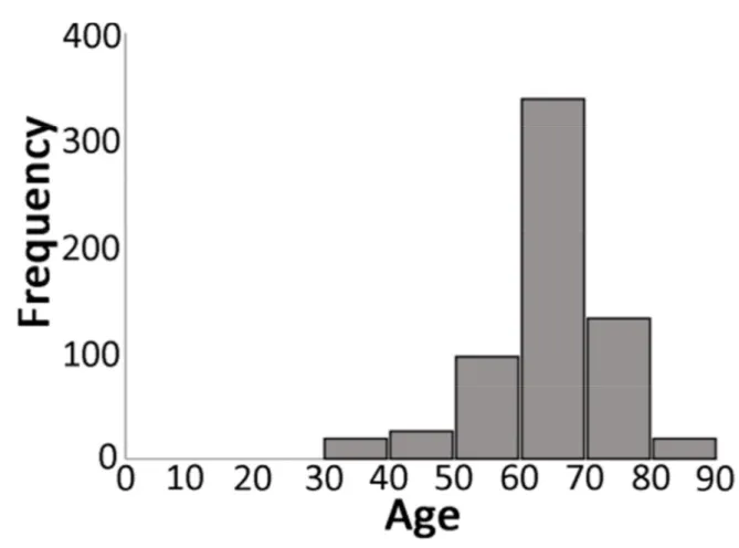
Table 3 shows detailed data of the included studies. Eighteen publications4-6, 12-26 were included in the present review, and the 19 patients of the present case series were also included in the analyses. These 19 studies totaled the placement of 712 implants (45 in men, 644 in women, 23 not available) in 186 patients (8 men, 167 women, 11 not available). It was possible to distinguish some patients presenting primary SS (21 patients, 118 implants) and secondary SS (31 patients, 191 implants). At the placement of the implants the mean age of the patients was 62.5±9.3 years (min-max, 38-85; 630 implants, 166 patients). Most implants were placed in the seventh decade of life (Figure 3).
Information on follow-up was provided for 705 implants (mean 72.5±59.2 months, min-max 5-341.2), of which 29 failed (4.1%). The failure rate was higher for implants installed in the maxilla (4.82%; 11/228) in comparison to implants placed in the mandible (3.49%; 13/373), but the difference was not statistically significant (p = 0.416; Pearson chi-squared test). Patients presenting primary SS had lower implant failure rate (2.54%, 3/118) than patients presenting secondary SS (6.52%, 12/184), but the difference was not statistically significant (p = 0.120; Pearson chi-squared test).
Implants failed at a mean time of 12.9±31.7 months (min-max, 1-160; n=24) after implant placement. Considering the implants with information on the precise time of failure, 79.2% (19/24) occurred within 6 months after installation surgery or at the abutment connection. One implant failed at 9 months, two at 16 months, and one implant each on 24 and 160 months after installation. There was no information on time of failure for the 5 failed implants of the study of Albrecht et al. (2016).
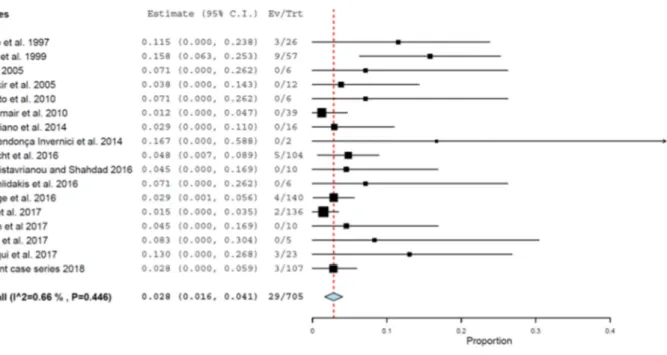
The probability of failure (Figure 4) was 2.8% (95% CI 1.6%, 4.1%, standard error = 0.006, p < 0.001; heterogeneity: τ2 = 0.000, Chi2 = 16.106, p = 0.446, I2 = 0.66%) for the observed mean follow-up of these studies altogether. Only the clinical cases for which the follow-follow-up was informed were included in this analysis.
DISCUSSION
The present study aimed to assess the clinical outcome of dental implants in a series of patients with SS, as well as to review the cases of SS patients receiving dental implants described in the literature. In relation to previous reviews on the subject,27, 28 the present study performed a more careful systematic search of the literature, thus resulting in greater number of included studies. Systematic reviews are insufficiently informative if they do not include all available current evidence. This failure to rigorously synthesize the totality of relevant evidence may have a detrimental effect on treatment decisions and future research planning.29 Moreover, the present study performed a more in-depth and detailed statistical analysis of the compilation of included studies in comparison to these previous reviews.
After reviewing 712 implants placed in 186 SS patients, it was observed that the failure rate is fairly low in this population: 4.1% over a mean of 72.5±59.2 months of follow-up. Moreover, in the majority of cases in which an implant failed to integrate, the cause is unknown. Most of the implant failures occurred within 6 months after implant installation, in agreement with other studies in which most of the lost implants fail within a couple of months after placement surgery.30, 31 In general, SS patients reported a significant improvement in the quality of life after oral rehabilitation with dental implants, with regard to satisfaction, chewing, self-assurance and self-appearance.6, 16, 20 It is, however, important to stress that oral functioning is impaired in patients with SS and continues to be impaired in patients with implant-retained prosthetics.5
Only few studies reported data on MBL5, 15, 19, 20, 22, 25 and still most of the publications provided only vague information such as ”good marginal bone levels”,22 “there was no peri-implant bone loss”,25 “no bone loss”,19 “stable bone levels”.15 Only two previous studies provided figures,5, 20 as well as the present one. The results of these two publications5, 20 suggested that MBL is not a major problem in SS patients. The present study, however, observed a very fast initial MBL in comparison to the general population followed up for at least 20 years in another study.31 This loss showed to be considerable in short- and medium-term observations, with many implants showing
MBL of 2 mm during the first years after installation and other many presenting MBL around 4 mm within the first 10 years of follow-up. Korfage et al.5 had already noticed that there probably is an increased risk of peri-implant infections in SS patients in the long term. The salivary secretion and the related self-clearance of the oral cavity is reduced in SS patients, which results in debris being collected more quickly and remaining on the implant surfaces in SS subjects than in patients with no xerostomia 5. In the study of Korfage et al.,5 SS subjects presented more gingival swelling, bleeding and increased pocket probing depths than in the controls with no salivary problems. As a consequence, the marginal periimplant tissue is more prone to continuous inflammatory insults than the peri-implant tissue in healthy patients. The reported more frequent presence of peri-implant mucositis in SS patients may be associated with an increased prevalence of peri-implantitis later on.5
Smoking could have been a factor to influence implant failure,32 and MBL33 in these patients. The same is true for a series of other conditions such as bruxism,34 diabetes,35 and the intake of antidepressants36 or of medicaments to reduce the gastric acid production.37 Detailed information about these conditions and habits among the patients was, however, not provided for every patient by the publications. Another point to consider is that patients with secondary SS showed a higher implant failure rate than patients with primary SS. Even though the difference was not statistically significant, there might be some clinical relevance and significance. The usual intake of corticosteroids for the management of underlying rheumatic diseases in the patients with secondary SS could be partly associated with impaired osseointegration. Corticosteroids induce apoptosis of osteoblasts,38 reducing the number of pre-osteoblasts,39 and promoting differentiation of bone marrow stromal cells to adipocyte lineage cells.40 Moreover, corticosteroids increase the life span of osteoclasts41 and suppress bone formation via the osteoclast.42 Besides corticosteroids, methotrexate43 and biological-targeted therapies, including TNF-α inhibitors and rituximab,44 have been used in patients with primary SS-associated inflammatory arthritis. Methotrexate has been shown to be a potent inhibitor of osteoblast's proliferation and mitochondrial metabolism in vitro,45 and to have the potential to interfere with the osseointegration process.46 Concerning biological
agents, these have been found to have effects on human chondrocytes and osteocytes in vitro,47 and to present a significant suppression in bone turnover.48 These drugs can have some impact on wound healing and osseointegration, not only the underlying SS itself. The higher MBL in primary SS patients in comparison to secondary SS patients can be either a true finding, being related to the negative effects of the abovementioned drugs in primary SS patients, or be related to a shorter radiological follow-up for the secondary SS patients and a much smaller sample size at the last follow-up. More balanced groups are necessary in order to confirm this difference.
All in all, the findings from the present study suggest that SS itself does not impair the biology of osseointegration. It may, however, result in a higher MBL than in non SS patients, which may require an increased number of recall visits and shortened professional hygiene intervals.
The limitations of the present study include (1) the presence of confounding factors that may have affected and influenced the outcomes,49 (2) the retrospective design of the studies included in the systematic review, as well as the present case series of 19 patients, (3), the lack standardized timing for evaluating the radiographs and low percentage of MBL measurements due to the retrospective nature of the case series, and (4) the fact that most studies reported a small number of patients followed up for a limited period of time.
CONCLUSIONS
Dental implants should be considered by dentists as a viable treatment option in patients with SS, as the failure rate is fairly low: 4.2% over a mean of 73.1±59.8 months of follow-up. The SS patients may, however, present a higher MBL around implants than in in the general population.
ACKNOWLEDGEMENTS
We would like to thank Dr. Alan B. Carr, Dr. Johanna Callhoff, Dr. Katinka Albrech, and Dr. Anke Korfage for providing us some missing information about their studies.
Trial registration at the U.S. National Institutes of Health (clinicaltrials.gov): NCT02369562. Funding/grant support
This work was supported by research funds from the Scientific Research Council of Sweden (Vetenskapsrådet, Dnr 2015-02971). This work was supported by Folktandvården Skåne AB, Sweden. Declaration of conflicting interests
REFERENCES
1. Amerongen AV, Veerman EC. Saliva--the defender of the oral cavity. Oral Dis 2002;8:12-22. 2. Hockberg MC. Sjogren’s syndrome. In: Bennett JC, Plum F, eds. Cecil textbook of medicine.
20th ed. Philadelphia: WB Saunders; 1996:1488-90.
3. Moutsopoulos HM, Tzioufas AG. Sjogren’s syndrome. In: Klipple JH, Dieppe PA, eds. Practical Rheumatology. London: Mosby; 1995:394-6.
4. Binon PP. Thirteen-year follow-up of a mandibular implant-supported fixed complete denture in a patient with Sjogren's syndrome: a clinical report. J Prosthet Dent 2005;94:409-13.
5. Korfage A, Raghoebar GM, Arends S, et al. Dental Implants in Patients with Sjogren's Syndrome. Clin Implant Dent Relat Res 2016;18:937-45.
6. Albrecht K, Callhoff J, Westhoff G, Dietrich T, Dörner T, Zink A. The Prevalence of Dental Implants and Related Factors in Patients with Sjogren Syndrome: Results from a Cohort Study. J Rheumatol 2016;43:1380-5.
7. Moher D, Liberati A, Tetzlaff J, Altman DG, Grp P. Preferred Reporting Items for Systematic Reviews and Meta-Analyses: The PRISMA Statement. Ann Intern Med 2009;151:264-9, W64. 8. DerSimonian R, Laird N. Meta-analysis in clinical trials. Control Clin Trials 1986;7:177-88. 9. Wallace BC, Dahabreh IJ, Trikalinos TA, Lau J, Trow P, Schmid CH. Closing the Gap between
Methodologists and End-Users: R as a Computational Back-End. J Stat Softw 2012;49:1-15. 10. Binon PP, Fowler CN. Implant-supported fixed prosthesis treatment of a patient with
Sjogren's syndrome: a clinical report. Int J Oral Maxillofac Implants 1993;8:54-8.
11. Weinländer M, Krennmair G, Piehslinger E. Implant prosthodontic rehabilitation of patients with rheumatic disorders: a case series report. Int J Prosthodont 2010;23:22-8.
12. Krennmair G, Seemann R, Piehslinger E. Dental implants in patients with rheumatoid arthritis: clinical outcome and peri-implant findings. J Clin Periodontol 2010;37:928-36.
13. Aravena DM. Rehabilitación oral mediante implantes dentales en paciente con síndrome de Sjögren. Int J Med Surg Sci 2016;3:779-87.
14. Carr AB, Revuru VS, Lohse CM. Association of Systemic Conditions with Dental Implant Failures in 6,384 Patients During a 31-Year Follow-up Period. Int J Oral Maxillofac Implants 2017;32:1153-61.
15. Chatzistavrianou D, Shahdad S. Implant Treatment in Patients with Sjogren's Syndrome: A Review of the Literature and Two Clinical Case Reports. Eur J Prosthodont Restor Dent 2016;24:40-6.
16. Chochlidakis K, Ercoli C, Elad S. Challenges in implant-supported dental treatment in patients with Sjogren's syndrome: A case report and literature review. Quintessence Int 2016;47:515-24.
17. Corigliano M, Re M, Cipollina A, Crescentini F, Docaj D, Baldoni E. The implant treatment of two patients suffering from Sjögren’s syndrome with multifactorial regenerative protocol. Eur Sci J 2014;10:14-25.
18. Cuifen L, Guoguang P, Yuanhua F, Xingxiang H. [Dental implantation in a patient with Sjogren's syndrome: a case report]. Hua Xi Kou Qiang Yi Xue Za Zhi 2017;35:108-11.
19. de Mendonca Invernici M, Finger Stadler A, Vale Nicolau G, Naval Machado MA, Soares de Lima AA, Compagnoni Martins M. Management of Sjogren's Syndrome Patient: A Case Report of Prosthetic Rehabilitation with 6-Year Follow-Up. Case Rep Dent 2014;2014:761251. 20. Isidor F, Brøndum K, Hansen HJ, Jensen J, Sindet-Pedersen S. Outcome of treatment with
implant-retained dental prostheses in patients with Sjogren syndrome. Int J Oral Maxillofac Implants 1999;14:736-43.
21. Oczakir C, Balmer S, Mericske-Stern R. Implant-prosthodontic treatment for special care patients: a case series study. Int J Prosthodont 2005;18:383-9.
22. Payne AG, Lownie JF, Van Der Linden WJ. Implant-supported prostheses in patients with Sjogren's syndrome: a clinical report on three patients. Int J Oral Maxillofac Implants 1997;12:679-85.
23. Peron C, Javed F, Romanos GE. Immediate Loading of Tantalum-Based Implants in Fresh Extraction Sockets in Patient With Sjogren Syndrome: A Case Report and Literature Review. Implant Dent 2017;26:634-8.
24. Siddiqui Z, Wang Y, Makkad P, Thyvalikakath T. Characterizing Restorative Dental Treatments of Sjogren's Syndrome Patients Using Electronic Dental Records Data. Stud Health Technol Inform 2017;245:1166-9.
25. Spinato S, Soardi CM, Zane AM. A mandibular implant-supported fixed complete dental prosthesis in a patient with Sjogren syndrome: case report. Implant Dent 2010;19:178-83. 26. Weber B, Martinez M, Saavedra S, Urrutia C. Síndrome de Sjögren y tratamiento protésico
removible total con implantes mandibulares: caso clínico. Int J Odontostomatol 2008;2:71-6. 27. Almeida D, Vianna K, Arriaga P, Moraschini V. Dental implants in Sjogren's syndrome
patients: A systematic review. PLoS One 2017;12:e0189507.
28. Reichart PA, Schmidt-Westhausen AM, Khongkhunthian P, Strietzel FP. Dental implants in patients with oral mucosal diseases - a systematic review. J Oral Rehabil 2016;43:388-99. 29. Créquit P, Trinquart L, Yavchitz A, Ravaud P. Wasted research when systematic reviews fail to
provide a complete and up-to-date evidence synthesis: the example of lung cancer. BMC Med 2016;14:8.
30. Chrcanovic BR, Kisch J, Albrektsson T, Wennerberg A. Factors Influencing Early Dental Implant Failures. J Dent Res 2016;95:995-1002.
31. Chrcanovic BR, Kisch J, Albrektsson T, Wennerberg A. A retrospective study on clinical and radiological outcomes of oral implants in patients followed up for a minimum of 20 years. Clin Implant Dent Relat Res 2018;20:199-207.
32. Chrcanovic BR, Albrektsson T, Wennerberg A. Smoking and dental implants: A systematic review and meta-analysis. J Dent 2015;43:487-98.
33. Duan X, Wu T, Xu X, et al. Smoking May Lead to Marginal Bone Loss Around Non-Submerged Implants During Bone Healing by Altering Salivary Microbiome: A Prospective Study. J Periodontol 2017;88:1297-308.
34. Chrcanovic BR, Kisch J, Albrektsson T, Wennerberg A. Bruxism and dental implant treatment complications: a retrospective comparative study of 98 bruxer patients and a matched group. Clin Oral Implants Res 2017;28:e1-e9.
35. Chrcanovic BR, Albrektsson T, Wennerberg A. Diabetes and oral implant failure: a systematic review. J Dent Res 2014;93:859-67.
36. Chrcanovic BR, Kisch J, Albrektsson T, Wennerberg A. Is the intake of selective serotonin reuptake inhibitors associated with an increased risk of dental implant failure? Int J Oral Maxillofac Surg 2017;46:782-8.
37. Chrcanovic BR, Kisch J, Albrektsson T, Wennerberg A. Intake of Proton Pump Inhibitors Is Associated with an Increased Risk of Dental Implant Failure. Int J Oral Maxillofac Implants 2017;32:1097-102.
38. Weinstein RS. Glucocorticoid-induced osteoporosis. Rev Endocr Metab Disord 2001;2:65-73. 39. Smith E, Coetzee GA, Frenkel B. Glucocorticoids inhibit cell cycle progression in
differentiating osteoblasts via glycogen synthase kinase-3beta. J Biol Chem 2002;277:18191-7.
40. Pereira RC, Delany AM, Canalis E. Effects of cortisol and bone morphogenetic protein-2 on stromal cell differentiation: correlation with CCAAT-enhancer binding protein expression. Bone 2002;30:685-91.
41. Jia D, O'Brien CA, Stewart SA, Manolagas SC, Weinstein RS. Glucocorticoids act directly on osteoclasts to increase their life span and reduce bone density. Endocrinology 2006;147:5592-9.
42. Kim HJ, Zhao H, Kitaura H, et al. Glucocorticoids suppress bone formation via the osteoclast. J Clin Invest 2006;116:2152-60.
43. Mirouse A, Seror R, Vicaut E, et al. Arthritis in primary Sjogren's syndrome: Characteristics, outcome and treatment from French multicenter retrospective study. Autoimmun Rev 2019;18:9-14.
44. O'Neill ID, Scully C. Biologics in oral medicine: Sjogren syndrome. Oral Dis 2013;19:121-7. 45. Annussek T, Kleinheinz J, Thomas S, Joos U, Wermker K. Short time administration of
antirheumatic drugs - methotrexate as a strong inhibitor of osteoblast's proliferation in vitro. Head Face Med 2012;8:26.
46. Tavakoli M, Yaghini J, Abed AM, Malekzadeh M, Maleki D. Evaluation of Effect of Low-Dose Methotrexate on Osseointegration of Implants: A Biomechanical Study on Dogs. Open Dent J 2018;12:546-54.
47. Isyar M, Bilir B, Yilmaz I, et al. Are biological agents toxic to human chondrocytes and osteocytes? J Orthop Surg Res 2015;10:118.
48. Wheater G, Hogan VE, Teng YK, et al. Suppression of bone turnover by B-cell depletion in patients with rheumatoid arthritis. Osteoporos Int 2011;22:3067-72.
49. Chrcanovic BR, Kisch J, Albrektsson T, Wennerberg A. Analysis of risk factors for cluster behavior of dental implant failures. Clin Implant Dent Relat Res 2017;19:632-42.
Table 1. Marginal bone condition around 72 implants at the last radiological follow-up (mean of 125.2 months).
Marginal bone condition Number of implants (%)
Stable (0 mm) 2 (2.8%)
Bone loss up to 1 mm 14 (19.4%)
Bone loss between 1 and 2 mm 17 (23.6%)
Bone loss between 2 and 3 mm 21 (29.2%)
More than 3 mm of bone loss 18* (25.0%)
* Seven (9.7%) out of these 18 implants presented MBL of more than 4 mm
Table 2. Marginal bone condition around 72 implants (293 measurements) at different follow-up periods.
Follow-up period MBL*
mean±SD (min, max)
Number of measurements with MBL of at least 2 mm (%) <1 year -0.848±0.961 (-2.706, 0.215) (n=30**) 6/30** (20.0) 1 year – 3 years -1.508±0928 (-3.597, 0.346) (n=51) 11/51 (21.6) 3 years – 5 years -1.878±0.939 (-4.453, -0.410) (n=38) 14/38 (36.8) 5 years – 10 years -2.006±1.002 (-3.902, -0.076) (n=63) 33/63 (52.4) >10 years -2.956±1.318 (-7.571, -0.529) (n=39) 29/39 (74.4) MBL – marginal bone loss, SD - standard deviation
* Negative values mean bone loss
** There were 30 MBL measurements until 1 year of follow-up, excluding the initial MBL measurements of the 72 implants at baseline
Figure 1. Scatter plot of 293 marginal bone loss (MBL) measurements of 72 implants. The line represents the estimated MBL along the years of observation, according to the linear regression.
Figure 3. Distribution of implants according to the age of the patient at the time of implant placement surgery.
Figure 4. Probability of implant failure - only clinical cases with information about follow-up were included.