THESIS
THE AMYLOID BETA DIMER/TRIMER: A POTENT STIMULATOR OF NEURONAL AMYLOID BETA SECRETION
AND COFILIN-ACTIN ROD FORMATION
Submitted by Ian Marsden
Department of Biochemistry and Molecular Biology
In partial fulfillment of the requirements For the Degree of Master of Science
Colorado State University Fort Collins, Colorado
Fall 2010
Master’s Committee:
Department Chair: Shing P. Ho Advisor: James R. Bamburg Santiago M. Di Pietro
ii ABSTRACT
THE AMYLOID BETA DIMER/TRIMER: A POTENT STIMULATOR OF NEURONAL AMYLOID BETA SECRETION
AND COFILIN-ACTIN ROD FORMATION
Amyloid beta (Aβ) peptides, a heterogeneous mixture of 39-43 amino acid peptides produced from β- and γ-secretase cleavage of the amyloid precursor protein (APP), are one of the causative agents of Alzheimer disease (AD). Although sensitive enzyme-linked immunosorbent assays (ELISAs) for specific rodent Aβ peptides and for total and specific human Aβ peptides have been commercially available, no commercial assay for total rodent Aβ was available when we began these studies. Such an assay is desirable to determine the effects of the human Aβ peptides on production of Aβ from cultured rodent
neurons, the major model system used in AD research. Here we report an ELISA for total rodent Aβ and show that it can be used without interference from
physiologically relevant concentrations of human Aβ. We then apply the assay to measure the production of Aβ in cultured dissociated rat cortical neurons and rat and mouse hippocampal organotypic slices in response to oxidative stress or treatment with human Aβ dimer/trimer (Aβd/t) obtained from culture medium of Chinese hamster ovary cell line 7PA2 expressing a mutant form of human
iii
amyloid precursor protein. Neither of the treatments leads to accumulation of intracellular Aβ peptides. Peroxide increases Aβ secretion by about 2 fold, similar to results from previous reports that used an immunoprecipitation and western blot assay. Of greater significance is that physiologically relevant
concentrations (250 pM) of human Aβd/t increase rodent Aβ secretion by >3 fold over 4 days, providing support for an Aβ-mediated feed-forward model of AD progression. The over two fold increase in rodent Aβ secreted in response to human Aβd/t was nearly identical between organotypic hippocampal slices of TAU knock-out mice and TAU knock-out mice expressing the human tau transgene, demonstrating that tau plays no role in the enhanced production of Aβ.
Previous studies showed oligomers of synthetic amyloid beta (Aβ1-42)
induced cofilin activation and formation of cofilin-actin rods in a neuronal subpopulation of rat hippocampus primarily localized within the dentate gyrus. Here we demonstrate that Aβd/t at ~250 pM is more potent in rod induction in both dissociated hippocampal neuronal cultures and organotypic slices than is 1 µM synthetic Aβ as typically prepared oligomers, about a 4000 fold difference. Treatment of the Aβd/t fraction with an Aβ-neutralizing antibody eliminates its rod inducing activity. Traditionally prepared synthetic Aβ oligomers contain SDS-stable trimers and tetramers, but are devoid of dimers. When synthetic human Aβ was incubated under conditions that generate a tyrosine oxidized dimer, the concentration that was required to induce rods decreased dramatically. The oxidized dimer had a maximum rod-inducing activity at ~2 nM (10 ng/mL),
iv
suggesting it is the presence of the SDS-stable tyrosine oxidized Aβ dimer in a low-n state that is largely responsible for the potency of the secreted Aβd/t.
Aβd/t-induced rods are highly localized to the dentate gyrus and mossy fiber pathway and form more rapidly (significant over controls by 2 h compared to 8 h for those induced by synthetic Aβ-oligomers). Aβd/t-induced rods are
reversible, disappearing by 24 h after washout. Cofilin dephosphorylation in response to Aβd/t is greatest within the hippocampal regions of rod formation. Overexpression of cofilin phosphatases slingshot and chronophin increase rod formation when expressed alone and exacerbate rod formation when coupled with Aβd/t treatment both in dissociated neurons and organotypic slice cultures. Overexpression of the cofilin kinase, LIM kinase 1, inhibits Aβd/t-induced rod formation. Together these data support a mechanism through which Aβd/t
produces selective synaptic dysfunction affecting learning and memory at least in part via primary effects on cofilin regulation and rod formation in sensitive
iv
TABLE OF CONTENTS
Abstract ii
Table of Contents v
Chapter One: β-Amyloid-induced β-amyloid secretion: A feed-forward model for Alzheimer disease. 1
Preface and Acknowledgement 1
Introduction 2
Materials and Methods 5
Results 11
Discussion 29
Reference List 33
Chapter Two: Amyloid beta dimers/trimers potently induce cofilin-actin rods that are inhibited by maintaining cofilin-phosphorylation. 38
Preface and Acknowledgement 38
Abstract 39
Introduction 40
Materials and Methods 44
Results 52
Discussion 71
1
CHAPTER ONE
β-AMYLOID-INDUCED β-AMYLOID SECRETION: A FEED-FORWARD MODEL FOR ALZHEIMER DISEASE.
Preface and Acknowledgement
The work presented in this chapter has been submitted to Journal of Alzheimer Disease. The order and list of authors is as follows: Ian T. Marsden, Laurie S. Minamide, and James R. Bamburg. We would like to thank Dr. Dennis Selkoe, BWH, Harvard Medical School for the gift of 7PA2 cells and Drs. Chi Pak, O’Neil Wiggan, Barbara Bernstein and Ms. Alisa Shaw for valuable
discussions. The authors gratefully acknowledge funding from the NIH National Institute of Neurological Disorders and Stroke (NS40371 to J.R.B.)
Abstract
Amyloid beta (Aβ) peptides, a heterogeneous mixture of 39-43 amino acid peptides produced from β- and γ-secretase cleavage of the amyloid precursor protein (APP), are one of the causative agents of Alzheimer disease (AD). Although sensitive enzyme-linked immunosorbent assays (ELISAs) for specific rodent Aβ peptides and for total and specific human Aβ peptides have been
2
commercially available, no commercial assay for total rodent Aβ was available when we began these studies. Such an assay is desirable to determine the effects of the human Aβ peptides on production of Aβ from cultured rodent
neurons, the major model system used in AD research. Here we report an ELISA for total rodent Aβ and show that it can be used without interference from
physiologically relevant concentrations of human Aβ. We then apply the assay to measure the production of Aβ in cultured dissociated rat cortical neurons and rat and mouse hippocampal organotypic slices in response to oxidative stress or treatment with human Aβ dimer/trimer (Aβd/t). Neither of the treatments leads to accumulation of intracellular Aβ peptides. Peroxide increases Aβ secretion by about 2 fold, similar to results from previous reports that used an
immunoprecipitation and western blot assay. Of greater significance is that physiologically relevant concentrations (250 pM) of human Aβd/t increase rodent Aβ secretion by >3 fold over 4 days, providing support for an Aβ-mediated feed-forward model of AD progression. The over two fold increase in rodent Aβ secreted in response to human Aβd/t was identical between organotypic
hippocampal slices of TAU knock-out mice and TAU knock-out mice expressing the human tau transgene, demonstrating that tau plays no role in the enhanced production of Aβ.
Introduction
Alzheimer disease is the major form of dementia that affects the aged with about a 50% probability of occurrence in every person living to age 85 and
3
beyond (Alzheimer’s Association, 2010). The pathological hallmarks of the disease are extracellular amyloid plaques, composed primarily of the amyloid beta peptide, and hyperphosphorylated tau inclusions in the form of striated neuropil threads and neurofibrillary tangles (Bamburg & Bloom, 2009). Familial AD, representing 1% or less of AD cases, arises from mutations in genes
affecting the production or clearance in the brain of the 38-43 amino acid amyloid beta (Aβ) peptides (Tanzi & Bertram, 2005), which are excised from the
transmembrane amyloid precursor protein (APP) through the actions of β- and γ-secretases (Glenner & Wong, 1984; Price et al., 1995; Hardy & Selkoe, 2002; Mattson, 2004). However, Aβ peptides also accumulate in the other 99% of AD cases, called sporadic AD, although the mechanisms driving its production are unclear (Mattson, 2004; Tanzi & Bertram, 2005).
Different isoforms and different conformations or aggregation states of the Aβ peptides deliver different signals to neurons and have remarkably different neuronal and synapto-toxicities. The Aβ1-42 peptides are more amyloidogenic
than the Aβ1-40 peptides, and are more correlated with AD and its progression
(Finder & Glockshuber, 2007; Portelius et al., 2010). Fibrillar forms of the Aβ species are less toxic than the soluble oligomeric forms (Krafft & Klein, 2010). An oligomeric fraction, called Aβ-derived diffusible ligands (ADDLs), has effects on synapses at submicromolar concentrations (Krafft & Klein, 2010). However, an even more active form of Aβ with maximal activity at subnanomolar
concentrations, is secreted from a cultured cell line (7PA2 cells) expressing a mutated form of human APP (Walsh et al., 2002). This material contains
SDS-4
stable human Aβ (HAβ) dimers and trimers (HAβd/t), which can be isolated by gel filtration; the isolated HAβd/t has a marked effect on synaptic function, both in cultured slices and when injected into rodent brain (Cleary et al., 2005;
Townsend et al., 2006; Shankar et al., 2007; Freir et al., 2010). An SDS-stable HAβ dimer has been extracted from postmortem human AD brain and is also active at subnanomolar concentrations (Shankar et al. 2008). The presence of the SDS-stable HAβ dimer is strongly correlated with AD type dementia
(McDonald et al., 2010). Excessive production of HAβ from APP occurs in familial AD due to mutations in APP and its processing enzymes or in proteins that
normally clear the excess HAβ, but the causative factors leading to excess HAβ production in sporadic AD are much less understood. HAβ is known to inhibit axonal transport of mitochondria and vesicles containing neurotrophin receptors in mouse hippocampal neurons within 60 min of treatment (Vossel et al., 2010). The transport inhibition is dependent on the presence of the microtubule-binding and stabilizing protein tau. It has been proposed that stalled vesicles containing APP might be the sites for enhanced production of HAβ (Maloney et al., 2005), since up to 70% of the Aβ secreted from cells arises from β- and γ-secretase cleavage of APP within the lipid raft environment of endosomes (Koo & Squazzo, 1994; Ehehalt et al., 2003; Thomas et al., 2006; Cirrito et al., 2008). Although ELISAs have been available for quantifying individually either Aβ1-40 or Aβ1-42
from rodents or humans (Fukumoto et al., 1999; Gasparini et al., 2004; Covance BetaMark x-40 SIG-38950, x-42 SIG-38952), no single ELISA for total rodent Aβ (RAβ) was available when we began these studies. Here we report the
5
development of an ELISA for total RAβ using the sandwich method requiring two primary antibodies. The capture antibody is on the ELISA plate, and is specific for RAβ. The detection antibody binds to both RAβ and HAβ in solution and has a detection probe (HRP) attached. We then demonstrate that this assay can be used in the presence of physiologically relevant amounts of HAβd/t and apply the assay to cultures of rat cortical neurons and rat and mouse organotypic
hippocampal slices treated with HAβd/t and with peroxide, another neuronal stress agent previously shown to increase Aβ secretion.
Materials and Methods Reagents
Unless otherwise noted, all chemicals are reagent grade and were
obtained from Sigma-Aldrich Co. (St. Louis, MO), and all tissue culture reagents were obtained from Life Technologies (Carlsbad, CA). Synthetic human amyloid beta (HAβ1-42) was obtained from AnaSpec, Inc. (San Jose, CA), and synthetic
rodent amyloid beta (RAβ1-42 and RAβ1-40) was a gift from Covance (Princeton,
NJ). The HAβ monomer (HAβm) and dimer/trimer (HAβd/t) fractions were
prepared from the conditioned medium from Chinese hamster ovary (CHO) cells, clone 7PA2, expressing a mutant human APP (Walsh et al., 2002; a gift from Dennis Selkoe, Harvard Medical School), fractionated by size-exclusion chromatography as previously described (Townsend et al., 2007). Unless otherwise noted, it was used at the equivalent of 1x concentration (250 pM, the concentration of HAβd/t found in the conditioned medium). Medium from wild
6
type CHO cells was fractionated identically by size-exclusion, and fractions at the equivalent elution positions of HAβd/t were used as one control.
Dot-blot assay for quantifying Aβ in 7PA2 cell medium
HAβ was quantified in 7PA2 cell culture medium using dot blots with synthetic human β-amyloid peptide (HAβ1-42) as a standard as previously
described (Davis et al., submitted). Briefly, samples were applied to nitrocellulose (0.1 μm), the membrane was boiled 10 min in PBS, and HAβ was detected with 6E10 antibody (overnight at 4oC) followed by a goat-anti-mouse antibody
conjugated to DyLight 680 (1:15,000 for 45 min; Thermo Scientific, Rockford, IL). Spots were imaged with a LI-COR Odyssey Infrared Imaging System, and
intensities quantified using TotalLab software (Nonlinear Dynamics, Newcastle upon Tyne, UK).
Sandwich ELISA
Synthetic RAβ1-40 and RAβ1-42 were solubilized to 1 mg/mL in 0.1%
ammonium hydroxide. Aliquots (10μL) were dried in a speed-vac and pellets were stored at -80°C. Costar 96-well white solid plates coated with 100 μL of either 2, 5, or 10 μg/mL of capture antibody, a rabbit polyclonal raised against amino acids 1-16 of RAβ, were provided by Covance (SIG-39153). The
detection antibody is a horseradish peroxidase-conjugated mouse monoclonal, (SIG-39245; Covance) that is reactive to both HAβ and RAβ. Synthetic RAβ1-40
7
phosphate buffered saline containing Tween and BSA (PBSTB; Covance). Wells to be used on the assay plate were washed 1x with PBSTB and standards or samples (100 μL per well) were applied and plates incubated overnight at 4°C. Wells were washed 5x with 300 μL PBSTB. SuperSignal ELISA Pico
Chemiluminescent Substrate (Thermo Scientific) was added at 300 μL to each well. Chemiluminescence was quantified by photon counting between 5 and 10 minutes using a Perkin-Elmer Victor V multi-mode microplate reader operating at room temperature with no filter.
Human Aβ interference in rodent Aβ ELISA:
HAβ1-42 (AnaSpec) was solubilized with 1,1,1,3,3,3-Hexafluoro-2-propanol
to 1 mg/mL. Aliquots (10 μL) were allowed to air dry at room temperature and pellets were frozen at -80°C. Pellets were solubilized with 10 μL of DMSO, and diluted with Neurobasal containing B27 supplement to their desired
concentration. In one set of experiments RAβ1-42 was maintained at 150 pg/mL
and the amount of HAβ1-42 was varied from 10 pg/mL to 1 μg/mL. In a second
set of experiments HAβ1-42 was maintained at 1.9 ng/mL (about the highest
concentration of HAβd/t used in cell treatments) and RAβ was varied. Tubes containing mixed HAβ and RAβwere placed at 37°C for 72 hrs to mimic any co-oligomerization between human and rodent Aβ that might occur during the three days in which rodent cells are exposed to HAβ. Detection antibody at a final concentration of 1 μg/mL was added to the samples and after incubation for 30
8
min, samples were added to sandwich ELISA plates and processed as described for Sandwich ELISA.
DNA Assay
Calf Thymus DNA was solubilized with 100 mM Tris, 10 mM EDTA, pH 8.0 (TE buffer) and its final concentration determined spectrophotmetrically using an extinction coefficient of .02 μg/mL-1⋅cm-1 at 260nm. Cells grown in Lab-Tek 8 well
chamber slides were lysed with 200 μL of DNA lysis buffer (25 mM NaOH, 10 mM EDTA, pH 12.0), and wells were washed 2x with 300 μL of TE buffer which was added to the lysate. Standards and samples were diluted with TE buffer containing 1:10,000 SybrGreen I (Life Technologies, Carlsbad, CA). Lysates from the dissociated neuronal cell cultures were diluted 1:50 in TE buffer containing SybrGreen I. Organotypic mouse hippocampal slices were removed from the coverslip and lysed in 200 μL of DNA lysis buffer for 30 min at room temperature and 600 μL of TE buffer was added to lower the pH. Slice lysates were further diluted 1:200 in TE buffer with SybrGreen I. Samples and standards were added at 100 μL per well onto 96-well white solid plates (Costar, Corning Inc.). Fluorescence was quantified for 0.1 s per well on a Perkin-Elmer Victor V multi-mode microplate reader equipped with fluorescein filters.
To determine if all DNA was released from cells during lysis, slides were mounted with ProLong Gold Antifade reagent supplemented with 1:10,000 SybrGreen I to visualize any remaining dsDNA using a Nikon Diaphot equipped with a 20x (.75NA) air objective a fluorescein filter cube and a Photometrics
9
CoolSNAP cf CCD camera. Fluorescence intensity was measured across entire images using MetaMorph v7.03 (MDS Analytical Technologies, Toronto,
Canada). Wells containing no cells were used as negative controls.
Neuronal cell culture
Animal studies were performed according to The National Research Council’s guide for the care and use of laboratory animals using protocols approved by the Institutional Animal Care and Use Committee. E18 rat cortical and hippocampal neurons were obtained from timed-pregnant dams purchased from Harlan (Indianapolis, IN) and were prepared as previously described
(Minamide et al., 2000). After counting, 300,000 cells were plated per well onto 8 well chamber slides (Lab-Tek, Thermo Scientific, Portsmouth, NH) that had been coated with poly-D-lysine. Neurons were cultured in 400 μL of Neurobasal medium supplemented with 1x B27, 2 mM GlutaMAX, and 100 μg/mL Penicillin/Streptomycin.
Mouse N2a neuroblastoma cells were obtained from ATCC and cultured in Dubelco’s Modified Eagle Medium (D-MEM) with 4.5 g/L D-glucose, 2 mM L-glutamine, 110 mg/L sodium pyruvate, and 10% fetal bovine serum. Cells were plated at 5,000 cells per well onto 8 well chamber slides. One day after plating, cells were stressed with varying concentrations of hydrogen peroxide. After 3 days the medium was collected and diluted 1:10 with PBSTB and processed identically as that from cultures of dissociated neurons.
10
To determine the amount of internal RAβ within cortical neurons we cultured neurons identically as if media were to be assayed for RAβ content. After cells were stressed for 3 days we removed the media, and washed the cells with PBS. Cells were lysed in PBSTB containing 0.1% NP-40 for 5 min at room temperature and the lysate was processed identically to the medium using the RAβ ELISA.
Organotypic hippocampal slice cultures
Hippocampal slices (400 μm thick) were prepared from P6 Sprague Dawley rat pups (Stoppini et al., 1991) and cultured on membranes in 6 well dishes as previously described (Davis et al., 2009). Hippocampal slices (300 μm thick) on glass coverslips were prepared from P7 mouse pups (TAU-/- (B6.Cg-Mapttm1(EGFP)Klt, Jackson Labs, Bar Harbor, ME) and TAU-/- mice carrying the human tau transgene (B6.Cg- Mapttm1(EGFP)Klt Tg(MAPT)8cPdav/J). Washed coverslips (12x22mm) were treated with 2% 3-aminopropyltriethoxysilane in acetone (10 sec dip), rinsed, air dried and UV sterilized. Chick plasma (4 μL) was spread into a 5 mm diameter circle near one end of the cover slip, two slices were placed side by side on the plasma and the slices were covered with 8 μL of fresh plasma/thrombin mixture (20 μL chick plasma (Cocalico Biologicals Inc., Reamstown, PA) and 6 μL thrombin (150 NIH units/ml in water; MP Biomedicals, Inc.). After the plasma clotted, the coverslip was inserted into a flat sided tube (Nunclon Delta Tubes, Nalge Nunc, Rochester, NY) and 600 μL slice culture medium added. Tubes were placed at a 5o angle in a roller incubator (10
11
revolutions per hour) at 35oC. The original slice medium (per 205 mL: 50 mL
horse serum, 50 mL Hanks BSS, 4 mL 25% glucose, 100 mL minimum essential medium containing glutamax (250 μL/100 mL), HEPES (4.76g/ L) and Pen/Strep (1 mL)) was replaced on day 2 with Neurobasal A medium containing (per 50 mL): 48 mL Neurobasal A, 180 μL 25% glucose, 625 μL Glutamax 1, 1 mL B27 supplements and 250 μL Pen/Strep. The Neurobasal A medium was replaced every 2-3 days. Slices were allowed to recover for at least 1 week after
dissection before treatment.
Statistics
All experiments were performed a minimum of two times with at least triplicate samples for every point but only a single experiment is shown. Error bars on each plot are standard deviations across at least 3 samples, and p values were determined from paired two-tail t tests.
Results
Development of an ELISA for total rodent Aβ
Several ELISAs have been reported that measure a variety of Aβ species or conformations including assays for RAβ1-40 or RAβ1-42 (Fukumoto et al., 1999;
Walsh et al., 2000; Stenh et al., 2005; Xia et al., 2009). However, no commercial ELISA was available for measuring total RAβ. Such an assay could be used to quantify the effects of various stress agents on Aβ secretion from cultured neurons with less effort and expense than having to quantify each species
12
independently. Covance, a company that manufactured ELISA plates for assaying either RAβ1-40 or RAβ1-42, agreed to supply us with plates and reagents
to optimize a total RAβ ELISA. We developed a sandwich ELISA in which an antibody specific to total RAβ is bound to the plate (capture antibody) and a horseradish peroxidase (HRP) conjugated detection antibody, directed against an epitope well separated from the capture antibody, is preincubated with the sample. To determine the optimal concentration of capture antibody that allowed for maximal binding of RAβ to the plate, we coated plates with varying
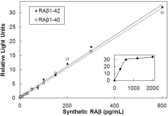
concentrations (2, 5, 10 μg/mL) of capture antibody. Synthetic RAβ1-40 and RAβ 1-42 were diluted (0-600 pg/mL), the HRP-conjugated detection antibody (1.6
μg/mL) was added and after 10 min the samples were placed in wells of the 96 well plate. Plates coated with 5 or 10 μg/mL capture antibody gave identical curves that were linear between 10-600 pg/mL Aβ and both RAβ1-40 and RAβ1-42
were recognized with equal affinity (Figure 1.1). The plate coated with 2 μg/mL of capture antibody became saturated above 400 μg/mL using either Aβ species (data not shown). Thus, all reported assays were performed on plates coated with 5 or 10 μg/mL capture antibody.
Determining the maximum level of HAβ that does not interfere in RAβ ELISA Rodent and human Aβ peptides differ in sequence in only three residues (Arg5, Tyr10, and His13 in HAβ is replaced by Gly5, Phe10, and Arg13 in RAβ) that are all within the epitope recognized by the capture antibody. By inference this means that the epitope recognized by the detection antibody is subject to
13
Figure 1.1. RAβ ELISA standard curves comparing RAβ1-40 and RAβ1-42. There is
no significant difference between the two RAβ species in this assay. The assay is linear between 10 pg/mL and 600 pg/mL. Points are triplicate samples; error bars = standard deviation. Inset: the assay plateaus at RAβ concentrations above 600 ng/mL
14
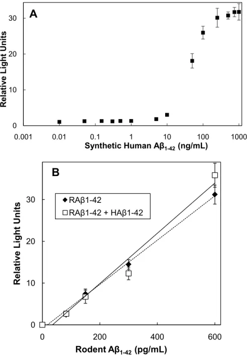
interference by Aβ from non-rodent species. To be able to utilize the RAβ ELISA in the presence of HAβ, the maximum level of HAβ that could be tolerated in the assay needed to be determined. A constant level (150 pg/mL) of RAβ1-42 was
maintained in the assay while HAβ1-42 was added from 10 pg/mL to 1 μg/mL
(Figure 1.2A). HAβ1-42 at 5 ng/mL or less had no effect on the ability of the
ELISA to correctly quantify the RAβ. The increased signal obtained at higher concentrations of HAβ could arise either from some weak affinity of the capture antibody for the HAβ or from co-oligomerization between RAβ and HAβ that might occur at higher Aβ concentrations (Fung et al., 2004; Jankowsky et al., 2007).
We also performed the RAβ ELISA using a constant amount of HAβ (1.9 ng/mL, approximately the maximum concentration in the HAβd/t fraction) with increasing concentrations of RAβ (from 10 pg/mL to 1μg/mL). At concentrations of RAβ below 600 pg/mL, the presence of the HAβ had no effect on the standard curve, demonstrating the ELISA is specific for RAβ within its linear region (Figure 1.2B).
Measurement of secreted RAβ from a rodent cell line
It was previously reported that peroxide induced secretion of Aβ from human neuroblastoma SH-SY5Y cells through JNK-dependent activation of γ-secretase (Shen et al., 2008). Therefore, we first utilized a mouse
neuroblastoma cell line (N2a) to apply the RAβ ELISA. Mouse N2a cells were cultured in 8 well chamber slides and were stressed using varying levels of
15
Figure 1.2. Effects of HAβ on the RAβ ELISA. (A) Effects of variable amounts of HAβ1-42 on the ability of the ELISA to detect a fixed amount (150 pg/ml) of RAβ 1-42. Samples were incubated at 37°C to allow possible co-oligomerization to occur
before the addition of detection antibody. Points are averages of triplicate samples; error bars = standard deviation (less than size of symbol for
concentrations below 10 ng/mL). (B) Effects of a fixed amount of HAβ1-42 (1.9
ng/ml) on the RAβ ELISA standard curve. Samples of each concentration were incubated at 37°C for 3 days before assay to mimic the incubation conditions for secreted RAβ in the presence of the HAβd/t. Points are averages of triplicate samples; error bars = standard deviation.
0 10 20 30 0.001 0.01 0.1 1 10 100 1000 R el at iv e Li ght U ni ts
Synthetic Human Aβ1-42(ng/mL)
0 10 20 30 0 200 400 600 R el at iv e Li ght U ni ts Rodent Aβ1-42 (pg/mL) RAβ1-42 RAβ1-42 + HAβ1-42
A
B
16
hydrogen peroxide (0.5 mM to 10 mM) for 3 days. Medium was harvested and assayed for RAβ in triplicate wells. There was a significant increase (p≤0.05) in secreted RAβ levels for peroxide concentrations above 0.5 mM when compared to untreated controls with a maximum of over 5 fold obtained when treating with 2 mM peroxide (Figure 1.3). However, it was obvious from phase microscopy observation of the wells that significant cell loss occurred, especially at peroxide concentrations above 2 mM.
To normalize Aβ secretion to cell number we applied an assay to measure DNA levels in cultured cells, using the DNA binding dye SybrGreen I, which does not react with nucleotides, RNA, single-stranded nucleic acids, and proteins, as may occur with other DNA binding dyes (Kricka, 2002). DNA standard curves generated from calf thymus DNA were linear between 1-300 ng/mL (Figure 1.4). To normalize the Aβ secreted by the peroxide treated N2a cells to cell number, medium was removed from each well and analyzed for total RAβ and the remaining cells were lysed, the lysates diluted, and the DNA quantified. When normalized for secreted RAβ on a per cell basis, secretion of RAβ increased with increasing peroxide up to a maximum at 2 mM (Figure 1.3).
Application of the RAβ ELISA to primary neurons
Primary E18 rat cortical neurons were cultured in 8 well chamber slides for 3 days, and were left untreated or treated with varying concentrations of
peroxide. Three days later culture medium was removed and assayed for RAβ. Neurons were washed once with PBS, lysed, and the lysates were used to
17
Figure 1.3. N2a cells were treated with different concentrations of hydrogen peroxide and the total RAβ secreted was determined after 3 days and normalized to levels secreted from untreated cells. An apparent decline in secreted Aβ
occurred at peroxide concentrations above 2 mM. However, after correcting for cell loss by normalizing secreted Aβ to DNA in each well, the production of Aβ reached a plateau at or above 2 mM peroxide. * values of p ≤ 0.05 compared to untreated samples. 0 1 2 3 4 5 6 0 0.5 1 2 5 10 R el at iv e [ Aβ ] S ecr et ed Hydrogen Peroxide (mM) Aβ Aβ/DNA
∗ ∗
∗
∗
∗
∗
18
Figure 1.4. Standard curves for DNA using calf thymus DNA dissolved in TE as the standard. Standards and cell lysates are diluted with TE and SybrGreen I and then assayed using a microtiterplate reader operating in the fluorescence mode. The assay is linear between 1 ng to 300 ng/mL of DNA, but linearity can be expanded by adjusting the intensity of fluorescence or exposure time. Points are triplicate samples; error bars = standard deviation.
0 1 2 3 0 50 100 150 200 250 300 R el at iv e F lu or escen ce DNA (ng/mL)
19
quantify either DNA (using the DNA Lysis Buffer) or the internal pool of RAβ using PBSTB w/ 0.1% NP-40).
Untreated neurons secreted 63 ± 5 pg (n=3) of Aβ per well equating to 157.0 pg/mL over the course of three days. By normalizing the concentration of Aβ to the DNA content of the well (1.72 ± .09 μg), we calculate that there are 0.036 pg Aβ/ng of DNA (Figure 1.5A). To determine the number of molecules secreted by each cell one can assume that there is 6.5 pg of DNA/cell (Ausubel et al., 1994); therefore, over a three day period, each cell secretes 220 ag of RAβ (or ~29,000 molecules), which equals about 6-7 RAβ peptides per minute/neuron under non-stress growth conditions.
Cortical neurons were stressed for 3 days with various concentrations (1 μM – 10 mM) of hydrogen peroxide, media were collected and RAβ contents were measured and normalized to DNA (Figure 1.5A). Concentrations of peroxide over 2 mM resulted in increasing cell death, observed by a decline in overall DNA content (data not shown). When compared to untreated neurons there was a significant (p≤0.05) increase in RAβ secreted only at or above peroxide concentrations of 100 μM, with a maximal secretion observed at 2 mM peroxide, resulting in a 2.4 ± 0.2 fold increase over untreated neurons (Figure 1.5A). This fold increase is similar to the increase (2.4 ± 0.6 fold over controls) observed in chick tectal neurons exposed to 10-20 μM peroxide for 20 h
(Goldsbury et al., 2008). It is important to note that we elected to keep the B27 supplements in the neuronal culture medium because the supplements contain factors in addition to anti-oxidants (including catalase) that help keep the cells
20
Figure 1.5. Effects of peroxide on secretion of RAβ from primary cortical
neurons. (A) Normalized dose-response of RAβ secretion to hydrogen peroxide in rat E18 cortical neurons cultured in 8 well chamber slides. After 3 days of exposure, medium was removed from each well and cells were lysed with DNA lysis buffer. Medium was analyzed to determine the concentration of secreted RAβ and the cell lysate was analyzed to determine the DNA content. Values were normalized to untreated wells. Points are averages of triplicate samples; error bars = standard deviation, * values with p ≤0.05 compared to untreated. RAβ secretion peaks with the addition of 2 mM peroxide. (B) Time course of RAβ secreted from rat E18 cortical neurons in response to 2 mM peroxide treatment. Medium was removed for RAβ assay and cells were lysed for DNA measurement at the times indicated. Points are averages of triplicate samples, error bars = standard deviation, * values with p ≤0.05 compared to 0 time.
0 0.5 1 1.5 2 2.5 1 10 100 1000 10000 R el at iv e [ Aβ] Secr et ed [Hydrogen Peroxide] (µM) 0 0.5 1 1.5 2 2.5 0 1 4 12 31 50 72 96 R el at iv e [ Aβ ] S ecr et ed
Time Post Treatment (hrs)
B
A
∗
∗
∗
∗
∗
∗
∗
∗
∗
∗
21
viable over the three day culture period. Thus, much higher concentrations of peroxide were required in these experiments than were needed to induce secretion in chick tectal neurons.
An increase in the internal pool of RAβ has been reported in neurons responding to certain types of stress (Hasegawa et al., 2005) and has been associated with synaptic dysfunction in mouse models of AD (Oddo et al., 2003; Casas et al., 2004). Thus, we quantified the internal pool of RAβ in lysed neurons untreated or treated with peroxide. The internal RAβ was not significantly
different between untreated and peroxide-treated neurons. Untreated neurons had 4.3 ± 0.6 pg of RAβ per ng DNA and 2 mM peroxide-treated neurons contained 4.9 ± 0.8 pg of RAβ per ng DNA.
We next determined the time course of RAβ production in cortical neurons stressed with 2 mM peroxide. Neurons were plated identically as above and stressed with peroxide, with medium being collected after peroxide treatment at 24 h intervals up to 4 days. Although increased secretion was observed as early as 12 h after treatment, the increase was not statistically significant (p ≤ 0.05) until 50 hrs, with maximal secretion obtained at 72 hrs (Figure 1.5B).
Because dissociated neurons might behave differently from neurons that maintain their connections with glia and other neurons, we also determined how peroxide affected the secretion of RAβ from rat hippocampal organotypic slices. Slices that had been grown on membranes for > 1 week (to allow recovery from the stress of preparation) were treated with 2 mM peroxide and after three days the culture medium was harvested and assayed for RAβ. The slices were lysed
22
for DNA quantification. When RAβ levels are compared to untreated slices on a per slice basis there is a 2.1 ± 0.3 fold increase; however when normalized to DNA, the fold increase is 1.5 ± 0.3, which is still significant but less than from the 2.4 fold increase observed for dissociated cortical neurons (Figure 1.6). Each untreated slice secretes 85 ± 8 pg (n=3) of RAβ over the course of 3 days.
HAβ dimer/trimer induces secretion of RAβ
Although synthetic HAβ peptides are usually used at concentrations well above 10 nM (45 ng/mL) to obtain any physiological or morphological changes in neurons, the secreted form of HAβ containing SDS-stable dimers and trimers (HAβd/t) (Walsh et al., 2002) and HAβ dimers extracted from postmortem AD brain (Shankar et al., 2008) are active at concentrations in the pM range (Freir et al., 2010), well below the levels of HAβ that interfere in our rodent ELISA. Thus we can for the first time directly assay the effects of physiologically relevant amounts of HAβ on RAβ secretion.
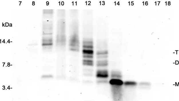
The conditioned medium from 7PA2 and wild type CHO cells was fractionated as previously described (Townsend et al., 2007). Fractions that contained primarily monomer (Aβm) and Aβd/t were obtained (Figure 1.7). The amount of HAβ in each fraction was quantified using a dot blot assay, because after adherence of the HAβ species the membrane could be boiled to expose the epitopes for detection. This step is essential for quantification of HAβ because oligomers are inefficiently measured by ELISA (Stenh et al., 2005), but it is not required for the RAβ assays, including the ELISA, because RAβ does not
23
Figure 1.6. Comparison of effects of hydrogen peroxide (2 mM), HAβm, and HAβd/t on RAβ secretion from dissociated cortical neurons and organotypic hippocampal slices. Media from both neuronal and slice cultures were harvested 3 days after peroxide addition, RAβ levels were analyzed by ELISA, normalized to DNA, and expressed relative to untreated wells. Bars are average values from triplicate samples; error bars = standard deviation; * values with p ≤ 0.05
compared to their untreated control. 0 0.5 1 1.5 2 2.5
Dissociated Cortical Neurons Organotypic Hippocampal Slices
R el at iv e [ Aβ ] S ecr et ed
Untreated Peroxide HAβm HAβd/t
∗
∗
∗
24
Figure 1.7. Western blot showing fractions of the HAβm and HABd/t from 10x concentrated culture medium of 7PA2 cells. The elution positions of monomer, dimer and trimer are shown. A total of 800 µlwas removed from each fraction and stored at –80°C.The remaining 200 µl was lyophilized, resuspended
in2x sample buffer, and electrophoresedon a 10–20% Tris-Tricine gel. Proteins were transferred onto 0.1 µm nitrocelluloseand detected, after boiling the blot for 10 min, by Western blotting for Aβ with6E10 mouse monoclonal antibody and DyLight secondary antibodies detected with a Li-Cor OdysseyInfrared Imaging System. Fractions enriched in monomer (fractions 14-16) or dimer/trimer (fractions 12 and 13) were pooled separately, lyophilized, andstored at –80°C. They were used at the equivalent of 1x to treat cells. The amounts of HAβ in each fraction were determined by a dot blot assay described elsewhere (Chapter 2).
25
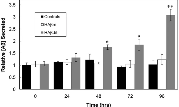
oligomerize at its secreted concentrations. The HAβm and HAβd/t fractions, as well as their respective controls (equivalent fractions from gel filtration of wt CHO cell medium), were added to 3 day old cultures of rat cortical neurons. Secreted rodent Aβ was quantified at 0, 24, 48, 72 and 96 h and normalized to the amount of DNA in each sample (Figure 1.8). The presence of HAβ did not interfere in the RAβ ELISA, as previously shown in Figure 1B, and confirmed here by the lack of change between untreated and HAβ-treated groups at time zero (Figure 1.8). Neurons treated with the HAβd/t, but not monomer or fractions from control CHO cell medium, induced secretion of RAβ to more than a 3-fold increase over controls by 4 days. Because of the possible co-oligomerization between the HAβd/t and the RAβ that could alter the assay results, we boiled some of the samples to enhance epitope exposure before performing the RAβ ELISA, but no differences between the boiled and unboiled samples were detected (data not shown). By 96 h the amount of RAβ was nearly identical to the amount of human Aβd/t added, so if co-oligomerization had occurred we should have observed an enhanced signal after boiling.
The effects of HAβm and HAβd/t on the secretion of RAβ from rat hippocampal organotypic slices was also determined. Slices were grown essentially as described in the peroxide experiments, and were treated with HAβm or HAβd/t and harvested 3 days after insult (Figure 1.6). When RAβ levels are compared to untreated slices on a per slice basis, there is no change in RAβ with HAβm treatment (0.9 ± 0.2 fold change), however HAβd/t treatment caused a significant (p≤0.05) 1.8 ± 0.2 fold increase in secreted RAβ. When
26
Figure 1.8. Time course of RAβ production from rat cortical neurons, untreated or treated with HAβm or HAβd/t fractionated from 7PA2 culture medium. Rat cortical neurons grown 3 days were left untreated or were treated with gel filtered fractions containing HAβm (3.6 ng/mL), HAβd/t (1.1 ng/mL) or equivalent
volumes of the same fractions from wild type CHO cell culture medium (controls). Medium was removed at 0, 24, 48, 72, and 96 hrs after HAβ addition, and
analyzed for RAβ. Cells were lysed and DNA measured for normalization. Values were then expressed relative to the untreated samples at time=0. All controls using fractionated medium from wild type CHO cells were not
significantly different from the untreated controls of the same time point. Bars are average values from triplicate samples; error bars = standard deviation; * values with p ≤ 0.05, or ** p ≤ 0.05, compared to their untreated control.
0 0.5 1 1.5 2 2.5 3 3.5 0 24 48 72 96 R el at iv e [ Aβ ] S ecr et ed Time (hrs) Controls HAβm HAβd/t
∗
∗
∗∗
27
organotypic hippocampal slices are compared to cortical neurons treated with HAβd/t there is a similar fold increase in RAβ secretion with both types of neurons.
There was no significant change in the amount of RAβ in the intracellular pool measured in lysates of cells treated with HAβd/t. Normalized per ng of DNA, untreated neurons had 5.1 ± 0.4 pg of RAβ, cells treated with HAβm had 4.8 ± 0.3 pg, and cells treated with HAβd/t had 5.4 ± 0.5 pg of rodent Aβ. These values were similar to the intracellular RAβ amounts reported above for the control and peroxide treated neurons (4.3 to 4.9 ± 0.8 pg).
To determine if the effects of HAβd/t on RAβ secretion are dependent on the microtubule protein tau, organotypic hippocampal slices from transgenic mice with either tau null (TAU-/-) or TAU-/- carrying a human tau transgene were used. These slices were cultured in pairs on glass coverslips in 0.6 mL of medium, which minimized the amount of HAβd/t required. Slices were stressed with 2 mM peroxide, HAβm (at 3.6 ng/ml), or HAβd/t (1.1 ng/ml) after slices had stabilized. After 3 days of treatment RAβ levels were quantified and normalized to DNA content (Figure 1.9). Both tau null slices and slices expressing the transgenic human tau showed a nearly identical increase in levels of RAβ when stressed with peroxide (1.6 ± 0.2 and 1.5 ± 0.2, respectively). Slices treated with HAβm showed no change in RAβ levels when compared with untreated slices. RAβ secreted from slices treated with HAβd/t increased 2.1 ± 0.3 fold (tau null) and a 2.3 ± 0.3 fold (transgenic human tau). Taken together, these results suggest that Aβ secretion is not dependent on tau.
28
Figure 1.9. The presence or absence of tau has no effect on RAβ secretion induced by either peroxide or HAβd/t. Triplicate roller tubes containing two organotypic hippocampal slices from mice of genotype mouse TAU-/- or mouse TAU-/- with a human tau transgene were cultured for 10 or more days and then treated with 2 mM peroxide, HAβm (3.6 ng/mL) or HAβd/t (1.1 ng/mL). After 3 days, medium was harvested for measurement of secreted RAβ and tissue was lysed to measure DNA for normalization. Bars are averages of triplicate samples, error bars = standard deviations; * values with p≤0.05 compared with mouse TAU-/- control. 0 0.5 1 1.5 2 2.5
Control Peroxide HAβm HAβd/t
R el at iv e [ Aβ ] S ecr et ed Treatment Conditions Mouse TAU Null
Human TAU
∗
∗
29 Discussion
Transgenic mice expressing human mutant APP are commonly used for AD research, but these animals also express endogenous mouse APP, which limits their usefulness when trying to measure effects on APP processing and Aβ release since quantifying mixed species of Aβ is more complex. In addition, HAβ peptides oligomerize and oligomers are not efficiently measured by a typical ELISA (Stenh et al., 2005), unless samples are denatured by boiling or treated with denaturants, both of which may decrease the accuracy of their
determination. Furthermore, a goal of our work is to apply an ELISA for
measuring RAβ secreted in response to treatment of cells with HAβ oligomers, something that is not feasible to do in cells secreting HAβ. Thus we developed an assay for total RAβ which can be used in the presence of physiologically relevant concentrations of HAβ oligomers. The advantage of the use of rodent neurons from non-transgenic animals is several fold: (1) they are easier to obtain and maintain than transgenic animals (Castrop, 2010); (2) rodent neurons
(hippocampal and cortical) are the standard model system for studying the behavioral effects of HAβ treatment, both electrophysiologically and
morphologically (Wang et al., 2004; Cleary et al., 2004; Shankar et al., 2007; Shankar et al., 2008; Freir et al., 2010); (3) the RAβ they produce does not oligomerize eliminating any need for boiling or denaturing higher order
complexes before analysis (Atwood et al., 2004; Marksteiner & Humpel, 2008); (4) both rat and mouse Aβ peptides have the identical sequences and either species can be used for these assays (Johnstone et al., 1991).
30
Chick neurons produce Aβ with sequence, isoform patterns, and
oligomerization patterns identical to HAβ (Esselmann et al., 2004; Carrodeguas et al., 2005), making them a useful system for some studies, but not for
experiments in which treatment with HAβ is desired. Previously it was shown that 10-20 μM peroxide treatment causes a 2.4 fold increase in secreted Aβ after 20 hours in chick tectal neurons using an immunoprecipitation and Western blotting assay (Goldsbury et al., 2008). Only monomeric Aβ was quantified because it was the only species to show up on the western blots, perhaps because SDS-stable oligomers would not be visualized without boiling the membrane. In our assays, which were performed in the presence of the B27 supplement, we obtained no effect on RAβ production until we exceeded 100 μM hydrogen peroxide, demonstrating the protective effect of the B27 antioxidants and explaining why we required using much higher levels of peroxide than in previous studies in which neurons were maintained for shorter time periods (less than 24 h) (Goldsbury et al., 2008).
Conventional approaches to studying Aβ secretion in cultured neurons focus on overexpressing human APP, and measuring HAβ secreted into the extracellular environment (Busciglio et al., 1993; Suzuki et al., 1994). Here we show that this method might cause artificially high levels of secreted HAβ, simply due to the fact that there is a positive feedback loop where HAβd/t secreted can cause a further increase in HAβ secretion. Furthermore when using an ELISA to quantify the amount of HAβ secreted, low values are often observed because antibodies recognize different oligomers with varying affinities (Stenh et al.,
31
2005); however RAβ does not oligomerize making it easier to get accurate values on its secreted level. When using the ELISA to look at HAβ-induced RAβ
secretion it is necessary to use the highly active naturally secreted Aβ dimer containing fractions (either from AD brain or 7PA2 conditioned medium) since synthetic HAβ is required at concentrations well above those that interfere with the RAβ ELISA.
HAβm does not show any synaptic detrimental effects and it has a role in neuroprotection, perhaps acting as a scavenger for metal induced oxidative stress (Zou et al., 2002). Here we show that HAβm does not cause an increase in RAβ secretion providing additional support that HAβm is not a pathogenic species responsible for AD (Giuffrida et al., 2009). Recently other small Aβ oligomers, specifically the dimer and the trimer, have been proposed as the species responsible for AD, and the synaptic deficits associated with the disease (Walsh et al., 2002; Cleary et al., 2004; Shankar et al., 2008; Freir et al., 2010). The dimer is found in AD brain but not in brains of stroke patients or patients diagnosed with diseases unrelated to HAβ overproduction (Shankar et al., 2008), and the dimer is correlated with severity of AD dementia (McDonald et al., 2010). Furthermore, synthetic HAβ does not form dimers when the oligomers are
prepared under traditional incubations conditions (Dahlgren et al., 2002; Stine et al., 2003) and these oligomers are at least 100-200 fold less potent than naturally secreted HAβ at decreasing long-term potentiation (Wang et al., 2004).
HAβ rapidly inhibits fast axonal transport, however in tau null neurons transport is not inhibited following HAβ treatment (Vossel et al., 2010) suggesting
32
that HAβ requires tau for its effects on vesicle transport. Between 40 and 70% of the Aβ secreted by neurons is produced following endocytosis of APP (Cirrito et al., 2008). Following endocytosis much of the APP and vesicular Aβ is trafficked to lysosomes and digested after endosome-lysosome fusion (Lorenzen et al., 2010). It has been previously suggested that inhibition of vesicle transport may be one means to generate enhanced Aβ production (Maloney et al., 2005). Thus, we determined if tau-dependent blockage is required to get enhanced RAβ
secretion by comparing the levels of secreted RAβ from HAβd/t-treated organotypic hippocampal slices obtained from TAU-/- mice and TAU-/- mice expressing a human tau transgene. We found no significant differences in RAβ secreted in response to HAβd/t from organotypic slices from either mouse,
suggesting that tau-dependent transport inhibition per se plays no significant role in HAβd/t-induced RAβ secretion.
33 Reference List
Alzheimer’s Association (2010) Alzheimer’s Disease Facts and Figures, Alzhemiers Dement 6:158-94
Atwood CS, Perry G, Zeng H, Kato Y, Jones WD, Ling K-Q, Huang X, Moir RD, Wang D, Sayre LM, Smith M, Chen SG, Bush AI (2004) Copper
mediates dityrosine cross-linking of Alzheimer’s amyloid-beta. Biochemistry 43:560-8.
Ausubel FM, Brent R, Kingston RE, Moore DD, Seidman JG, Smith JA, Struhl K (1994) Current Protocols in Molecular Biology, Vol 3, Appendix A.1- B.1, John Wiley & Sons, New York.
Bamburg JR, Bloom GS (2009) Cytoskeletal pathologies of Alzheimer disease. Cell Motil Cytoskeleton 66:635-49.
Busciglio J, Gabuzda DH, Matsudaira P, Yankner BA (1993) Generation of beta-amyloid in the secretory pathway in neuronal and nonneuronal cells. Proc Natl Acad Sci USA 90:2092-6.
Carrodeguas JA, Rodolosse A, Garza MV, Sanz-Clemente A, Pérez-Pé R, Lacosta AM, Domínguez L, Monleón I, Sánchez-Díaz R, Sorribas V, Sarasa M (2005) The chick embryo appears as a natural model for research in beta-amyloid precursor protein processing. Neuroscience 134:1285-300.
Castrop H (2010) Genetically modified mice-successes and failures of a widely used technology. Pflugers Arch 459:557-67
Cirrito JR, Kang J-E, Lee J, Stewart FR, Verges DK, Silverio LM, Bu G,
Mennerick S, Holtzman DM (2008) Endocytosis is required for synaptic activity-dependent release of amyloid-beta in vivo. Neuron 58:42-51. Cleary JP, Walsh DM, Hofmeister JJ, Shankar GM, Kuskowski MA, Selkoe DJ,
Ashe KH (2004) Natural oligomers of the amyloid-beta protein specifically disrupt cognitive function. Nat Neurosci 8:79-84.
Dahlgren KN, Manelli AM, Stine WB, Baker LK, Krafft G, LaDu MJ (2002) Oligomeric and fibrillar species of amyloid-beta peptides differentially affect neuronal viability. J Biol Chem 277:32046-53.
Ehehalt R, Keller P, Haass C, Thiele C, Simons K (2003) Amyloidogenic
processing of the Alzheimer beta-amyloid precursor protein depends on lipid rafts. J Cell Bio 160:113-23.
34
Esselmann H, Maler JM, Kunz N, Otto M, Paul S, Lewczuk P, Rüther E, Kornhuber J, Wiltfang J (2004) Lithium decreases secretion of Aβ1- 42 and C-truncated species Aβ1-37/38/39/40 in chicken telencephalic
cultures but specifically increases intracellular Aβ1-38. Neurodegener Dis 1:236-41.
Finder VH, Glockshuber R (2007) Amyloid-beta aggregation. Neurodegener Dis 4:13-27.
Freir DB, Fedriani R, Scully D, Smith IM, Selkoe DJ, Walsh DM, Regan CM (2010) Aβ oligomers inhibit synapse remodelling necessary for memory consolidation. Neurobiol Aging.
Fukumoto H, Tomita T, Matsunaga H, Ishibashi Y, Saido TC, Iwatsubo T (1999) Primary cultures of neuronal and non-neuronal rat brain cells secrete similar proportions of amyloid beta peptides ending at Aβ40 and Aβ42. Neuroreport 10:2965-9.
Fung J, Frost D, Chakrabartty A, McLaurin J (2004) Interaction of human and mouse Aβ peptides. J Neurochem 91:1398-403.
Gasparini L, Rusconi L, Xu H, Soldato P del, Ongini E (2004) Modulation of beta-amyloid metabolism by non-steroidal anti-inflammatory drugs in neuronal cell cultures. J Neurochem 88:337-48.
Giuffrida ML, Caraci F, Pignataro B, Cataldo S, De Bona P, Bruno V, Molinaro G, Pappalardo G, Messina A, Palmigiano A, Garozzo D, Nicoletti F, Rizzarelli E, Copani A (2009) Beta-amyloid monomers are neuroprotective. J
Neurosci 29:10582-7.
Glenner GG, Wong CW (1984) Alzheimer’s disease: initial report of the purification and characterization of a novel cerebrovascular amyloid protein. Biochem Biophys Res Commun 120:885-90.
Goldsbury C, Whiteman IT, Jeong EV, Lim YA (2008) Oxidative stress increases levels of endogenous amyloid-beta peptides secreted from primary chick brain neurons. Aging Cell 7:771-5.
Hardy J, Selkoe DJ (2002) The amyloid hypothesis of Alzheimer’s disease: progress and problems on the road to therapeutics. Science 297:353-6.
35
Hasegawa T, Ukai W, Jo D, Xu X, Mattson M, Nakagawa M, Araki W, Saito T, Yamada T (2005) Homocysteic acid induces intraneuronal
accumulation of neurotoxic Aβ42: Implications for the pathogenesis of Alzheimer's disease. J Neurosci Res 80:869–76.
Jankowsky JL, Younkin LH, Gonzales V, Fadale DJ, Slunt HH, Lester HA, Younkin SG, Borchelt DR (2007) Rodent Aβ modulates the solubility and distribution of amyloid deposits in transgenic mice. J Biol Chem
282:22707-20.
Johnstone E, Chaney M, Norris F, Pascual R, Little S (1991) Conservation of the sequence of the Alzheimer’s disease amyloid peptide in dog, polar bear and five other mammals by cross-species polymerase chain reaction analysis. Brain Res Mol Brain Res 10:299-305.
Koo EH, Squazzo SL (1994) Evidence that production and release of amyloid beta-protein involves the endocytic pathway. J Biol Chem 269:17386-9. Krafft G, Klein WL (2010) ADDLs and the signaling web that leads to
Alzheimer’s disease. Neuropharmacology 59:230-42.
Kricka LJ (2002) Stains, labels and detection strategies for nucleic acids assays. Ann Clin Biochem 39:114-129.
Lorenzen A, Samosh J, Vandewark K, Anborgh PH, Seah C, Magalhaes AC, Cregan SP, Ferguson SSG, Pasternak SH (2010) Rapid and direct transport of cell surface APP to the lysosome defines a novel selective pathway. Mol Brain 3:11.
Marksteiner J, Humpel C (2008) Beta-amyloid expression, release and
extracellular deposition in aged rat brain slices. Mol Psychiatry 13:939-52. Mattson MP (2004) Pathways towards and away from Alzheimer’s disease.
Nature 430:631-9.
McDonald JM, Savva GM, Brayne C, Welzel AT, Forster G, Shankar GM, Selkoe DJ, Ince PG, Walsh DM (2010) The presence of sodium dodecyl sulphate-stable Aβ dimers is strongly associated with Alzheimer-type dementia. Brain 133:1328-41.
Minamide LS, Striegl AM, Boyle JA, Meberg PJ, Bamburg JR (2000)
Neurodegenerative stimuli induce persistent ADF/cofilin-actin rods that disrupt distal neurite function. Nat Cell Biol 2:628-36.
Portelius E, Bogdanovic N, Gustavsson MK, Volkmann I, Brinkmalm G, Zetterberg H, Winblad B, Blennow K (2010) Mass spectrometric
36
characterization of brain amyloid beta isoform signatures in familial and sporadic Alzheimer’s disease. Acta Neuropathol 120:185-93.
Price DL, Sisodia SS, Gandy SE (1995) Amyloid beta amyloidosis in Alzheimer’s disease. Curr Opin Neurol 8:268-74.
Shankar GM, Bloodgood BL, Townsend M, Walsh DM, Selkoe DJ, Sabatini BL (2007) Natural oligomers of the Alzheimer amyloid-beta protein induce reversible synapse loss by modulating an NMDA-type glutamate receptor-dependent signaling pathway. J Neurosci 27:2866-75.
Shankar GM, Li S, Mehta TH, Garcia-Munoz A, Shepardson NE, Smith I, Brett FM, Farrell MA, Rowan MJ, Lemere CA, Regan CM, Walsh DM, Sabatini BL, Selkoe DJ (2008) Amyloid-beta protein dimers isolated directly from Alzheimer’s brains impair synaptic plasticity and memory. Nat Med 14:837-42.
Shen C, Chen Y, Liu H, Zhang K, Zhang T, Lin A, Jing N (2008) Hydrogen
peroxide promotes Abeta production through JNK-dependent activation of gamma-secretase. J Biol Chem 283:17721-30.
Stenh C, Englund H, Lord A, Johansson A-S, Almeida CG, Gellerfors P,
Greengard P, Gouras GK, Lannfelt L, Nilsson LNG (2005) Amyloid-beta oligomers are inefficiently measured by enzyme-linked immunosorbent assay. Ann Neurol 58:147-50.
Stine WB, Dahlgren KN, Krafft GA, LaDu MJ (2003) In vitro characterization of conditions for amyloid-beta peptide oligomerization and fibrillogenesis. J Biol Chem 278:11612-22.
Stoppini L, Buchs PA, Muller D (1991) A simple method for organotypic cultures of nervous tissue. J Neurosci Methods 37:173-82.
Suzuki N, Cheung T, Cai X, Odaka A, Otvos L, Eckman C, Golde T, Younkin S (1994) An increased percentage of long amyloid beta protein secreted by familial amyloid beta protein precursor (beta APP717) mutants. Science 264:1336-1340.
Tanzi RE, Bertram L (2005) Twenty years of the Alzheimer’s disease amyloid hypothesis: a genetic perspective. Cell 120:545-55.
Thomas RS, Liddell JE, Murphy LS, Pache DM, Kidd EJ (2006) An antibody to the beta-secretase cleavage site on amyloid-beta-protein precursor inhibits amyloid-beta production. J Alzheimers Dis 10:379-90.
37
Townsend M, Mehta T, Selkoe DJ (2007) Soluble Aβ inhibits specific signal transduction cascades common to the insulin receptor pathway. J Biol Chem 282:33305-12.
Townsend M, Shankar GM, Mehta T, Walsh DM, Selkoe DJ (2006) Effects of secreted oligomers of amyloid beta-protein on hippocampal synaptic plasticity: a potent role for trimers. J Physiol 572:477-92.
Vossel KA, Zhang K, Brodbeck J, Daub AC, Sharma P, Finkbeiner S, Cui B, Mucke L (2010) Tau reduction prevents Aβ-induced defects in axonal transport. Science 330:98.
Walsh DM, Klyubin I, Fadeeva JV, Cullen WK, Anwyl R, Wolfe MS, Rowan MJ, Selkoe DJ (2002) Naturally secreted oligomers of amyloid beta protein potently inhibit hippocampal long-term potentiation in vivo. Nature 416:535-9.
Walsh DM, Tseng BP, Rydel RE, Podlisny MB, Selkoe DJ (2000) The
oligomerization of Aβ-protein begins intracellularly in cells derived from human brain. Biochemistry 39:10831-10839.
Wang Q, Walsh DM, Rowan MJ, Selkoe DJ, Anwyl R (2004) Block of long-term potentiation by naturally secreted and synthetic amyloid β-peptide in hippocampal slices is mediated via activation of the kinases c-Jun N-terminal activated protein kinase as well as metabotropic glutamate receptor type 5. J Neurosci 24:3370 -3378.
Xia W, Yang T, Shankar G, Smith IM, Shen Y, Walsh DM, Selkoe DJ (2009) A specific enzyme-linked immunosorbent assay for measuring beta-amyloid protein oligomers in human plasma and brain tissue of patients with Alzheimer disease. Arch Neurol 66:190-9.
38
CHAPTER TWO
AMYLOID BETA DIMERS/TRIMERS POTENTLY INDUCE COFILIN-ACTIN RODS THAT ARE INHIBITED BY MAINTAINING COFILIN
PHOSPHORYLATION
Preface and Acknowledgement
The work presented in this chapter was submitted to Molecular
Neurodegeneration. The order and list of authors is as follows: Richard C. Davis, Ian T. Marsden, Michael T. Maloney, Laurie S. Minamide, Marcia Podlisny, Dennis J. Selkoe, and J.R. Bamburg. My contributions to the work include: (1) the purification of the Aβ monomer and dimer/trimer from 7PA2 conditioned medium; (2) the quantification of the Aβ secreted by these cells using a dot-blot assay; (3) purification and testing of the dityrosine cross-linked synthetic human Aβ1-42 dimer.
The authors gratefully acknowledge funding from the NIH National Institute of Neurological Disorders and Stroke (NS40371 to J.R.B., NS43115 to RCD, MTM, and JRB) and a Core Infrastructure Grant for Microscope Imaging from Colorado State University. We are grateful for technical assistance of Chi Pak and Alisa Shaw and valuable discussions from these same individuals and from Barbara Bernstein, Kevin Flynn, and O’Neil Wiggan.
39 Abstract
Previously we showed oligomers of synthetic amyloid beta (Aβ1-42)
induced cofilin activation and formation of cofilin-actin rods in a neuronal subpopulation of rat hippocampus primarily localized within the dentate gyrus. Here we demonstrate that CHO cell (7PA2) secreted Aβ dimer/trimer (Aβd/t) at ~250 pM is more potent in rod induction in both dissociated hippocampal neuronal cultures and organotypic slices than 1 µM synthetic Aβ as typically prepared oligomers, about a 4000 fold difference. Treatment of the Aβd/t fraction with an Aβ-neutralizing antibody eliminates its rod inducing activity. Traditionally prepared synthetic Aβ oligomers contain SDS-stable trimers and tetramers, but are devoid of dimers. When synthetic human Aβ was incubated under conditions that generate a tyrosine oxidized dimer, the concentration that was required to induce rods decreased dramatically. The oxidized dimer had a maximum rod-inducing activity at ~2 nM (10 ng/mL), suggesting it is the presence of the SDS-stable tyrosine oxidized Aβ dimer in a low-n state that is largely responsible for the potency of the secreted Aβd/t. Aβd/t-induced rods are highly localized to the dentate gyrus and mossy fiber pathway and form more rapidly (significant over controls by 2 h compared to 8 h for those induced by synthetic Aβ-oligomers). Aβd/t-induced rods are reversible, disappearing by 24 h after washout. Cofilin dephosphorylation in response to Aβd/t is greatest within the hippocampal regions of rod formation. Overexpression of cofilin phosphatases slingshot and chronophin increase rod formation when expressed alone and exacerbate rod formation when coupled with Aβd/t treatment both in dissociated neurons and
40
organotypic slice cultures. Overexpression of the cofilin kinase, LIM kinase 1, inhibits Aβd/t-induced rod formation. Together these data support a mechanism through which Aβd/t produces selective synaptic dysfunction affecting learning and memory at least in part via primary effects on cofilin regulation and rod formation in sensitive hippocampal regions.
Introduction
Proteolytic cleavage of amyloid precursor protein (APP) by β- and γ-secretases gives rise to Aβ peptides ranging in length from 39-43 amino acids (Glenner & Wong, 1984; Hardy, & Selkoe, 2002; Mattson, 2004; Price, et al., 1995; Sisodia & Price, 1995; Tanzi, & Bertram, 2005). Early onset familial AD is linked with high penetrance to mutations that lead to increased production of the most amyloidogenic species, Aβ1-42 (Chartier-Harlin et al., 1991; Goate et al.,
1991; Murrell et al., 1991; Price et al., 1995). The “amyloid hypothesis” proposes that increasing cerebral accumulation of Aβ over years to decades exacerbates cognitive decline, neurodegeneration, and senile plaque deposition associated with AD. Elevated Aβ can result from mutations or allele expression patterns (or both) that enhance its production/aggregation or decrease its clearance/
degradation (Hardy & Selkoe, 2002).
The concept that different isoforms and/or conformations of Aβ deliver independent signals to neurons is widely supported. Although the term Aβ is used to describe a spectrum of peptide species, the effects of different Aβ peptide species on neuronal function or morphology are not the same (Maloney
41
& Bamburg, 2007; Heredia et al., 2006). Emphasis has been placed recently on the characterization of small soluble oligomeric forms of Αβ, sometimes referred to as Aβ-derived diffusible ligands (ADDLs) (Kraaft and Klein, 2010). ADDLs are toxic to cultured neurons at nanomolar concentrations (Lambert et al., 1998) and at 500 nM they prevent high frequency stimulation-induced long-term potentiation (LTP) measured from the dentate gyrus in acute hippocampal slices (Wang et al., 2002). Furthermore, ADDLs have been linked to hippocampus-dependent
temporal memory deficits in mice. Deletion of the BACE1 gene in Tg6799 mice, expressing mutant forms of human APP and presenilin-1, lowered the
concentration of ADDLs to wild type levels and rescued temporal memory
deficits, implying a direct role of Aβ formation in memory loss (Ohno et al., 2006; Kimura et al., 2010). ADDLs bind to synaptic sites on cultured hippocampal neurons (Gong et al., 2003; Lacor et al., 2004) where they impair insulin receptor signaling (Zhao et al., 2008) and where stimulation by insulin prevents the
pathogenic binding of ADDLs (De Felice et al., 2009).
An even more potent synaptic-inhibitory preparation of Aβ containing SDS-stable dimers and trimers (Aβd/t) has been obtained from culture medium of a CHO cell line (7PA2) expressing a mutant human APP (Walsh et al., 2002). When used at their physiologically relevant (subnanomolar) concentrations to treat hippocampal slices, the Aβd/t fraction and a fraction of Aβ dimer obtained from postmortem human AD brain markedly inhibited the development of long-term potentiation and enhanced long-long-term depression (LTD), electrophysiological correlates of learning and memory defects in intact animals (Cleary et al., 2005;
42
Shankar et al., 2008). Single intracerebral ventricular (i.c.v.) infusions into adult rat brain of either gel filtered Aβd/t from 7PA2 cells or Aβ dimer from human AD brain caused transient memory and learning deficits (Walsh et al., 2002; Cleary et al., 2005; Townsend et al., 2006; Shankar et al., 2008; Freir et al., 2010). Infusion (i.c.v.) of Aβd/t into adult rat brain several hours after training inhibits synaptic remodeling that accompanies learning and memory consolidation by preventing a transient increase in the number of synapses in the dentate gyrus (Freir et al., 2010). Although their mechanism is unknown, the SDS-stable Aβd/t or dimer fractions cause synaptic dysfunction at sub-nanomolar concentrations, which are 103- 104 fold lower than commonly used traditionally prepared
oligomeric forms of synthetic Aβ, and 102-103 fold lower than concentrations of
ADDLs. In this regard it is significant that the presence of the SDS-stable Aβ dimer is strongly associated with Alzheimer-type dementia (McDonald et al., 2010).
In addition to the classical hallmarks of AD pathology, amyloid plaques and phospho-tau-containing neuropil threads and neurofibrillary tangles,
histopathological structures involving actin and the actin-binding protein, cofilin, have been identified in AD brain (reviewed in Bamburg & Bloom, 2009). Rod-shaped arrays of cofilin-saturated actin bundles (cofilin-actin rods) are induced in cultured hippocampal neurons and organotypic hippocampal slice cultures in response to mitochondrial dysregulation (ATP-depletion) (Minamide et al., 2000; Huang et al., 2008; Davis et al., 2009; Bamburg et al., 2010), oxidative stress (Minamide et al., 2000; Kim et al., 2009), excitotoxic glutamate (Minamide et al.,
43
2000), extracellular ATP (Homma et al., 2008), overexpression of cofilin (Bernstein et al., 2006), and exposure to Aβ oligomers (Maloney et al., 2005; Davis et al., 2009), each of which is a potential mediator of synaptic loss
observed in both familial and sporadic AD (reviewed in Ohm et al., 2007). Rods contain actin and cofilin in a 1:1 complex (Minamide et al., 2010), they form in tandem arrays (striations) within neurites, and they serve as sites for
accumulation of phosphorylated tau (Whiteman et al., 2009), suggesting that they may play a role in formation of striated neuropil threads, the major tau pathology in human AD brain (Velasco et al., 1998). Cofilin-actin rods can grow to
completely occlude the neurites in which they form (Minamide et al., 2000) and thus inhibit vesicular transport (Maloney et al., 2005; Jang et al., 2005) and cause microtubule loss (Minamide et al., 2000). Because an early indication of AD is blockage in axonal transport that leads to axonal swellings (reviewed in Stokin and Goldstein, 2006; Velasco et al., 1998) and synaptic loss (Davies et al., 1987), we investigated the ability of the physiologically relevant amounts of Aβd/t to induce cofilin activation (dephosphorylation) and rod formation in rat hippocampal neurons and organotypic slices. The Aβd/t dose, time course, reversibility of Aβd/t rod formation, and the location of rods in the hippocampus, all suggest that cofilin-actin rods are likely mediators of Aβd/t in synaptic
44 Materials and Methods
Reagents
All chemicals are reagent grade and were obtained from Sigma-Aldrich Co. and all tissue culture reagents were from Life Technologies (Invitrogen, Carlsbad, CA). Synthetic Aβ peptide (Aβ1-42) and a scrambled peptide with the
Aβ1-42 amino acid composition were purchased from AnaSpec, Inc. (San Jose,
CA). Amyloid beta monomer and dimer/trimer fractions were prepared from the culture medium of CHO 7PA2 cells (Walsh et al., 2002) as previously described (Cleary et al., 2005; Shankar et al., 2007), and unless noted otherwise were used at 1x concentration (equivalent to their secreted concentration in the medium). Similar fractions obtained from culture medium of wild type CHO cells were used as controls.
Culture treatments of dissociated neurons and slices
Synthetic Aβ oligomer was made by solubilizing the peptide in
hexafluoroisopropanol and drying in 10 μg aliquots. Each 10 μg of synthetic Aβ 1-42 was solubilized in 10 μL of DMSO, diluted with 78.6 μL of sterile Ham’s F-12
(to yield a 25 μM stock) and incubated 24 h at 4ºC (Dahlgren et al., 2002; Stine et al., 2003; Maloney et al., 2005). Scrambled peptide was prepared identically and both scrambled peptide and synthetic Aβ1-42 oligomer were added to a final
concentration of 1 μM. The secreted Aβ fractions (monomer or d/t) or the corresponding fractions from control medium, were prepared as described (Cleary et al., 2005), and after gel filtration were freeze-dried to remove the