ABNORMAL CHEST X-RAY EVALUATION
by
Wendy M. Johnson
ARTHUR LAKES LIBRARY
COLORADO SCHOOL OF
mm
INFORMATION TO ALL USERS
The qu ality of this repro d u ctio n is d e p e n d e n t upon the q u ality of the copy subm itted. In the unlikely e v e n t that the a u th o r did not send a c o m p le te m anuscript and there are missing pages, these will be note d . Also, if m aterial had to be rem oved,
a n o te will in d ica te the deletion.
uest
ProQuest 10783802
Published by ProQuest LLC(2018). C op yrig ht of the Dissertation is held by the Author. All rights reserved.
This work is protected against unauthorized copying under Title 17, United States C o d e M icroform Edition © ProQuest LLC.
ProQuest LLC.
789 East Eisenhower Parkway P.O. Box 1346
A thesis submitted to the Facutly and Board of Trustees of the Colorado School of Mines in partial fulfillment of the requirements for the degree of Master of Science (Mine
Safety and Health).
Golden, Colorado Date
ii/zhsr
Signed: Approved: Wendy r. Robert H. Ki: Thesis Advisor Golden, Colorado Date / / / * 3/£f~
Dr. Tibor Rozgani Professor and Head, Department of MiningARTHUR LAKES LIBRARY COLORADO SCHOOL OF MINES GOLDEN, CO 80401
ABSTRACT
This thesis project was a follow-up to the Beryllium Health Surveillance Program (BHSP) at Rocky Flats Environmental Technology Site (RFETS) in Golden, Colorado. The purpose of this study was to determine the final medical outcome of additional lung disorders not associated with chronic beryllium disease. This information was compared to the radiologist report, using the International Labor Organization (ILO) classification system, to evaluate the effectiveness of the chest x-ray as a screening tool for pre-existing pulmonary function disorders. Secondly, the final medical diagnosis for each participant was complied into a database and compared to three databases in an effort to determine the incidence of pulmonary disease at RFETS.
The screening tool evaluation results indicated that if a disorder was diagnosed by the B-reader from the chest x-ray, the same disorder was subsequently confirmed by the personal physician 70 percent or more of the time for all categories except
density/nodule/mass disorders. Specifically, chronic obstructive pulmonary disease was confirmed 82.8 percent of the time, restrictive pulmonary disease was confirmed 74.4 percent of the time, and the miscellaneous diagnosis was confirmed 87 percent of the time. Density/nodule/mass disorders were confirmed only 35 percent of the time. Based on the percentage of confirmed diagnosis for each disorder the chest x-ray as diagnosed using the ILO classification system by a certified B-reader appears to be a good screening tool, if a 70 percent or less accuracy is required, for chronic obstructive pulmonary disease,
restrictive pulmonary disease, and miscellaneous disorders. The ILO classification system was not effective in identifying and diagnosing density/nodule/mass disorders that may be indications of potential lung cancer.
When confidence intervals were calculated it was confirmed that the overall
effectiveness of the chest x-ray is an adequate screening tool for the specific lung disorders in this study except the density/nodule/mass disorder. The chi-square test determined that agreement between the diagnosis of the radiologist and the participant’s personal physician is highly dependent on the specific lung disorder.
The second part of this thesis was to determine if the incidence of lung disorders at RFETS was less than the incidence in the State of Colorado. Unfortunately, database information was not found that made this comparison possible.
TABLE OF CONTENTS
Page
ABSTRACT... iii
LIST OF TABLES... -... vii
LIST OF FIGURES... ix
ACKNOWLEDGEMENTS... x
Chapter 1. INTRODUCTION...1
1.1 Background... 2
1.2 Purpose and Scope... 7
1.3 Hypothesis... 7
Chapter 2. METHODOLOGY... 9
Chapter 3. LITERATURE REVIEW...12
3.1 Pulmonary System...12
3.2 Lung Disorders...13
3.2.1 Chronic Obstructive Pulmonary Diseases...13
3.2.2 Restrictive Pulmonary Diseases... 14
3.2.3 Density/Nodule/Mass... 16
3.2.4 Miscellaneous Disorders...17
3.3 Chest Radiography... 17
Chapter 4. RESULTS... 23
Chapter 5. SCREENING TOOL EVALUATION... 27
5.1 Database Results...27
TABLE OF CONTENTS
Page
5.2 Statistical Evaluation...31
5.3 Analysis of the Database... 39
Chapter 6. DATABASE COMPARISONS... 41
6.1 Chronic Obstructive Pulmonary Disease...41
6.2 Restrictive Pulmonary Disease... 42
6.3 Lung Cancer...43
Chapter 7. SUMMARY AND CONCLUSIONS... 45
LIST OF TABLES
Page
1. Respiratory Tract Diseases Caused by Occupational Agents...3
2. ILO Classification of Small Opacities... 20
3. ILO Classification of Pleural Thickening and Calcification...21
4. Symbols for the ILO Classification Comments...22
5. Verification of Diagnosed Chronic Obstructive Pulmonary Disease... 28
6. Verification of Diagnosed Restrictive Pulmonary Disease... 29
7. Verification of Diagnosed Density/Nodule/Mass... 29
8. Verification of Diagnosed Miscellaneous Disorders... 30
9. Verification of Diagnosed All Four Lung Disorders...30
10. Standard Deviation of Four Lung Disorders... 31
11. Chi-Square Analysis Observed Values... 35
12. Chi-Square Analysis Observed and Expected Values... 38
13. Colorado Division of Workers’ Compensation Lost Time Claims for Obstructive Pulmonary Disease...42
14. Colorado Division of Workers’ Compensation Lost Time Claims for Restrictive Pulmonary Disease... 43
15. Average Annual Incidence Rate of Lung Cancer per 100,000 Population for Overlapping 3-Year Time Periods...44
LIST OF TABLES
Page
A -l. Abnormal Chest X-Ray Data October 1991 through January 1993... 52 A-2. Total Participants With a Final Medical Diagnosis... 61 A-3. Total Participants With a Final Medical Diagnosis Distribution by Age... 67 A-4. Total Participants With a Final Confirmed Medical Diagnosis
Distribution by Age...72 A-5. Total Participants With a Final Confirmed Medical Diagnosis
of Chronic Obstructive Pulmonary Disease Distribution by Age...76 A-6. Total Participants With A Final Confirmed Medical Diagnosis
of Restrictive Pulmonary Disease Distribution by Age...77 A-7. Total Participants With A Final Confirmed Medical Diagnosis
of Density/Nodule/Mass Distribution by Age... 79 A-8. Total Participants With A Final Confirmed Medical Diagnosis
of Miscellaneous Disorders Distribution by Age... 81
LIST OF FIGURES
Page
1. Graphical Representation of Confirmed Versus Unconfirmed Diagnosis
by Number of Participants and Lung Disorder... 25
2. Graphical Representation of Confirmed Versus Unconfirmed Diagnosis by Percent Diagnoses and Lung Disorder... 26
B -1. Sample ILO Classification Recording Form... 85
B-2. Letter to Participants...86
B-3. Release of Information Form... 87
ACKNOWLEDGMENTS
The author wishes to express sincere appreciation to Dr. Robert King, Engineering Department, Dr. Stephen Daniel, Chemistry and Geochemistry Department, and Dr.
William Astle, Mathematical and Computer Sciences Department for serving as graduate committee members. In addition, she would also like to thank Bill Stange with the Health Effects Group at Rocky Flats Environmental Technology Site for the opportunity to participate in the research effort.
Chapter 1.
INTRODUCTION
People are exposed on and off the job to a variety of hazardous substances. Most of these substances do not present a hazard under ordinary circumstances, but they all have the potential for being injurious depending on the concentration, the route of exposure, and the duration of the exposure. This concept has been recognized since the time of Paracelsus (1493 - 1541) when he stated that "All substances are poisons; there is none which is not a poison. The right dose differentiates a poison from a remedy (Amdur and others, 1993)."
Four routes exist as possibilities of hazardous substance exposure (1) ingestion, (2) skin absorption, (3) injection (i.e., penetrating wound, needle prick, etc.), and (4)
inhalation. Of these four modes of entry, inhalation is considered in this thesis. Inhalation is important because of the rapidity with which a hazardous substance can be absorbed in the lungs, pass into the bloodstream, or cause damage to the delicate tissue of the lungs. Nearly all materials that are airborne can be inhaled. Frequently, pulmonary diseases are irreversible or fatal because of the delicate tissue structure of the lungs. Currently,
nonspecific pulmonary diseases are considered the major occupational health hazard in the United States (Saric, 1992).
Occupational pulmonary diseases are a preventable source of morbidity and mortality. A significant number of chronic obstructive pulmonary diseases and
pneumoconioses are caused by well-established agents and reducing the incidence of these diseases is an important and achievable goal (Markowitz, 1992). Table 1 (Markowitz,
1992) lists some common agents and the impairment associated with the resulting disease. It has been estimated that 15 percent of lung cancer in men and 5 percent in women can be attributed to occupational exposures (Coultas and Samet, 1992). Prevention of these diseases requires recognition, evaluation, and control of workplace hazards. Currently, these diseases are recognized but more evaluation is necessary to adequately assess the controls. If workplace respiratory exposures are lowered, it can be assumed that, this will reduce the overall occurrence of work-related pulmonary disorders.
1.1 Background
The focus of this thesis is on the Beryllium Health Surveillance Program (BHSP) at Rocky Flats Environmental Technology Site (RFETS) in Golden, Colorado. EG&G Rocky Flats, Inc., initiated the program as a health surveillance tool for retired, former, current Rocky Flats' employees, subcontractors, and Department of Energy (DOE)
employees. It is a voluntary program that is currently funded by DOE. During the course of the BHSP it was determined that there was a significant portion of the study population that had potentially been exposed to occupational hazards other than beryllium.
This thesis project is a follow-up to the BHSP to determine the final medical outcome of those additional lung disorders not associated with chronic beryllium disease. The final medical outcome is important to the study to determine how accurate the chest x-rays are in determining a pre-existing lung disorder that may result in the employee being more susceptible to a workplace exposure. Beryllium is a good example of certain people being more or less susceptible than others to various levels of exposure.
Table 1
Respiratory Tract Diseases Caused by Occupational Agents
Part of the Respiratory System
Disease Example of Causal Occupational Agent
Upper Respiratory Tract Nasal perforation Nasal cancer Oropharyngeal cancer Laryngeal spasm/edema
Cancer of the Larynx Upper respiratory irritation
Chromium Wood dust, chromium, nickel Asbestos Ammonia Asbestos Ammonia Airways Chronic obstructive pulmonary
disease Chronic bronchitis
Asthma
Cotton dust Many dusts
Formaldehyde, isocyanates, wood dust
Parenchyma Pneumonitis/pulmonary edema Pneumoconiosis
Cadmium, ammonia, chlorine, nitrogen oxide Silica, asbestos, beryllium Allergic alveolitis Animal proteins, nitrogen oxides,
various fungi Cancer Asbestos, nickel, arsenic,
chromium, uranium Infectious disease Legionnaire’s disease,
leptospirosis Pleura Fibrosis Cancer Effusion Asbestos Asbestos Asbestos
Beryllium can cause an allergic-like reaction in certain people with a predisposition for beryllium sensitivity. This is why prescreening in the workplace is so important in jobs that may require some type of exposure.
The BHSP is currently being conducted at the RFETS which was established in 1952 on a site 16 miles northwest of downtown Denver in Jefferson County. The plant site occupies 11 square miles, including a 384-acre production facility surrounded by an undeveloped preserve that serves as a buffer area. For almost 40 years, RFETS had two main missions. The first was the production of "triggers" for nuclear weapons. The plutonium triggers, also known as pits, are the first stage fission bombs used to set off the second-stage fusion reaction of a hydrogen bomb. The second mission was processing of retired weapons for plutonium recovery. This process required dismantling the
components it originally produced to retrieve and recycle the materials of value
(ChemRisk, 1992). Today, RFETS has an environmental cleanup mission and no longer produces any parts for nuclear weapons.
Beryllium was used in the research and development process of weapon parts starting in 1953. Early process operations involved pressed-powder beryllium which had been cut in the shape of a bowl. These bowls were heat-treated and then machined to the required dimensions in Building 444. Beryllium became part of the primary production line in 1958, at which point the plant began casting its own beryllium ingots. The ingots were cut up into puck-like billets, then heated and rolled to the desired thickness
(ChemRisk, 1992).
Early beryllium machining operations were not enclosed. Local exhaust, "elephant trunks", were used for ventilation near beryllium machines and open hoods. Originally,
the "elephant trunks" were cyclone separator units placed at each machine to filter the air at the point of operation. In 1974, the cyclone separator units were taken out of service and replaced by an overhead duct system. The overhead duct system was an external chip cyclone with a HEPA filtration unit. The last ventilation upgrade was in 1986. The HEP A filtration units were upgraded with one additional stage of HEPA filtration. The new system included two types of conveyance systems, a low vacuum local exhaust system to carry the fine particulates and a high vacuum local exhaust to carry the heavier particulates. Each subsystem had its own cyclone separator, which was then connected to its own HEPA filtration unit (ChemRisk, 1992).
The BHSP was initiated to detect possible long term health effects of beryllium to people who have worked at RFETS. Inhalation of beryllium dust and fumes is the most common cause of beryllium poisoning (Eisenbud and Lisson 1983). The effects of inhaling high levels of beryllium can range from mild inflammation of pulmonary
membranes to a severe pneumonia reaction. Sometimes a metallic taste also may signal beryllium poisoning. An allergic-like recurrence of these symptoms may show up later, after exposure to much lower levels of beryllium. Chronic beryllium disease may not show up for 20 years after the last exposure to beryllium. Chronic beryllium disease is
characterized by granulomas in the lungs. Symptoms are characterized by coughing, difficulty in breathing, chest pain and tightness, general weakness, and tiredness.
Pulmonary dysfunction and systemic effects, such as heart enlargement, enlargements of the liver and spleen, cyanosis, and the appearance of kidney stones are also characteristic of chronic beryllium disease (Plog, 1988).
The RFETS BHSP began in June of 1991. The program provided health surveillance testing for all employees who believed they may have been exposed to
beryllium while employed at RFETS. Three techniques were utilized to identify
participants who were potentially exposed to beryllium: 1) a lymphocyte proliferation test was used to detect beryllium sensitization, 2) posterior and anterior chest x-rays were taken and evaluated by board certified radiologists who were certified B-readers according to the International Labor Organization (ILO) classification system for radiographs of pneumoconioses. The ILO classification is a standardized internationally accepted system that identifies radiographic changes of lung disorders and provides a means of comparison between chest x-rays (Potchen and others, 1993), and 3) medical evaluations were
conducted at two major medical centers with experience in the diagnosis of chronic beryllium disease.
The Medical Director at EG&G Rocky Flats received information provided by the certified ILO classification system B-reader radiologist and then proceeded to divide the chest x-rays into three groups specifically for the purpose of the RFETS study: 1) Category "A" chest x-rays were considered to be normal, meaning that no lung
abnormalities were noted by the certified B-reader using the ILO classification system; 2) Category "B" chest x-rays were considered abnormal chest x-rays, meaning that the B- reader noted an abnormality that was not associated with chronic beryllium disease; and 3) Category "C" chest x-rays were considered as a probable diagnosis of chronic beryllium disease..
All participants with a category "B" chest x-ray were notified by the EG&G Medical Director of the findings, provided a copy of the B-reader chest x-ray report, and advised to seek medical assistance from their personal physicians. Current employees who received a Category "B" chest X-ray report also met with a RFETS Occupational Health Physician regarding their findings, and were referred to their personal physicians if
appropriate. This thesis study was initiated by EG&G Rocky Flats to determine the medical outcome in relation to information found on those Category "B" chest x-rays.
1.2 Purpose and Scope
This thesis study was a follow-up to the RFETS's original Beryllium Health
Surveillance Program. The purpose was to determine a final medical diagnosis, from each participant’s personal physician, for each abnormal (Category "B") chest x-ray not related to chronic beryllium disease. The scope of this study was to first determine the final medical diagnosis from each participant's personal physician. This information was then compared to the radiologist report to evaluate the effectiveness of the chest x-ray as a screening tool for pre-existing pulmonary function disorders. Secondly, the final medical diagnosis for each participant was complied into a database. This database was evaluated and compared to three other databases in an effort to determine the incidence of
pulmonary disease at RFETS. The databases used were the following: 1) Colorado Division of Workers' Compensation Lost Time Claims for Obstructive Pulmonary Disease (Rohrback, 1994), 2) Colorado Division of Workers' Compensation Lost Time Claims for Restrictive Pulmonary Disease (Rohrback, 1994), and 3) Colorado Central Cancer Registry (Karp and others, 1991).
1.3 Hypothesis
The two-part hypothesis statement of this study is as follows: 1) The chest x-ray is a good screening tool for detection of pulmonary function disorders in the workplace, and 2) The abnormal chest x-rays incidence rate in this study will be less than the
occurrence of abnormal chest x-rays from lung disorders (e.g., chronic obstructive pulmonary disease, asbestosis, silicosis, lung cancer, etc.), when compared to available databases.
The author formulated the second part of the hypothesis based on the idea that a large number of the population in this study were laborers and were probably in better physical condition when compared to other occupations.
Chapter 2.
METHODOLOGY
The following methodology was used to conduct this thesis study:
1) All abnormal chest x-rays (Category “B”), from the BHSP, taken between
October 1991 and January 1993 were used as the study population. An abnormal chest x-ray was defined as a chest x-ray that did not show any evidence of chronic beryllium disease and was not considered normal as evaluated by a certified B-reader based on the ILO classification system.
The ILO classification system determines chronic beryllium disease by radiographic features, which include granular (up to 1mm in diameter), nodules (1 mm to 5 mm in diameter), and linear features specific to chronic beryllium disease. A normal chest x-ray is determined by the absence of any radiographic abnormalities. Figure B-l is a sample of the form the B- reader used to document the findings of the chest x-ray.
2) The database for the study was then developed with the categories of
interest to RFETS. The categories included: study number, age, sex, chest x-ray date, date the chest x-ray was read by the B-reader, if the participant was a smoker or nonsmoker, chronic obstructive pulmonary disease, restrictive pulmonary disease, nodule/density/mass determination, and
miscellaneous disorders. The personal identification information (i.e., age, sex, chest x-ray date, smoker information, etc.) was taken from a
questionnaire that the participant filled out as part of the BHSP.
3) Each participant in the BHSP who was determined to have an abnormal
chest x-ray (Category “B”) for that period was sent a letter (Figure B-2) from F.J. Furman, M.D., Medical Director (EG&G Rocky Flats), a release of information form (Figure B-3), and a letter from the United
Steelworkers of America (Figure B-4). The letter from Dr, Furman was an explanation of the follow-up study and a request to participate in the
program. The release of information form specifically asked for the information regarding the participant's chest x-ray, with atypical results, that was taken as part of the BHSP. The United Steelworkers of America is the primary union at RFETS and their letter encouraged all BHSP participants with atypical chest x-ray findings to participate in the follow- up study.
4) All participants who sent back the release of information form were first
entered into the database and then contacted by telephone. The interviewer asked if they had contacted their personal physicians concerning the final medical diagnosis of their abnormal chest x-rays as determined by the B- reader during the BHSP. If the participants contacted their personal physician and were aware of the final medical diagnosis, this information was recorded. Personal physicians were contacted only when the
participants did not know or were unsure of their medical diagnoses as they related to the specific chest x-rays taken for the BHSP.
participants with a correct final diagnosis versus an incorrect final
diagnosis. Distribution by age and lung disorder was completed for each participant with a correct diagnosis.
A search was also conducted to find appropriate databases for comparison. This was done through literature searches in the major Colorado university libraries, telephone interviews with the Colorado Division of Workers' Compensation, Colorado Department of Health, and the Colorado Central Cancer Registry.
Once the data were evaluated, the first hypothesis was tested by assessing the effectiveness of the ILO classification system as a pre-screening tool for lung disorders in the workplace. The first hypothesis was tested through a percentage response by lung disorder, confidence interval calculation and then by a chi-square test for an overall assessment of effectiveness for all of the disorders. The second hypothesis was tested by assessing the database against existing databases. This was an attempt to determine an incident rate at RFETS as compared to similar sites.
Chapter 3.
LITERATURE REVIEW
The following sections provide a brief overview of the technical background necessary for this study.
3.1 Pulmonary System
The pulmonaiy system is composed of two main areas: 1) the upper respiratory tract airways - the nose, throat, trachea, and major bronchial tubes leading to the lungs; and 2) the alveoli where the actual transfer of gases across thin cell walls takes place. Air first enters the respiratory system through the nose or mouth. Air then passes through the glottis and the larynx and enters the tracheobronchial tree (Gray, 1977). After passing through the conducting airway, the inspired air enters the alveoli, where it comes into contact with the pulmonaiy capillaries. The pulmonary capillaries are responsible for the rapid exchange of oxygen and carbon dioxide between the atmosphere and the blood (Levitzky, 1991).
The vital capacity of the lungs range from about 0.5 liters at rest to 3.3 liters at deepest inspiration. During an 8-hour day a person breathes about 8500 liters of air (Thibodeau and others, 1992), with approximately 10,000 liters of air inspired into the lungs every day (Levitzky, 1991). The inspired air may contain dust, pollen, ash, bacteria, or hazardous chemicals all of which could damage the delicate lung tissues. Particles
smaller than about 5 micrometers in diameter are the most likely to enter the deeper lung or the alveolar sac and not be exhausted from the lungs (Fishman 1992). The following sections briefly describe lung disorders that were identified in this study.
3.2 Lung Disorders
The abnormal chest x-rays in this study were separated into four categories: 1) chronic obstructive pulmonary diseases, 2) restrictive pulmonary diseases, 3)
density/nodule/mass (potential lung cancer), and 4) miscellaneous disorders (broken ribs, surgery, etc.). These four categories are described below.
3.2.1 Chronic Obstructive Pulmonary Diseases
Chronic obstructive pulmonary disease (COPD) is often used to describe conditions of people who have emphysema, bronchial asthma, or a mixture of the two. COPD is the progressive, irreversible obstruction of the airways. Occupational asthma is generally defined as variable air flow limitation caused by a specific agent in the workplace (Alberts and Brooks, 1992). Emphysema is a lung disorder characterized by trapping of air in alveoli of the lung that causes them to rupture and fuse to other alveoli (Thibodeau and others, 1992). People with COPD have difficulty breathing, especially during
expiration.
COPD afreets approximately 14 million people in the United States. Of these 14 million, 12 million have bronchial asthma and 2 million have emphysema. COPD is currently the fifth leading cause of death in the United States (Owens, 1991). The overall prevalence of occupational asthma is not known but has been estimated to represent 2
percent of all asthmatics. In Japan, it is estimated that 15 percent of the asthma in men is due to occupational exposure (Alberts and Brooks, 1992).
A number of different conditions may cause obstruction of the airways. Cigarette smoke is the most common air pollutant that can trigger a constriction of bronchial airways. In obstructive disorders, the total lung capacity may be normal, but the time it takes to inhale or exhale maximally is significantly increased (Thibodeau and Patton,
1992).
Emphysema may result from the progression of chronic bronchitis or other
conditions as air becomes trapped within alveoli and causes them to become abnormal and much larger than normal. As the alveoli enlarge, their walls rupture and reduce the total surface area of the lung, making breathing difficult.
Asthma is an obstructive disorder characterized by recurring spasms of the smooth muscle in the walls of the bronchial air passages. The muscle contractions narrow
airways, making breathing difficult. Inflammation (edema and excessive mucus production) usually accompany the spasms, further obstructing the airways.
3.2.2 Restrictive Pulmonary Diseases
Restrictive pulmonary diseases involve restriction of the alveoli. Restrictive pulmonary disease differs from obstructive pulmonary disease because it inhibits
inspiration, and reduces pulmonary volumes and capacities. Restrictive pulmonary disease results from an inflammation or fibrosis (scarring) of lung tissue caused by exposure to asbestos, coal, silica, or dust (Schwarz and King, 1993).
Pneumoconiosis is a term that describes various pulmonary diseases of dust inhalation exposure that result in restrictive pulmonary diseases (NIOSH, 1977). The typical pathological condition in pneumoconiosis is the production o f fibrotic tissue in the alveolar sacs. The fibrotic tissue reduces the efficiency of the lungs by making them less resilient, and by reducing the effective working surface for gaseous exchange.
Asbestosis is probably the most common pneumoconiosis, caused by the inhalation of asbestos fibers. Asbestosis is a diffuse, interstitial, nonmalignant scarring of the lungs. Pleural findings include fibrotic scar tissue also called plaques, calcified plaques, diffuse thickening, and mesothelioma. The severity of asbestosis is related to the type of asbestos, the duration of exposure, the amount of exposure, and when the person was first exposed. Radiographically, small linear irregular plaques are located usually on the lower lateral chest wall and on the central portion of the diaphragm. Less than 5 percent of plaques occur in subjects with less than 20 years exposure (Baum and Wolinsky, 1989).
Generally, a large cumulative exposure is necessary for the development of asbestosis. The latency of the disease is approximately 20 years, but may be as early as 15 years in highly exposed workers (Epler, 1992).
Coal workers' pneumoconiosis (CWP), miners' black lung, is associated with chronic overexposure to coal dust. Varying degrees of fibrotic changes and symptoms can occur with CWP. It can develop from simple fibrosis to progressive massive fibrosis and result in a severe restriction of lung capacity (Parkes, 1982). CWP has declined during the past 50 years due to the control of coal dust in mines. The current prevalence rate in working miners is approximately 10 percent, which is a marked improvement from 75 percent in retired miners (Epler, 1992).
Silicosis is classified as a lung disease caused by the inhalation of free silica dust. This disease develops because silica is toxic to lung macrophage cells. The most common criteria used in diagnosing silicosis are the results obtained from pulmonary function tests, chest x-rays, and occupational exposure histories. A chest x-ray is a moderately good indicator o f the degree to which tissue reacts to exposure of free silica. Unfortunately, a silicosis chest x-ray is very similar to other pneumoconioses. In the United States the occurrence of silicosis has declined significantly. Occasionally, isolated acute cases are reported, usually related either to some unusual household situation, such as inhalation of domestic scouring powder, or to ignoring or not taking effective preventive measures in the workplace as regulated by the government (Epler, 1992).
3.2.3 Density/Nodule/Mass
A density/nodule/mass description on a chest x-ray is generally considered a
potential indication of lung cancer. This study was focussed on the confirmation of only a density/nodule/mass description, as described by the B-reader, and did not determine if the study participant had a confirmed case of lung cancer.
Lung cancer, once a rare disease, is now one of the leading causes of death in the United States. The most common predisposing conditions associated with lung cancer are cigarette smoking and exposure to "second-hand" cigarette smoke. The risk of lung cancer, from an occupational exposure, for current smokers is increased approximately 20 times and for former smokers the risk is 10 times that of a nonsmoker (Coultas and Samet,
Lung cancer is a malignancy of pulmonary tissue that not only destroys the gas- exchange tissues of the lungs but like other cancers may also invade other parts of the body. Lung cancer most often develops in damaged or diseased lungs (Pare, 1983).
Associations of specific occupations with excess lung cancer risk were well recognized even before the epidemic of lung cancer caused by smoking was identified during the 1950s. Agricola, in the 1500s, documented the high incidence of fatal pulmonary disease in underground miners from dusty conditions and gaseous emissions. In the late 1800s, Harting and Hesse showed that these miners developed respiratory malignancy, and then in 1913 Amstein reported the malignancy as lung cancer (Coultas and Samet, 1992). Other case histories reported in the early 1900s indicated that workers exposed to arsenic, asbestos, chromium, nickel, and coal combustion products were at an increased risk for lung cancer (Pare, 1983).
3.2.4 Miscellaneous Disorders
Participants in this category had a number of conditions, such as, broken ribs, enlarged heart, scars from chest surgery, bullet wounds, and partial lung removal. These conditions were not considered individually because they are not directly related to an occupational exposure.
3.3 Chest Radiography
Lung diseases are generally diagnosed by a physical examination, pulmonary function test, exposure history, and a chest x-ray. A chest x-ray is important in the detection, differential diagnosis, and monitoring of lung disorders. The chest x-ray in
many cases provides the only objective evidence that a pulmonary disorder is present when symptoms of abnormality are minimal or absent (Newell and Tarver, 1993).
The chest x-rays in the Beryllium Health Effects Study were evaluated based on the ILO classification. This system was originally developed in 1930 to describe
pneumoconiosis in coal miners (NIOSH, 1977). The ILO classification provides a simple reproducible means of systematically recording changes in the lung associated with the inhalation of dusts. The ILO classification provides two pieces of information,
categorization of the size or form of opacities and an indication of their profusion or extent in the lung fields. It is most useful in relating the radiographic features to dust exposures and changes in lung function (Weill and Tumer-Warwick, 1981).
A set of standard chest x-rays is used for this classification. These chest x-ray films provide examples of abnormalities and show differing levels of profusion of small opacities. Standard chest x-rays are also included for such features as pleural thickening and cardiac abnormalities (Parkes, 1982). The chest x-rays in the BHSP were compared to the standard chest x-ray films by a National Institute of Occupational Safety and Health (NIOSH) certified B-reader.
Basically the ILO classification system identifies the radiographic features by small (less than 1 centimeter) and large (greater than 1 centimeter) opacities. Therefore, simple pneumoconiosis is diagnosed when none of the opacities exceeds 1 centimeter in diameter, and a complicated pneumoconiosis is diagnosed when one or more of the opacities exceed 1 centimeter in diameter. The small opacity category is further characterized by the type, profusion, and extent (NIOSH, 1977).
The latest version of the ILO classification system was published in 1980 (Graham, 1992). The system first requires the B-reader to determine if the chest x-ray film is normal or abnormal. If the chest x-ray film is abnormal, then the B-reader determines if the
abnormalities are consistent with pneumoconiosis. Next the B-reader rates the chest x-ray film as to the primary and secondary size and shape of the opacities, their locations within six different lung zones, and the profusion, which indicates the density of changes. The two types of opacities are either rounded, designated p, q, and r, depending on the size, or irregular, designated s, t, and u. The ILO classification system also requires an evaluation as to whether large opacities are present and an estimation of the extent. Pleural changes are graded as to thickness, extent, and whether or not the pleura are calcified. Finally, there are 22 designations that allow the B-reader to indicate other diseases that may be present, such as potential lung cancer (density/nodule/mass), heart disease, and
emphysema.
Figure B-l is an example of the form filled out by the B-reader during the
evaluation of the chest x-ray. Tables 2 through 4 are definitions of the terms used in the ILO classification (Sperber, 1990).
Table 2
ILO Classification of Small Opacities Classification
Identification
Definition of Classification
P diameter up to about 1.5 mm
q diameter exceeding about 1.5 mm and
up to about 3 mm
r diameter exceeding about 3 mm and
up to about 10 mm
s width up to about 1.5 mm
t width exceeding 1.5 mm and up to
about 3 mm
u width exceeding 3 mm and up
Table 3
ILO Classification of Pleural Thickening and Calcification
Pleural Thickening Pleural Calcification
Width: On the lateral chest wall, the measurement of the maximum width of the shadow is made from the inner line of the chest wall to the inner margin of the shadow seen most sharply at the
parenchymal-pleural boundary.
a = maximum width up to about 5 mm b = maximum width over about 5 mm and up to about 10 mm
c = maximum width over about 10 mm If pleural thickening can be seen face on, this should be recorded, even if it can also be seen in profile. Pleural thickening that can only be seen face on generally has no measurable width. Extent: This means maximum length, or sum of maximum lengths either in profile or face on.
1 = total length equivalent to up to one quarter of the projection of the lateral chest wall
2 = total length exceeding one quarter but not one half of the projection of the lateral chest wall
3 = total length exceeding one half of the pro jection of the lateral chest wall
Site and extent are recorded for each lung separately.
1 = an area of calcified pleura with greatest diameter up to about 20 mm, or a number of such areas the sum of whose greatest diameters does not exceed about 20 mm.
2 = an area of calcified pleura with greatest diameter exceeding about 20 mm and up to about
100 mm, or a number of such areas the sum of whose greatest diameters exceeds about 20 mm but does not exceed about 100 mm.
3 = an area of calcified pleura with greatest diameter exceeding about 100 mm, or a number of such areas the sum of whose greatest diameters exceeds about 100mm.
Table 4
Symbols for the ILO Classification Comments
ILO Classification Symbol Symbol Definition ax coalescence of small opacities
bu bulla(e)
ca cancer of lung of pleura cn calcification in small opacities
C O abnormal cardiac size or shape
cp cor pulmonale
cv cavity
di marked distortion of intrathoracic organs
ef effusion
em definite emphysema
es eggshell calcification of lymph nodes fr fractured rib(s)
hi enlargement of hilar or other nodes ho honeycomb lung
id ill-defined diaphragm ih ill-defined heart outline kl septal (Kerley) lines
od other significant abnormality
Pi pleural thickening in the interlobular fissure or mediastinum
px pneumothorax
r p rheumatoid pneumoconiosis
Chapter 4.
RESULTS
A total o f 2,097 chest x-rays were taken, as part of the BHSP, between October 1991 and January 1993. Of these 2,097 chest x-rays 337 males and 30 females were identified with abnormal chest x-rays (Category "B") not associated with chronic beryllium disease.
Letters were sent to the 367 participants and response was received from 340 participants, 93 percent of the total. Table A-l contains a summary of the responses found in this study which are as follows: 35 percent of the population received a final medical diagnosis from their personal physician, 25 percent of the abnormal chest x-rays were considered normal by the participant’s personal physician, 17 percent of the
participants never communicated with their personal physicians concerning the abnormal chest x-ray, 2 percent of the population was deceased, 14 percent of the population listed no forwarding address, and 7 percent of the population had no response at all.
The total population that received a final medical diagnosis was 220. Of the 220 participants, agreement between the diagnosis of the radiologist and the participant’s personal physician was observed in 58 percent of the population and disagreement was observed in 42 percent of the population. Table A-2 describes the 220 participants with a final medical diagnosis, both those which agreed and those which disagreed with the finding of the radiologist. Table A-3 lists the 220 participants with a final medical
diagnosis, that both the radiologist and the participant’s personal physician agreed upon, distributed by age. Table A-4 includes those participants with a confirmed final
diagnosis distributed by age. Tables A-5 through A-8 are participants with a final diagnosis, that both the radiologist and personal physician agreed on, distributed by age and condition, i.e., chronic obstructive pulmonary disease, restrictive pulmonary disease, density/nodule/mass, and miscellaneous disorders.
Figure 1 is a graphical representation of the confirmed versus unconfirmed diagnosis for chronic obstructive pulmonary disease, restrictive pulmonary disease, density/nodule/mass disorder, and miscellaneous disorders. As can be seen in Figure 1, the certified B-reader using the ILO classification was most accurate in diagnosing chronic obstructive pulmonary disease (24 correct versus 5 not incorrect), restrictive pulmonary disease (32 correct versus 11 incorrect), and miscellaneous disorders (80 correct versus 12 incorrect). The density/nodule/mass disorder has a correct diagnosis confirmed only 37 times versus an incorrect diagnosis 67 times. Figure 2 also represents the confirmed versus unconfirmed diagnosis for each lung disorder by percent as opposed to number of participants.
N umber of P a rt ic ip a n ts Figure 1
Graphical Representation of Confirmed Versus Unconfirmed Diagnosis by Number of Participants and Lung Disorder
C O P D R P D D/N/M
Lung D is o rd e rs
M isc.
Confirmed Diagnosis U n c o n firm ed D ia g n o s is
Figure 1 Acronym Definitions
COPD - Chronic Obstructive Pulmonary Disease RPD - Restrictive Pulmonary Disease
D/N/M - Density/Nodule/Mass Disorder Misc. - Miscellaneous
Figure 2
Graphical Representation of Confirmed Versus Unconfirmed Diagnosis by Percent Diagnoses and Lung Disorder
COPD RPD D/N/M Misc.
Lung Disorders
H Confirmed Diagnosis Q Unconfirmed Diagnosis
Figure 2 Acronym Definitions
COPD - Chronic Obstructive Pulmonary Disease RPD - Restrictive Pulmonary Disease
D/N/M - Density/Nodule/Mass Disorder Misc. - Miscellaneous
Chapter 5.
SCREENING TOOL EVALUATION
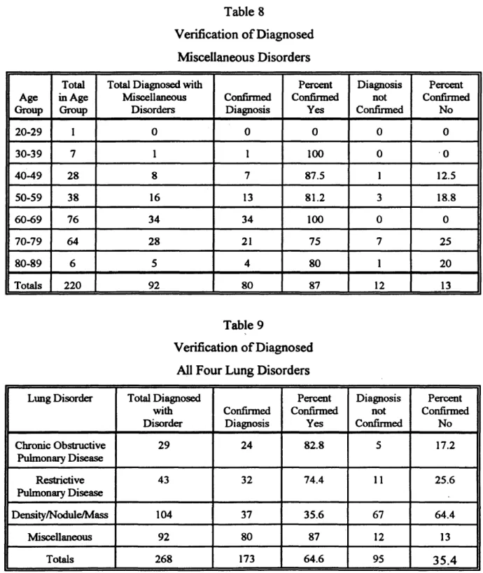
The first hypothesis of this thesis is that the chest x-ray is a good screening tool for the detection of pulmonary function disorders in the workplace. This was evaluated by confirming the B-readers diagnosis with the participant's personal physician's diagnosis. Tables 7 through 10 present a summary of the results for each disorder, i.e., chronic obstructive pulmonary disease, restrictive pulmonary disease, density/nodule/mass disorders, and miscellaneous. This evaluation included the 220 participants who had a follow-up examination with their personal physician as a result of their abnormal chest x- ray from the BHSP.
5.1 Database Results
Results indicate that if a disorder was diagnosed by the B-reader from the chest x- ray, the same disorder was subsequently confirmed by the personal physician 70 percent or more of the time for all categories except density/nodule/mass disorders. Specifically, chronic obstructive pulmonary disease was confirmed 82 .8 percent of the time, restrictive pulmonary disease was confirmed 74.4 percent of the time, and the miscellaneous
diagnosis was confirmed 87 percent of the time. Density/nodule/mass disorders were confirmed only 35 percent of the time. In general, the interviewer observed that a final medical diagnosis was received from the participant's personal physician within
Flats. Based on the percentage of confirmed diagnosis for each disorder the chest x-ray as diagnosed using the ILO classification system by a certified B-reader appears to be a good screening tool for chronic obstructive pulmonary disease, restrictive pulmonary disease, and miscellaneous disorders. The ILO classification system was not effective in
identifying and diagnosing density/nodule/mass disorders that may be indications of potential lung cancer.
Table 5
Verification of Diagnosed Chronic Obstructive Pulmonary Disease
Age Group
Total in Age Group
Total Diagnosed with Chronic Obstructive Pulmonary Disease . Confirmed Diagnosis Percent Confirmed Yes Diagnosis not Confirmed Percent Confirmed No 20-29 1 0 0 0 0 0 30-39 7 1 1 100 0 0 40-49 28 0 0 0 0 0 50-59 38 1 1 100 0 0 60-69 76 14 11 78.6 3 21.4 70-79 64 13 11 84.6 2 15.4 80-89 6 0 0 0 0 0 Totals 220 29 24 82.8 5 17.2
Table 6
Verification of Diagnosed Restrictive Pulmonary Disease
Age Group
Total in Age Group
Total Diagnosed with Restrictive Pulmonary Disease Confirmed Diagnosis Percent Confirmed Yes Diagnosis not Confirmed Percent Confirmed No 20-29 1 0 0 0 0 0 30-39 7 0 0 0 0 0 40-49 28 5 3 60 2 40 50-59 38 7 5 71.4 2 28.6 60-69 76 13 10 76.9 3 23.1 70-79 64 16 12 75 4 25 80-89 6 2 2 100 0 0 Totals 220 43 32 74.4 11 25.6 Table 7 Verification of Diagnosed Density/Nodule/Mass Age Group Total in Age Group
Total Diagnosed with Density/Nodule/Mass Confirmed Diagnosis Percent Confirmed Yes Diagnosis not Confirmed Percent Confirmed No 20-29 1 1 0 0 1 100 30-39 7 5 2 40 3 60 40-49 28 16 3 18.8 13 81.2 50-59 38 21 6 28.6 15 71.4 60-69 76 39 16 41 23 59 70-79 64 21 10 47.6 11 52.4 80-89 6 1 0 0 1 100 Totals 220 104 37 35.6 67 64.4
Table 8 Verification of Diagnosed Miscellaneous Disorders Age Group Total in Age Group
Total Diagnosed with Miscellaneous Disorders Confirmed Diagnosis Percent Confirmed Yes Diagnosis not Confirmed Percent Confirmed No 20-29 1 0 0 0 0 0 30-39 7 1 1 100 0 0 40-49 28 8 7 87.5 1 12.5 50-59 38 16 13 81.2 3 18.8 60-69 76 34 34 100 0 0 70-79 64 28 21 75 7 25 80-89 6 5 4 80 1 20 Totals 220 92 80 87 12 13 Table 9 Verification of Diagnosed All Four Lung Disorders
Lung Disorder Total Diagnosed with Disorder Confirmed Diagnosis Percent Confirmed Yes Diagnosis not Confirmed Percent Confirmed No Chronic Obstructive Pulmonary Disease 29 24 82.8 5 17.2 Restrictive Pulmonary Disease 43 32 74.4 11 25.6 Density/Nodule/Mass 104 37 35.6 67 64.4 Miscellaneous 92 80 87 12 13 Totals 268 173 64.6 95 35.4
5.2 Statistical Evaluation
As can be seen from the data, based on percentages for each lung disorder, the chest x-ray appears to be a good screening tool when comparing specific disorders, if a 70 percent or less accuracy is required. In an attempt to evaluate the chest x-ray as a
screening tool in general for the four disorders in this study, confidence intervals were calculated for the confirmed diagnoses of each lung disorder and a chi-square (X2) test was used for evaluation of the relationship between a confirmed and unconfirmed diagnosis (Munro and others, 1986).
Confidence intervals are used to determine the confidence that a population of values falls within certain boundaries. The following calculations are an estimate that given a certain mean for all four lung disorders that the population of values will fall between the established boundaries.
Table 10
Standard Deviation of Four Lung Disorders
Lung Disorder N P Percentage of Confirmed Diagnosis o standard deviation Chronic Obstructive Pulmonary Disease 29 0.828 0.070
Restrictive Pulmonary Disease 43 0.744 0.067
Density/Nodule/Mass 104 0.356 0.047
Miscellaneous 92 0.870 0.035
(2) p is the percentage of confirmed diagnoses and was calculated as follows:
p = number of confirmed diagnoses
number of total diagnosed with disorder
(3) a is the standard deviation and is a measure of variability. The standard
deviation represents the average amount by which results vary from the central tendency or mean.
q2 = e(i-e)
N(4) 95 percentile confidence intervals for the percentage of confirmed
diagnoses were calculated as follows for each lung disorder and for all of the lung disorders:
Chronic Obstructive Pulmonary Disease
95% = 0.828 ± (1.96)(0.070) 95% = 0.828 ± 0.137
0.691 £ 0.828 <; 0.965
Restrictive Pulmonary Disease
95% = 0.744 ± (1.96)(0.067) 95% = 0.744 ±0.131 0.613 <; 0.744 <; 0.875 Density/Nodule/Mass Disorders 95% = 0.356 ± (1.96)(0.047) 95% = 0.356 ±0.092 0.264 <; 0.356 <; 0.448 Miscellaneous Disorders 95% = 0.870 ± (1.96)(0.035) 95% = 0.870 ± 0.069 0.801 ^ 0.870 ^ 0.939
All Lung Disorders
95% = 0.646
±
(1.96)(0.029)95% * 0.646 ± 0.057 0.589 <; 0.646 s 0.703
The 95 percentile confidence interval calculation confirmed the original conclusion that the chest x-ray is a good screening tool for all of the lung disorders (i.e., chronic obstructive pulmonary disease, restrictive pulmonary disease, and miscellaneous disorders) except the density/nodule/mass disorders.
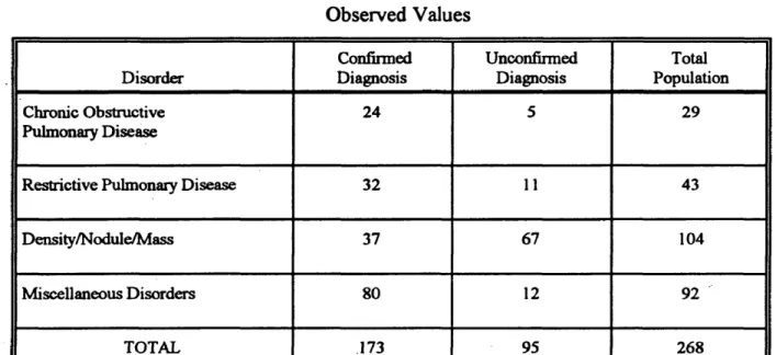
The chi-square test is used to evaluate the relationship between the expected number of people that fall into a category and the actual, or observed number of people that fall into a category. The idea of the chi-square test is that the null hypothesis is true, if the observed frequencies do not deviate too much from their expected values.
The null hypothesis for this evaluation is the following: A diagnosis that both the radiologist and participant's personal physician agree upon is independent of the type of lung disorder in this study, i.e., chronic obstructive pulmonary disease, restrictive
Table 11 Chi-Square Analysis Observed Values Disorder Confirmed Diagnosis Unconfirmed Diagnosis Total Population Chronic Obstructive Pulmonary Disease 24 5 29
Restrictive Pulmonary Disease 32 11 43
Density/Nodule/Mass 37 67 104
Miscellaneous Disorders 80 12 92
TOTAL 173 95 268
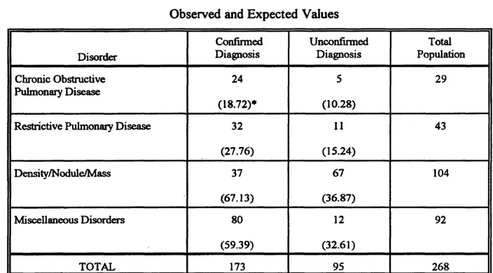
(5) The expected frequency for the chi-square analysis is calculated using the
observed frequency with the following equation, where:
fo = observed frequency (values listed above in Table 11)
df = degrees of freedom (columns - l)(rows-l) ( 2 - l ) ( 4 - l ) = 3
fe = expected frequency (values calculated below)
fe — fo (observed frequency of the lung disorder) x fo (observed frequency of the diagnosis) / Total Population
(6) Chronic Obstructive Pulmonary Disease Expected Value Calculation
fe = (29 x 173)/268 = 18.72 (expected frequency with a confirmed diagnosis of chronic obstructive pulmonaty disease)
fe = (29, x 95)/268 = 10.28 (expected frequency with an
unconfirmed diagnosis of chronic obstructive pulmonary disease)
(7) Restrictive Pulmonary Disease Expected Value Calculation
fe = (43 x 173)/268 = 27.76 (expected frequency with a confirmed diagnosis of restrictive pulmonary disease)
fe = (43 x 95)/268 = 15.24 (expected frequency with an
unconfirmed diagnosis of restrictive pulmonary disease)
(8) Density/Nodule/Mass Disease Expected Value Calculation
fe = (104 x 173)/268 = 67.13 (expected frequency with a confirmed diagnosis of density/nodule/mass disorder)
fe = (104 x 95)/268 = 36.87 (expected frequency with an unconfirmed diagnosis of density/nodule/mass disorder)
(9) Miscellaneous Disorder Expected Value Calculation
fe = (92 x 173)/268 = 59.39 (expected frequency with a confirmed diagnosis of miscellaneous disorder)
fe = (92 x 95)/268 = 32.61 (expected frequency with an
unconfirmed diagnosis of miscellaneous disorder)
Table 12
Chi-Square Analysis Observed and Expected Values
Disorder Confirmed Diagnosis Unconfirmed Diagnosis Total Population Chronic Obstructive 24 5 29 Pulmonary Disease (18.72)* (10.28)
Restrictive Pulmonary Disease 32 11 43
(27.76) (15.24) Density/Nodule/Mass 37 67 104 (67.13) (36.87) Miscellaneous Disorders 80 12 92 (59.39) (32.61) TOTAL 173 95 268
‘Expected values are presented in parenthesis
(10) Chi-Square Analysis Calculation
X2 = E (fo - fe)2 fe X2 = (24 - 18.72)2/18.72 + (32 - 27.76)2/27.76 + (37 - 67.13)2/67.13 + (80 - 59.39)759.39 + (5 - 10.28)710.28 + (11 - 15.24)715.24 + (67 - 36.87)736.87 + (12 - 32.61)732.61 X2= 1.49 + 0.65 + 13.52 + 7.15 + 2.71 + 1.18 + 24.62 + 13.03 X2 = 64.35
ARTHUR LAKES LIBRARY COLORADO SCHOOL OF MINES 001DEN.C0 80401
When comparing the chi-square value of 64.35 to the Distribution of X2
Probability Table (Munro and others, 1986), using three degrees of freedom it is evident that 64.35 is highly significant beyond the .001 level (the tabled value for .001 is 16.266). Therefore, the null hypothesis, that a diagnosis that both the radiologist and the
participant's personal physician agree upon is independent of the lung disorder, must be rejected. The conclusion from this test is that agreement between the radiologist and personal physician is highly dependent on the lung disorder. Therefore, based on this statistical test the chest x-ray is a good screening tool for some lung disorders, but not all.
5.3 Analysis of the Database
When examining the data without any statistical evaluation it appears that the chest x-ray is a good screening tool, if a 70 percent or less accuracy is required, for chronic obstructive pulmonary disease, restrictive pulmonary disease, and miscellaneous disorders. Upon further evaluation of the data collected in this study, with the use of 95 percentile confidence intervals it was confirmed that the chest x-ray is a good screening tool for specific lung disorders (i.e., chronic obstructive pulmonary disease, restrictive pulmonary disease, and miscellaneous disorders) except the density/nodule/mass disorder.
The chi-square test revealed that the chest x-ray, as diagnosed by the ILO
classification system is highly dependent on the specific lung disorder. In other words, it is more likely that the radiologist and personal physician will agree on a diagnosis with some lung disorders and not with others.
The interviewer did observe during the study that a large number of participants who had been diagnosed by the radiologist with a density/nodule/mass disorder that their
personal physician concluded that the radiologist was seeing the participant's nipples on the chest x-ray. This may have been one of the reasons that the density/nodule/mass disorder was confirmed at such a lower rate when compared to the other lung disorders.
As discussed earlier, the ILO classification system was developed as a tool for diagnosis of Coal Workers Pneumoconiosis. It is a useful tool for the evaluation of effects of exposure to dusty atmospheres (Albin and others, 1992). The ILO
classification provides two pieces of information, categorization of the size or form of opacities from dust exposure and an indication of their profusion or extent in the lung fields. It is most useful in relating the radiographic features to dust exposures and
changes in lung function (Weill and Tumer-Warwick, 1981) not to potential lung cancer. When a B-reader identifies a density/nodule/mass disorder there is a possibility that this diagnosis is a lung cancer or the observed abnormality may be a benign spot on the lung that has no significance.
According to the United States Preventive Services Task Force (1990) in
Washington, D.C. chest x-rays lack sufficient accuracy to be used as a screening tool for potential lung cancers. Accuracy of the chest x-ray is limited by the technology and by variation in interpretation among the B-readers. These standard chest x-ray films used for the ILO classification system provide examples of abnormalities specific to
pneumoconioses and include such features as pleural thickening and cardiac abnormalities (Parkes, 1982).
The chest x-ray as an overall screening tool for workplace exposures should be used only when its limitations are understood by the user and the information is used in conjunction with a full physical examination.
Chapter 6.
DATABASE COMPARISONS
The second hypothesis of this study was that if the incidence of lung disorders (e.g., chronic obstructive pulmonary disease, restrictive pulmonary disease,
density/nodule/mass disorders, and miscellaneous disorders) is less than the State of Colorado incidence of similar disease. Unfortunately, data on the incidence and prevalence of occupational lung disease is essentially nonexistent (Markowitz, 1992). The following sections describe several databases that are maintained, but did not offer much in the way of comparison for incidence and prevalence rates.
6.1 Chronic Obstructive Pulmonary Disease
The Colorado Division of Workers’ Compensation tracks respiratory disease as it relates to lost time claims (Rohrback, 1994). Table 13 is a summary of the number of cases of obstructive pulmonary disease. A comparison could not be made between these data and the Rocky Flats data because the workers' compensation data track acute obstructive pulmonary disease cases and the Rocky Flats data consists of chronic obstructive pulmonary disease cases.
Table 13
Colorado Division of Workers’ Compensation Lost Time Claims for
Obstructive Pulmonary Disease
Nature of Injury Number of Cases
Respiratory (Fumes) 167
Upper Respiratory Condition 5
Respiratory System Condition Unspecified 2
Pneumonia, Bronchitis, Asthma 8
6.2 Restrictive Pulmonary Disease
The Colorado Division of Workers' Compensation also tracks respiratory disease as it relates to lost time claims (Rohrback, 1994). Table 14 contains a summary of the number of cases of restrictive pulmonaiy disease per 10,000 workers in 1992.
The total number of cases of restrictive pulmonary disease during 1992 was nine. The total number of confirmed cases of restrictive pulmonary disease between October
Table 14
Colorado Division of Workers’ Compensation Lost Time Claims for
Restrictive Pulmonary Disease
Nature of Injury Number of Cases
Asbestosis 4
Black Lung 1
Silicosis 3
Pneumoconiosis 1
6.3 Lung Cancer
Since 1979, the State of Colorado has been tracking the cancer incidence in Colorado through the Colorado Central Cancer Registry (CCCR). The CCCR estimates that it registers approximately 90 percent of the cancer diagnosed in Colorado and 100 percent of the cancers diagnosed in the Denver Metropolitan Area. The most recent published report is from 1991 and covers 1979 to 1988 (Karp and others, 1991). Table
Table 15
Average Annual Incidence Rate1 of Lung Cancer per 100,000 Population
for Overlapping 3-Year Time Periods
Year Sex 1979-81 1980-82 1981-83 1982-84 1983-85 1984-86 1985-87 1986-88 U.S.2 1986-1988 M ale All Races 69.2 70.2 68.5 67.3 65.0 64.9 64.6 63.6 83.2 Female All Races 23.0 25.3 25.8 25.9 27.9 28.3 31.0 31.8 38.4
1 Age-Adjusted to 1970 United States Standard Population by Direct Method.
Chapter 7.
SUMMARY AND CONCLUSIONS
This thesis study was a follow-up to the RFETS's original Beryllium Health Surveillance Program. The purpose was to determine a final medical diagnosis for each abnormal (Category "B") chest x-ray not related to chronic beryllium disease. This was accomplished by first determining the final medical diagnosis from each participant's personal physician. This information was then compared to the radiologist's report to evaluate the effectiveness of the chest x-ray as a screening tool for pre-existing
pulmonary function disorders. Next, the final medical diagnosis for each participant was complied into a database. This database was evaluated and compared to other available databases in an effort to determine the incidence of pulmonary disease at RFETS.
Based on the data in this study the chest x-ray, as diagnosed by a certified B- reader using the ILO classification system, is a good screening tool, if a 70 percent or less accuracy is required, when compared on a percentage basis for chronic obstructive pulmonary disease, restrictive pulmonary disease, and certain miscellaneous disorders. However, the ILO classification system was not an effective tool for identifying and diagnosing density/nodule/mass disorders that may be indications of lung cancer.
The 95 percentile confidence interval calculation confirmed that the overall effectiveness of the chest x-ray is an adequate screening tool for the lung disorders (i.e., chronic obstructive pulmonary disease, restrictive pulmonary disease, and miscellaneous
disorders) except for the density/nodule/mass disorder. In addition, the chi-square test determined that agreement between the diagnosis of the radiologist and the participant’s personal physician is highly dependent on the specific lung disorder.
The second part of this study was intended to determine if the incidence of lung disorders (e.g., chronic obstructive pulmonary disease, restrictive pulmonary disease, density/nodule/mass disorders, and miscellaneous disorders) in the RFETS groups was greater or smaller than the incidence of lung disorders in Colorado. Unfortunately, no databases were found that would provide this type of comparison.
This study indicates that the following research studies should be done in the future:
• A database of occupational lung disorders should be developed to
support future studies. Currently no one database exists that provides information on the incidence and prevalence of occupational lung disorders nationally or at the state level.
• The final medical diagnosis that was confirmed for each participant in this
study should be further evaluated by an occupational physician. An occupational physician is trained differently from a personal physician and may have a different opinion concerning the diagnosis.
• The chest x-rays in this study were analyzed by two radiologists. It
would be interesting to cross check their work and see if one radiologist was more or less accurate than the other.
but based on the history of the ILO classification it seems that the restrictive lung disorders should be the easiest to identify.
This study did not look at any participants in Category “A” that were considered to have a normal chest x-ray by the radiologist, but their personal physician found a problem during the follow-up.
A prospective study needs to be conducted on employees working at RFETS to track exposures now and their effects into the future.
Exposure histories need to be compiled for all job classifications at RFETS from the start-up of operations in 1952 to the present.
REFERENCES CITED
Alberts, W. Michael, MD and Stuart M. Brooks MD, 1992, June, Advances in Occupational Asthma: Clinics in Chest Medicine, v. 13, no. 2.
Albin, M., G. Engholm, K.Frostrom, S. Kheddache, S.Larsson, and L.Swantesson, 1992, Chest X-Ray Films from Construction Workers: International Labour Office Classification Compared with Routine Readings: British Journal of Industrial Medicine v. 49, pp 862-868.
Amdur, Mary O., John Doull, and Curtis D. Klaassen, 1993 Casarett and Doull's
Toxicology, The Basic Science of Poisons: New York, McGraw-Hill, Inc., 4th ed.
Baum, Gerald, and Emanual Wolinsky, 1989, Textbook of Pulmonary Diseases: Boston, Mass., Little, Brown and Company, 4th ed.
ChemRisk, A Division of McLaren/Hart, 1992, Rockv Flats History. Rocky
Flats Toxicological Review and Dose Reconstruction: Health Studies on Rocky Flats, Report E92-023.
Coultas, David B., MD, and Jonathan M. Samet, MD, 1992, June, Occupational Lung Cancer: Clinic in Chest Medicine, v. 13, no 2, p. 341.
Eisenbud, M. and J. Lisson, 1983, Epidemiological Aspects of Beryllium Induced Non- Malignant Lung Disease: A 30 Year Update: Journal of Occupational Medicine: v. 25 pp 196-202.
Epler, Gary R., M.D., 1992, November, Clinical Overview of Occupational Lung Disease. Radiological Clinics of North America, v. 30, no. 6, p. 1121.
Fishman, Alfred P., 1992. Update: Pulmonary Diseases and Disorders: McGraw-Hill, Inc.
Graham, William G.B. M.D., 1992, June, Silicosis: Clinics in Chest Medicine, v. 13, no. 2, p. 253.
Gray, Henry, 1977, Anatomy. Descriptive and Surgical: New York, New York, Bounty Books.
Karp, Shelley, Finch, Jack L., Bott, Robin, Young, Walter. 1991. Cancer in Colorado 1979 to 1988 Prevention. Incidence. Survival and Mortality: Denver, Colorado, American Cancer Society.
Levitzky, Michael G., 1991, Pulmonary Physiology (3rd ed.): New York, New York, McGraw-Hill,Inc.
Markowitz, Steven, 1992, Primary Prevention of Occupational Lung Disease: A View from the United States: Israel Journal of Medical Sciences, v.28, p. 513.
Munro, Barbara, Hazard, Visintainer, Madelon A., Page, Ellis Batten, 1986, Statistical Methods for Health Care Research. Philadelphia, Pennsylvania, J.B. Lippincott Company.
Newell, John D. Jr., and Tarver, Robert D., 1993, Thoracic Radiology: New York, Raven Press.
NIOSH (National Institute for Occupational Safety and Health), 1977, June, Occupational Diseases. A Guide to Their Recognition (Revised ed.): Washington D.C., U.S. Department of Health, Education, and Welfare.
Owens, Gregory R., 1991, October 21, Public Screening for Lung Disease: Experience with the NIH Lung Health Study: American Journal of Medicine, v. 91 (suppl 4A), p. 4A-37S.
Pare, J.A. Peter, 1983, Synopsis of Diseases of the Chest. W.B. Saunders Company. Parkes, W. Raymond, 1982, Occupational Lung Disorders (2nd ed.): Boston, Mass,
Butterworths.
Plog, Barbara A., 1988, Fundamentals of Industrial Hygiene (3rd ed.): Chicago, Illinois, National Safety Council.
Protchen, E. James, MD, Grainger, Ronald G., MD, FRCP, FRCR, DMRD, Greene, Reginald, MD, 1993, Pulmonary Radiology: Philadelphia, Pennsylvania, W.B. Saunders Company.
Rohrback, Dan, 1994, July 26, Memorandum. Department of Labor and Employment, Division of Workers' Compensation, Denver, Colorado.
Saric, M., 1992, Occupational and Environmental Exposures and Nonspecific Lung Disease-A Review of Selected Studies: Israel Journal of Medical Sciences, v. 28, p. 509.
Schwarz, Marvin I. and King, Talmadge E., 1993, Interstitial Lung Disease (2nd ed.): Mosby Year Book.
Sperber, Miriam, M.D., 1990. Radiologic Diagnosis of Chest Disease: Springer-Verlag, New York.
Thibodeau, Gary A. and Patton, Kevin T., 1992, The Human Body in Health & Disease. Mosby Year Book.
United States Preventive Services Task Force, 1990, Screening for Lung Cancer: AFP, v. 41, no. 6.
Weill, Hans and Tumer-Warwick, Margaret, 1981, Occupational Lung Diseases: New York, Marcel Dekker, Inc.
Table A -l
Abnormal Chest X-Ray Data October 1991 through January 1993
S t u d y R e s u l t s S t u d y # A g e S e x S m o k e r C h r o n i c O b s t r u c t i v e P u l m o n a r y D i s e a s e R e s t r i c t i v e P u l m o n a r y D i s e a s e D e n s i t y / N o d u l e / ' M a s s M i s c e l l a n e o u s C o n f i r m e d I 3 7 M Y Y F i n a l 2 6 0 M Y M e d i c a l 3 6 5 M Y Y D i a g n o s i s 4 6 4 M Y Y 5 6 9 M Y Y 6 6 8 M Y 7 4 4 M Y Y 8 5 8 M Y 9 6 5 M Y Y 1 0 7 6 M Y Y Y 1 1 3 3 M • Y 1 2 7 2 M Y 1 3 6 4 M Y Y 1 4 7 5 M Y 1 5 6 8 M Y Y 1 6 4 7 F Y 1 7 6 4 M Y Y 1 8 5 2 M Y Y 1 9 7 6 M Y Y 2 0 7 6 M Y Y 2 1 7 0 M Y Y Y 2 2 7 5 M Y Y Y 2 3 6 0 M Y Y Y 2 4 7 4 M Y Y 2 5 6 9 M Y Y 2 6 7 1 M Y 2 7 6 7 M Y Y Y 2 8 6 8 M Y Y Y Y 2 9 6 4 M Y 3 0 5 6 M Y Y 3 1 6 7 F Y Y 3 2 6 1 M Y Y 3 3 7 0 M Y Y 3 4 6 9 M Y Y Y 3 5 6 9 M Y Y 3 6 5 6 M Y Y Y 3 7 7 6 M Y Y Y 3 8 5 8 M Y Y 3 9 7 4 M Y Y Y 4 0 7 1 M Y 4 1 6 1 M Y 4 2 4 7 M Y Y 4 3 5 4 M Y 4 4 7 4 M Y (continued)
Table A -l
Abnormal Chest X-Ray Data October 1991 through January 1993
S t u d y R e s u l t s S t u d y # A g e S e x S m o k e r C h r o n i c O b s t r u c t i v e P u l m o n a r y D i s e a s e R e s t r i c t i v e P u l m o n a r y D i s e a s e D e n s i t y / N o d u l e / M a s s M i s c e l l a n e o u s C o n f i r m e d 4 5 7 1 M Y Y F i n a l 4 6 6 1 M Y Y Y M e d i c a l 4 7 7 3 M Y Y D i a g n o s i s 4 8 4 8 M Y Y ( c o n t i n u e d ) 4 9 6 7 M Y Y 5 0 5 9 M Y 5 1 6 0 M Y Y 5 2 7 5 M Y Y 5 3 7 4 M Y Y Y 5 4 3 9 M Y Y 5 5 6 2 M Y Y Y 5 6 8 1 M Y Y 5 7 6 8 M Y Y Y Y 5 8 6 1 M Y Y 5 9 7 1 M Y Y 6 0 4 8 M Y Y 6 1 4 6 F Y 6 2 6 8 M Y Y Y 6 3 7 2 M Y Y 6 4 5 1 M Y 6 5 6 7 M Y Y 6 6 6 2 M Y Y 6 7 4 5 M Y Y 6 8 8 0 M Y Y Y 6 9 7 5 M Y Y 7 0 5 3 M Y Y 7 1 7 9 M Y 7 2 4 4 F Y Y 7 3 5 2 F Y Y 7 4 4 6 M Y 7 5 7 1 M Y 7 6 5 8 M Y Y 7 7 6 2 M Y Y 7 8 7 5 M Y Y 7 9 6 5 M Y Y 8 0 7 8 M Y Y 8 1 5 9 M Y Y Y 8 2 8 2 M Y 8 3 7 6 M Y Y Y 8 4 7 3 M Y Y 8 5 5 6 M Y Y 8 6 6 9 M Y Y 8 7 8 3 M Y Y 8 8 6 6 M Y Y (continued)