ACTA UNIVERSITATIS
UPSALIENSIS
Digital Comprehensive Summaries of Uppsala Dissertations
from the Faculty of Medicine 1548
Induction and repair of clustered
DNA damage sites after exposure
to ionizing radiation
ANDRIS ABRAMENKOVS
ISSN 1651-6206 ISBN 978-91-513-0591-2
Dissertation presented at Uppsala University to be publicly examined in Rudbecksalen, Rudbecklaboratoriet, Dag Hammarskjölds v 20, Uppsala, Monday, 29 April 2019 at 13:00 for the degree of Doctor of Philosophy (Faculty of Medicine). The examination will be conducted in English. Faculty examiner: Professor Emeritus Peter O'Neill (Department of Oncology, University of Oxford).
Abstract
Abramenkovs, A. 2019. Induction and repair of clustered DNA damage sites after exposure to ionizing radiation. Digital Comprehensive Summaries of Uppsala Dissertations
from the Faculty of Medicine 1548. 54 pp. Uppsala: Acta Universitatis Upsaliensis.
ISBN 978-91-513-0591-2.
The mechanisms that maintain genomic stability safeguard cells from constant DNA damage produced by endogenous and external stressors. Therefore, this thesis aimed to specifically address questions regarding the requirement and involvement of DNA repair proteins in the repair of various types of radiation-induced DNA damage.
The first aim was to determine whether the phosphorylation of DNA-PKcs, a major kinase involved in non-homologous end joining pathway, can be utilized to score the DNA double-strand break (DSB) content in cells. DNA-PKcs phosphorylated (pDNA-PKcs) at T2609 was more sensitive to the cellular DSB content than ɣH2AX, as analyzed by flow cytometry. Further, pDNA-PKcs at T2609 could discriminate between DSB repair-compromised and normal cells, confirming that the pDNA-PKcs can be used as a DSB repair marker. In paper II, the DSB repair was assessed in cells with reduced levels of DNA-PKcs. The reduction in DNA-PKcs resulted in decreased cell survival and unaffected DSB repair. These results clearly indicate that DNA-PKcs plays an additional role in promoting cell survival in addition to its function in DSB repair. The second part of the thesis focused on the characterization of complex DNA damage. DNA damage was investigated after exposure to α-particles originating from Ra-223. The Ra-223 treatment induced a nonrandom DSB distribution consistent with damage induced by high-linear energy transfer radiation. The exposure to Ra-223 significantly reduced cell survival in monolayers and 3D cell structures. The last paper unraveled the fate of heat-sensitive clustered DNA damage site (HSCS) repair in cells. HSCS repair was independent of DSB repair, and these lesions did not contribute to the generation of additional DSBs during repair. Prolonged heating of DNA at relatively low temperatures induced structural changes in the DNA that contributed to the production of DNA artifacts.
In conclusion, these results demonstrate that DNA-PKcs can be used to monitor DSB repair in cells after exposure to ionizing radiation. However, the functions of DNA-PKcs are not limited to DSB repair, as it can promote cell survival through other mechanisms. The complexity of the DNA damage produced by high-LET radiation is a major contributor to cell death. However, not all clusters produced in irradiated cells are converted into DSBs during repair.
Keywords: NHEJ, DSB repair, clustered DNA damage, DNA repair, DNA-PKcs, HSCS,
Ra-223, ionizing radiation
Andris Abramenkovs, Department of Immunology, Genetics and Pathology, Medical Radiation Science, Rudbecklaboratoriet, Uppsala University, SE-751 85 Uppsala, Sweden.
© Andris Abramenkovs 2019 ISSN 1651-6206
ISBN 978-91-513-0591-2
List of Papers
This thesis is based on the following papers, which are referred to in the text by Roman numerals.
I Abramenkovs A, Stenerlöw B. (2017) Measurement of
DNA-Dependent Protein Kinase Phosphorylation Using Flow Cytom-etry Provides a Reliable Estimate of DNA Repair Capacity.
Ra-diation Research, 188(6): 597 – 604.
II Gustafsson AS, Abramenkovs A, Stenerlöw B. (2014) Suppres-sion of DNA-dependent protein kinase sensitize cells to radiation without affecting DSB repair. Mutation Research, 769: 1 – 10. III Abramenkovs A, Spiegelberg D, Nilsson S, Stenerlöw B. The
α-emitter Ra-223 induces clustered DNA damage and signifi-cantly reduces cell survival. Manuscript.
IV Abramenkovs A, Stenerlöw B. (2018) Removal of
heat-sensi-tive clustered damaged DNA sites is independent of double-strand break repair. PLoS ONE 13(12):e0209594.
Contents
Introduction ... 9
Ionizing radiation ... 9
DNA ... 10
DNA damage ... 11
Factors influencing DNA damage induction ... 12
DNA repair ... 13
Repair of non-DSB lesions ... 13
DSB signaling ... 14
ATM and ATR ... 15
Mediators of DSB repair ... 15
DSB repair ... 15
Non-homologous end joining ... 16
Homologous recombination ... 18
Alternative repair mechanisms ... 19
Cancer ... 19
Prostate cancer ... 20
Cancer diagnosis and treatment ... 21
Aim ... 23
Results ... 24
Paper I ... 24
Measurement of DNA-Dependent Protein Kinase Phosphorylation Using Flow Cytometry Provides a Reliable Estimate of DNA Repair Capacity ... 24
Aim and background ... 24
Methods ... 24
Results ... 25
Discussion ... 26
Paper II ... 27
Suppression of DNA-dependent protein kinase sensitizes cells to radiation without affecting DSB repair ... 27
Aim and background ... 27
Methods ... 27
Results ... 28
Paper III ... 30
The α-emitter Ra-223 induces clustered DNA damage and significantly reduces cell survival ... 30
Aim and background ... 30
Methods ... 31
Results ... 31
Discussion ... 33
Paper IV ... 33
Removal of heat-sensitive clustered damaged DNA sites is independent of double-strand break repair ... 33
Aim and background ... 33
Methods ... 34 Results ... 35 Discussion ... 36 Concluding remarks ... 37 Future studies ... 39 Acknowledgements ... 41 References ... 44
Abbreviations
53BP1 p53-Binding protein 1 8-oxoG 8-Oxoguanine
ADP Adenosine diphosphate ADT Androgen deprivation therapy
alt-NHEJ Alternative non-homologous end joining AP Apurinic/Apyrimidinic site
APE Apurinic/Apyrimidinic endonuclease AR Androgen receptor
ARv7 Androgen receptor variant 7 ATM Ataxia telangiectasia mutated
ATR Ataxia telangiectasia and RAD3-related protein BER Base excision repair
Bp Base pair
BRCA1 Breast cancer type 1 susceptibility protein CtIP C-terminal-binding protein 1-interacting protein DDB DNA damage-binding protein
DNA Deoxyribonucleic acid
DNA-PKcs Deoxyribonucleic acid-dependent protein kinase catalytic subunit
DSB Double-strand break
ɣH2AX phosphorylated H2A histone family member X H Histone
H2AX H2A histone family member X HR Homologous recombination HSCS Heat-sensitive cluster sites
KU70 X-ray repair cross-complementing 6 KU80 X-ray repair cross-complementing 5 LET Linear energy transfer
MMR Mismatch repair
Mre11 Meiotic recombination 11 homolog A Mut Mutator
NBS1 Nijmegen breakage syndrome 1 NER Nucleotide excision repair NHEJ Non-homologous end joining
PARP1 Poly [adenosine diphosphate – ribose] polymerase 1 PCNA Proliferating cell nuclear antigen
PIKK Phosphatidylinositol 3-kinase-related kinase Pol Polymerase
RAD DNA repair protein RNA Ribonucleic acid RPA Replication protein A SSB Single-strand break
ssDNA Single-stranded deoxyribonucleic acid TFIIH Transcription factor II human
UV Ultraviolet
XLF X-ray repair cross-complementing factor 4-like factor XPC Xeroderma pigmentosum, complementation group C XRCC X-ray repair cross-complementing protein
Introduction
Ionizing radiation
All organisms are constantly exposed to low levels of ionizing radiation. The danger associated with exposure to ionizing radiation stems from its ability to ionize atoms and molecules that form important cellular structures. Ionization events can change molecules either directly or indirectly by producing radicals that can alter surrounding molecules (1).
In biological systems, the most deleterious alterations related to ionizing radiation exposure are changes in the DNA sequence. However, some biolog-ical effects can also be observed in cells that reside nearby exposed cells, and this is referred to as the bystander effect (2).
The linear energy transfer (LET) is an important factor that determines the cell damage pattern and the consequences of that damage. LET is defined as the rate of energy loss of an ionizing particle along its track. Irradiation with heavy charged particles is characterized by an increase in LET at the very end of the particle’s track (3). Consequently, on a macroscopic scale, the increase in LET results in a higher dose deposited at the end of the particle’s track, which differs from the dose distribution of low-LET radiation such as X-rays and ɣ-radiation (Figure 1).
Figure 1. Schematic representation of depth – dose distribution of photon and heavy charged particle radiation.
DNA
DNA was isolated for the first time in 1869 by Friedrich Miescher (4); how-ever, at the time, the importance of DNA was not fully recognized, and only in 1944 was DNA identified as a carrier of genetic information (5). The mo-lecular composition of DNA was clarified in 1909, when it was shown that the DNA molecule was composed of 4 different nucleobases – adenine, guanine, cytosine and thymidine (6). Further studies of the DNA molecule by X-ray crystallography revealed that DNA is a double helix with inward facing bases and an outward facing sugar backbone (7). In this model, the bases are stacked parallel to each other within a distance of 3 – 4 Å, and the interaction between bases is limited to hydrogen bonding between cytosine and guanine or adenine and thymine. As the interactions between the bases are governed by noncova-lent interactions, the strands can be thermally or mechanically separated (8) without causing any damage to the macromolecule.
In eukaryotic cells, DNA is wound around proteins to form nucleosomes (9). The proteins found in nucleosomes are called histones, and each nucleo-some is an octamer consisting of a pair of H2A, H2B, H3 and H4 histones (10,
11) and 146 base pairs (bp) of DNA (12). The nucleosomes are connected
together with a DNA fragment that is associated with histone H1 (13). The nucleosomes that have undergone successive folding events create larger structures called chromosomes.
Histone binding to DNA is mainly governed by noncovalent interactions such as hydrogen bonds and salt bridges (12, 14). Any DNA sequence is able to bind with histones; however, some more flexible DNA sequences exhibit stronger interactions (15). Long amino acid side chains on histones can un-dergo posttranslational modifications such as methylation, acetylation, phos-phorylation, ubiquitination and ADP-ribosylation (16-20). The histones and their modifications are key factors that determine DNA accessibility and thus gene transcription (21). More specifically, the modifications of histone tails affect the electrostatic interactions between DNA and histones (22), which changes the DNA binding strength and determines whether a gene will be ex-pressed or silenced. The modifications of histone tails are subsequently passed on to daughter cells during mitosis and are referred to as epigenetic inher-itance. The flexibility of histone modifications allows an organism to develop various types of cells and tissues by altering the gene expression and molecu-lar landscape of the cell (23). In the nucleus, transcriptionally repressed re-gions are referred to as heterochromatin, while transcriptionally active rere-gions are called euchromatin. Regions of euchromatin and heterochromatin can eas-ily be discriminated by electron microscopy. However, macroscopic observa-tions of euchromatin and heterochromatin regions are not consistent with mo-lecular analysis, which has identified at least five different states of chromatin organization (24).
DNA damage
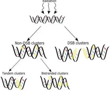
Every day, cells are subjected to DNA damage originating from endogenous events and external stressors, e.g., ionizing radiation and chemical exposure. The majority of the damage induced by ionizing radiation is similar to damage originating from endogenous processes in terms of the chemical adducts pro-duced (25). Ionizing radiation and endogenous processes can induce a vast array of DNA lesions, which include single-strand breaks (SSBs), double-strand breaks (DSBs), abasic sites, base and sugar lesions, protein crosslinks and interstrand crosslinks (26). Ionizing radiation induces clustered DNA damage sites at a much higher frequency than endogenous processes (27). Clustered DNA damage sites are complex lesions that have 2 or more DNA lesions within a distance of 20 - 30 nucleotides (28, 29) that can be further divided into DSB and non-DSB cluster sites (Figure 2). The damage in clus-tered DNA damage sites can be found in a bistranded and/or tandem configu-ration. In bistranded clusters, the lesions are located on the opposite strands, while in the tandem clusters, the lesions are distributed on the same strand. The simplest bistranded cluster site is the double-strand break (DSB), which is composed of two single-strand breaks within a distance of 14 nucleotides (30). Simple DSBs or DSBs within cluster sites are the most toxic lesions as-sociated with exposure to ionizing radiation (31). Non-DSB cluster sites, as
Figure 2. Clustered DNA damage sites can be separated in non-DSB cluster damage sites and DSB containing cluster sites. Further, the non-DSB cluster sites can be di-vided in clusters containing damage in tandem or bistranded configurations.
their name implies, are complex lesions that do not harbor a DSB. These clus-ter sites can contain one or several SSBs, apurinic/apyrimidinic (AP) sites and base modifications.
A specific type of non-DSB cluster site that is converted into DSBs upon heating is the heat-sensitive cluster site (HSCS). The repair mechanism for and the nature of these lesions has hitherto remained elusive; however, HSCSs are converted into DSBs at elevated temperatures (32). HSCS are produced after both high-LET and low-LET irradiation (33-35). The importance of the contribution of HSCSs to DSB yields was revealed when DNA extraction after irradiation was performed at 50°C and 4°C. The extraction at elevated tem-peratures yields 40 – 50% more DSBs in the irradiated samples than extraction performed at 4°C (32, 35). Further experiments revealed that HSCSs are re-paired within 1 hour after irradiation, are independent of DNA-PKcs, PARP1, and XRCC1 (36) and are not related with AP sites (37). As of now, the con-version of HSCSs into DSBs is controversial (35, 37). Therefore, understand-ing the conversion of HSCSs into DSBs durunderstand-ing the repair process in cells is crucial to correctly determine non-homologous end joining (NHEJ) repair ki-netics and the consequences of DNA repair protein deficiencies.
Factors influencing DNA damage induction
The quantity and complexity of DNA damage after exposure to ionizing radi-ation depend on biological, chemical and physical factors. The most promi-nent factors are DNA packaging and radiation quality, i.e., LET and the oxy-gen concentration. It is important to note that these factors can interact recip-rocally, which is briefly discussed below.
Irradiation with high-LET radiation results in more complex DNA damage clusters than exposure to low-LET radiation (Figure 3). Furthermore, in con-trast to low-LET irradiation, exposure to high-LET irradiation induces a nonnormal distribution of DSBs (38, 39). The complexity of DSBs increases with increasing LET (40, 41).
The compactization of DNA in the nucleus leads to a 5-fold decrease in the number of DSBs after irradiation, and an even more pronounced effect is ob-served when the chromosomes are condensed, in which a 50-fold reduction in DSB induction is observed (42). The protective effect of DNA condensation results from its reduction in the number of water molecules, which in turn decreases the number of radicals produced in the vicinity of the DNA (43); this effect is observed after both high-LET and low-LET irradiation (42). Low-LET irradiation at a reduced oxygen concentration results in a decrease in DNA damage that is mediated via the reduction of oxygen-generated per-oxy radicals (44). However, the reduced per-oxygen concentrations have no or a minimal effect on high-LET-induced DNA damage (45).
DNA repair
Repair of non-DSB lesions
Due to the large number of different adducts that can be formed in DNA, more than 130 different proteins have been identified that mediate its repair in hu-man cells (46). The repair of nonclustered and non-DSB clustered lesions usu-ally involves base excision repair (BER), nucleotide excision repair (NER) and mismatch repair (MMR).
Base lesions that do not distort the DNA helix are recognized by DNA gly-cosylases, which cleave the N-glycosyl bond, leaving an AP site. The AP sites are then processed by apurinic/apyrimidinic endonucleases (APEs), which in-duce an SSB. Alternatively, a base lesion can be processed by bifunctional DNA glycosylase, which cleaves the N-glycosyl bond as well as the AP site, directly producing an SSB. After the formation of an SSB, BER is initiated in a form of short-patch or long-patch repair (47). During short-patch repair, pol-ymerase β (polβ) inserts the nucleotide, and ligase IIIα/XRCC1 ligates the SSB. In long-patch repair, a DNA fragment of 7 – 14 nucleotides is resynthe-sized at the AP site by polβ, polδ or polε (47, 48). The displaced original DNA Figure 3. The high-LET radiation induce spatially correlated complex DSBs while low-LET radiation produce randomly distributed DSBs. Representative images of cells irradiated with alpha particles (high-LET) and gamma radiation (low-LET). The red dots represent DSBs (53BP1) and DNA is shown in blue (DAPI).
strand is then removed by flap endonuclease 1 (49). Finally, the strand is re-joined by ligase I/PCNA or ligase IIIα/XRCC1 (50, 51).
NER repairs base modifications that result in distorted DNA. The repair is initiated by XPC-RAD32 and the DDB2/DDB1 complex (52). Lesions de-tected during transcription are sensed by RNA polymerase, which stalls tran-scription, and repair proteins are recruited to the damage site. After lesion de-tection, the surrounding DNA is melted and processed by the TFIIH complex. The TFIIH complex contains 10 different proteins that unwind and incise the DNA (53). Then, polδ, polε, and polκ resynthesize the damaged DNA frag-ment (54), which is subsequently rejoined via either ligase IIIα or ligase I (55).
MMR is responsible for the removal of incorrect base pairing produced during cytosine deamination that created uracil or is the result of a misplaced nucleotide by the polymerase. The repair is initiated via one of the MutS hom-ologs (56), which recruits MutLα to incise the DNA, creating a SSB (57). Fur-thermore, the strand is excised by exonuclease 1, and polδ synthesizes a new complementary DNA fragment (58, 59). The repair is completed when ligase I rejoins the DNA strand (60).
SSBs can be repaired via several repair pathways as their formation is an intermediate step during BER, NER and MMR. The repair starts with recog-nition of the SSB by PARP1. After modification of the broken strand, repair is then mediated by the repair proteins involved in BER (61); however, com-ponents from other repair pathways can contribute.
The repair of non-DSB clustered lesions depends on the configuration of the lesions, their type, the distance between the lesions and their complexity. The lesions positioned in a tandem (62) and bistrand (63) configuration are repaired less efficiently. Unlike lesions distributed in tandem, the bistranded clusters have the potential to become DSBs if they are closely spaced and pro-cessed at the same time (64). As DSBs threaten the survival of cells, the repair of clustered lesions follows a specific hierarchy to minimize the production of these highly toxic lesions. The first lesions removed from the cluster sites are SSBs and AP sites, followed by 8-oxoG (65, 66). There are additional diffi-culties during repair when the lesions are closely spaced, resulting in delayed processing (67).The repair and conversion of non-DSB cluster sites into DSBs have been studied in isolated systems or in plasmids transfected into cells. However, at this point, it is not clear how cluster damage sites are repaired in different chromatin regions and how well their processing in isolated systems represents their processing in live cells.
DSB signaling
DSB repair is a complex process that requires lesion recognition, signal prop-agation, recruitment and the retention of the appropriate repair proteins at damage sites. DSB sensing is poorly understood; however, the MRN complex,
KU70/80, RPA and PARP1 have been proposed to be the major DSB sensors (68).
ATM and ATR
One of the major signal transducers in DSB repair is ataxia telangiectasia mu-tated (ATM). ATM is extremely sensitive to ionizing radiation, and its maxi-mum activation is reached when cells are exposed to 0.4 Gy (69). ATM is normally a dimer that dissociates into monomers and autophosphorylates after exposure to ionizing radiation (69). The actual changes that occur in ATM after exposure are not fully understood; however, it has been suggested that the MRN complex is required for the complete activation of ATM (70). After its activation, ATM targets and phosphorylates more than 700 molecules in-volved in cell cycle regulation, chromatin remodeling, and DNA damage re-pair (71). Importantly, ATM activates DNA-PKcs via phosphorylation, and DNA-PKcs phosphorylates ATM, leading to its inactivation (72).
Ataxia telangiectasia and Rad3-related protein (ATR) autophosphorylates after it is recruited to the DNA damage site and interacts with RPA (73). ATR responds to a wide variety of lesions and is not specific to DSB repair alone. The activation of ATR also occurs during replicative stress or after UV irra-diation (71, 74). ATM and ATR can phosphorylate each other in response to DNA damage, clearly indicating the importance of these kinases in signal transduction (72, 74).
Mediators of DSB repair
Prominent DSB markers are p53-binding protein 1 (53BP1) and phosphory-lated H2AX (ɣH2AX). 53BP1 is an essential mediator of DSB repair that re-localizes to DSB sites and facilitates the recruitment of chromatin remodeling proteins (75), activates checkpoint signaling (76), and participates in the choice of a DSB repair pathway (77). 53BP1 binds to H4K20me2, which is modulated by H4K16 acetylation levels near DSBs (78). Specifically, the fo-cal accumulation of 53BP1 at the DSB site is dependent on H2K15ub (79).
H2AX phosphorylation at S139 (ɣH2AX) is well established marker of DSBs. H2AX is phosphorylated by ATM or DNA-PKcs after the detection of a DSB (80, 81). Phosphorylated H2AX molecules can span megabases away from the DSB (82), and the major role of ɣH2AX in the DSB regions is to prevent the dissociation of repair proteins from the break sites (83).
DSB repair
In response to DSBs, cells activate complex networks of repair and signaling pathways that are responsible for cell cycle arrest, chromatin remodeling and repair. The key DSB repair pathways in mammalian cells are non-homologous end joining (NHEJ), homologous recombination (HR) and alternative end
joining. The first proteins to bind at the DNA termini are the KU70/80 heter-odimer, PARP1 and the MRN complex, which is composed of Mre11, NBS1 and RAD50 (68, 84). After MRN and KU70/80 bind to the DNA, the correct repair pathway must be selected. The decision to use NHEJ or HR is depend-ent on the cell cycle stage; HR is utilized during S and G2 phases, while NHEJ operates in all stages of the cell cycle (85). The main proteins involved in the pathway choice are 53BP1 and BRCA1. 53BP1 prevents DSB end processing and promotes NHEJ, while BRCA1 promotes end processing and thus HR (86).
Non-homologous end joining
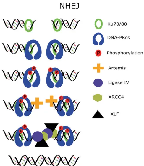
The most prominent DSB repair pathway in human cells is NHEJ, and the knockout of core NHEJ factors leads to severe radiosensitivity. During NHEJ repair, DSBs are resolved in several steps, and lesions are repaired with a half-life of 7.5 min and 160 min (36). The repair starts with the binding of KU70/80 to the ends of broken DNA, followed by the recruitment of DNA-PKcs to au-tophosphorylate and activate Artemis, which trims the DNA ends if necessary. The DNA ends are ligated by the ligase IV, XLF and XRCC4 complex (Figure 4).
KU70/80
KU70/80 is a heterodimer composed of KU68 and KU86 that forms a ring-like structure (87). The nucleus of human cells contains approximately 500 000 KU70/80 molecules (88). KU70/80 has a very high binding constant for DNA ends, which has been estimated to be 2.4 × 10⁹ M-1 (89). After binding
to the DNA ends, KU70/80 rotates inwards, creating a space for other repair proteins to bind (90). In addition to protecting the DNA ends from degrada-tion, KU70/80 has a weak AP lyase function that is important for the removal of AP sites located near DSBs (91). The deficiency of KU70/80 in human cells leads to a lethal phenotype, which can potentially be explained by the role of KU70/80 in telomere maintenance (92).
DNA-PKcs
Following KU70/80 binding, DNA-PKcs is recruited to the end of the DNA. DNA-PKcs is a member of the phosphatidylinositol 3 kinase-like protein ki-nase (PIKK) family (93). The kiki-nase activity of DNA-PKcs in DSB repair is stimulated by KU70/80 and dsDNA (94). When activated, DNA-PKcs inter-acts with all major NHEJ components and phosphorylates KU70/80 (95), XLF (96), XRCC4 (97), ligase 4 (98) and Artemis (99).
An important function of DNA-PKcs is its autophosphorylation, resulting in conformational changes within the protein that disrupt its interaction with KU70/80 (100). The autophosphorylation of DNA-PKcs at the ABCDE clus-ter involves threonines at positions 2609, 2620, 2638 and 2647 and serines at positions 2612 and 2624. Defects within the ABCDE cluster result in reduced
DSB end processing and increased retention of DNA-PKcs in complex with KU70/80 (101). The mutation of serines in another autophosphorylation clus-ter, PRQ, at positions 2023, 2029, 2041, 2053 and 2056 results in increased end processing, suggesting that the activation of the PRQ cluster counteracts the activation of the ABCDE cluster (102). DNA-PKcs in S phase is phos-phorylated at T2609, while the phosphorylation of S2056 is blocked by BRCA1 (103), indicating the potential role of T2609 in DSB end processing to initiate HR. DNA-PKcs is also autophosphorylated at other residues; how-ever, the functions of those other autophosphorylation sites are not well un-derstood. The knockout of DNA-PKcs in human cells results in compromised DSB repair (35).
In addition to its function in NHEJ, DNA-PKcs plays an important role in mitosis, in which DNA-PKcs phosphorylated at T2609 and S2056 localizes at centrosomes (104, 105). Furthermore, DNA-PKcs phosphorylated at T2609 is found in the midbody and kinetochores (105). The inhibition of DNA-PKcs during mitosis leads to atypical nuclear morphology, the misalignment of chromosomes and abnormal chromosome separation (104). Additional func-tions of DNA-PKcs have been identified and include the maintenance of ge-nomic stability (106), fragmentation of the Golgi (107) and cell motility (108).
DNA processing in NHEJ
Approximately 10% of DSBs induced by ionizing radiation require processing by Artemis (109). Artemis has endonuclease and exonuclease activities that are regulated by DNA-PKcs (99). At the DSB ends, Artemis exhibits an en-donuclease activity that trims the DNA ends in the 3´ to 5´ direction (110). Artemis has been suggested to play a role in the pathway choice, because the knockout of Artemis rescues BRCA1-deficient cells treated with a PARP1 in-hibitor (111), thus allowing the normal progression of HR. Defects in Artemis lead to reduced cell survival following exposure to ionizing radiation and se-vere combined immune deficiency (112, 113).
After the DNA end is bridged together by the core NHEJ proteins, the pol-ymerases fill in the missing nucleotides. The major polpol-ymerases involved in NHEJ are pol µ and pol λ (114, 115). Interestingly, cells deficient in pol µ or pol λ demonstrate no or mild radiosensitivity (116).
Ligation
After the processing of DSB ends, the ligase IV/XRCC4/XLF complex rejoins the broken DNA ends. In the complex, XRCC4 stabilizes and stimulates the activity of ligase IV (117) and directly interacts with XLF, which orchestrates the correct positioning of the DNA ends before ligation (118). Deficiency in ligase IV, XRCC4 or XLF leads to reduced DSB repair and an increased sen-sitivity to DSB-inducing agents (119-122).
Figure 4. Simplistic representation of DSB repair via NHEJ. The repair process is initiated by KU70/80 binding which rotates inwards on DNA strand and facilitates binding of DNA-PKcs which then autophosphorylates. Afterwards Artemis process the DSB ends if necessary and Ligase IV/XRCC4/XLF complex rejoins the DNA ends.
Homologous recombination
Unlike NHEJ, HR is active only when a sister chromatid is present as a tem-plate. Repair via HR is more precise in terms of preserving the DNA sequence surrounding the DSB than NHEJ. However, DSB repair by HR is much slower than repair via NHEJ and has been estimated to take approximately 7 hours (123).
HR is initiated by MRN and CtIP, which resect the DNA at DSB sites (124). The single-stranded DNA is then quickly covered by RPA, which pro-tects the DNA from enzymatic degradation and annealing (125). In the next step, RAD51 replaces RPA (126) and, together with RAD54, initiates the in-vasion of the sister chromatid (127). After localizing the complementary DNA fragment, pol η extends the DNA strand (128), and ligase I rejoins the DNA (129).
Alternative repair mechanisms
Single-strand annealing
Single-strand annealing is a subpathway of HR that requires resection, and RAD52 facilitated annealing of DSB ends (130). The single-strand DNA over-hang is trimmed via the ERCC1/XPF complex (131). Lastly, the repair is com-pleted by filling in the missing nucleotides if necessary, and the DNA ends are ligated. As of now, the ligases and polymerases directly involved in SSA have not been identified.
Alternative NHEJ
When the functions of core NHEJ proteins are compromised in rodent cell lines, the repair of DSBs is not fully blocked, indicating the existence of an alt-NHEJ pathway. Repair by alt-NHEJ involves PARP1, the XRCC1-ligase III complex, and pol θ (132, 133). Rejoining via alt-NHEJ relies on shorter DNA overhangs than SSA.
The discrepancies in repair mechanisms in different model organisms have been poorly discussed in the scientific literature, and only in recent years have few papers addressed this issue. Rodent cells are able to repair DSBs to some extent in KU70/80- and DNA-PKcs-deficient cells. However, this is not the case with human cells for several reasons. First, the knockout of KU70/80 in human cells generates a lethal phenotype, and alt-NHEJ is strongly inhibited in the presence of KU70/80 (133, 134). Second, the knockout of DNA-PKcs in human cells results in compromised DSB repair, while in rodent cells, the repair is merely reduced (36). Third, translocations previously attributed to alt-NHEJ in mouse cells, however, are mediated by NHEJ in human cells (135).
Cancer
More than 14.1 million new cancer cases are reported every year worldwide (136). Cancer is a multifactorial disease characterized by a loss of normal cell proliferation. The major factors that cause cancer are genetic factors, smoking, aging, obesity, alcohol consumption, infection, a lack of exercise and expo-sure to carcinogens, ionizing radiation and UV radiation (136, 137).
Tumor tissue is highly complex and composed of normal cells and hetero-geneous populations of malignant cells. The heterohetero-geneous populations of tu-mor cells exhibit different molecular properties and can therefore show a var-iable response to cancer treatment. Malignant cells acquire one or several of the following characteristics during cancer progression: abnormal prolifera-tive signaling, the evasion of growth suppression, immortalization, escape from cell death, neoangiogenetic signaling, invasiveness, the capacity to form metastasis, atypical epigenetics, the stimulation of inflammatory response, ge-netic instability and the avoidance of immunogenic destruction (138).
A hyperactive DNA damage response has been observed in premalignant lesions, clearly showing that the cells are experiencing elevated DNA damage and genomic instability at the early stages of tumor formation (139). In gen-eral, cancer cells have higher mutation rates than normal cells (140), which eventually leads to the disruption of proteins and pathways involved in the maintenance of genomic stability. Alternatively, tumor cells disable the sys-tems that monitor genomic integrity that would normally trigger apoptosis or senescence in a normal cell (138).
Cancer progression and aggressiveness have been associated with deficien-cies in proteins involved in NHEJ. The overexpression of KU70/80 (141), DNA-PKcs (142), ligase IV (143), XLF (144) and XRCC4 (145) is linked with reduced overall survival. NHEJ proteins control important functions in cells that are essential for cancer progression, thus making them viable targets for cancer treatment. However, only a few agents targeting the major protein kinase in NHEJ, DNA-PKcs, have been enrolled in clinical trials as of now (146).
Prostate cancer
Prostate cancer is responsible for the highest number of reported cancer cases in men, with more than 1.09 million cases reported every year worldwide (147). The treatment of prostate cancer involves radiation therapy, chemother-apy and surgery. When low-risk prostate cancer is detected, patients undergo watchful waiting instead of receiving aggressive treatments (148) that cause unnecessary side effects without providing any health benefits. When ad-vanced prostate cancer is accompanied by metastatic lesions, androgen depri-vation therapy (ADT) is prescribed. ADT only slows down the progression of the disease, and in the majority of cases, prostate cancer eventually develops resistance. The resistance associated with ADT is dependent on changes asso-ciated with androgen receptor (AR), including the upregulation of AR (149), mutations resulting in its constitutive activity (150), mutations affecting lig-and binding (151) or AR gene amplification (152). AR variant 7 (ARv7) is a truncated form of the full protein that is constitutively active, and the expres-sion of ARv7 drives the proliferation of prostate cells even when the ligands that activate AR are not available (153, 154). The expression of ARv7 is
as-sociated with a more aggressive disease that results in a reduced overall sur-vival (153). Approximately 90% of castration-resistant prostate cancers (CRPCs) result in bone metastases that are incurable as of now (155, 156). Bone metastases are painful lesions that present as bone fractures or nerve compressions and significantly reduce quality of life (156, 157).
Cancer diagnosis and treatment
Cancer is usually discovered during routine examination or other tests such as blood tests, positron emission tomography, computed tomography, magnetic resonance imaging, ultrasound, X-ray or endoscopic investigations. Often, a biopsy is acquired from the tumor site to confirm the malignant nature of the tumor. After acquisition of the tumor sample, the malignancy is assessed. Can-cer progression is divided into 5 different stages, in which stage 0 is associated with premalignancy and stage I – stage III characterize the invasiveness of the cancerous cells in local tissue. Stage IV indicates the most advanced stage of the disease accompanied by distant metastases (158). The stage of the disease is the most important factor that determines the future prognosis. The spread of cancer is a major factor contributing to mortality and morbidity (159). After diagnosis, proper treatment modalities are prescribed that include surgery, radiotherapy, chemotherapy, immunotherapy, brachytherapy or a combination of treatments. Tumor resection is only possible if the malfor-mation is localized and resides in a favorable position. However, when the disease has progressed too far or the tumor is located in an inoperable position, chemotherapy or radiation therapy is utilized to attempt to cure the disease. Radiation therapy is an essential tool in cancer treatment that is given in ~ 50% of cases, and radiation therapy is often combined with surgery or chem-otherapy (160).
The most commonly used type of radiation in cancer treatment is high-en-ergy X-rays; however, in recent years, particle therapy has gained popularity. The major advantage of particle therapy is its ability to reduce the dose to normal tissue (161), which is critical in cases in which the tumor is localized close to radiosensitive tissue or in pediatric cancer cases in which the reduc-tion of secondary cancers and the risk of cognitive effects is of the utmost importance.
When cancer has reached a more advanced stage, the tumor cells spread to distant organs such as bone, the liver, lungs and the brain. As of now, meta-static cancer is not curable. However, several treatments focusing on alleviat-ing symptoms associated with cancer progression are employed in clinics in-cluding surgery, chemotherapy, hormone therapy, radiation therapy and tar-geted therapy.
A treatment targeting bone metastases in prostate cancer has been devel-oped that is based on the bone-seeking properties of an α-emitter, Ra-223
(162). The redistribution of Ra-223 from the bloodstream to active bone re-modeling sites occurs within 2 hours after intravenous injection (163). Ra-223 treatment increases median survival and improves the quality of life of cancer patients exhibiting bone metastases (164). Unfortunately, Ra-223 does not ac-cumulate in metastatic regions found in other types of tissues, limiting the applicability of Ra-223 treatment.
Aim
The overall aim of this thesis was to further expand our knowledge on clus-tered DNA damage, repair and the consequences after exposure to ionizing radiation. In the thesis, the following issues were addressed more specifically: First, using flow cytometry, the potential use of phosphorylated
DNA-PKcs as a marker of DSB repair kinetics after irradiation to provide alternative ways to estimate DSB repair kinetics was evaluated. Second, the consequences of DNA-PKcs knockdown and inhibition
on cell survival and DSB repair were assessed.
Third, DNA damage and repair induced by Ra-223 exposure in dif-ferent prostate cancer cell lines were described.
Fourth, the importance of NHEJ inhibition during the repair of HSCS was investigated, and HSCS were further characterized.
Results
Paper I
Measurement of DNA-Dependent Protein Kinase
Phosphorylation Using Flow Cytometry Provides a Reliable
Estimate of DNA Repair Capacity
Aim and background
Several techniques have been developed to analyze the DSB content in cells after radiation exposure, including pulsed-field gel electrophoresis (PFGE), foci scoring assays, live cell imaging, comet assays and flow cytometry. The analysis of DSB repair by biophysical assays, such as PFGE, provides the most reliable estimate of DSB repair kinetics in cells; however, this method is laborious and low-throughput. Alternatively, DSBs can be detected by meas-uring the phosphorylation or accumulation of DSB repair proteins at DSB sites using fluorescence microscopy or flow cytometry. The drawback of the clas-sical foci scoring assay is its inability to discriminate early repair events taking place within 1 hour postirradiation, similar to ɣH2AX intensity measurements with flow cytometry. To address these issues, more complex systems to assess foci dynamics in live cells have been developed. However, advanced methods require more sophisticated setups and analysis that are not available in all la-boratories.
The major protein kinase modulating the repair of DSBs in cells is DNA-PKcs, and upon irradiation, DNA-PKcs is quickly phosphorylated at several sites. One of the most prominent phosphorylation sites in DNA-PKcs is T2609, which regulates end processing activities at DSBs. T2609 foci can be easily detected after irradiation using microscopy; however, the feasibility of the use of T2609 as a DSB marker in flow cytometric evaluation has not been investigated. Therefore, the aim of the study was to determine the properties of DNA-PKcs phosphorylation in irradiated cells and to evaluate the potential use of DNA-PKcs phosphorylated at T2609 as a DSB marker.
Methods
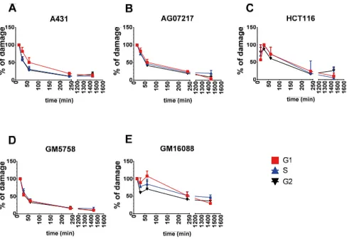
The DSB repair capacity of ɣ-irradiated wild-type A431, GM5758, AG07217, and HCT116 cells and ligase IV hypomorphic GM16088 cells was assessed
using PFGE, foci scoring assays and flow cytometry. Cell cycle phases were determined by flow cytometry using costaining with DAPI. The foci were scored manually in ImageJ, statistical analysis was performed using Bonfer-roni correction when necessary, and p-values less than 0.05 indicated statisti-cal significance.
Results
DNA-PKcs was quickly phosphorylated at position T2609 after irradiation, reaching its maximum signal intensity within 15 – 30 min of irradiation (Fig-ure 5). After reaching its maximum signal intensity, DNA-PKcs was rapidly dephosphorylated, and a notable signal reduction was observed within one hour postirradiation in all wild-type cells. The ligase IV hypomorphic cells displayed reduced dephosphorylation of DNA-PKcs. Unexpectedly, the dephosphorylation kinetics of DNA-PKcs were similar in all cell cycle stages. Next, the levels of DNA-PKcs dephosphorylated at T2609 in the G1 phase of the cell cycle were compared to the DSB repair kinetics obtained from the PFGE assay (Figure 6). DNA-PKcs dephosphorylation at T2609 followed
Figure 5. Kinetics of DNA-PKcs phosphorylation and dephosphorylation at different cell cycle stages in wild-type cells (A) A431, (B) AG07217, (C) HCT116, (D) GM5758 and Ligase IV hypomorphic cells (E) GM16088 after irradiation with 10 Gy of gamma radiation. Data from 3 – 8 independent experiments are shown and error bars repre-sent SEM.
similar kinetics as the DSB repair kinetics observed by the PFGE assay, alt-hough the repair kinetics determined with flow cytometry were slower than those determined by PFGE.
Discussion
The levels of DNA-PKcs phosphorylated at T2609 were able to discriminate between DSB repair-compromised and normal cells, clearly showing that phosphorylated DNA-PKcs T2609 correlates with the DSB content in cells. The phosphorylation of H2AX is a widely used DSB marker; however, H2AX phosphorylation does not reflect DSB repair within the first hour of repair. Thus, the use of DNA-PKcs phosphorylated at T2609 as a DSB marker at early times during repair has clear advantages over the use of ɣH2AX.
Furthermore, DNA-PKcs is phosphorylated at T2609 in the M phase, which is unrelated to DNA damage. It is worth noting that the signal intensity of T2609 phosphorylation in the M phase is low and does not interfere with the analysis. Additionally, DNA-PKcs phosphorylation levels in each cell cy-cle phase should be separated when DSB repair is assessed at time points longer than 4 hours, because the phosphorylation of DNA-PKcs varies over the cell cycle.
Figure 6. Comparison of the DNA-PKcs phosphorylation levels and DSB amount in (A) A431, (B) AG07217, (C) HCT116, (D) GM5758 and (E) GM16088 cells. Data from 3 – 8 independent experiments are shown and error bars represent SEM. Dotted line represents the repair in G1 cell cycle stage and is transferred from figure 5.
DNA-PKcs is the key kinase regulating DSB repair in cells in the G1 and G2 phases. Unexpectedly, we observed similar kinetics for the dephosphory-lation of residue T2609 in the S and the G1/G2 phases. Previously, it was observed that DNA-PKcs interacted with BRCA1 in the S phase, which blocked its phosphorylation at S2056, while the phosphorylation of T2609 was not affected (103). As of now, the role of DNA-PKcs phosphorylation and its involvement in DSB repair in the S phase are not well understood; therefore, DSB repair scoring with phosphorylated DNA-PKcs in the S phase should be used with caution.
Paper II
Suppression of DNA-dependent protein kinase sensitizes cells to
radiation without affecting DSB repair
Aim and background
Dysfunction in normal DNA repair processes is an important factor driving cancer progression. Changes in DNA repair protein expression influence can-cer aggressiveness and resistance to treatment. The high expression of DNA-PKcs, a major PIKK involved in NHEJ, leads to reduced overall survival and poor prognosis (165, 166), while the downregulation of DNA-PKcs expres-sion has been suggested to increase the risk of cancer development (167). In addition to its functions in DSB repair, DNA-PKcs is an important player that orchestrates events in mitosis.
The aim of this study was to determine whether the reduced survival of irradiated cells with reduced levels of DNA-PKcs is related to defective DNA repair or mediated by the alternative functions of DNA-PKcs.
Methods
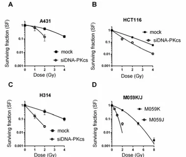
DNA-PKcs knockdown in the A431, HCT116, and H314 cancerous cell lines and the GM5758 normal cell line was achieved using transfection of small siRNA fragments. To study survival and DSB repair, wild-type DNA-PKcs-expressing M059K cells and DNA-PKcs-deficient M059J cells were used. The relative protein content in the cells was determined using western blot. The survival of cells was assessed using a clonogenic assay. The cells were synchronized with nocodazole, and mitotic cells were detected using phos-phorylated H3 as a marker. DSB repair was scored using a foci scoring assay and PFGE. In all experiments, cells were irradiated with gamma radiation.
Results
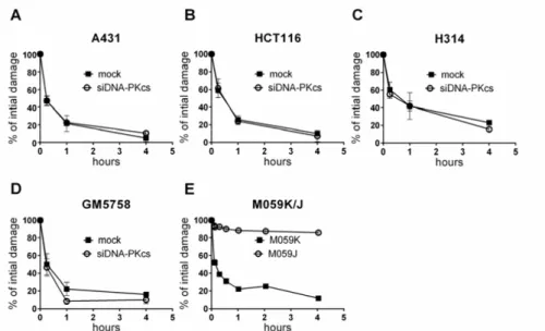
After transfection, DNA-PKcs levels in the cells were reduced to 5 – 20% of the normal levels. The knockdown of DNA-PKcs in combination with radia-tion exposure resulted in a significant reducradia-tion in cell survival, which was comparable to the cell survival observed in cells lacking DNA-PKcs (Figure 7). To further investigate whether the reduction in DNA-PKcs affected DSB repair, PFGE was performed (Figure 8). In all cell lines, DNA-PKcs knock-down did not affect the DSB repair kinetics. In contrast, DNA-PKcs-deficient
cells showed reduced DSB repair, indicating that DSB repair is differentially affected when DNA-PKcs is knocked down or not expressed. The PFGE ex-periments did not indicate any DSB repair defect in mock-treated cells and in cells with reduced DNA-PKcs levels (Figure 8). Results from the PFGE ex-periments were consistent with those from the foci scoring assay, in which DNA-PKcs phosphorylated at T2609 and S2056 as well as 53BP1 and the ɣH2AX foci count were not significantly different at 1, 4 and 24 hours postir-radiation in mock-treated and DNA-PKcs-depleted cells.
Figure 7. Knockdown of DNA-PKcs results to similar sensitivity to ionizing radiation as knockout of DNA-PKcs. Clonogenic survival of mock treated and DNA-PKcs knockdown (A) A431, (B) HCT116, (C) H314 cells and (D) DNA-PKcs deficient M059J and respective wild-type cells M059K. Colonies with more than 50 colonies were scored 10 – 15 days after irradiation. Error bars represent SD from at least 3 independent experiments.
Then, we examined how DNA-PKcs depletion affects cell cycle progression. Cells with low levels of DNA-PKcs exhibited an increased amount of phos-phorylated H3, which marks cell accumulation at M phase (Figure 9 A). The Figure 9. Suppression of DNA-PKcs leads to accumulation of cells in mitosis. (A) A431 cells were irradiated with 5 Gy and after 24 hours cells with positive phosphor-ylated H3 marker were scored. Error bars represent SD from 2 independent experi-ments where >250 cells were scored each time. (B) A431 cells were treated with no-codazole for 6 hours, irradiated, incubated for 18 hours without nono-codazole and then cells positive for phosphorylated H3 were scored. Error bars represent SD of two independent experiments where >400 cells were scored in each experiment.
Figure 8. Reduction of DNA-PKcs does not change DSB repair kinetics. Comparison of DSB repair of DNA-PKcs siRNA treated (A) A431, (B) HCT116, (C) H314, (D) GM5758 cells and DNA-PKcs deficient (E) M059J cells. The DNA damage is normal-ized to the t=0. Error bars represent SD from 3 independent experiments.
irradiation of synchronized cells in the G2/M phase further increased the ac-cumulation of cells in M phase (Figure 9 B).
Discussion
The reduction in DNA-PKcs levels in cells had no effect on DSB repair even when the cells were irradiated with high doses of ionizing radiation, suggest-ing that only a few molecules of DNA-PKcs present in the nucleus are enough to repair the lesions. However, the depletion of DNA-PKcs had a prominent effect on cell survival, which is consistent with the notion that low levels of DNA-PKcs expression in cancers correlate with the overall increased survival of patients (142). The increased accumulation of DNA-PKcs knockdown cells in mitosis indicated that DNA-PKcs is involved in processes important for cell survival independent of its function in DSB repair. This was further supported by the fact that interference with normal DNA-PKcs functioning resulted in abnormal cell division and reduced the metastatic capacity of tumors (105,
165). Taken together, these results suggest that DNA-PKcs has an important
role beyond its function in DSB repair. Identification of the alternative roles of DNA-PKcs in cell survival regulation and cancer progression could poten-tially identify new targets and strategies for drug development and cancer treatment and therefore should be further explored.
Paper III
The α-emitter Ra-223 induces clustered DNA damage and
significantly reduces cell survival
Aim and background
The most commonly diagnosed cancer in men worldwide is prostate cancer. The treatment of prostate cancer involves surgery, radiation therapy and an-drogen deprivation therapy (ADT). ADT is usually prescribed when the dis-ease has reached a more advanced stage and is accompanied by metastases. The major target of ADT is androgen receptor (AR), which is often mutated, amplified or upregulated. ADT only delays the progression of prostate cancer, and given enough time, castration-resistant prostate cancer emerges. Prostate cancers in late stages are incurable as of now, and metastases originating from prostate cancer can be found in bone, the lungs, the liver and the brain. Bone metastases are painful lesions that significantly affect a patient’s quality of life. To combat the progression of bone metastasis, Ra-223 treatment has been developed. Ra-223 is an α-emitter that accumulates in bone undergoing active remodeling, which also coincides with metastatic sites. Ra-223 treatment has proven to be efficacious in the treatment of bone metastasis in clinics, yet the
characteristics of Ra-223 binding to hydroxyapatite, a mineral found in bones, and the damage produced in cells have not been described. Therefore, the aim of this study was to investigate Ra-223 binding to hydroxyapatite, DNA dam-age, DNA repair and the cellular consequences of Ra-223 exposure.
Methods
In this study, PC3 and DU145 cells, which are ARv7-negative prostate cancer cells, and 22RV1 cells, which are ARV7 positive cells, were used. The ex-pression of ARv7 was validated using western blot. Cell survival was deter-mined using a clonogenic assay in which colonies of more than 50 cells were considered viable. The DNA damage distribution in cells was measured via PFGE or visualized by microscopy using 53BP1 and ɣH2AX as DSB markers. DSB repair was determined using flow cytometry in which the levels of DNA-PKcs phosphorylated at T2609 were scored. The affinity of Ra-223 was esti-mated in a hydroxyapatite-coated Petri dish using LigandTracer technology.
Results
Ra-223 exposure sensitized cells to the same extent independent of ARv7 ex-pression (Figure 10 A). As alpha particles have a limited range in water and tissue, we investigated whether Ra-223 exposure also reduced the growth of 3D cell structures. Indeed, exposure to 100 kBq/ml significantly reduced cell survival, while higher concentrations of 250 and 500 kBq/ml effectively in-hibited the growth of spheroids (Figure 10 B).
Unlike the DSBs induced by X-rays, the alpha particles originating from the Ra-223 decay chain produced a nonrandom distribution of DSBs (Figure 11 A). The majority of the DSBs induced by Ra-223 treatment were repaired Figure 10. Ra-223 treatment reduces cell survival in monolayers and spheroids. (A) Ra-223 reduces cell survival similarly in ARv7 positive cells 22RV1 and ARv7 nega-tive cells PC3 and DU145. (B) The Ra-223 treatment decreases spheroid growth in 22RV1 cells. Error bars represent SEM from 3 independent experiments.
within 4 hours when 6 – 11% of the initial amount of the damage was detected (Figure 11 B).
Binding studies revealed that Ra-223 quickly binds to the hydroxyapatite and saturates the surface within 2 hours (Figure 12 A). The estimated affinity was 19.2 ± 6.5 e-18 (mean ± SD, n=9). The Ra-223-bound hydroxyapatite surface prevented spheroid expansion on the surface and reduced the cell sur-vival >100-fold more than that observed in the unbound Ra-223 treatment (Figure 12 B and C).
Figure 11. 223 induced clustered DNA damage and repair. (A) Exposure to Ra-223 leads to production of non-random DSB distribution. DU-145 cells were exposed to 1MBq/ml Ra-223 for 6 hours or irradiated with X-rays. Error bars represent SEM from 2 independent experiments. (B) The DNA-PKcs phosphorylation levels is dras-tically reduced after exposure to Ra-223. DU-145 and PC3 cells were exposed to 300 kBq/ml Ra-223 for 1 hour allowed to repair for indicated time. The signal intensity is normalized to 15 min time point. The error bars represent SEM from 3 independent experiments.
Figure 12. Ra-223 coated hydroxyapatite surface significantly reduces cell survival. (A) Extravasation of 22RV1 cells are reduced on Ra-223 treated hydroxyapatite sur-face. The 22RV1 spheroids were placed on hydroxyapatite coated dish and let adhere. On the next day, the spheroids were treated with Ra-223 and every two days area of the extravasating cells were measured. The error bars represent SEM from 3 inde-pendent experiments. (B) Cell killing effect of Ra-223 is enhanced >100-fold when Ra-223 is bound to hydroxyapatite. Less than survival values represent experiments with no colonies detected and specified value is included in calculations.
Discussion
ARv7 expression confers resistance to ADT and promotes progression of dis-ease. Here, we show that treatment with Ra-223 reduces cell survival inde-pendent of ARv7 expression. The damage induced by Ra-223 treatment was consistent with the damage pattern produced by high-LET irradiation, indicat-ing that the major contributor to the cell-killindicat-ing effect is the production of DNA damage that is difficult to repair. Although the cells were able to rejoin DSBs produced by Ra-223 exposure, the cell-killing effect potentially arose from misrepaired DSBs. The cell-killing effect associated with Ra-223 treat-ment was observed in monolayers as well as in 3D cell structures and was further amplified when Ra-223 was bound to the hydroxyapatite surface. For the first time, we illustrate that Ra-223 treatment can prevent the progression of bone metastasis independent of the molecular properties of the cells and stop the seeding of new bone metastasis. Ra-223 quickly associated with the bone-like structure, similarly as observed in biodistribution studies (163). Here, we are the first to quantify the affinity of Ra-223 for hydroxyapatite, which was extremely high. The advantage of high affinity compounds is the quick relocalization of compounds from blood to their target. In clinics, this property is particularly important because it reduces the time Ra-223 spends in the bloodstream and therefore the dose to normal tissue.
Paper IV
Removal of heat-sensitive clustered damaged DNA sites
is independent of double-strand break repair
Aim and background
The quantification and understanding of molecular mechanisms behind DSB repair is of the utmost importance because these are the most toxic lesions that can be generated in cells. Additionally, to prompt DSBs, a vast variety of non-DSB clusters can be produced in DNA after ionizing radiation exposure. A non-DSB cluster is defined as a cluster of several lesions within 20 – 30 bp that do not contain DSBs. A large number of in vitro experiments have con-cluded that these non-DSB cluster sites can be translated into DSBs during repair; however, at this stage, it is not clear whether the generation of addi-tional DSBs also occurs in living cells.
A specific type of non-DSB cluster damage site is heat-sensitive cluster sites (HSCSs). At elevated temperatures, these lesions undergo transformation from non-DSB cluster sites to DSBs. The lesions found in the clusters or dur-ing the repair of HSCSs have not been identified as of yet. The release of DSBs
from HSCSs in cells has been controversial; therefore, the aim of this study was to clarify whether HSCSs are converted into DSBs in living cells and to further characterize the HSCS conversion process in vitro.
Methods
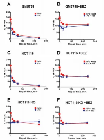
In this study, we examined DSB and HSCS repair in the normal fibroblast cell line GM5758 and DNA-PKcs knockout and the corresponding wild-type cells in the HCT116 colorectal cancer cell line treated with or without the DSB repair inhibitor NVP-BEZ235. DSB repair was assessed using PFGE and 53BP1 and ɣH2AX foci scoring. Additionally, the presence of HSCSs in the pBR322 plasmid was measured by linear gel electrophoresis. Heat-induced morphological changes were visualized in the pBR322 plasmid immobilized on mica using atomic force microscopy operating in liquid tapping mode.
Figure 13. Deficiency in DSB repair does not affect the repair of HSCS. Repair kinet-ics of DSB and HSCS repair in NVP-BEZ235 untreated GM5758, HCT116 and HCT116 DNA-PKcs KO cells (A, C, E) and NVP-BEZ235 treated cells (B, D, F). The relative amount of DSBs are normalized to the DSB amount at t = 0. Error bars rep-resent SD from 3 – 4 independent experiments.
Results
To investigate whether HSCS are converted into DSBs in human cells, HSCS repair kinetics after irradiation were observed in DSB repair-com-promised cells. Wild-type cells repaired the HSCSs and DSBs within 1 and 4 hours, respectively (Figure 13 A and C). However, a deficiency in DNA-PKcs abolished DSB repair but had no effect on HSCS repair (Figure 13 E). The wild-type cells treated with the DSB repair inhibitor exhibited proficient HSCS repair and compromised DSB repair, similar to the results seen in the DNA-PKcs-deficient cells (Figure 13 B, D and F). Furthermore, no additional, excess DSBs was observed in DSB repair inhibitor-treated or DNA-PKcs-de-ficient cells over a time period of 4 hours, during which all HSCSs were re-moved. The results of the 53BP1 and ɣH2AX foci scoring assay were in agree-ment with the results of PFGE experiagree-ments, in which the NVP-BEZ235-treated cells displayed delayed foci formation and increased foci retention without an effect on the maximum foci count. The inhibition of DSB repair by lowering of the repair temperature delayed HSCS repair without generating excess DSBs.
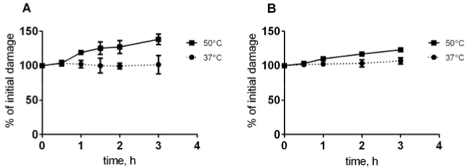
Next, we examined the kinetics of HSCS conversion into DSBs. At physio-logical temperatures, there was a significant conversion of HSCSs into DSBs within 3 hours (Figure 14), while treatment at 50°C resulted in a gradual in-crease in additional DSBs.
The analysis of DNA morphology via atomic force microscopy revealed that prolonged heating resulted in the aggregation of DNA (Figure 15). After closer inspection, some DNA regions were similar to denatured DNA, sug-gesting that melting of DNA sequences can occur at 50°C.
Figure 14. Reduced release of HSCSs into DSBs at physiological temperatures. HCT116 (A) and GM5758 (B) cells were irradiated with 40 Gy followed by DNA ex-traction a Figure 14. Reduced release of HSCSs into DSBs at physiological tempera-tures. HCT116 (A) and GM5758 (B) cells were irradiated with 40 Gy followed by DNA extraction and heating in 37°C and 50°C. The error bars represent SD from two to three independent experiments and all data is normalized to the starting level.
Discussion
Research focused on the translation of clustered DNA damage into DSBs after exposure to ionizing radiation in cells has been challenging. In this study we show that HSCSs are not converted into DSBs in live cells. This conclusion was further strengthened by the fact that no increase in the number of DSBs occurred when DNA was heated at physiological temperatures up to 3 hours, a time period during which HSCSs are repaired in live cells. Furthermore, it has been shown that only the initial number of DSBs correlates with cell sur-vival, while no association between cell survival and HSCS number has been found (168), indicating that HSCSs might not be toxic per se. Surprisingly, the prolonged heating of DNA at 50°C changed the structure of DNA, which was a consequence of DNA melting and denaturation, showing that some ar-tifacts can be produced by the prolonged exposure of DNA to heat. Therefore, we conclude that the heating of DNA should be avoided when DSB analysis is performed using PFGE.
Figure 15. Representative images of (A) control and (B) 200 Gy gamma irradiated pBR322 heated at 50°C.
Concluding remarks
The overall goal of this thesis was to expand the knowledge of clustered DNA damage and repair. More specific efforts were directed towards understanding ionizing radiation-induced DNA damage and how this damage is resolved in cells in terms of DNA-PKcs expression and modifications.
In recent years, particle irradiation and treatments have gained popularity in clinics. It is well established that particle irradiation is able to induce DNA damage that is difficult to repair, which becomes more complex with increas-ing LET. An additional focus of these studies was to investigate the complex-ity and repair of DNA damage induced by Ra-223, a radionuclide used in the treatment of metastatic prostate cancer exhibiting bone metastases.
The majority of the knowledge of cluster DNA damage sites comes from studying the lesions in isolation. However, the consequences of clustered DNA damage site repair in living cells are poorly understood. The last part of this work addressed the repair of specific nonclustered DSB sites and HSCSs, the conversion of HSCSs into DSBs during repair and how the HSCS content in cells affects DSB repair estimates.
The major findings of this thesis were as follows:
DNA-PKcs phosphorylation at T2609 increased with increasing radiation dose.
The DNA-PKcs phosphorylation kinetics at T2609 represent early repair events better than ɣH2AX.
The kinetics of DNA-PKcs dephosphorylation at T2609 should be scored in each cell cycle phase separately if the DNA damage is determined after repair times longer than 4 hours.
The kinetics of DNA-PKcs dephosphorylation were delayed in ligase IV hypomorphic cells.
The reduced amount of DNA-PKcs decreased cell survival after ionizing radiation exposure without significantly affecting DSB repair.
The depletion of DNA-PKcs resulted in cell accumulation in mitosis after irradiation.
The inhibition of DNA-PKcs activity had a different effect than the re-duction of the DNA-PKcs levels.
Ra-223 reduced cell survival independently of ARv7 expression in mon-olayers.
Ra-223 induced DNA damage that was consistent with damage induced by high-LET radiation.
The majority of the DSBs induced via Ra-223 exposure were repaired within 4 hours.
Ra-223 strongly binds to hydroxyapatite.
Cells exposed to Ra-223-bound hydroxyapatite exhibited an enhanced cell-killing effect.
HSCSs were not converted into DSBs in live cells, and the repair of HSCSs was independent of DSB repair.
The conversion of HSCSs in physiological conditions was slower than the repair of HSCSs.
The prolonged heating of DNA at 50°C can alter DNA structure. The studies presented in this thesis illustrate that the phosphorylation of DNA-PKcs at T2609 can be used as a marker of DSB repair during flow cytometric evaluation. It was shown that DNA-PKcs expression levels affect cell survival independent of its role in DSB repair. Furthermore, complex DNA damage associated with Ra-223 exposure is responsible for cell death independent of the molecular properties of the cell. Lastly, the heating of irradiated DNA dur-ing extraction should be avoided because it changes the structure of DNA and produces additional DSBs that do not contribute to the yield of DSBs in living cells.
Future studies
The increased use of hadron therapy in cancer treatment has been shown to reduce normal tissue exposure, thus minimizing the side effects associated with radiation exposure. With increasing LET, it is possible to overcome the challenges related to the reduced efficacy of conventional photon therapy in the hypoxic regions of the tumor. The main cell-killing effects achieved in hadron therapy are due to the increased complexity of the DNA damage. The ability of nonclustered DNA damage sites to convert into DSBs has been well established in isolated DNA; however, in cells, the DNA is wrapped around histones, which could potentially alter how these lesions are repaired or mod-ified. The identification of the repair pathways that resolve non-DSB cluster sites and the clarification of the requirements to collapse these lesions in DSBs could potentially lead to new approaches in cancer treatment. Therefore, fu-ture studies to address these questions in live cells are necessary.
In the first paper, we identified that the phosphorylation of DNA-PKcs co-incides with the number of DSBs found in cells after exposure to low-LET. However, DNA-PKcs dephosphorylated at T2609 displayed similar kinetics in all phases of the cell cycle. Hitherto, it is not clear what functions the DNA-PKcs has in S phase and how representative is the DNA-DNA-PKcs phosphorylation at T2609 of DSB repair in the S phase. Thus, more extensive studies investi-gating the role of DNA-PKcs in DSB repair in S phase cells should be per-formed.
The second paper illustrated the importance of DNA-PKcs in cell survival after ionizing radiation injury. Surprisingly, in cells with low levels of DNA-PKcs, the kinetics of DSB repair were not affected even after exposure to rel-atively high doses of radiation, which leads to several questions that should be addressed in future studies. First, does the DNA-PKcs level affect the fi-delity of DSB repair? Second, is the reduced survival solely the result of dys-function in mitosis? Third, how exactly does DNA-PKcs promote survival?
In the third paper, we demonstrated that cell survival is not dependent on the molecular characteristics of high-LET-irradiated cells, suggesting that the complexity of DNA damage by high-LET irradiation is responsible for the cell-killing effect. Interestingly, it was noted that the DSB repair was not dras-tically different from the repair of low-LET-irradiated cells, which is in con-trast to the general notion in the scientific literature. Therefore, in further stud-ies, whether the DSB repair kinetics are similar following low-LET and high-LET irradiation should be addressed, and a comprehensive comparison with
PFGE and flow cytometric evaluation should be made. Furthermore, studies should focus on the spatial distribution of the generated DSBs surrounding the core of the tracks to further elaborate the damage produced in chromatin struc-tures. Additional studies should be performed to evaluate Ra-223 treatment in other types of cancers that metastasize to the bone in a clinical setting.
The last paper evaluated the repair of HSCSs after irradiation. Our study indicated that the process is independent of DSB repair and does not contrib-ute towards the generation of excess DSBs. Because the lesions generating HSCSs and the proteins involved in the repair of these lesions are not known, future studies should focus on answering these questions. Understanding these processes could provide possible new targets for small inhibitors that, in com-bination with radiation therapy, could increase the efficacy of cancer treat-ment.