A pilot study: Double-blinded
local injection of
active/non-active agents:
Normal response and importance of
expectations
Olivia Edvinsson
Johanna Ekelund
Supervisor: Maria Pigg
Master Thesis in Odontology (30p)
Malmö University
Programme of dentistry
Faculty of Odontology
2
ABSTRACT
Aim
To investigate the amount of anesthetic effect that can be achieved following injection with active (lidocaine) or non-active (saline) agent and to examine if the participants correctly can identify which injection they received when there is a 50/50 chance that they have received lidocaine or saline.
Materials and Method
20 healthy volunteers were randomized in two groups. One group got injection with active agent and one with non-active agent. The participants were instructed that chances of
receiving active versus non-active agent were equal, 50 %. The participants were exposed to a specific painful stimulus before and after the injection and they had to rate their pain score on an 0-10 NRS-scale. The following day, the participants were asked what agent they thought they had received.
Students T-test was used to calculate the difference in pain intensity between pre- and post-injection in both groups. Fisher's exact test was used to calculate qualitative variables. P<0.05 was considered statistically significant.
Results
There was a statistically significant difference between the active (NRS 2.9) and the non-active (NRS 0.0) group regarding the change in pain intensity rating from pre-injection to post-injection. All participants could correctly identify which injection they received.
Conclusion
No anesthetic effect could be measured after injection with non-active substance in healthy individuals when there was a 50/50 level of uncertainty that the individual would receive the active agent. All individuals could correctly determine whether they received active
3
SAMMANFATTNING
Syfte
Att undersöka hur stor bedövningseffekt som kan uppnås efter injektion med aktiv (lidokain) eller in-aktiv substans (koksaltlösning) samt att undersöka om deltagarna korrekt kan
identifiera vilken injektion de fått, när de vet att det är en 50/50 chans att de har fått lidokain eller koksalt.
Material och metod
20 friska frivilliga deltagare randomiserades in i två grupper. En grupp fick injektion med aktiv substans och en med in-aktiv substans. Deltagarna informerades om att chansen att få aktiv eller in-aktiv substans var lika, 50 %. Deltagarna utsattes för ett specifikt smärtsamt stimulus före och efter injektionen. De graderade sedan sin smärtintensitet på en 0-10 NRS-skala. Följande dag tillfrågades deltagarna vilken substans de trodde de hade fått.
Students T-test användes för att beräkna skillnaden i smärtintensitet före och efter injektion i båda grupperna. Fischer’s exakta test användes för att beräkna kvalitativa data. P <0,05 ansågs vara statistiskt signifikant.
Resultat
Det var en statistiskt signifikant skillnad mellan den aktiva (NRS 2,9) och den in-aktiva (NRS 0,0) gruppen gällande förändringen i smärtintensitet före injektion och efter injektion. Alla deltagare kunde korrekt identifiera vilken injektion de fått.
Slutsats
Ingen bedövningseffekt kunde mätas efter injektion med inaktiv substans hos friska individer när deltagarna visste att det var en 50/50 möjlighet att de skulle få den aktiva substansen. Alla individer kunde korrekt avgöra om de fått injektion med aktivt bedövningsmedel eller in-aktivt koksalt.
4
Table of Contents
ABSTRACT ... 2 SAMMANFATTNING ... 3 INTRODUCTION ... 6 Pain ... 6 Atypical odontalgia ... 6 Etiology ... 7 Epidemiology ... 7 Prognosis ... 8 Clinical management ... 8 Diagnostics ... 8 Aim ... 9 Research question ... 9 Hypothesis ... 10MATERIALS AND METHOD ... 11
Participants ... 11 Inclusion criteria ... 11 Exclusion criteria ... 11 Ethical considerations ... 11 Study design ... 11 Statistical methods ... 13 Quantitative variables ... 13 Qualitative variables ... 13 RESULTS ... 14 Sample characteristics ... 14 Quantitative variables ... 14 Qualitative variables ... 15 DISCUSSION ... 17
Materials and method ... 17
Participants ... 17
Gender and pain experience ... 17
Study design ... 17
Statistical methods ... 18
5
Quantitative variables ... 18
Qualitative variables ... 18
Clinical aspects ... 19
Pain relief and placebo effect ... 19
Future studies ... 20
CONCLUSION ... 21
6
INTRODUCTION
Patients coming to the dental clinic complaining about pain are part of a dentist's everyday life. In most cases, there is a clear pathological cause to the pain condition, such as pulpitis or apical periodontitis. Sometimes, however, it is not that simple and no clear pathological signs are found either in the clinical or radiological examination. In these cases, other pain
etiologies than inflammatory nociceptive pain should be considered. Diagnostic methods that distinguish different pain etiologies are required, since different types of pain conditions are treated in different ways.
Pain
Pain is defined by IASP (International Association for the Study of Pain) as “An unpleasant
sensory and emotional experience associated with actual or potential tissue damage, or described in terms of such damage (1).”
Pain is a subjective experience and cannot be measured by objective methods (2). The pain experience is extremely complex and is not only determined of the degree of nociceptive input but also emotional and cognitive factors. Research has shown that not only sensory, but also emotional and cognitive brain regions are involved in determining the pain experience (3).
One way to divide pain into categories is as follows:
• Nociceptive pain is caused by activation of nociceptors, our pain receptors, and is associated with tissue damage or inflammatory responses, such as pulpitis or apical periodontitis.
• Peripheral neuropathic pain is caused by injury/damage in a peripheral nerve. • Central neuropathic pain is due to damage in the central nerve system.
• Centrally disturbed pain modulation is due to disturbance in the central pain transmission.
• Psychogenic pain arises exclusively from psychological causes.
• Idiopathic pain is pain that cannot be attributed to any of the other categories (2).
Atypical odontalgia
Atypical odontalgia (AO) is a pain condition without clear clinical or radiological signs of pathology. This condition poses a clinical challenge and often leads to treatment of teeth, but this does not result in the expected pain relief (4,5). To date, there are no evidence-based diagnostic criteria available (6,7), and improved diagnostic methods for this condition are required. One diagnostic method used today is double-blinded local injections. AO and the effect of double-blinded injections will be discussed in this paper (4,5).
AO is defined as a subgroup of persistent idiopathic facial pain (PIFP) by International
Headache Society (IHS) and they share the definition “Persistent facial and/or oral pain, with
varying presentations but recurring daily for more than 2 hours per day over more than 3 months, in the absence of clinical neurological deficit.” Furthermore, IHS suggests that ”The term atypical odontalgia has been applied to a continuous pain in one or more teeth or in a tooth socket after extraction, in the absence of any usual dental cause (6).” The term atypical
odontalgia and its definition is debated in the literature, and several other terms with slightly different criteria is used, among these PDAP (persistent dento-alveolar pain disorder) and PTTN (painful post-traumatic trigeminal neuropathy) (8,9).
7
Etiology
The mechanisms behind AO have been frequently discussed the past years, and is not yet fully understood (5). This lack of knowledge is one of the reasons to the difficulties in diagnosing and treating the patients suffering from AO (10). Baad-Hansen has summarized possible etiologies in a review article. The theory that has received most support is that AO is a neuropathic pain condition (5).
Neuropathic pain is defined by IASP as “Pain caused by a lesion or disease of the
somatosensory nervous system(1).” The mechanisms behind neuropathic pain can be of
peripheral, central or of combined origin. Neuropathic pain differs from nociceptive pain since the cause for pain is pathology of the neural tissue itself, and not pathology of the structures that the nerve innervates. The function of nociceptive pain is to work as a warning signal for noxious stimulation, that tissue damage has or is about to occur. This is not the function of neuropathic pain. Neuropathic pain indicates a structural or functional abnormality in the neural tissue. Various events or injuries can cause neuropathic pain, for example
infections, trauma, surgery, tumour and nerve compression (4).
AO is sometimes described as a burning and pressing pain. These characteristics correspond well to deafferentation pain, a type of neuropathic pain caused by severe damage to the peripheral nerve branches. Patients suffering from AO often report that the pain developed in connection with pulpectomies or tooth extraction, which in fact represents deafferentation procedures. According to this, a neuropathic pain condition is the most likely etiology for AO (4,5). Furthermore, animal studies support this hypothesis. These studies have shown neural changes following pulpectomy (11). These changes are usually reversible, but might in some cases cause development of neuropathic pain (12). It is not uncommon for endodontic treatments to cause non-odontogenic pain conditions. This implicates that pain relief cannot be expected with retreatment of painful teeth without clinical or radiographic signs of pathology, since the pain can be of neuropathic origin, caused by the primary endodontic treatment (13).
In spite of the fact that neuropathy is the most likely etiology to AO, for a pain to be diagnosed as neuropathic, the pain onset must have been preceded by damage or disease to the neural system. In some cases of AO, no damage to the neural tissues can be identified. In these cases, an idiopathic etiology is the most correct way to describe the pain condition (5). Psychogenic etiology has also been discussed, seeing that patients with AO often struggles with psychological disorders. However, it is unclear whether the depression is the reason for the pain condition or the pain condition the reason for the depression. Most likely the chronic pain condition is associated with development of depression, and not vice versa (4,5).
Epidemiology
The prevalence of AO is unknown (13). Women are affected to a higher extent than men (5). No studies have described the prevalence on a population level. The existing epidemiological studies have only described the prevalence of persisting pain after different clinical
procedures.
Nixdorf et al. have performed a systematic review to investigate the frequency of
nonodontogenic pain in patients who have undergone root canal treatment. They defined nonodontogenic pain as “dentoalveolar pain present for 6 months or more after endodontic
8
treatment without evidence of dental pathology.” The study concluded that 3.4 % of the
patients that had experienced root canal treatment had persistent nonodontogenic pain (13).
Prognosis
An observational study performed by Pigg et al. has shown that the treatment results vary. A few of the patients with AO achieves total pain relief, but for most patients AO is a chronic pain condition (14).
Clinical management
AO poses a clinical difficulty as it is difficult to distinguish it from odontogenic tooth ache, since the symptoms are very similar (4,5). Percussion tenderness, palpation tenderness and tenderness to chewing and cusp loading can be provoked in some patients with AO at the painful tooth (15).
Differential diagnoses to AO is odontogenic pain (pulpitis, periapical periodontitis, cracked tooth syndrome etc), sinusitis, myofascial pain of masticatory muscles,
arthralgia/osteoarthritis, trigeminal neuralgia, migraine and other headache conditions. To avoid misdiagnosis and unnecessary treatment, a thorough examination is necessary. A detailed anamnesis, clinical and radiological examination of the teeth and surrounding tissues and TMD examination should be performed to be able to exclude various differential
diagnoses (5).
• Anamnesis: A detailed anamnesis can facilitate exclusion of the differential diagnoses sinusitis, trigeminal neuralgia and various headache conditions, since the
symptomatology differs from AO.
• Clinical examination: A clinical examination should be performed to exclude
odontogenic painby pulp vitality testing and looking for caries, probing depth, cracks and other pathology in the area (5).
• Radiological examination: CBCT is better than intraoral X-ray to detect apical destruction and can thus be used as a complement to intraoral X-ray to improve the identification of patients without apical bone destruction. This may facilitate the distinction between AO and symptomatic apical periodontitis. If no apical destruction is apparent on either conventional X-ray or CBCT the suspicions of AO increases (16). MRI can also be used to distinguish AO and odontogenic pain, as it can provide information about the inflammatory activity around the teeth in the pain region. One study has shown that in most patients with AO, MRI does not reveal any changes, which indicates that there is no ongoing inflammation (17).
• TMD-examination: A TMD-examination should be performed to exclude myofascial pain or arthralgia with referred pain (5).
Diagnostics
To date, there are no evidence-based diagnostic criteria or treatment guidelines for AO available (6,7). The condition is diagnosed by exclusion of pathologic signs after clinical and radiological examination (10). However, there are some diagnostic methods that can be useful in diagnosing neuropathic pain. Somatosensory testing is one of them, since somatosensory changes are an essential part of neuropathic pain. Quantitative somatosensory testing (QST) is a method that can detect somatosensory changes and can therefore be useful in the diagnosis of neuropathic pain (12,18,19). A full QST includes 13 tests according to a protocol
developed and recommended by the DFNS (German Research Network on Neuropathic Pain). The method examines sensitivity to mechanical and thermal stimuli and the results are
9
compared to a reference interval of data from healthy individuals (19). The protocol has been tested intraorally with good reliability in healthy subjects without pain and in patients with AO (20,21). Studies using QST have shown abnormal sensory function of the pain site in patients with AO when compared to healthy reference standards (12,21). In the present study, a QST-based stimulus was used in a modified manner, to investigate the normal response of local anaesthesia with lidocaine and placebo.
Another diagnostic method that can be helpful in exclusion of local inflammatory causes to the pain condition is anaesthetic blocks (5,12). The mechanism behind neuropathic pain conditions are complex, in many cases both peripheral and central mechanisms may contribute. Anaesthetic blocks can aid the clinician in getting a better understanding of the neural mechanisms in the specific case. The anaesthetic block can give rise to a wide range of different responses: Complete, partial or no pain relief. The amount of pain relief reflects the contribution of peripheral and central input to the AO pain.
A study by List et al. investigated the pain-relieving effect of local anaesthesia in patients with AO. This was a double-blind controlled cross-over study comparing the effect of lidocaine and normal saline (placebo). It showed significant pain-relieving effects from both lidocaine and placebo injections, but the effect was significantly greater for lidocaine. The pain relief following lidocaine was not total. This indicates that AO pain not only depends on peripheral but also on central mechanisms (12).
Why did injection with saline give pain relief? Patients have an expectation that medical treatment will have a positive impact on the medical state, treatment outcome is therefore not only the result of specific drug effects but also contains an effect of expectations (22). The placebo effect is the non-specific, psychological or psychophysiological therapeutic effect achieved by treatment with placebo (25). The neurobiological mechanisms behind the non-specific placebo effect are not yet fully understood, but it is a complex procedure that
involves neurotransmitters and activation of areas in the brain (23). Placebo is defined as any form of therapy that is prescribed to treat symptoms or illness, which in fact have no specific effect (24,25). All medical interventions contain placebo effect to some degree. The type of administration is essential for the degree of placebo, for example, injections with placebo give greater pain relief than tablets with placebo (22). The influence of placebo on the result of a diagnostic procedure such as the above-mentioned double-blind diagnostic anaesthetic procedure has not been investigated previously.
Aim
The study was a pilot study performed to provide a basis for sample calculation and to
evaluate the method for a future study. The aim was (i) to investigate the amount of anesthetic effect that can be achieved, measured in self-reported pain intensity to a standardized stimulus following injection with active agent (lidocaine) or non-active agent (placebo). The purpose was also (ii) to investigate whether the participants can correctly identify if they got injection with anesthesia or placebo when they know that there is a 50/50 chance that they have received lidocaine or saline.
Research question
1. A: How much anesthetic effect can be achieved, measured by self-reported pain intensity to a standardized stimulus after injection with non-active substance when there is a 50/50 level of uncertainty that the individual will receive the active agent in a double-blinded fashion?
10
B: How much anesthetic effect can be achieved, measured by self-reported pain intensity to a standardized stimulus after injection with active substance when there is a 50/50 level of uncertainty that the individual will receive the active agent in a double-blinded fashion?
2. What is the distribution of participants' perception of what agent they have received when there is a 50/50 uncertainty that the individual has received an active agent in a double-blinded fashion?
Possible outcomes:
a. Believes they received active agent and this is correct b. Believes they received active agent but this is not correct c. Believes they received non-active agent and this is correct d. Believes they received non-active agent but this is not correct
Hypothesis
Both lidocaine and placebo injections will result in an anaesthetic effect, but the effect is more pronounced for lidocaine. Everyone who receives lidocaine will correctly assume that they have received active agent. Since all treatments have a certain degree of placebo (22), a minor proportion of the individuals who receive saline will incorrectly believe they have received lidocaine.
11
MATERIALS AND METHOD
Participants
20 healthy individuals, 14 women and 6 men, were recruited from Malmö University. The age range were 18-56 years, mean age 38.8, standard deviation 11.3. This was a convenience sample; the participants were personally asked by the study examiners (OE and JE) for their willingness to participate. No consideration was given to either age or sex.
Inclusion criteria
The inclusion criteria were: Students or staff from Malmö University, good health, without dental pain.
Exclusion criteria
The exclusion criteria were: Dental pain, chronic pain in the orofacial region (>3 months), pregnancy, allergy to local anaesthesia, medication with antidepressant or antiepileptic and professional knowledge in the area of anaesthesia. (Dental staff, dental students and dental hygienist students above semester 2 were excluded)
To ensure that all criteria for study participation were fulfilled, the participants completed a questionnaire concerning general and orofacial health.
In addition, the participants were asked about chronic pain in other regions than the orofacial region and TMD-symptoms (<3 months).The individuals were screened for TMD-pain by the following anamnestic questions:
1. Does it hurt in your temple, face, jaw joints or jaws once a week or more often? 2. Does it hurt when you chew or open your mouth once a week or more often? This was done to characterize the sample and was not used as an exclusion criteria.
Ethical considerations
A local ethical application was approved by the local ethics review board at Malmö University. All participants signed informed consent forms.
Study design
The study was an experimental, double-blinded, randomized study comparing the effect on an active local anaesthetic agent (active group) to a non-active agent (non-active group). The outcome measures included both qualitative and quantitative variables.
20 volunteers were randomly divided in two groups. https://www.randomizer.org/ was used to perform the randomization. The active group (N=10) got an injection with 2 ml lidocaine solution (Xylocain, AstraZeneca, Södertälje Sverige) (10 mg/ml) without vasoconstrictor. The non-active group (N=10) got an injection with 2 ml normal saline solution (9 mg/ml NaCl) (placebo).
Examiner 1 (JE) informed and instructed the participants about the process, that there was a 50/50 chance to get injection of active or non-active agent. The participants were instructed to only answer the examiners' questions and not give any additional information about the sensory experience. Examiner 1 then applied a specific stimulus using a 512 mN mechanical stimulator (the Pin-Prick TM, manufactured by the Johannes Gutenberg University of Mainz, Mainz, Germany), about 2 mm from the gingival margin at the attached gingiva at the midline
12
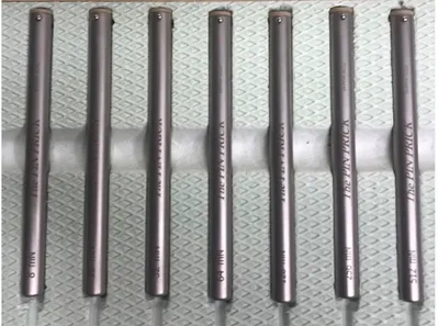
of the first upper right premolar. A Pin-Prick stimulator is a calibrated instrument with a weighted needle that exerts a specific force, see figure 1 and 2. The stimulator was held in place for 2 seconds and only one time. The participant then rated their pain intensity score on a 0-10 numeric rating scale, NRS with the endpoints 0=No pain and 10= The worst pain imaginable. The participant was also asked to state whether the stimulus was perceived as sharp or dull.
The method using Pin-Prick to create pain stimulus is technically sensitive. To ascertain that the procedure was performed in a correct way and to ensure reproducibility examiner 1 was trained by a person with experience in intraoral QST and practiced before the examinations. Examiner 1 practiced on examiner 2 who confirmed that the stimulus was experienced the same each time.
Figure 1: The Pin-Prick stimulators with different forces. The one to the right, 512 mN, was used in this study.
Figure 2: The Pin-Prick 512 mN were placed for 2 second at the attached gingiva at the first upper right premolar.
In the next step, examiner 2 (OE) injected either active agent or non-active agent. The injection was placed in the mucogingival fold adjacent to the first upper right premolar. The
13
injections were selected randomly, and neither the examiners or the participants knew if the injection contained active or non-active agent.
Five minutes passed, and then the participant met examiner 1 again and was exposed to the same stimulus with Pin-Prick 512 mN and again rated the pain intensity score on a 0-10 NRS scale. The participant also stated whether the stimulus was perceived as sharp or dull.
The following day, the participants were contacted to answer if they thought they had received active or non-active agent.
The procedure was double-blinded and a third person helped with the blinding process and prepared the injections. Examiner 1 and 2 did not communicate during the trial to minimize bias. A sealed coded number list was used during the trial to ensure the blinding. The seal was broken when all data was collected from all 20 participants.
Statistical methods
Descriptive statistics were used to describe the sample characteristics.
Quantitative variables
Mean values and standard deviation were calculated for quantitative, continuous variables.
Paired sample T-test was used to calculate differences in pain intensity between pre- and post- injection measurements. Independent sampled T-test was used to calculate differences
between the groups regarding pain intensity before and after injection, respectively. P<0.05 was considered statistically significant.
Qualitative variables
Fischer’s exact test was used to compare qualitative, categorical data. Differences between the groups regarding chronic pain, TMD pain and intake of analgesics were compared to ensure that the two test groups were comparable. Fischer's exact test was also used for comparison between the groups on whether the Pin-Prick stimuli was felt as sharp or dull. P<0.05 was considered statistically significant.
14
RESULTS
Sample characteristics
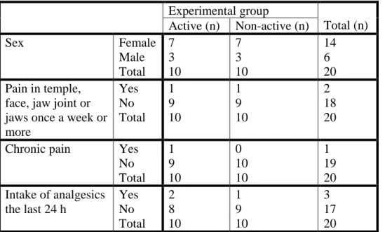
All subjects completed the study. All the participants considered their general health as good. Table 1 shows the distribution of the individuals in the two groups regarding sex, TMD pain, chronic pain and intake of analgesics the last 24 h. Fischer’s exact test was used to calculate the comparability of the two groups. As seen, there were no statistically significant
differences between the group that received active agent and the group that received non-active agent for any of the background variables.
Experimental group Total (n) Active (n) Non-active (n) Sex Female Male Total 7 3 10 7 3 10 14 6 20 Pain in temple,
face, jaw joint or jaws once a week or more Yes No Total 1 9 10 1 9 10 2 18 20 Chronic pain Yes
No Total 1 9 10 0 10 10 1 19 20 Intake of analgesics the last 24 h Yes No Total 2 8 10 1 9 10 3 17 20
Table 1: Sample characteristics. There were no significant differences between groups for any variable (all P=1.0).
Quantitative variables
The pre-injection pain intensity to Pin-Prick stimulus ranged from NRS 0 to NRS 7, on average NRS 3.1 (SD 1.9). There was a significant difference (P<0.0001) in pain intensity rating between pre- and post- injections. The mean post-injection pain intensity to Pin-Prick stimulus was NRS 1.6 (SD 2.1). The mean pain relief was 1.5 (SD 2.3).
Table 2 shows the difference between the active group and the non-active group in pain intensity to Pin-Prick stimulus before and after injection.
15
Table 2: Difference between the active group and the non-active group in pain intensity to Pin-Prick stimulus, expressed in NRS 0-10, before and after injection. The green staples represent the pain intensity in NRS with the blue line representing each respective groups’ standard deviation. There was no difference between the groups regarding the pain intensity to Pin-Prick before injection (P=0.9). There was a significant difference (P<0.0001) between groups regarding pain intensity to Pin-Prick stimulus after injection. In 9 out of 10 cases the pain score on Pin-Prick stimulus was rated to NRS 0 after injection with lidocaine. The remaining case was rated to NRS 1.
There was a statistically significant difference (P<0.0001) between the groups regarding the change in pain intensity rating from pre- to post-injection. The change in pain intensity rating between pre- and post- injection was NRS 2.9 (SD 2.3) for the active group and NRS 0.0 (SD 1.2) for the non-active group. This means that the group that received injection with active substance received total anaesthetic effect, and the group that received injection with non-active substance didn´t receive anaesthetic effect at all.
Qualitative variables
Table 3 shows the overall differences between the groups in how rating of stimulus quality changed from pre- to post- injection.
There was a no difference between groups in the proportion of subjects that rated the stimulus quality as going from sharp to dull, see Table 4.
All the 20 participants could correctly identify whether they received injection with active or non-active agent. 3 3,1 0,1 3,1 -1 0 1 2 3 4 5 6 NR S 1 -10
Pain intensity to Pin-Prick stimulus +/- 1 S. D.
Active Non-active Active Non-active
16 Experimental group
Post-injection rating of pain quality
Total
dull sharp
Active Pre-injection rating of pain quality
dull 5 1 6
sharp 4 0 4
Total 9 1 10
Non-active Pre-injection rating of pain quality
dull 3 4 7
sharp 1 2 3
Total 4 6 10
Total Pre-injection rating of pain quality
dull 8 5 13
sharp 5 2 7
Total 13 7 20
Table 3: Differences between the active group and the non-active group in how rating of stimulus quality changed from pre- to post injection. There were no significant differences between groups for any variable (P=1.0).
Experimental group Total Active Non-active Pain quality change from sharp to dull Yes No Total 4 6 10 1 9 10 5 15 20
Table 4: Difference between the active group and the non-active group in the proportion for subjects that rated the quality as going from sharp to dull. There were no significant
17
DISCUSSION
Materials and method Participants
This study aimed to evaluate the response to local anaesthesia in individuals free from chronic orofacial pains, the inclusion and exclusion criteria were therefore determined to minimize factors that may contribute to the pain experience. The characteristics of the participants that are described in the result (gender, presence of orofacial pain, presence of chronic pain and intake of analgesics) are factors that might affect the pain experience to some degree. Gender can affect the pain experience since women generally have lower pain threshold than men (26). Chronic pain conditions can affect the pain experience since the perception threshold in these individuals often is lowered due to central sensitisation (2).Since there were no
significant differences in characteristics between the active group and the non-active group, we are reasonably certain that the groups are comparable, and that these characteristics had minimal impact on the result.
One thing that might have affected the result is if the participants had earlier experiences of local anesthesia or not. An individual who has received local anesthesia before probably knows what sensory effect (numbness) can be expected and after having received the experimental injection directly forms an opinion on whether pain perception will change or not. By including previous experience of dental anaesthesia as an exclusion criterion in this study, the participants expectations would not have affected the result. But from another point of view, patients with AO have probably experienced local anaesthesia multiple times before the diagnostic double-blinded blockades, so excluding subjects with previous experience of dental anaesthesia would not reflect the reality.
The participants for this study were recruited at convenience. The participants were overall younger than the population in which a diagnostic anaesthetic procedure is most likely to be applied. This might be a possible source of error since it may not be representative for the general population and therefore it lowers the external validity.
Gender and pain experience
Women report pain more often, have a lower pain threshold and a lower pain tolerance than men. Possible reasons for this have been researched and it has been concluded that biological, psychological and social factors all contribute to the observed difference between the genders (26). This study included more women than men. For the gender difference to not affect the outcome, a 50/50 gender distribution would be optimal. However, there was an even gender distribution between the active and the non-active group. Furthermore, this study evaluated differences in pain rating not between different individuals, but in the same individual before and after the injection. Considering this the gender distribution is unlikely to have affected the outcome.
Study design
This study aimed to evaluate how much anaesthetic effect that can be achieved after injection with non-active/active substance when there is a 50/50 level of uncertainty that the individual will receive the active agent. The study was designed with two parallel groups to minimize the risk to affect the participants expectations. It was not designed as a cross over, since the first injection might affect the expectation of the second injection.
18
There is no standardized way to measure the level of anaesthetic effect. In this study the anaesthetic effect was measured as a difference in self-reported pain intensity to a specific
Pin-Prick stimulus, before and after the injection. Pain intensity is a highly individual
experience and is therefore difficult to grade by objective measures. One way to measure pain intensity is by numeric rating scale (NRS). This scale is a recognized method and is often recommended for measurement of pain intensity (27). Therefore, NRS was used in this study. The method using Pin-Prick stimulus is tested intraorally with good reliability (20). Because the needle is weighted, the stimulus is reproducible and is performed with the same force every time. The method is technically sensitive and requires proper handling as the wrong angulation of the instrument may change the force applied. To minimize this source of error, the examiner practiced the method before the examinations.
Statistical methods
Which analytical methods that can be used depends on the variables scale level. For example, mean values can only be calculated of interval scales. It is not optimal to make a statistical calculation of the values from an ordinal scale, which NRS can be classified as. Ordinal scale values can be ranked but it cannot be assumed that the difference between 1 and 2 is equal to the difference between 2 and 3. However, it is not unusual to make such statistical analyzes on ordinal scales even if it might not be statistically correct (28).
Results
Quantitative variables
In the hypothesis it was expected that both the non-active group and the active group would perceive an anesthetic effect. The group that received injection with non-active substance didn´t achieve any anesthetic effect and this part of the hypothesis can thus be falsified. In the hypothesis the effect was thought to be more pronounced for the group that received active agent. Since all the participants that received injection with active substance did achieve pronounced anesthetic effect, the hypothesis corresponds well to the result and can be verified.
This result contradicts the result of an earlier performed study, that could demonstrate a placebo effect after injection with non-active agent in patients suffering from AO (12).Our sample consisted only of healthy individuals, so our result only indicates that healthy
individuals don´t get any anesthetic effect of saline when there is a 50/50 level of uncertainty that the individual will receive the active agent. It is important to understand the difference between spontaneous chronic pain and provoked nociceptive pain. In this study we used Pin-Prick to provoke pain, and this activates nociceptive Aδ fibers (29). In chronic pain
conditions, such as AO, other pain mechanisms are involved. This could explain the difference in results between this study and the study made by List et al(12). The design of the study and the instructions to the participants might also have contributed to the
differences.
Qualitative variables
Since it is difficult to rate pain on a numerical scale, stimulus quality was also used as a measure of outcome. This aimed to get an additional way to measure the anesthetic effect. According to a forced-choice procedure, the participants had to say whether they experienced the stimuli as sharp or dull, both before and after the injection. The change from experienced stimulus quality from sharp before injection to dull after injection could be considered a sign of perceived anesthesia. There was no significant difference between the groups in how the
19
participants experienced the stimulus of going from sharp to dull. On the other hand, in the active group only four participants experienced the stimulus quality as sharp before injection and all of these four experienced that the stimulus quality was changed to dull after injection. In the non-active group three individuals experienced the stimulus as sharp prior to injection and only one of them considered the stimulus quality asdull after injection. In this aspect, there was a difference between the groups which was not statistically analyzed. Therefore, a change in stimulus quality from sharp to dull can be considered a sign of anesthetic effect. However, since so many perceived the pre-injection stimulus as dull, this measure does not appear to be an optimal way to measure the anesthetic effect.
All 20 participants could correctly identify if they got injection with active or non-active agent. In our hypothesis, we expected that all the participants receiving injection with active agent would correctly assume that they had received lidocaine. This corresponds well to the result. We also expected that some of the participants receiving injection with non-active agent would incorrectly believe that they had received lidocaine. This part of the hypothesis was falsified, and as a result, we were not able to evaluate how the participants beliefs about the received agent was associated to the reduction in pain elicited by a Pin-Prick stimulus.
Clinical aspects
Double-blinded blocks are used in clinical dental practice as part of the investigation when AO is suspected. Two injections are injected on two different occasions. One injection contains an active anaesthetic agent and the other injection contains non-active normal saline solution. These are usually injected in a double-blinded procedure, which means that neither the clinician or the patient knows which injection is administered at what occasion. The patient evaluates the pain before injection and at fixed intervals after injection, for example 30 min, 1 h, 2h, 4 h, 6 h, 12 h post-injection.
There is no clear script when using double blind anaesthetic blocks in the practice on patients suspected of having AO. The instructions can be given to the patient in different ways. One way to inform the patient is that one out of two different anesthetic agents is going to be injected. Another way to inform the patient is to say that you will administer one out of two different injections where one contains saline and the other contains anesthetics. The first type of instruction could be considered an ethical problem, since care should be given in agreement with the patient, and this procedure includes deception of the patient.From an ethical
perspective deception in investigation or treatment of patients should be avoided unless there is a scientific support for the procedure. Therefore, it is important to examine these patient-related procedures scientifically.
In this study, the participants were told that they would receive one injection that may contain either non-active saline or active anesthetics. The participants initially knew that there was a 50/50 chance of getting injection either with active or non-active substance. If the participant instead would have been told that they would receive one out of two different anesthetic agents, there might have been a placebo effect. This could be a subject for future studies.
Pain relief and placebo effect
The placebo effect is dependent on the expectations. Verbal instructions that pain relief will occur by a specific treatment may be enough to create positive expectations and consequently decrease the actual pain experience (30). Therefore, it can be assumed that the instructions given to the patient when using double-blind blocks in investigating patients with suspected AO play a major part in the response to the injections.
20
Future studies
The study was a pilot study performed to provide a basis for sample calculation and to evaluate the method for a future study. Because no placebo effect could be measured in the group that received non-active agent, the method should be improved in a new pilot study before the study is done with a bigger sample. As mentioned earlier, one suggestion for a future study is to present the instructions to the participants in another way. One example is by telling the participants that they will receive one out of two different anaesthetic agents. A possible research question could be:How much anesthetic effect can be achieved, measured by self-reported pain intensity to a standardized stimulus after injection with non-active substance when the individuals believe they will receive active anesthetic agent or after injection with active substance when the individuals believe they will receive non-active agent respectively?
The participants in this study could correctly determine whether they received injection with lidocaine or saline. This indicates that saline might not be an optimal control in double-blind blockades. Other alternatives to saline should be considered for future studies, for example lidocaine in low concentration.
Another improvement of the method could be to make repeated measures to improve
reliability, alternatively to use a stimulus that induces a stronger pain response, since the Pin-Prick 512 mN stimulus generally gave a relatively low pain intensity. Some of the individuals graded the pain intensity to the stimulus before the injection very low. No side effects such as bleeding from the stimulation site were observed, however using a stronger stimulus was avoided because of the risk of tissue damage.
Since this study was a pilot study, one of the goals was to evaluate the method. We aimed to assess if this way to measure the amount of anesthetic effect was suitable. In this study, a change from experienced stimulus quality from sharp before injection to dull after injection was considered a sign of perceived anesthesia. Since this method proved to be inadequate, a future study should aim to develop a good way measure the degree of anesthetic effect.
21
CONCLUSION
No anesthetic effect can be achieved, measured by self-reported pain intensity to a
standardized stimulus after injection with non-active substance, when there is a 50/50 level of uncertainty that the individual will receive the active agent in a double-blinded fashion. Total anesthetic effect can be achieved, measured by self-reported pain intensity to a standardized stimulus after injection with active substance when there is a 50/50 level of uncertainty that the individual will receive the active agent in a double-blinded fashion. Individuals can correctly determine whether active anesthesia or non-active saline have been injected when there is a 50/50 level of uncertainty that the individual will receive the active agent in a double-blinded fashion.
22
REFERENCES
(1) IASP. Pain terms. 2012; Available at: http://www.iasp-pain.org/Taxonomy#Pain. Accessed 2016-11-02, 2016.
(2) Fredenberg S, Vinge E, Karling M. Smärta och smärtbehandling. In: Ramström H, editor. Läkemedelsboken 2011-2012. 18th ed. Stockholm: Läkemedelsverket; 2011. p. 877-904. (3) Peters ML. Emotional and Cognitive Influences on Pain Experience. Mod Trends Pharmacopsychiatri 2015;30:138-152.
(4) Okeson JP. Bell´s orofacial pains: The clinical management of orofacial pain. 6th ed. Chicago: Quintessence books; 2005.
(5) Baad-Hansen L. Atypical odontalgia - pathophysiology and clinical management. J Oral Rehabil 2008 Jan;35(1):1-11.
(6) Headache Classification Committee of the International Headache Society (IHS). The International Classification of Headache Disorders, 3rd edition (beta version). Cephalalgia 2013 Jul;33(9):629-808.
(7) Abiko Y, Matsuoka H, Chiba I, Toyofuku A. Current evidence on atypical odontalgia: diagnosis and clinical management. Int J Dent 2012;2012:518548.
(8) Fact sheets in English revised in 2016 for the 2013-2014 global year against orofacial pain, IASP. Persistent Idiopathic Facial Pain (Previously “Atypical Facial Pain”). 2016; Available at:
https://s3.amazonaws.com/rdcms-iasp/files/production/public/Content/ContentFolders/GlobalYearAgainstPain2/20132014Orof acialPain/FactSheets/Persistent_Idiopathic_Facial_Pain_2016.pdf, 2018.
(9) Fact sheets in English revised in 2016 for the 2013-2014 global year against orofacial pain, IASP. Painful postTraumatic Trigeminal Neuropathy (PTTN). 2016; Available at:
https://s3.amazonaws.com/rdcms-iasp/files/production/public/Content/ContentFolders/GlobalYearAgainstPain2/20132014Orof acialPain/FactSheets/PTTN_2016.pdf, 2018.
(10) Forssell H, Jaaskelainen S, List T, Svensson P, Baad-Hansen L. An update on pathophysiological mechanisms related to idiopathic oro-facial pain conditions with implications for management. J Oral Rehabil 2015 Apr;42(4):300-322.
(11) Holland GR. Periapical neural changes after pulpectomy. Oral Surg Oral Med Oral Pathol Oral Radiol Endod 1995 Dec;80(6):726-734.
(12) List T, Leijon G, Helkimo M, Oster A, Svensson P. Effect of local anesthesia on atypical odontalgia--a randomized controlled trial. Pain 2006 Jun;122(3):306-314.
(13) Nixdorf DR, Moana-Filho EJ, Law AS, McGuire LA, Hodges JS, John MT. Frequency of nonodontogenic pain after endodontic therapy: a systematic review and meta-analysis. J Endod 2010 Sep;36(9):1494-1498.
23
(14) Pigg M, Svensson P, Drangsholt M, List T. Seven-year follow-up of patients diagnosed with atypical odontalgia: a prospective study. J Orofac Pain 2013 Spring;27(2):151-164. (15) Pigg M, Baad-Hansen L, Drangsholt M, Svensson P, List T. Clinical findings in atypical odontalgia : reliability of dental examination. J Dent Res 2015;94:421.
(16) Pigg M, List T, Petersson K, Lindh C, Petersson A. Diagnostic yield of conventional radiographic and cone-beam computed tomographic images in patients with atypical odontalgia. Int Endod J 2011 Dec;44(12):1092-1101.
(17) Pigg M, List T, Abul-Kasim K, Maly P, Petersson A. A comparative analysis of magnetic resonance imaging and radiographic examinations of patients with atypical odontalgia. J Oral Facial Pain Headache 2014 Summer;28(3):233-242.
(18) Jensen TS, Gottrup H, Sindrup SH, Bach FW. The clinical picture of neuropathic pain. Eur J Pharmacol 2001 Oct 19;429(1-3):1-11.
(19) Rolke R, Baron R, Maier C, Tolle TR, Treede RD, Beyer A, et al. Quantitative sensory testing in the German Research Network on Neuropathic Pain (DFNS): standardized protocol and reference values. Pain 2006 Aug;123(3):231-243.
(20) Pigg M, Baad-Hansen L, Svensson P, Drangsholt M, List T. Reliability of intraoral quantitative sensory testing (QST). Pain 2010 Feb;148(2):220-226.
(21) Baad-Hansen L, Pigg M, Ivanovic SE, Faris H, List T, Drangsholt M, et al. Intraoral somatosensory abnormalities in patients with atypical odontalgia--a controlled multicenter quantitative sensory testing study. Pain 2013 Aug;154(8):1287-1294.
(22) Werner M, Strang P, Hedner T. Farmakologisk smärtbehandling. Smärta och smärtbehandling. 1st ed. Stockholm: Liber; 2003. p. 99-161.
(23) Kaptchuk TJ, Miller FG. Placebo Effects in Medicine. N Engl J Med 2015 Jul 2;373(1):8-9.
(24) Lund K, Petersen GL, Erlandsen M, De Pascalis V, Vase L, Jensen TS, et al. The
magnitude of placebo analgesia effects depends on how they are conceptualized. J Psychosom Res 2015 Dec;79(6):663-668.
(25) Boström H. Placebo och placeboeffekter. In: Boström H, Dahlgren H, editors. Placebo. 1st ed. Stockholm: Liber/SBU; 2000. p. 11-19.
(26) Ramirez-Maestre C, Esteve R. The role of sex/gender in the experience of pain:
resilience, fear, and acceptance as central variables in the adjustment of men and women with chronic pain. J Pain 2014 Jun;15(6):608-618.e1.
(27) Hjermstad MJ, Fayers PM, Haugen DF, Caraceni A, Hanks GW, Loge JH, et al. Studies comparing Numerical Rating Scales, Verbal Rating Scales, and Visual Analogue Scales for assessment of pain intensity in adults: a systematic literature review. J Pain Symptom Manage 2011 Jun;41(6):1073-1093.
24
(28) Bjørndal A, Hofoss D. Statistik för hälso- och sjukvårdspersonal. 1st ed. Stockholm: Universitetsforlaget; 1998.
(29) List T, Mojir K, Svensson P, Pigg M. A new protocol to evaluate the effect of topical anesthesia. Anesth Prog 2014 Winter;61(4):135-144.
(30) Peters ML. Emotional and Cognitive Influences on Pain Experience. Mod Trends Pharmacopsychiatry 2015;30:138-152.