Journal of the American Heart Association
J Am Heart Assoc. 2021;10:e017579. DOI: 10.1161/JAHA.120.017579 1
ORIGINAL RESEARCH
Multicohort Metabolomics Analysis
Discloses 9-Decenoylcarnitine to Be
Associated With Incident Atrial Fibrillation
Lars Lind , MD, PhD; Samira Salihovic , PhD; Johan Sundström , MD, PhD; Corey D. Broeckling , PhD; Patrik K. Magnusson, PhD; Jessica Prenni, PhD; Tove Fall , PhD; Johan Ärnlöv , MD, PhD
BACKGROUND: The molecular mechanisms involved in atrial fibrillation are not well known. We used plasma metabolomics to
investigate if we could identify novel biomarkers and pathophysiological pathways of incident atrial fibrillation.
METHODS AND RESULTS: We identified 200 endogenous metabolites in plasma/serum by nontargeted ultra-performance
liq-uid chromatography coupled to time-of-flight mass spectrometry in 3 independent population-based samples (TwinGene, n=1935, mean age 68, 43% females; PIVUS [Prospective Investigation of the Vasculature in Uppsala Seniors], n=897, mean age 70, 51% females; and ULSAM [Uppsala Longitudinal Study of Adult Men], n=1118, mean age 71, all males), with available data on incident atrial fibrillation during 10 to 12 years of follow-up. A meta-analysis of ULSAM and PIVUS was used as a discovery sample and TwinGene was used for validation. In PIVUS, we also investigated associations between metabolites of interest and echocardiographic indices of myocardial geometry and function. Genome-wide association studies were performed in all 3 cohorts for metabolites of interest. In the meta-analysis of PIVUS and ULSAM with 430 incident cases, 4 metabolites were associated with incident atrial fibrillation at a false discovery rate <5%. Of those, only 9-decenoylcarnitine was associated with incident atrial fibrillation and replicated in the TwinGene sample (288 cases) following adjustment for tradi-tional risk factors (hazard ratio, 1.24 per unit; 95% CI, 1.06–1.45, P=0.0061). A meta-analysis of all 3 cohorts disclosed another 4 significant metabolites. In PIVUS, 9-decenoylcarnitine was related to left atrium size and left ventricular mass. A Mendelian randomization analysis did not suggest a causal role of 9-decenoylcarnitine in atrial fibrillation.
CONCLUSIONS: A nontargeted metabolomics analysis disclosed 1 novel replicated biomarker for atrial fibrillation,
9-Decenoylcarnitine, but this acetylcarnitine is likely not causally related to atrial fibrillation. Key Words: atrial fibrillation ■ carnitine ■ epidemiology ■ gene ■ metabolomics
A
trial fibrillation (AF) is a very common cardio-vascular disorder. AF at extreme pulse rates is a potentially life-threatening condition, but in the majority of subjects a stable heart rhythm can be man-aged by appropriate medication. However, AF is still associated with a substantial increased risk of cardio-embolic stroke, as well as of heart failure.1,2Traditional risk factors for atherosclerotic cardiac dis-ease, such as hypertension, diabetes mellitus, obesity, and smoking, are risk factors also for AF. In addition, apart
from these established clinical risk factors, a number of biomarkers have also been identified as potential AF risk factors. NT-proBNP/BNP (N-terminal pro–brain natriuretic peptide),3 cardiac troponins,4 white blood cell count,5 low testosterone levels in men,6 advanced glycation end-prod-ucts and their receptor,7 FGF23 (fibroblast growth factor 23)8 and C-reactive protein,9 FABP4 (fatty acid-binding protein 4), GDF15 (growth differentiation factor 15), and interleukin-610 have all been identified as biochemical AF risk factors, but none of those are used in clinical practice.
Correspondence to: Lars Lind, MD, PhD, Department of Medical Sciences, Dag Hammarskjöldsv. 10 B, Uppsala Science Park, 75237 Uppsala, Sweden. E-mail: lars.lind@medsci.uu.se
Supplementary Material for this article is available at https://www.ahajo urnals.org/doi/suppl/ 10.1161/JAHA.120.017579 For Sources of Funding and Disclosures, see page 9.
© 2021 The Authors. Published on behalf of the American Heart Association, Inc., by Wiley. This is an open access article under the terms of the Creative Commons Attribution-NonCommercial-NoDerivs License, which permits use and distribution in any medium, provided the original work is properly cited, the use is non-commercial and no modifications or adaptations are made.
JAHA is available at: www.ahajournals.org/journal/jaha
Recent technological advances have made mea-surements of small molecules in biological samples, so called metabolomics, available for use in epidemiological studies. Using this approach, we and others have found lysophosphatidylcholine 18∶1 and 18∶2, monoglyceride 18∶2, and sphingomyelin (SM) 28∶1 to be related to in-cident coronary heart disease,11 SM (32:1) to be associ-ated with incident stroke,12 and urobilin and SM (30:1) to be linked to incident heart failure,13 showing metabolo-mics to be a way forward to discover novel biomarkers for cardiovascular disease. Previous studies investigating the association of plasma metabolomics and incident AF in the community are scarce.14–16
The aim of the present study was to identify novel biomarkers for incident AF using large-scale metabolo-mics data in 3 independent Swedish cohorts, in which we have recorded incident AF for 10 to 12 years. For this primary aim, we used a discovery/validation approach in independent cohorts. Because AF has been linked to specific myocardial pathology such as enlargement of the left atrium,17 a low left ventricular (LV) ejection fraction and thick LV walls,18 as well as an impaired transmitral early/late ratio,19 we also investigated how metabolites of interest were associated with myocardial geome-try and function at echocardiography as a mechanistic
explorative aim. Furthermore, we used Mendelian ran-domization analysis to evaluate causality.
METHODS
The data that support the findings of this study are available from the corresponding author upon reason-able request.
Samples
In the PIVUS (Prospective Investigation of the Vasculature in Uppsala Seniors), all 70-year-old resi-dents of Uppsala County, Sweden, were invited to participate in a health survey and detailed clinical as-sessment between 2001 and 2004, as described in detail previously.20 Of 2025 invited, 1016 (50.2%) par-ticipated in the baseline assessment within 1 month of their 70th birthday. Following exclusion of 38 subjects with known AF at baseline and subjects without me-tabolomics measurements, 897 subjects with metabo-lomic data were used in the analyses.
In the ULSAM (Uppsala Longitudinal Study of Adult Men), all men born in Sweden between 1920 and 1924 and living in Uppsala were invited to par-ticipate in a health assessment between 1970 and 1973, as described in detail previously.21,22 Of 1681 invited, 1221 (72.6%) participated in the follow-up assessment at age 70 between 1991 and 1995 that serves as the baseline examination for the present study. Following exclusion of 20 subjects with known AF at baseline and subjects without metabolomics measurements, 1118 subjects with metabolomic data were used in the analyses.
TwinGene is a longitudinal study of 12 591 men and women nested within the Swedish Twin Registry.23 Metabolomics was performed in a subset of TwinGene using a case-cohort design, where all incident cases of type 2 diabetes mellitus (n=218), coronary artery disease (n=282), ischemic stroke (n=186), and dementia (n=114) before December 31, 2010 were included, and a sub-cohort (controls) of 1643 individuals (43% women) strat-ified on age and sex was included. In the analyses, we used data on incident AF in both cardiovascular disease cases and controls. A total of 1935 individuals without prevalent AF at baseline were used in the analyses.
A flow chart of the 3 samples is given in Figure 1. Approval from the Ethics Committee at Uppsala University was obtained for the PIVUS and ULSAM stud-ies and from Karolinska Institutet for the TwinGene cohort. All participants gave their informed consent to the study.
Traditional Cardiovascular Risk Factors
The investigations in PIVUS and ULSAM were per -formed using standardized methods including
CLINICAL PERSPECTIVE
What Is New?
• In a multicohort analysis of metabolomics, we showed that 1 acetylcarnitine, 9-decenoylcar-nintine, was related to incident atrial fibrillation using a discovery/validation approach in inde-pendent samples.
What Are the Clinical Implications?
• Although a Mendelian randomization analysisdid not show a clear causal role of this metabo-lite in atrial fibrillation, it is worthwhile to further investigate the role of acetylcarnitines in the pathogenesis of atrial fibrillation.
Nonstandard Abbreviations and Acronyms
AC acetylcarnitinesACADM acyl-CoA dehydrogenase medium
chain gene
FDR false discovery rate
PIVUS Prospective Investigation of the Vasculature in Uppsala Seniors
SM sphingomyelin
ULSAM Uppsala Longitudinal Study of Adult Men
J Am Heart Assoc. 2021;10:e017579. DOI: 10.1161/JAHA.120.017579 3
Lind et al Metabolomics and Atrial Fibrillation
measurements of blood pressure (mean of 2 measure-ments in the supine position), biochemistry (lipids and glucose), and anthropometry. Participants in TwinGene went to their local healthcare center for a health checkup. In all cohorts, information on lifestyle and medication at baseline was collected through ques-tionnaires. Diabetes mellitus was defined as having a fasting plasma glucose >7.0 mmol/L or taking antidia-betic medication.
Blood Sampling
In ULSAM and PIVUS, participants were investigated in the morning after an overnight fast. Venous blood samples were frozen immediately after separation of plasma (ULSAM) or serum (PIVUS) and stored at −80°C until analysis. Participants in TwinGene were sent blood sampling kits and then they went to their local health-care center for blood sampling. Participants were in-structed to perform the sample collection in the morning after an overnight fast, and serum samples were sent by overnight mail to the Karolinska Biobank where they were frozen at −80°C until analysis. However, some nonfasting individuals contributed with samples, and these were excluded in the present study.
Metabolomics
Metabolomics profiling in ULSAM, PIVUS, and TwinGene was performed using a Waters Acquity ultra-performance liquid chromatography system coupled to a Waters Xevo G2 Quadrupole Time-Of-Flight-Mass Spectrometry platform at Colorado State
University (Fort Collins, CO, USA). Data acquisition using positive electrospray ion mode with a mass-to-charge ratio range of 50–1200 at 5 scans per second was alternately performed at collision energies of 6 V and 15–30 V. Details on sample handling and data processing by XCMS in R24 have been published previ-ously.11,25 In total, 10 162 (ULSAM), 9755 (TwinGene), and 7522 (PIVUS) features were detected and adjusted for factors of external variability (plate effect, analysis date, retention, time drift, and sample collection) by analysis of variance-type standardization and/or log-transformation; by removal of spectra with abnormal intensities and/or low interduplicate correlations and/ or retention times. This procedure has been described in detail (https://github.com/andga n/metab olomi cs_ pipeline). For each feature, retention time, m/z, and fragmentation pattern were compared with in-house and public database reference libraries and matched according to Metabolomics Standard Initiative guide-lines. Metabolites in common among ULSAM, PIVUS, and TwinGene were identified by matching m/z and retention time, followed by manual inspection of spec-tra. The metabolomic data are expressed in arbitrary units. Only the 200 endogenous metabolites being de-tected and annotated in all 3 samples are considered in this study. These metabolites are given together with a pathway analysis in Tables S1 through S2 and Figure S1.
Further confirmation of the metabolites of interest was performed to ensure data quality. The experi-mental confirmations were performed using the same method on the same instrument (ultra-performance
Figure 1. Flow chart on the use of the 3 samples.
A meta-analysis of all 3 cohorts was also performed as a secondary analysis. AF indicates atrial fibrillation; PIVUS, Prospective Investigation of the Vasculature in Uppsala Seniors; and ULSAM, Uppsala Longitudinal Study of Adult Men.
liquid chromatography-Quadrupole Time-Of-Flight Mass Spectrometry) as the samples from PIVUS, TwinGene and ULSAM. When synthetic stan-dards were available, metabolites were annotated by matching of mass spectra and retention time to the mass spectra and retention time obtained from experiments of external standards (Metabolomics Standard Initiative level 1). When synthetic stan-dards were not available, experimental confirmations were performed by parallel reaction monitoring of the different metabolites in question using random serum samples from the 3 cohorts or by experimen-tally obtained mass spectra obtained from public database reference libraries such as, for example, METLIN (Metabolomics Standard Initiative level 2). The confirmatory annotation information is provided in Figures S2 through S6.
Follow-Up
During the follow-up periods, incident cases of AF (International Classification of Diseases, Eighth
Revision [ICD-8] code 427.9, International Classification of Diseases, Ninth Revision [ICD-9] 427D, and International Classification of Diseases, Tenth Revision
[ICD-10] I48) were obtained by record linkage with the Swedish Patient register (inpatient) and the Swedish Cause of Death registry. The participants were fol-lowed to the first AF event, death, or censor date for the registers.
Echocardiography
A 2-dimensional echocardiography was performed with an Acuson XP124 cardiac ultrasound unit (Acuson, Mountain View, CA, USA) in the PIVUS study only. A 2.5 MHz transducer was used.
LV dimensions were measured with M-mode on-line from the parasternal projection, using a leading edge to leading edge convention. Measurements included left atrial diameter, interventricular septal thickness, posterior wall thickness, and LV diameter in end diastole and end systole. LV relative wall thick-ness (RWT) was calculated as (interventricular septal thickness + posterior wall thickness)/LV diameter in end diastole.
LV mass was determined from the Penn conver-sion. LV mass was then indexed for height2.7 to obtain LV mass index.
LV volumes were calculated according to the Teichholz formula (7*D3/(2.4 + D) and from those values LV ejection fraction was calculated.
Statistical Analysis
Prevalent cases of AF known at baseline were deleted from the analyses. In the discovery step, we analyzed the relationships between 200 metabolites and incident AF
one by one in the PIVUS and ULSAM separately using Cox proportional hazard regression analysis. Adjustments were performed for age and sex. Using the betas, SEs, and P values from these analyses, we performed an inverse-variance weighted, fixed-effects, meta-analysis of the PIVUS and ULSAM results. Metabolites passing the P value threshold estimated to yield a false discovery rate (FDR, Benjamini and Hochberg) of 5% in this meta-analysis was taken further to the replication step.
TwinGene was used for validation. Cox proportional hazard regression analysis weighted for the inverse of the sampling probability was used to relate the 200 me-tabolites to incident AF one by one adjusting for age and sex and in the second step also adjusted for the tradi-tional risk factors systolic blood pressure, diabetes mel-litus, smoking, low-density lipoprotein- and high-density lipoprotein-cholesterol, body mass index, as well as myocardial infarction and heart failure occurring before the AF event. Metabolites showing an FDR of 5% were considered significant in this validation analyses.
As a secondary analysis to maximize statistical power, we also performed an inverse weighted meta-analysis of all 3 cohorts using the same procedure as described previously. In this case, we adjusted for age and sex, as well as for the traditional cardiovascular risk factors, and used a strict Bonferroni adjustment for 200 tests (P=0.00025) for the age- and sex-adjusted P value in order to take into account the multiple testing, because no replication was performed in this secondary analysis.
In a mechanistic, exploratory analysis, we related replicated metabolites to left atrial diameter and differ-ent indices of LV geometry and function in the PIVUS cohort. These analyses were performed by linear re-gression with 2 levels of adjustment, first with age and sex only and then using also traditional risk factors, as previously. In this exploratory analysis, we reported associations at nominal significance (P<0.05).
In a publicly available database of genome-wide association studies (GWAS) for metabolites (http:// mips.helmh oltz-muenc hen.de/proj/GWAS/gwas/index. php?task=advan ced_search), we did not find any signif-icant associations for 9-Decenoylcarnitine, our validated top finding. We therefore meta-analyzed GWAS results from ULSAM, PIVUS, and TwinGene for this metabo-lite. TwinGene and PIVUS participants were genotyped with Illumina Human OmniExpress (≈700 000 single-nu-cleotide polymorphisms [SNPs]), and ULSAM partici-pants were genotyped with Illumina Human Omni2.5M (≈2 500 000 SNPs). Samples were excluded based on call rate <95%, extreme heterozygosity (>3 SD from the mean), sex discordance, duplicated samples, close rel-atives, or ethnic outliers. Variants with a call rate <0.99 and SNPs with effective allele frequency <1% were excluded from the scaffold before imputation. All the samples underwent the same quality control and impu-tation of polymorphic 1000 genome CEU SNPs (Phase
J Am Heart Assoc. 2021;10:e017579. DOI: 10.1161/JAHA.120.017579 5
Lind et al Metabolomics and Atrial Fibrillation
I, version 3) performed using IMPUTE2. SNPTEST 2.5 was used for the GWAS analyses. Only SNPs with ef-fective allele frequency >0.05 and IMPUTE2 info >0.4 were included in the analyses. A cutoff of P<5×10⁻⁸ was used to denote genome-wide significance.
The P values reported in the tables and text are the original P values, not being adjusted for FDR. Analyses were performed using Stata 14.1 if not otherwise stated (Stata Corp., College Station, TX, USA).
Results
Basic characteristics of the 3 samples are given in Table 1.
Incident Cases of Atrial Fibrillation
During a follow-up period of a median 10.0 years in PIVUS (range 0.3–10.9 years), 148 incident cases of AF occurred during 8726 person-years at risk. In ULSAM 282 incident cases were recorded during median fol-low-up of 12.9 years (range 0.1–17.3 years, 13 373 per-son-years at risk), and in TwinGene 288 incident cases occurred during median follow-up of 10.1 years (range 0.1–12.6 years, 18 386 person-years at risk).
Primary Analysis
In a meta-analysis of the PIVUS and ULSAM sam-ples (discovery samsam-ples), 3 metabolites were associ-ated with incident AF with FDR <5%. Of those, only
9-decenoylcarnitine (hazard ratio, 1.24 per unit; 95% CI, 1.06–1.45, P=0.0061) was associated with inci-dent AF in the TwinGene sample (replication sample) also following adjustment for traditional risk factors (for details see Table 2 and Figure 2). The mean val-ues (and SD) for 9-decenoylcarnitine in the 3 cohorts were 13.4 (0.83) in TwinGene, 10.8 (1.11) in ULSAM, and 12.8 (0.85) in PIVUS, all expressed in arbitrary units.
Because antihypertensive treatment per se, and in particular beta-blocking agents, as well as heart rate could theoretically influence both metabolites and risk of incident AF, we performed a sensitivity analysis in the PIVUS sample, the only sample with data on these potential confounders. However, neither the addition of heart rate, nor antihypertensive treatment or beta-blocking agents to the multiadjusted models changed the relationships between the metabolites and incident AF to any substantial degree.
Secondary Analysis
In the exploratory meta-analysis of the 3 samples, 5 metabolites, including 9-Decenoylcarnitine, were re-lated to incident AF when adjusted for age and sex and when the P value was adjusted for 200 tests according to Bonferroni (P<0.00025; l-octanoylcarnitine, bilirubin, urobilin, and SM (28:1)(inverse)). All of those showed FDR <5% when further adjusted for traditional risk fac-tors (see Table 3 for details).
Table 1. Means (SD) or Proportions of Cardiovascular Risk Factors in the 3 Cohort Studies
PIVUS (n=897) ULSAM (n=1118) TwinGene (n=1935)
Age, y 70.1 (0.1) 71.2 (0.6) 68.3 (8.2)
Sex (% female) 51 All males 43
Smokers, % 11 21 14
High-density lipoprotein-cholesterol, mmol/L 1.52 (0.42) 1.28 (0.35) 1.35 (0.40) Low-density lipoprotein-cholesterol, mmol/L 3.40 (0.88) 3.89 (0.89) 3.75 (1.02)
Body mass index, kg/m2 26.9 (4.3) 26.3 (3.4) 26.3 (4.0)
Diabetes mellitus, % 11 15 12
Systolic blood pressure, mm Hg 149 (22) 146 (18) 142 (20)
Antihypertensive medication, % 31 42 25
Heart rate, beats/min 62 (9)
Prevalent myocardial infarction, % 7.2 9.0 8.3
Prevalent stroke, % 3.8 3.5 5.6
Prevalent heart failure, % 5.8 1.7 4.5
Left atrial diameter, mm 39 (7)
Left ventricular end-diastolic diameter, mm 47 (6)
Relative wall thickness 0.44 (0.08)
Left ventricular mass index, g/m2.7 43 (13)
N-terminal pro–brain natriuretic peptide, pg/mL Median: 106 Interquartile range: 62–174
PIVUS indicates Prospective Investigation of the Vasculature in Uppsala Seniors; and ULSAM, Uppsala Longitudinal Study of Adult Men.
Mechanistic Analyses
9-decenoylcarnitine was related to left atrium diameter, relative wall thickness, and LV mass index in the age- and sex-adjusted analyses, but following adjustment also for the traditional risk factors, these relationships were of borderline significance (P=0.06–0.08, see Table 4 for details).
Mendelian Randomization Analysis
In our own GWAS analyses, we found several genetic SNPs in the region of the acyl-CoA dehydrogenase medium chain gene (ACADM, chromosomal position chr1: 76190036–76253260) associated to 9-decenoyl-carnitine at a genome-wide level (P<5×10⁻⁸). The strongest association was seen for rs74339586 (also denoted rs121226481 in some databases), position Chr1:76222640 (using HRCh37, hg19), being located in an intron of ACADM (see Table 5 for details). According to Phenoscanner (http://www.pheno scann er.medsc hl.cam.ac.uk) and the GWAS catalogue (https://www.
ebi.ac.uk/gwas/), no phenotype has previously been associated with that SNP.
Other SNPs in the ACADM locus have been linked to other acetylcarnitines (AC), such as rs1146588 (chr1:76229787), being related to hexanoylcarnitine, octanoylcarnitine, cis-4-decenoyl carnitine, decanoyl-carnitine, as well as isobutyrylcarnitine. Our top hit for 9-decenoylcarnitine was in close linkage disequilibrium (R2 0.92) with that SNP (https://snipa.helmh oltz-muenc hen.de/snipa 3/?task=varia nt_browser).
In a Mendelian randomization analysis, our top hit for 9-Decenoylcarnitine, rs74339586, was not asso-ciated with AF (beta .0069, SE .0081, P value=0.39) using publicly available results of the largest published GWAS for AF by Roselli et al.26
For the SNPs within the region of ACADM (±50k bp) only 1 SNP showed P<0.01 (rs76671446, effect allele A, position chr1: 76251884, beta −0.062, SE 0.023, P value=0.0089) in the GWAS for AF. Our top hit was not in close linkage disequilibrium with that locus.
DISCUSSION
The primary analysis of the present study showed 1 replicated metabolite, 9-Decenoylcarnitine, to be re-lated to incident atrial fibrillation. A meta-analysis of all 3 cohorts disclosed additional 4 metabolites that were associated with AF. 9-decenoylcarnitine was further-more related to left atrial size at echocardiography, but our Mendelian randomization analysis did not support a causal role for circulating 9-decenoylcarnitine in the development of incident AF.
Comparison With the Literature
There have been a few small-scale studies comparing the metabolomics profile of patients with prevalent AF with that of controls without AF.27,28 However, because patients with AF most often receive medications with possible effects on the metabolome, such studies are hard to interpret.
We have identified 3 studies that have reported asso-ciations between untargeted circulating metabolomics
Table 2. Associations Between Metabolites and Incident Atrial Fibrillation in the TwinGene Sample for the 3 Metabolites That Showed a False Discovery Rate <0.05 in the Meta-Analysis of the PIVUS and ULSAM Results
Metabolite
Age and Sex Adjusted Multiple Adjusted
HR 95% CI Lower Limit 95% CI Higher Limit P Value HR 95% CI Lower Limit 95% CI Higher Limit P Value 9-decenoylcarnitine 1.20 1.03 1.41 0.015 1.24 1.06 1.45 0.0061 Bilirubin 1.05 .91 1.20 0.51 1.01 .87 1.14 0.95 L-octanoylcarnitine 1.11 .97 1.28 0.12 1.15 .98 1.34 0.074
Associations are given at 2 levels of adjustment. Multiple adjustment includes age, sex, systolic blood pressure, diabetes mellitus, smoking, low-density lipoprotein- and high-density lipoprotein-cholesterol, and body mass index. The HRs are given for a unit change in metabolite levels. HR indicates hazard ratio; PIVUS, Prospective Investigation of the Vasculature in Uppsala Seniors; and ULSAM, Uppsala Longitudinal Study of Adult Men.
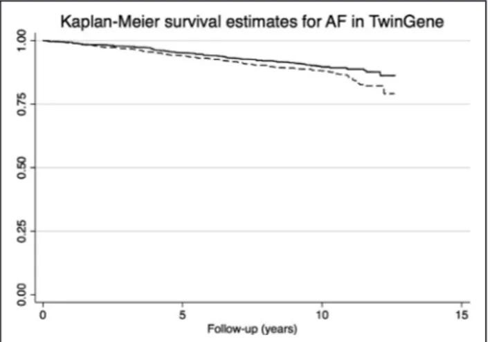
Figure 2. Kaplan-Meier survival curve for atrial fibrillation (AF) in the TwinGene sample regarding 9-decenoylcarnitine levels.
The dashed line indicates subjects with levels of 9-decenoylcarnitine above the median and the solid line indicates subjects below the median (P<0.015 for difference between the 2 groups).
J Am Heart Assoc. 2021;10:e017579. DOI: 10.1161/JAHA.120.017579 7
Lind et al Metabolomics and Atrial Fibrillation
and incident AF. The first, performed in 1919 Blacks in the ARIC (Atherosclerosis Risk in Communities) study, identified associations between 2 conjugated bile acids (glycolithocholate sulfate and glycocholenate sulfate) and incident AF.16 However, in a subsequent analysis in a larger portion of the ARIC study (n=3922) only gly-cocholenate sulfate was confirmed as an AF predictor. Also, that study identified 2 metabolites related to py-rimidine metabolism (pseudouridine and uridine) and 1 metabolite related to polyamine metabolism, acisoga, as AF predictors.14 In contrast, none of 217 metabolites were associated with incident AF at the prespecified Bonferroni corrected level of significance in 2458 par-ticipants in the Framingham heart study.15 Thus, none of the 6 metabolites disclosed in the present study were identified in previous studies relating untargeted metabolomics to incident AF.
9-decenoylcarnitine belongs to the family of ACs being oxidative metabolites with a fatty acid esteri-fied to a carnitine molecule.29 These compounds are generated by the enzymes CPT1 and CPT2 (carnitine palmitoyltransferase 1 and 2), in order to facilitate the transport of fatty acids across the mitochondrial mem-branes to the matrix space. There, the AC carnitines are split into acyl-CoA and carnitine. The carnitine
molecule is shuttled back across the membrane while acyl-CoA is used for ATP production by the beta-oxi-dation pathway.
The heart tissue could use different sources for en-ergy production. However, after a normal mixed meal, 60% to 70% of the ATP production in myocardial tissue is due to beta-oxidation of fatty acids. Thus, ACs have a major role in the daily energy production is the heart.
Medium- and long-chain ACs have been found to be linked to visceral obesity,30 and intentional weight loss is known to affect levels of ACs.31 Decenoylcarnitine and other ACs have also been linked to an impaired glucose tolerance in previous studies.32 Of particular interest is that 2 other medium-chained ACs (deca-noylcarnitine and octa(deca-noylcarnitine) have been asso-ciated with cardioembolic stroke,33 a diagnosis mainly caused by AF.
In our secondary analysis, another medium-chained AC, L-octanoyncarnitine, was related to incident AF, suggesting that it may be ACs as a group rather than specific ACs that could be of interest for AF.
As reviewed by Huang et al,34 it is commonly believed that for the maintenance of AF both a trig-ger and a substrate is needed. The trigtrig-ger could be an increased tone in the autonomic nerve system or
Table 3. Associations Between Metabolites and Incident Atrial Fibrillation in a Meta-Analysis of the TwinGene, ULSAM, and PIVUS Samples
Metabolite
Age and Sex Adjusted Multiple Adjusted
HR 95% CI Lower Limit 95% CI Higher Limit P Value HR 95% CI Lower Limit 95% CI Higher Limit P Value 9-decenoylcarnitine 1.18 1.09 1.27 0.000012 1.17 1.08 1.26 0.000042 Bilirubin 1.14 1.07 1.22 0.000013 1.11 1.04 1.20 0.0025 Sphingomyelin (28:1) 0.77 0.69 0.87 0.000029 0.80 0.70 0.91 0.0014 L-octanoylcarnitine 1.15 1.07 1.24 0.000045 1.16 1.08 1.25 0.000053 Urobilin 1.11 1.05 1.17 0.00020 1.09 1.02 1.16 0.0047
Associations are given at 2 levels of adjustment. Multiple adjustment includes age, sex, systolic blood pressure, diabetes mellitus, smoking, low-density lipoprotein- and high-density lipoprotein-cholesterol, and body mass index. Only associations with an age- and sex-adjusted P value<0.00025 (Bonferroni adjustment) are shown. The HRs are given for a unit change in metabolite levels. HR indicates hazard ratio; PIVUS, Prospective Investigation of the Vasculature in Uppsala Seniors; and ULSAM, Uppsala Longitudinal Study of Adult Men.
Table 4. Relationships Between 9-Decenoylcarnitine Levels and Indices of Myocardial Function and Geometry in the PIVUS Study
Age- and Sex-Adjusted Analysis Multiple-Adjusted Analysis
Beta SE P Value Beta SE P Value
Left atrial diameter 0.67 0.24 0.0064 0.39 0.21 0.063
Left ventricular end-diastolic diameter
0.31 0.20 0.11 0.131 0.191 0.49
Relative wall thickness 0.008 0.003 0.019 0.006 0.003 0.079
Left ventricular mass index
1.62 0.52 0.0019 0.79 0.42 0.064
Left ventricular ejection fraction
0.001 0.003 0.86 0.001 0.003 0.81
The betas are given for a unit change in 9-decenoylcarnitine levels. PIVUS indicates Prospective Investigation of the Vasculature in Uppsala Seniors.
changed dynamics of Ca2+. The substrate is typically an enlarged left atrium with fibrosis but also includes changed properties of ion channels. GWAS studies on AF have disclosed a number of genes being related to the disease.25 A pathway analysis on those genes showed striated muscle development and cardiac muscle contraction to be the major pathways. Because 9-Decenoylcarnitine is involved in the generation of ATP from fatty acids, it is likely that this AC is involved in this cardiac muscle contraction pathway.
SM is the second most abundant phospho-lipid in human plasma and is present in all cellular membranes, in the myelin sheath, and in plasma lipoproteins. SM metabolites that are present or derived from lipoproteins have been implicated in various vascular cell changes that contribute to atherogenesis.35
Bilirubin and urobilin were also identified in the secondary analysis. Both are breakdown products of hemoglobin and have recently been linked to incident heart failure.13 However, the inclusion of heart failure before AF in the models had only a marginal effect on the effect estimate, suggesting that the link between SM and incident AF is not mediated by heart failure to a major degree.
An important risk factor for AF is heart failure,36 as well as myocardial infarction. Both of these disorders lead to cardiac remodeling with fibrosis development, and heart failure is a common complication of a myo-cardial infarction.
In the present study, we adjusted the analyses for both heart failure and myocardial infarction that pre-sented before the AF. However, these analyses was based on hospitalized cases for both diseases, and si-lent myocardial infarction and less severe heart failure, not demanding in-hospital care, will not be covered by our approach. It could therefore not be ruled out completely that our top finding, 9-Decenoylcarnitine, is linked to AF through an association with mild heart failure or silent myocardial infarction.
Because of a lack of publicly available genetic instru-ments for 9-Decenoylcarnitine, we performed a me-ta-analysis of GWAS results from the 3 samples (around 4000 individuals) to search for a powerful genetic
instrument to be used in Mendelian randomization anal-ysis. We identified an SNP in the ACADM gene being associated with 9-decenoylcarnitine with a rather low P value (10−20). In the Mendelian randomization analysis, we used a large GWAS study on AF with >65 000 cases as outcome data. It should be acknowledged that our own GWAS could have been underpowered in order to be able to show causality for 9-Decenoylcarnitine, and we have to await future larger GWASs for metabolomics to finally solve the causality issue. Because of the lack of causality for 9-decenoylcarnitine so far, this metab-olite is not likely to be an important biomarker for AF to be used in the clinic in the future but might nevertheless shed a light on the role of carnitines in AF.
The strengths of the present study are that metab-olomics was measured with the same instruments in 3 independent cohorts in combination with genetic analyses, which enabled us to use a strict discovery/ validation approach in combination with a Mendelian randomization analysis.
A limitation is the amount of only 200 annotated me-tabolites, so it is not a true untargeted approach even though the selection of metabolites were solely based on the availability of reliable annotations. Another lim-itation is that we almost exclusively have individuals with European descent in our Swedish samples, so the results have to be confirmed in other geographical and ethnic groups. Because the present study consists of mainly elderly subjects, further studies on this topic are needed in younger samples. In epidemiological stud-ies, the incidence rate of AF is almost always underes-timated, because many cases with mild paroxysmal of AF will not demand hospital care.
Two cohorts used plasma, and 1 study used serum for the metabolomics analyses. This might have cre-ated slightly different values for the metabolites, although the values between the 3 cohorts were har-monized before statistical analyses were performed (11,25). If anything, using a mix of plasma and serum in the cohorts could only drive the associations toward the null hypothesis and underestimate the true asso-ciation between the metabolites and AF. In this case, no false positive associations would be induced by the mix of plasma/serum.
All population-based epidemiological studies are suffering from a selection bias toward healthy sub-jects. However, as long as there is a wide range in the exposure variable, a selection bias is usually not a major problem when estimating the degree of associ-ation between a certain exposure and an outcome. In the case of metabolites being the exposures, as in this case, it is unclear if all metabolites have an appropriate wide range. It was therefore appropriate that we used 3 different study samples and a discovery/validation approach to ensure that a possible selection bias in 1 of the samples did not produce false positive results.
Table 5. Genome-Wide Association Study (GWAS) for 9-Decenoylcarnitine as Being Related to Incident Atrial Fibrillation
rs-Number
rs74339586 (Also Denoted rs121226481)
Position (hg19) 1:76222640
Effect/other allele (EAF) A/G (0.28) Beta, SE, and P value −0.280, 0.025, P<1.0 x 10⁻²¹
Nearest gene ACADM
The most significant locus is given.
J Am Heart Assoc. 2021;10:e017579. DOI: 10.1161/JAHA.120.017579 9
Lind et al Metabolomics and Atrial Fibrillation
In conclusion, a metabolomics analysis dis-closed 1 novel replicated biomarker for AF, 9-Decenoylcarnitine. The genetic studies did, how-ever, not suggest a causal role in AF for circulating 9-Decenoylcarnitine.
ARTICLE INFORMATION
Received May 19, 2020; accepted November 16, 2020. Affiliations
From the Department of Medical Sciences (L.L., J.S.)and Department of Medical Sciences, Molecular Epidemiology and Science for Life Laboratory (S.S., T.F.), Uppsala University, Uppsala, Sweden; School of Medical Sciences, Örebro University, Örebro, Sweden (S.S.); Uppsala Clinical Research Center, Uppsala University, Uppsala, Sweden (J.S.); Proteomics and Metabolomics Facility, Colorado State University, Fort Collins, CO (C.D.B.); Department of Medical Epidemiology and Biostatistics (MEB), Karolinska Institutet, Stockholm, Sweden (P.K.M.); Department of Horticulture and Landscape Architecture, Colorado State University, Fort Collins, CO (J.P.); Division of Family Medicine and Primary Care, Department of Neurobiology, Care Sciences and Society, Karolinska Institutet, Huddinge, Sweden (J.Ä.); and School of Health and Social Sciences, Dalarna University, Falun, Sweden (J.Ä.).
Sources of Funding
The study was supported by the Swedish Heart and Lung Foundation and Uppsala University Hospital (ALF-medel).
Disclosures None. Supplementary Material Tables S1–S2 Figures S1–S6 REFERENCES
1. Pistoia F, Sacco S, Tiseo C, Degan D, Ornello R, Carolei A. The epide-miology of atrial fibrillation and stroke. Cardiol Clin. 2016;34:255–268. DOI: 10.1016/j.ccl.2015.12.002.
2. Ling LH, Kistler PM, Kalman JM, Schilling RJ, Hunter RJ. Comorbidity of atrial fibrillation and heart failure. Nat Rev Cardiol. 2016;13:131–147. DOI: 10.1038/nrcar dio.2015.191.
3. Patton KK, Heckbert SR, Alonso A, Bahrami H, Lima JA, Burke G, Kronmal RA. N-terminal pro-B-type natriuretic peptide as a predictor of incident atrial fibrillation in the Multi-Ethnic Study of Atherosclerosis: the effects of age, sex and ethnicity. Heart. 2013;99:1832–1836. DOI: 10.1136/heart jnl-2013-304724.
4. Rienstra M, Yin X, Larson MG, Fontes JD, Magnani JW, McManus DD, McCabe EL, Coglianese EE, Amponsah M, Ho JE, et al. Relation be-tween soluble ST2, growth differentiation factor-15, and high-sensitivity troponin I and incident atrial fibrillation. Am Heart J. 2014;167(1):109– 115.e2. DOI: 10.1016/j.ahj.2013.10.003.
5. Rienstra M, Sun JX, Magnani JW, Sinner MF, Lubitz SA, Sullivan LM, Ellinor PT, Benjamin EJ. White blood cell count and risk of incident atrial fibrillation (from the Framingham Heart Study). Am J Cardiol. 2012;109:533–537. DOI: 10.1016/j.amjca rd.2011.09.049.
6. Alonso A, Yin X, Roetker NS, Magnani JW, Kronmal RA, Ellinor PT, Chen LY, Lubitz SA, McClelland RL, McManus DD, et al. Blood lipids and the incidence of atrial fibrillation: the Multi-Ethnic Study of Atherosclerosis and the Framingham Heart Study. J Am Heart Assoc. 2014;3:e001211. DOI: 10.1161/JAHA.114.001211.
7. Raposeiras-Roubín S, Rodino-Janeiro BK, Grigorian-Shamagian L, Seoane-Blanco A, Moure-González M, Varela-Román A, Álvarez E, González-Juanatey JR. Evidence for a role of advanced glycation end products in atrial fibrillation. Int J Cardiol. 2012;157:397–402. DOI: 10.1016/j.ijcard.2011.05.072.
8. Mathew JS, Sachs MC, Katz R, Patton KK, Heckbert SR, Hoofnagle AN, Alonso A, Chonchol M, Deo R, Ix JH, et al. Fibroblast growth
factor-23 and incident atrial fibrillation: the Multi-Ethnic Study of Atherosclerosis (MESA) and the Cardiovascular Health Study (CHS). Circulation. 2014;130:298–307. DOI: 10.1161/CIRCU LATIO NAHA.113.005499.
9. Smith JG, Newton-Cheh C, Almgren P, Struck J, Morgenthaler NG, Bergmann A, Platonov PG, Hedblad B, Engström G, Wang TJ, et al. Assessment of conventional cardiovascular risk factors and multiple bio-markers for the prediction of incident heart failure and atrial fibrillation. J
Am Coll Cardiol. 2010;56:1712–1719. DOI: 10.1016/j.jacc.2010.05.049.
10. Lind L, Sundström J, Stenemo M, Hagström E, Ärnlöv J. Discovery of new biomarkers for atrial fibrillation using a custom-made proteomics chip. Heart. 2017;103:377–382. DOI: 10.1136/heart jnl-2016-309764. 11. Ganna A, Salihovic S, Sundström J, Broeckling CD, Hedman AK,
Magnusson PK, Pedersen NL, Larsson A, Siegbahn A, Zilmer M, et al. Large-scale metabolomic profiling identifies novel biomarkers for inci-dent coronary heart disease. PLoS Genet. 2014;10:e1004801. 12. Lind L, Salihovic S, Ganna A, Sundstrom J, Broeckling CD, Magnusson
PK, Pedersen NL, Siegbahn A, Prenni J, Fall T, et al. A multi-cohort metabolomics analysis discloses sphingomyelin (32:1) levels to be in-versely related to incident ischemic stroke. J Stroke Cerebrovasc Dis. 2020;29:104476.
13. Stenemo M, Ganna A, Salihovic S, Nowak C, Sundström J, Giedraitis V, Broeckling CD, Prenni JE, Svensson P, Magnusson PKE, et al. The metabolites urobilin and sphingomyelin (30:1) are associated with incident heart failure in the general population. ESC Heart Fail. 2019;6:764–773.
14. Alonso A, Yu B, Sun YV, Chen LY, Loehr LR, O’Neal WT, Soliman EZ, Boerwinkle E. Serum metabolomics and incidence of atrial fibrillation (from the Atherosclerosis Risk in Communities Study). Am J Cardiol. 2019;123:1955–1961.
15. Ko D, Riles EM, Marcos EG, Magnani JW, Lubitz SA, Lin H, Long MT, Schnabel RB, McManus DD, Ellinor PT, et al. Metabolomic profiling in relation to new-onset atrial fibrillation (from the Framingham Heart Study). Am J Cardiol. 2016;118:1493–1496.
16. Alonso A, Yu B, Qureshi WT, Grams ME, Selvin E, Soliman EZ, Loehr LR, Chen LY, Agarwal SK, Alexander D, et al. Metabolomics and inci-dence of atrial fibrillation in African Americans: the Atherosclerosis Risk in Communities (ARIC) Study. PLoS One. 2015;10:e0142610.
17. Psaty BM, Manolio TA, Kuller LH, Kronmal RA, Cushman M, Fried LP, White R, Furberg CD, Rautaharju PM. Incidence of and risk factors for atrial fibrillation in older adults. Circulation. 1997;96:2455–2461. 18. Vaziri SM, Larson MG, Benjamin EJ, Levy D. Echocardiographic
pre-dictors of nonrheumatic atrial fibrillation. The Framingham Heart Study.Circulation. 1994;89:724–730.
19. Vasan RS, Larson MG, Levy D, Galderisi M, Wolf PA, Benjamin EJ. National Heart, Lung, and Blood Institute, National Institutes of Health. Doppler transmitral flow indexes and risk of atrial fibrillation (the Framingham Heart Study). Am J Cardiol. 2003;91:1079–1083.
20. Lind L, Fors N, Hall J, Marttala K, Stenborg A. A comparison of three different methods to evaluate endothelium-dependent vasodila-tion in the elderly: the Prospective Investigavasodila-tion of the Vasculature in Uppsala Seniors (PIVUS) study. Arterioscler Thromb Vasc Biol. 2005;25:2368–2375.
21. Hedstrand H. A study of middle-aged men with particular reference to risk factors for cardiovascular disease. Ups J Med Sci Suppl. 1975;19:1–61.
22. Ärnlov J, Ingelsson E, Sundström J, Lind L. Impact of body mass index and the metabolic syndrome on the risk of cardiovascular disease and death in middle-aged men. Circulation. 2010;121:230–236. DOI: 10.1161/CIRCU LATIO NAHA.109.887521.
23. Zagai U, Lichtenstein P, Pedersen NL, Magnusson PKE. The Swedish Twin Registry: content and management as a research in-frastructure. Twin Res Hum Genet. 2019;22:672–680. DOI: 10.1017/ thg.2019.99.
24. Smith CA, Want EJ, O’Maille G, Abagyan R, Siuzdak G. XCMS: Processing mass spectrometry data for metabolite profiling using Nonlinear peak alignment, matching, and identification. Anal Chem. 2006;78:779–787.
25. Ganna A, Fall T, Salihovic S, Lee W, Broeckling CD, Kumar J, Hägg S, Stenemo M, Magnusson PKE, Prenni JE, et al. Large-scale non-tar-geted metabolomic profiling in three human population-based stud-ies. Metabolomics. 2015;12:4. DOI: 10.1007/s1130 6-015-0893-5. 26. Roselli C, Chaffin MD, Weng LC, Aeschbacher S, Ahlberg G, Albert
CM, Almgren P, Alonso A, Anderson CD, Aragam KG, et al. Multi-ethnic
genome-wide association study for atrial fibrillation. Nat Genet. 2018;50:1225–1233. DOI: 10.1038/s4158 8-018-0133-9.
27. Zhou J, Sun L, Chen L, Liu S, Zhong L, Cui M. Comprehensive metabo-lomic and proteomic analyses reveal candidate biomarkers and related metabolic networks in atrial fibrillation. Metabolomics. 2019;15:96. DOI: 10.1007/s1130 6-019-1557-7.
28. Lai S, Hua X, Gao R, Zeng L, Song J, Liu J, Zhang J. Combinational bio-markers for atrial fibrillation derived from atrial appendage and plasma metabolomics analysis. Sci Rep. 2018;8:16930. DOI: 10.1038/s4159 8-018-34930 -6.
29. Reuter SE, Evans AM. Carnitine and acylcarnitines: pharmacoki-netic, pharmacological and clinical aspects. Clin Pharmacokinet. 2012;51:553–572. DOI: 10.1007/BF032 61931.
30. Baek SH, Kim M, Kim M, Kang M, Yoo HJ, Lee NH, Kim YH, Song M, Lee JH. Metabolites distinguishing visceral fat obesity and ath-erogenic traits in individuals with overweight. Obesity (Silver Spring). 2017;25:323–331. DOI: 10.1002/oby.21724.
31. Kang M, Yoo HJ, Kim M, Kim M, Lee JH. Metabolomics identifies in-creases in the acylcarnitine profiles in the plasma of overweight subjects
in response to mild weight loss: a randomized, controlled design study.
Lipids Health Dis. 2018;17:237. DOI: 10.1186/s1294 4-018-0887-1.
32. Mai M, Tönjes A, Kovacs P, Stumvoll M, Fiedler GM, Leichtle AB. Serum levels of acylcarnitines are altered in prediabetic conditions. PLoS One. 2013;8:e82459. DOI: 10.1371/journ al.pone.0082459.
33. Seo WK, Jo G, Shin MJ, Oh K. Medium-chain acylcarnitines are associ-ated with cardioembolic stroke and stroke recurrence. Arterioscler Thromb
Vasc Biol. 2018;38:2245–2253. DOI: 10.1161/ATVBA HA.118.311373.
34. Huang X, Li Y, Zhang J, Wang X, Li Z, Li G. The molecular genetic basis of atrial fibrillation. Hum Genet. 2020;139:1485–1498. DOI: 10.1007/ s0043 9-020-02203 -w.
35. Kikas P, Chalikias G, Tziakas D. Cardiovascular implications of sphingo-myelin presence in biological membranes. Eur Cardiol. 2018;13:42–45. DOI: 10.15420/ ecr.2017:20:3.
36. Alonso A, Krijthe BP, Aspelund T, Stepas KA, Pencina MJ, Moser CB, Sinner MF, Sotoodehnia N, Fontes JD, Janssens AC, et al. Simple risk model predicts incidence of atrial fibrillation in a racially and geograph-ically diverse population: the CHARGE-AF consortium. J Am Heart
Assoc. 2013;2:e000102. DOI: 10.1161/JAHA.112.000102.
SUPPLEMENTAL MATERIAL
1,2-dilinolenoyl-sn-glycero-3-phosphocholine
1,2-dilinoleoyl-sn-glycero-3-phosphocholine
1,2-dioleoyl-sn-glycero-3-phosphocholine,
1,2-dipetroselenoyl-sn-glycero-3-phosphocholine
1,2-dipalmitoleoyl-sn-glycero-3-phosphocholine
1,2-dipalmitoyl-sn-glycero-3-phosphocholine
1,3,7-Trimethyluric acid
1-O-1'-(Z)-octadecenyl-2-hydroxy-sn-glycero-3-phosphoethanolamine
1-Stearoyl-2-Hydroxy-sn-Glycero-3-Phosphoethanolamine
1-arachidoyl-2-hydroxy-sn-glycero-3-phosphocholine
1-linoleoyl-2-stearoyl-sn-glycerol
1-oleoyl-2-hydroxy-sn-glycero-3-phosphocholine
1-oleoyl-2-hydroxy-sn-glycero-3-phosphoethanolamine
1-oleoyl-2-palmitoyl-sn-glycero-3-phosphocholine
1-palmitoyl-2-docosahexaenoyl-sn-glycero-3-phosphocholine
1-palmitoyl-2-hydroxy-sn-glycero-3-phosphocholine
1-palmitoyl-2-hydroxy-sn-glycero-3-phosphoethanolamine
1-stearoyl-2-arachidonoyl-sn-glycero-3-phosphoethanolamine, C16-20:4
Phosphatidylcholine
1-stearoyl-2-hydroxy-sn-glycero-3-phosphocholine
1-vaccenoyl-2-palmitoyl-sn-glycerol
10-nitro-9E-octadecenoic acid, 9-nitro-9E-octadecenoic acid
17-phenyl trinor Prostaglandin E2, 17-phenyl trinor Prostaglandin D2
2,6 dimethylheptanoyl carnitine
2-Ketohexanoic acid
3-Indolepropionic acid
3-Pyridylacetic acid, trigonelline
3a,6b,7b-Trihydroxy-5b-cholanoic acid
4-Androsten-11Beta-ol-3, 17-dione, 11_-Hydroxy-4-androstene-3,17-dione
6-hydroxy-5-cholestanol, cholesterol
7-Ketocholesterol
9-Decenoylcarnitine
Acetaminophen
Alpha-Linolenic acid
Alpha-Tocopherol
Arachidonic acid
Arachidonic acid ethyl ester
Arachidonic acid methyl ester
Barogenin
Betaine
Biliribin I
Biliribin II
Biliverdin hydrochloride a
Biliverdin hydrochloride b
C12 Carnitine
C16 Carnitine
C16-20:5 Phosphatidylcholine
Caffeine
Ceramide phosphoethanolamine(33:1), Sphingomyelin(30:1)
Ceramide phosphoethanolamine(34:1)
Ceramide phosphoethanolamine(35:1), Sphingomyelin(32:1)
Ceramide phosphoethanolamine(35:2), Sphingomyelin(32:2)
Ceramide phosphoethanolamine(36:1), Sphingomyelin(33:1)
Ceramide phosphoethanolamine(37:2), Sphingomyelin(34:2)
Ceramide phosphoethanolamine(38:2)
Chenodeoxycholic acid
Choline
Cinnamic acid and derivatives
Corticosterone
Cortisol
Creatinine
D-Urobilinogen, I-Urobilin
D-erythro-sphingosine
DL-2-Aminooctanoic acid
Decanoyl-L-carnitine
Deoxycholic acid
Deoxycholic acid glycine conjugate
Deoxycholic acid related metabolite
Dodecanedioic acid
Dodecanoic acid
Fatty acid C16:0
Fatty acid 22:5
Fatty acid C18:4
Fatty acid C19:2
Fatty acid C20:2
Fatty acid C20:3
Fatty acid C20:4
Fatty acid C20:5 methyl ester
Fatty acid C22:4
Fatty acid C22:6
Fatty acid C22:6 methyl ester
Flavone
Gamma-Caprolactone
Gamma-Tocopherol
Gamma-glutamyl-Leucine
Geranyl acetoacetate
Glycocholic acid
Heptadecanoic aicd
Hippuric acid
Hyodeoxycholic acid
Indoleacetic acid
Indolelactic acid
L-Acetylcarnitine
L-Aspartyl-L-phenylalanine
L-Carnitine
L-Leucine, L-Norleucine
L-Octanoylcarnitine
L-Phenylalanine
L-Proline
L-Tryptophan
L-Tyrosine, o-Tyrosine
Linoleic acid
Linoleyl carnitine
Lyso-PAF C-18
Lysophosphatidylcholine(0:0/16:0)
Lysophosphatidylcholine(0:0/16:1)
Lysophosphatidylcholine(0:0/18:0)
Lysophosphatidylcholine(0:0/18:2)
Lysophosphatidylcholine(0:0/20:4)
Lysophosphatidylcholine(0:0/20:5)
Lysophosphatidylcholine(16:1/0:0)
Lysophosphatidylcholine(18:1)a
Lysophosphatidylcholine(18:2/0:0)
Lysophosphatidylcholine(18:3)
Lysophosphatidylcholine(18e:0/0:0)
Lysophosphatidylcholine(20:1)
Lysophosphatidylcholine(20:2)
Lysophosphatidylcholine(20:3)a
Lysophosphatidylcholine(20:3)b
Lysophosphatidylcholine(20:4/0:0)
Lysophosphatidylcholine(20:5/0:0)
Lysophosphatidylcholine(22:5)a
Lysophosphatidylcholine(22:5)b
Lysophosphatidylethanolamine(16:0)
Lysophosphatidylethanolamine(18:0)
Lysophosphatidylethanolamine(18:1)
Lysophosphatidylethanolamine(18:2)
Lysophosphatidylethanolamine(20:4)
N-(15Z-tetracosenoyl)-sphinganine-1-phosphocholine
N-(9Z-octadecenoyl)-sphing-4-enine-1-phosphocholine
N-(octadecanoyl)-sphing-4-enine-1-phosphocholine
N-palmitoyl-D-erythro-sphingosylphosphorylcholine
Oleamide
Oleoyl-L-carnitine hydrochloride
Ornithine
Palmitic acid
Palmitoleic acid
Pantothenic acid
Paraxanthine, Theophylline
Pentadecanoic acid
Phenylalanylphenylalanine
Phosphatidylcholine(28:1)
Phosphatidylcholine(28:2)
Phosphatidylcholine(29:1)
Phosphatidylcholine(30:1)
Phosphatidylcholine(30:2)
Phosphatidylcholine(32:1)
Phosphatidylcholine(33:1), Phosphoethanolamine(36:1)
Phosphatidylcholine(34:0)
Phosphatidylcholine(34:0), Phosphoethanolamine(37:3)
Phosphatidylcholine(34:2), Phosphoethanolamine(37:2)
Phosphatidylcholine(34:3)
Phosphatidylcholine(34:4)
Phosphatidylcholine(34:5), Phosphoethanolamine(37:5)
Phosphatidylcholine(35:2), Phosphoethanolamine(38:2)
Phosphatidylcholine(35:4), Phosphoethanolamine(38:4)
Phosphatidylcholine(36:1)
Phosphatidylcholine(36:3), Phosphoethanolamine(39:3)
Phosphatidylcholine(36:5), Phosphoethanolamine(39:5)
Phosphatidylcholine(36:6)
Phosphatidylcholine(37:5), Phosphoethanolamine(40:0)
Phosphatidylcholine(38:2)
Phosphatidylcholine(38:3)
Phosphatidylcholine(38:4)
Phosphatidylcholine(38:5), Phosphoethanolamine(41:6)
Phosphatidylcholine(38:7)
Phosphatidylcholine(40:5)
Phosphatidylcholine(40:6)
Phosphatidylcholine(42:7)
Phosphatidylcholine(O-18:1/0:0), Phosphatidylcholine(P-18:1/0:0)
Phosphatidylethanolamine(19:1), Phosphatidylcholine(16:2)
Phosphatidylserine(18:0)
Piperine
Propranolol
Prostaglandin J2
Salicylic acid, Aspirin
Sphingomyelin(40:2)
Sphingomyelin(41:2)
Sphingomyelin(42:3)
Sphingomyelin(d18:2/18:1)
Stachydrine
Stearic acid
Treprostinil
Uric acid
Vitamin D3 and derivatives
Caprolactam
cis-5-Tetradecenoylcarnitine
cis/trans-Oleic acid
dehydroepiandrosterone sulfate (sodium salt)
monoacylglycerol(14:0)
monoacylglycerol(16:0)
monoacylglycerol(16:1)
monoacylglycerol(18:0)
monoacylglycerol(18:1)
monoacylglycerol(18:2)
monoacylglycerol(20:5)
myristic acid
sodium glycochenodeoxycholate
sum of Hexoses
Table S2. Metabolome pathway analysis was performed for 200 metabolites used in the present study using MetaboAnalyst 4.0. The analysis combines pathway enrichment analysis with pathway topology in order to simplify biological interpretation and pathway visualization. Metabolites from curated human metabolic pathways (Kyoto Encyclopedia of Genes and Genomes) were included in an over representation analysis which uses a hypergeometric test to evaluate whether the
metabolite set of interest is represented more than expected by chance within that specific list.
Total pathway metabolites Metabolite hits Raw
P-value −log10(P) Impact Caffeine metabolism 10 4 0.001198 2.9214 0.69231 Biosynthesis of unsaturated fatty acids 36 7 0.002147 2.6683 0 Phenylalanine metabolism 10 3 0.013076 1.8835 0.35714 Phenylalanine, tyrosine and tryptophan biosynthesis 4 2 0.015477 1.8103 1 Linoleic acid metabolism 5 2 0.024913 1.6036 1 Glycerophospholipid metabolism 36 5 0.03791 1.4212 0.28914 Primary bile acid biosynthesis 46 5 0.09152 1.0385 0.06847 Sphingolipid metabolism 21 3 0.095475 1.0201 0.04462 Neomycin, kanamycin and gentamicin biosynthesis 2 1 0.10304 0.98699 0 Aminoacyl-tRNA biosynthesis 48 5 0.1054 0.97715 0 alpha-Linolenic acid metabolism 13 2 0.14792 0.82999 0.33333 Glycine, serine and threonine metabolism 33 3 0.25206 0.59849 0.05034 Arginine and proline metabolism 38 3 0.32597 0.48683 0.20055 Valine, leucine and isoleucine biosynthesis 8 1 0.35328 0.45188 0 Ubiquinone and other terpenoid-quinone biosynthesis 9 1 0.38767 0.41153 0 Fatty acid biosynthesis 47 3 0.45786 0.33927 0.01473 Steroid hormone biosynthesis 85 5 0.47227 0.32581 0.06857 Porphyrin and chlorophyll metabolism 30 2 0.47781 0.32074 0.12753 Glycosylphosphatidylinositol (GPI)-anchor biosynthesis 14 1 0.53432 0.2722 0.00399 Arginine biosynthesis 14 1 0.53432 0.2722 0.06091 Arachidonic acid metabolism 36 2 0.57743 0.2385 0.3135 Fatty acid degradation 39 2 0.62187 0.2063 0 Starch and sucrose metabolism 18 1 0.62616 0.20331 0.13851 Pantothenate and CoA biosynthesis 19 1 0.64617 0.18965 0.00714 Tryptophan metabolism 41 2 0.64948 0.18743 0.14305 Tyrosine metabolism 42 2 0.66269 0.17869 0.13972 Ether lipid metabolism 20 1 0.66512 0.1771 0.14458 Glutathione metabolism 28 1 0.7847 0.1053 0 Inositol phosphate metabolism 30 1 0.80728 0.092977 0 Purine metabolism 65 2 0.87027 0.060344 0.01651 Fatty acid elongation 39 1 0.88316 0.053959 0 Valine, leucine and isoleucine degradation 40 1 0.8895 0.050852 0 Steroid biosynthesis 42 1 0.90118 0.045187 0.0282
0.4
0.6
Pathway Impact
-log10(p)
0.0
0.2
0.8
1.0
0.0
0.5
1.0
1.5
2.0
2.5
3.0
Caffeine metabolism Biosynthesis of unsaturated fatty acids Phenylalanine metabolism Glycerophospholipid metabolism Phenylalanine, tyrosine and tryptophan biosynthesis Linoleic acid metabolism alpha‐Linolenic acid metabolism Primary bile acid biosynthesis Sphingolipid metabolism Arachidonic acid metabolism Arginine and proline metabolism2006009195_00186999 Time 2.55 2.60 2.65 2.70 2.75 2.80 2.85 2.90 % 0 2.55 2.60 2.65 2.70 2.75 2.80 2.85 2.90 % 1 2.55 2.60 2.65 2.70 2.75 2.80 2.85 2.90 % 9
120207_TWINGENE_000802 Scan ES+
85.029 1.02e4 Area
2.76 464
120207_TWINGENE_000802 Scan ES+
314.233 3.08e3 Area
2.78 126
120207_TWINGENE_000801 Scan ES+
314.233 2.98e4 Area 2.76 1495 2006009195_00186999 m/z 80 100 120 140 160 180 200 220 240 260 280 300 320 % 0 100 m/z 80 100 120 140 160 180 200 220 240 260 280 300 320 % 0 100 m/z 80 100 120 140 160 180 200 220 240 260 280 300 320 % 0 100
120207_TWINGENE_000802 370 (2.762) Scan ES+
9.18e3 85.0289 314.2316 149.0597 98.9634 121.0281 170.0972 203.0712 257.1935 288.1602 227.1404 331.2064
120207_TWINGENE_000802 371 (2.769) Scan ES+
7.88e3 85.0291 314.2317 299.1375 149.0606 97.9701 121.0292 203.0695 170.0941 255.1594 331.2116
120207_TWINGENE_000801 371 (2.766) Scan ES+
5.07e4 102.1282 84.9602 314.2330 117.1398 303.1958 141.9586 167.0717 257.1953 288.1636 212.1405 MS (6 eV) 9-decenoylcarnitine M+H = 314.233 HMDB0013205 idMSMS (15-30 eV) 9-decenoylcarnitine M+H = 314.233 HMDB0013205 idMSMS (15-30 eV) 9-decenoylcarnitine M-C4H5O2+ = 85.029 HMDB0013205
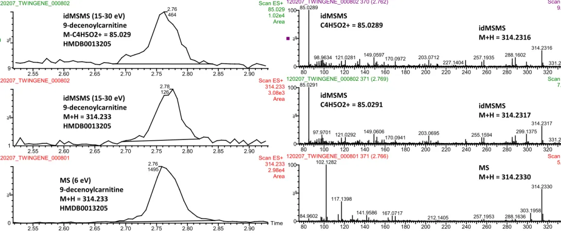
Figure S2. 9-decenoylcarnitine was annotated at metabolomics standards initiative (MSI) level 2 by database matching with accurate mass and
fragmentation schemes reported for other acylcarnitines. These fragmentation schemes include the precursor of 85 fragment, or by detecting the neutral losses of either M-59 or M-161. We conclude that we selectively detect 9-decenoylcarnitine as shown in MS (collision induced dissociation at 6 eV) and idMSMS (collision induced dissociation ramp 15-30 eV) chromatograms obtained from an authenthic sample from TWINGENE. We consistently observe M+H = 314.233 in both MS and idMSMS mode as well as the major fragment C4H5O2+ = 85.0289 eluting at 2.76 minutes using UPLC-QTOFMS operated in positive ESI. The corresponding mass spectra are shown to the right.
MS M+H = 314.2330 idMSMS M+H = 314.2317 idMSMS M+H = 314.2316 idMSMS C4H5O2+ = 85.0289 idMSMS C4H5O2+ = 85.0291
m/z 80 100 120 140 160 180 200 220 240 260 280 300 % 0 100 m/z 80 100 120 140 160 180 200 220 240 260 280 300 % 0 100 m/z 80 100 120 140 160 180 200 220 240 260 280 300 % 0 100 193.0868 102.1288 165.0557 133.0655 288.2163 229.1433 273.1649 301.1619
120717_PIVUS_000702 329 (2.458) Scan ES+
3.50e4
288.2175 193.0863
120.0816 167.1036
144.1027 229.1440 273.1610 301.1627
120717_PIVUS_000701 328 (2.447) Scan ES+
1.18e5 288.2174 167.1048 109.1016 137.9879 239.0919 179.0713 221.0809 261.0756279.0845 301.1149 Time 2.30 2.35 2.40 2.45 2.50 2.55 2.60 2.65 2.70 % 0 2.30 2.35 2.40 2.45 2.50 2.55 2.60 2.65 2.70 % 1 2.30 2.35 2.40 2.45 2.50 2.55 2.60 2.65 2.70 % 5 4.06e4 Area 1752
120717_PIVUS_000702 Scan ES+
288.218 9.27e3 Area
2.46 463
120717_PIVUS_000701 Scan ES+
288.218 1.20e5 Area 2.45 6064 MS (6 eV) L-Octanoylcarnitine M+H = 288.218 HMDB0000791 idMSMS (15-30 eV) L-Octanoylcarnitine M+H = 288.218 HMDB0000791 idMSMS (15-30 eV) L-Octanoylcarnitine M-C4H5O2+ = 85.029 HMDB0000791
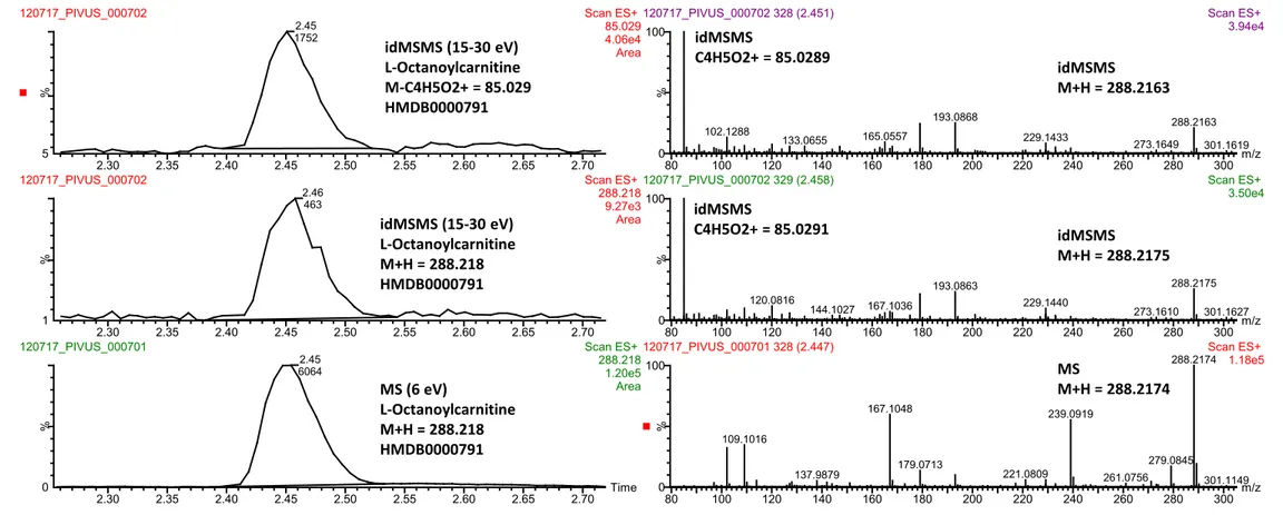
Figure S3. L-Octanoylcarnitine was annotated at MSI level 2 by database matching with accurate mass and fragmentation schemes reported for other
acylcarnitines. These fragmentation schemes include the precursor of 85 fragment, or by detecting the neutral losses of either M-59 or M-161. We conclude that we selectively detect 9-decenoylcarnitine as shown in MS (collision induced dissociation at 6 eV) and idMSMS (collision induced dissociation ramp 15-30 eV) chromatograms obtained from an authenthic sample. We consistently observe M+H = 288.218 in both MS and idMSMS mode as well as the major fragment C4H5O2+ = 85.0289 eluting at 2.45 minutes using UPLC-QTOFMS operated in positive ESI. The corresponding mass spectra are shown to the right.
MS M+H = 288.2174 idMSMS M+H = 288.2175 idMSMS M+H = 288.2163 idMSMS C4H5O2+ = 85.0289 idMSMS C4H5O2+ = 85.0291
31650-ok m/z 50 100 150 200 250 300 350 400 450 500 550 600 650 700 % 0 100 m/z 50 100 150 200 250 300 350 400 450 500 550 600 650 700 % 0 100 m/z 50 100 150 200 250 300 350 400 450 500 550 600 650 700 % 0 100
120621_PIVUS_016702 321 (2.389) Scan ES+
1.22e4 585.2687
299.1393
97.9699 225.0998 347.2025 436.2005 539.2654
647.3016 701.4158
120621_PIVUS_016702 321 (2.389) Scan ES+
1.22e4
585.2687 299.1393
97.9699 225.0998 347.2025 436.2005 539.2654
647.3016 701.4158
120621_PIVUS_016701 321 (2.386) Scan ES+
3.96e4 102.1285 585.2705 141.9594 301.1628 172.9767 239.0897 436.1981 494.2236 645.2882 31650-ok Time 2.10 2.20 2.30 2.40 2.50 2.60 2.70 % 0 2.10 2.20 2.30 2.40 2.50 2.60 2.70 % 0 100 2.10 2.20 2.30 2.40 2.50 2.60 2.70 % 1
120621_PIVUS_016702 Scan ES+
299.14 9.86e3 Area
2.38 626
120621_PIVUS_016702 Scan ES+
585.271 1.33e4 Area
2.38 815
120621_PIVUS_016701 Scan ES+
585.271 3.56e4 Area 2.39 2298 MS (6 eV) Bilirubin M+H = 585.271 HMDB0000791 idMSMS (15-30 eV) Bilirubin M+H = 585.271 HMDB0000791 idMSMS (15-30 eV) Bilirubin M-C15H18N2O3+ = 299.14 HMDB0000791
Figure S4. Bilirubin was annotated at MSI level 2 by database matching with accurate mass and major fragment observed experimentally. We consistently detect bilirubin
as shown in MS (collision induced dissociation at 6 eV) and idMSMS (collision induced dissociation ramp 15-30 eV) chromatograms obtained from an authenthic sample. We consistently observe M+H = 585.271 in both MS and idMSMS mode as well as the major fragment M-C15H18N2O3+ = 299.14 eluting at 2.39 minutes using UPLC-QTOFMS operated in positive ESI. The corresponding mass spectra are shown to the right.
MS M+H = 288.2705 idMSMS M+H = 585.2687 idMSMS M+H = 585.2687 idMSMS Major fragment = 299.1393 idMSMS Major fragment = 299.1393
m/z 570 575 580 585 590 595 600 605 610 615 620 625 630 635 640 645 650 % 0 100 m/z 570 575 580 585 590 595 600 605 610 615 620 625 630 635 640 645 650 % 0 100 m/z 570 575 580 585 590 595 600 605 610 615 620 625 630 635 640 645 650 % 0 100 591.3146 2.23e3 583.3259 575.3256 593.3291 603.7915 595.3309 610.7521 627.3508 611.7710 632.2808 645.3555 HF_SS_20180326_018 358 (2.663) 1: TOF MS ES+ 128 ! 613.2960 ! 591.3231 ! 590.3170 ! 578.2306 593.4075! ! 612.2676 ! 638.7331 ! 614.3022 ! 638.6978 ! 621.3726 ! 648.6019 HF_SS_20180326_013 359 (2.671) 1: TOF MS ES+ 5.19e4 591.3199 ! 591.1842 ! 578.2380 593.3347 594.3364 595.3445 613.3078! ! 629.2683 646.2534! ! 635.2838 Time 2.20 2.40 2.60 2.80 3.00 3.20 % 0 100 2.20 2.40 2.60 2.80 3.00 3.20 % 0 100 2.20 2.40 2.60 2.80 3.00 3.20 % 0 100 591.3211.78e3 Area 2.45 119 HF_SS_20180326_018 Sm (Mn, 2x3) 1: TOF MS ES+ 591.321 182 Area 2.67 14 HF_SS_20180326_013 Sm (Mn, 2x3) 1: TOF MS ES+ 591.321 4.56e4 Area 2.67 4622 Standard Urobilin M+H = 591.3207 Serum Urobilin M+H = 591.3207 HMDB0004161 Serum Urobilin M+H = 591.3207 HMDB0004161 M+H = 591.3199 M+H = 591.3231 M+H = 591.3146
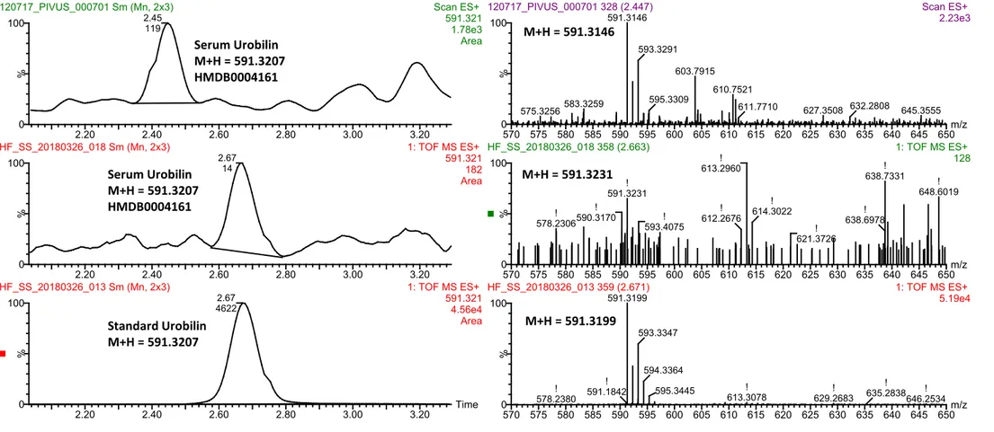
Figure S5. Urobilin was annotated at MSI level 1 by using an external standard. The data was acquired using UPLC-QTOFMS operated in positive electrospray
ionization (ESI) mode. As shown in the bottom chromatogram, the urobilin standard eluted at 2.67 minutes using the method applied to all serum samples in 2012. The mid chromatogram shows M+H for urobilin in a random serum sample run alongside the standard. The top chromatogram shows a sample that was run in 2012, as can be seen there has been a shift in the rt time over this long time period. The selected M+H mass is shown to the right of each chromatogram. The mass spectra of M +H = 591.321 in both serum samples matches well with the spectra of the SM 30:1 standard. The human metabolome database (HMDB) accession number is also provided.
Figure S6. Parallel reaction monitoring (PRM) of sphingolipid SM 28:1 annotated at level MSI 2 in random samples from the ULSAM and PIVUS cohort. The data was
acquired using UPLC-QTOFMS operated in positive electrospray ionization (ESI) mode. The selected precursor mass M+H = 661.528 is shown in the chromatogram. The corresponding mass spectra using collision induced dissociation (CID) at 30 eV are shown to the right.