R E S E A R C H
Open Access
Peak oxygen uptake in relation to total heart
volume discriminates heart failure patients from
healthy volunteers and athletes
Henrik Engblom
1, Katarina Steding
1, Marcus Carlsson
1, Henrik Mosén
1, Bo Hedén
1, Torsten Buhre
2, Björn Ekmehag
3,
Håkan Arheden
1*Abstract
Background: An early sign of heart failure (HF) is a decreased cardiac reserve or inability to adequately increase cardiac output during exercise. Under normal circumstances maximal cardiac output is closely related to peak oxygen uptake (VO2peak) which has previously been shown to be closely related to total heart volume (THV). Thus,
the aim of this study was to derive a VO2peak/THV ratio and to test the hypothesis that this ratio can be used to
distinguish patients with HF from healthy volunteers and endurance athletes. Thirty-one patients with HF of different etiologies were retrospectively included and 131 control subjects (60 healthy volunteers and 71 athletes) were prospectively enrolled. Peak oxygen uptake was determined by maximal exercise test and THV was
determined by cardiovascular magnetic resonance. The VO2peak/THV ratio was then derived and tested.
Results: Peak oxygen uptake was strongly correlated to THV (r2= 0.74, p < 0.001) in the control subjects, but not for the patients (r2 = 0.0002, p = 0.95). The VO2peak/THV ratio differed significantly between control subjects and
patients, even in patients with normal ejection fraction and after normalizing for hemoglobin levels (p < 0.001). In a multivariate analysis the VO2peak/THV ratio was the only independent predictor of presence of HF (p < 0.001).
Conclusions: The VO2peak/THV ratio can be used to distinguish patients with clinically diagnosed HF from healthy
volunteers and athletes, even in patients with preserved systolic left ventricular function and after normalizing for hemoglobin levels.
Introduction
Heart failure (HF) is a complex syndrome associated with a variety of etiologies and clinical presentations [1] which implies a major diagnostic challenge. The accu-racy of diagnosis by clinical means alone is often inade-quate [2,3]. According to the definition of HF, patients should exhibit typical symptoms or signs such as breath-lessness or fatigue at rest or during exercise, pulmonary congestion or ankle swelling, and objective evidence of cardiac dysfunction at rest [1]. However, ACC/AHA have identified 4 stages (A-D) with emphasis on the evolution and progression of HF, where stage A defines patients who are at high risk for developing HF but has no structural disorder of the heart and no signs or
symptoms of HF [4]. Thus, there is a need for diagnostic methods that can objectify early signs of decreased car-diac performance in order to optimize management and treatment to prevent or delay progression of the disease and consequently improve patient prognosis.
Cardiac performance can be assessed by maximal exer-cise testing with measurements of respiratory gas exchange to determine peak oxygen uptake (VO2peak),
shown to be closely correlated to maximal cardiac output [5,6]. This method is predominantly used in evaluation of patients with established HF under consideration for heart transplantation [7,8]. Experimental animal studies [9,10] and a recent study in humans [11] have shown that VO2peak is closely correlated to the total heart
volume (THV). Hence, a physiologically enlarged heart has a proportionally increased VO2peak. A decreased
car-diac reserve and inability to increase carcar-diac output to
* Correspondence: hakan.arheden@med.lu.se
1
Dept of Clinical Physiology, Lund University and Skåne University Hospital, Lund, Sweden
Full list of author information is available at the end of the article
© 2010 Engblom et al; licensee BioMed Central Ltd. This is an Open Access article distributed under the terms of the Creative Commons Attribution License (http://creativecommons.org/licenses/by/2.0), which permits unrestricted use, distribution, and reproduction in any medium, provided the original work is properly cited.
can be used to distinguish patients with HF from healthy volunteers and endurance athletes with physio-logically enlarged hearts.
Methods
Study population and design
This study follows the Declaration of Helsinki and was approved by the regional ethics committee. All study subjects gave their informed consent to participate in the study.
Thirty-one patients (5 females) who had been clinically diagnosed with HF with varying etiology were retrospec-tively enrolled and compared with 60 healthy volunteers (20 females) from the local community and 71 elite ath-letes (30 soccer players [12 females], 23 European hand-ball players [12 females] and 18 triathletes [6 females]) prospectively enrolled in the study. All underwent both CMR and maximal incremental exercise testing on erg-ometer cycle, with measurement of respiratory gas exchange, within a month for the patients and within a week for the healthy volunteers and athletes. Hemoglobin level was assessed within 2 weeks of the exercise test in patients and on the day of the exercise test in healthy volunteers and athletes who gave their consent for blood sampling.
CMR and analysis
CMR was performed on a 1.5 T system (Philips Intera CV; Philips, Best, The Netherlands) with a cardiac synergy coil. All subjects were placed in supine position. Cine short-axis gradient-recalled echo images covering the entire heart from base of the atria to the ventricular apex were acquired using a steady state free precession sequence - slice thickness = 8 mm, field-of-view = 340 mm, repetition time = 3.14 ms, echo time = 1.58 ms.
For viability assessment in the patients, late gadoli-nium enhancement (LGE) images were acquired 20–30 minutes after intravenous injection of an extracellular gadolinium-based contrast agent (gadoteric acid, Gd-DOTA, 0.2 mmol/kg, Guerbet, Gothia Medical AB, Billdal, Sweden). An inversion-recovery (IR) sequence was applied in the corresponding short-axis planes as for the cine images. Typical IR sequence parameters were:
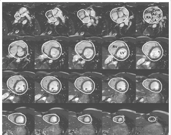
proximal parts of the great vessels covered by the peri-cardium. A region of interest was manually drawn around the pericardial border in all the short-axis images of the heart in end-diastole (Figure 1) using a freely available software (Segment 1.697; http://segment. heiberg.se) [16]. The summed area was then multiplied with slice thickness to obtain THV. The volumetric method used to determine THV has previously been applied in studies of cardiac pumping mechanics [15,17-21].
Furthermore, endocardial borders of the left ventricular (LV) myocardium were manually delineated in end-diasto-lic and end-systoend-diasto-lic short-axis images, enabling calculation of LV end-diastolic and end-systolic volumes (LVEDV and LVESV) as well as LV ejection fraction (LVEF). In addi-tion, the epicardial borders of the left ventricle were deli-neated for determination of LV mass (LVM).
Viability assessment
Myocardial scar/fibrosis was assessed by manual delinea-tion of the hyperenhanced myocardium in the LGE short-axis images.
Exercise test with respiratory gas analysis
A maximal incremental exercise test was performed using an electronically braked cycle ergometer (Siemens Ergomed 940, Upplands Väsby, Sweden) with respiratory gas analysis equipment (Oxycon Champion, Jaeger, Hochberg, Germany). The test protocols were chosen to yield exercise duration of approximately 8-12 minutes, yielding starting stress between 10-150 W with 10-30 W increment per minute. Peak oxygen uptake was defined as the highest value reached at the end of exercise and expressed both as ml min-1(VO2peak) and ml min-1kg-1
(VO2peakkg).
A 12-lead ECG was acquired throughout the test. Blood pressure was measured by sphygmanometer in the supine position before exercise, every two minutes during exercise on the ergometer cycle and in the supine position at rest after exercise.
VO2peak/THV ratio
A ratio between VO2peak and THV was derived.
to partly compensate for differences in oxygen delivery capacity in peripheral tissue in the presence of anemia. The VO2peak/THV ratio was also compared to levels of
plasma amino terminal pro-brain natriuretic peptide (pro-BNP) for the patients who had a pro-BNP taken within 4 months of the exercise test.
Statistical analysis
Values are shown as mean ± standard deviation. Uni-variate linear regression analysis was used to assess the correlation between VO2peak and THV as well as body
weight. Unpaired T-test was used to assess differences between variables where normal distribution was assumed. The Mann-Whitney test was used to assess differences where normal distribution could not be assumed. Forward stepwise multivariate regression ana-lysis was performed to assess the independent predictive
values of THV, LVEF, LVEDV, VO2peak, VO2peakkg
and the VO2peak/THV ratio for presence of HF.
Recei-ver operating characteristic (ROC) analysis was per-formed to test the ability of the VO2peak/THV ratio to
distinguish between HF patients and controls. All statis-tical analysis was performed using SPSS 16.0 (Chicago, IL, USA). A p-value of <0.05 was considered statistically significant.
Results
Subject characteristics are seen in Table 1. Twelve patients had been diagnosed with dilated cardiomyopathy, five patients with ischemic cardiomyopathy, three patients with suspected myocardial hemocromatosis, two patients with hypertrophic cardiomyopathy, two patients with con-genital heart disease, two patients with constrictive cardio-myopathy, one patient with post-partum cardiocardio-myopathy,
Figure 1 Delineation of total heart volume. Example of delineation of the pericardium from short-axis cardiac magnetic resonance images enabling determination of the total heart volume. The base is seen in the upper left corner and apex in the lower right corner. Ao = ascending aorta, LA = left atrium, LV = left ventricle, Pulm = pulmonary artery, RA = right atrium, RV = right ventricle.
one patient with drug-induced cardiomyopathy, and three patients with HF of unknown cause. In 16% (5/31) of the patients there were signs of scarred/fibrous myo-cardium on the LGE images. Hemoglobin levels were assessed the same day as the exercise test in 65% (39/60) of the healthy volunteers and in 73% (52/71) of 71 athletes (hemoglobin levels were not assessed in the triathletes) and within 2 weeks of the exercise test in 97% (30/31) of the patients.
jects with a considerable variation in VO2peak for a given
body weight.
Even though a significant difference in LVEF was found between the control subjects and the HF patients (p < 0.001; Figure 3A), there was a significant overlap with 29% (9/31) of the HF patient examinations showing LVEF > 50%. Figure 3B shows a comparison between control subjects and HF patients with LVEF > 50%, where the HF patients had a slightly higher LVEF (p = 0.04). Despite the higher LVEF in these patients, the VO2peak/THV ratio was significantly lower compared
to the control subjects (p < 0.001; Figure 3C).
VO2peak/THV ratio
Figure 4A shows the VO2peak/THV ratio for each
sub-ject group. The HF patients showed a significantly lower VO2peak/THV ratio (p < 0.001) than the other subject
groups, even after normalizing for hemoglobin levels (p < 0.001, Figure 4B).
When dividing the different subgroups with regard to VO2peak alone, there was still a significant difference
between healthy volunteers and HF patients (3102 ± 696
747*** 430*** VO2peakkg(ml min-1 kg-1) 41 ± 7 52 ± 7*** 16 ± 5*** LVEDV (ml) 183 ± 31 223 ± 39*** 280 ± 132*** LVM (g) 105 ± 25 130 ± 34*** 177 ± 87*** THV (ml) 793 ± 128 947 ± 169*** 1201 ± 460*** LVEF (%) 62 ± 6 58 ± 6** 37 ± 22***
DBP = diastolic blood pressure, LVEDV = left ventricular end-diastolic volume, LVEF = left ventricular ejection fraction, LVM = left ventricular mass, SBP = systolic blood pressure, THV = total heart volume, VO2peak = peak oxygen
uptake, **p < 0.01, ***p < 0.001 compared to the healthy volunteers.
Figure 2 Peak oxygen uptake in relation to total heart volume and body weight. Peak oxygen uptake (VO2peak) in comparison to A) total
heart volume (THV) and B) body weight. Filled circles denote control subjects, including healthy volunteers and athletes and open circles denote heart failure patients. Solid lines represent the regression line and dashed lines the 95% confidence interval. In the control group there was a strong correlation between VO2peak and THV. Even though there was also a significant correlation between VO2peak and body weight,
vs 1309 ± 430 ml/min; p < 0.001). However, there was a considerable overlap between the two groups (Figure 5A). Ten control subjects and 10 HF patients had overlapping VO2peak (Figure 5B). The VO2peak/THV ratio, however,
differed significantly between these two groups (p < 0.001) (Figure 5C). The LVEF for the overlapping healthy volunteers did not differ significantly from the overlap-ping HF patients (42 ± 25% vs 61 ± 5%; p = 0.11), whereas THV was significantly larger for the latter (640 ± 66 vs 1092 ± 336 ml/min; p < 0.001).
In 68% (21/31) of the HF patients a pro-BNP was taken 11 ± 25 days from the exercise test. There was a significant correlation between decreasing VO2peak/THV ratio and
increasing pro-BNP (r2= 0.42, p = 0.002).
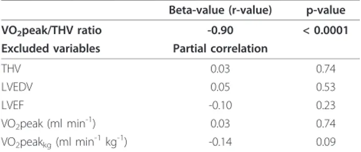
Furthermore, the VO2peak/THV ratio was the only
independent predictor of HF (p < 0.001; Table 2). The area under the curve (AUC) for the VO2peak/THV ratio
to diagnose HF was 1.0. With a cut-off value of 3.0, the VO2peak/THV ratio had a sensitivity of 100% and a
spe-cificity of 97%. Even though there was a slight decrease in VO2peak/THV ratio with increasing age, there was a
significant difference between patients and healthy volunteers for all age groups (Figure 6).
Discussion
This study shows that a VO2peak/THV ratio can be
used to distinguish patients with HF from healthy volunteers and athletes, even in patients with preserved
Figure 4 VO2peak/THV ratio in heart failure patients and control subjects. VO2peak/THV ratio in heart failure patients compared to healthy
volunteers and different groups of athletes. The VO2peak/THV ratio was significantly lower in the heart failure patients compared to the control
groups (A). This relationship remained unchanged after normalizing the results to hemoglobin levels (B). The solid line represents the mean value. ** p < 0.01 and *** p < 0.001 compared to healthy volunteers.
Figure 3 Left ventricular ejection fraction in relation to VO2peak/THV ratio. Left ventricular ejection fraction (LVEF) for A) all subjects and
B) control subjects and heart failure patients with LVEF > 50% and C) VO2peak/THV ratio for the control subjects and heart failure patients with
LVEF > 50%. In the heart failure patients with LVEF > 50% there was a significantly lower VO2peak/THV ratio despite a slightly higher LVEF
systolic LV function and after normalizing for hemoglo-bin levels.
Clinical use of the proposed VO2peak/THV ratio
It is expected that the VO2peak/THV ratio will drop in
the presence of HF, since cardiac output, and conse-quently VO2peak, is disproportional to the metabolic
requirements of the peripheral tissue in the situation of a failing heart. However, when and in which stage of HF development the VO2peak/THV ratio decreases is not
known. A decreased VO2peak/THV ratio could
poten-tially be seen in patients with no symptoms at rest and in the absence of significant structural cardiac abnormalities.
Figure 5 Peak oxygen uptake in comparison to VO2peak/THV ratio. Peak oxygen uptake (VO2peak) for the healthy volunteers and the HF
patients (A) which showed a considerable overlap (B). The VO2peak/THV ratio was, however, significantly lower in the 10 heart failure patients
compared to the 10 healthy volunteers with overlapping VO2peak (C). Dashed lines indicate the overlap between the two groups and the solid
Currently, maximal exercise test with analysis of respiratory gases and analysis of VO2peak is
predomi-nantly used in patients with terminal HF under consid-eration for heart transplantation [7]. The present study suggests that assessment of VO2peak in relation to THV
for determination of a VO2peak/THV ratio can be of
potential use even in non-terminal and early stage HF, maybe even before the patient exhibit symptoms, such as in stage A HF [4]. Another possible application of a VO2peak/THV ratio is in management of patients with
a history of endurance training and a sudden change in exercise capacity. These patients might perform within normal limits during a maximal exercise test with analy-sis of respiratory gases when compared to age and gen-der matched controls. However, in relation to THV the working capacity might be decreased which would be detected by a low VO2peak/THV ratio as an indicator of
early stage HF. This is supported by the findings that the healthy volunteers and the HF patients with overlap-ping VO2peak showed a significant difference in
VO2peak/THV ratio with almost no overlap, indicating
that the ratio has additional diagnostic value. Further-more, LVEF was not statistically significant different between the two overlapping groups. Thus, VO2peak/
THV provides diagnostic information beyond LVEF. Even though there was a significant difference between control subjects and HF patients with regard to LVEF in the present study, almost 30% of the patients had nor-mal LVEF. However, these patients had significantly lower VO2peak/THV ratio than the control subjects
with almost no overlap. Thus, VO2peak/THV ratio may
also be used as a marker of HF in patients with pre-served systolic LV function.
Heart failure is often described based on clinical symptoms as being mild, moderate, or severe. Mild HF is used for patients with no significant physical limita-tions due to dyspnoea or fatigue and severe HF is used for patients who are markedly symptomatic with a need for frequent medical attention. Moderate HF is used for the remaining patients. For clinical management of HF patients and for randomized clinical trials, the New York Heart Association (NYHA) functional classification [22,23] is often employed. However, the accuracy of diagnosing HF by clinical means alone is often inade-quate [2,3]. Hence, there is a need for objective mea-sures of cardiac performance in order to determine the efficiency of different therapeutic strategies in rando-mized clinical trials.
The clinical use a VO2peak/THV ratio could
poten-tially complement commonly used cardiac diagnostic biomarkers such as ECG and biochemical markers. A commonly used biochemical marker for severity of HF is pro-BNP, which has been shown to correlate with the severity of HF [24,25]. In the present study, the VO2peak/THV ratio was shown to correlate with
pro-BNP, indicating that the ratio can potentially serve as a measure for severity of HF.
Normalization of peak oxygen uptake to cardiac dimension rather than body weight
Lower limits for VO2peak are usually determined in
relation to body weight and used to determine if a patient is suitable for heart transplantation or not [26,27]. However, the present study show a considerable variability in VO2peak for a given body weight (Figure 2B).
Total heart volume, however, was shown to be closely correlated to VO2peak in healthy volunteers and
ath-letes but not in HF patients. Thus, for the non-failing heart, the VO2peak/THV ratio is preserved in the
pre-sence of a physiologically enlarged heart, whereas for the failing heart the VO2peak/THV ratio is not
pre-served as THV increases due to pathological enlarge-ment of the heart. This suggests that VO2peak should
be normalized to THV and not to body weight. This is
Table 2 Forward stepwise multivariate regression analysis for prediction of heart failure
Beta-value (r-value) p-value VO2peak/THV ratio -0.90 < 0.0001
Excluded variables Partial correlation
THV 0.03 0.74
LVEDV 0.05 0.53
LVEF -0.10 0.23
VO2peak (ml min-1) 0.03 0.74
VO2peakkg(ml min-1kg-1) -0.14 0.09 LVEDV = left ventricular end-diastolic volume, LVEF = left ventricular ejection fraction, THV = total heart volume, VO2peak = peak oxygen uptake.
Figure 6 VO2peak/THV ratio in different age groups. VO2peak/
THV ratio in healthy volunteers and in heart failure patients in different age groups. Even though there was a slight decrease in VO2peak/THV ratio with increasing age in the healthy volunteers,
VO2peak/THV ratio remained significantly higher in the healthy
volunteers compared to the heart failure patients. * p < 0.05 and *** p < 0.001
In the present study THV was determined by CMR, which is considered the gold standard for assessing car-diac dimensions and function [13]. In situations when CMR is not available or is contraindicated, i.e. in patients with biventricular pacing, THV could poten-tially also be determined by low dose-cardiac CT, pre-ferably gated. However, non-gated examinations would also be possible if CT thorax is performed for other rea-sons, since THV varies little during the cardiac cycle [15,17,18]. Three dimensional echocardiography might potentially also be used to determine THV, although this technique may suffer from incomplete coverage of the tissue within the pericardium. By using the THV, enlargement of all cardiac chambers including the atria is considered. Enlargement of the left atrium has been shown to provide prognostic value in HF patients with normal LVEF [30].
Limitations
The results of the present study should be interpreted in the light of some limitations. The number of patients in the present study is limited and the patients were retro-spectively included with clinically diagnosed HF and clinically determined etiology. Thus, no specific HF inclusion criteria based on biochemical markers, ECG or echocardiographic variables were applied. Furthermore, the patients had different etiologies and different stages of HF. However, this pilot study can be considered a proof of the concept study for determining a VO2peak/
THV ratio as an objective measure of cardiac capacity, independent of HF etiology.
Due to symptoms during the exercise test, HF patients might not reach VO2peak representative of the
indivi-dual’s actual maximal oxygen uptake capacity. For this reason VO2peak is referred to as peak and not maximal
oxygen uptake in the present study.
There was a significant difference in mean age between the patient population and the control groups. However, the slight decrease in VO2peak/THV ratio
found with increased age in the healthy volunteers (21-65 years) is negligible compared to the decrease in VO2peak/THV ratio found in the HF patients.
Conclusions
The VO2peak/THV ratio can be used to distinguish
patients with known HF from healthy volunteers and athletes, even in patients with preserved systolic LV function and after normalizing for hemoglobin levels.
Acknowledgements
We thank our technicians Ann-Helen Arvidsson and Christel Carlander for their invaluable assistance with data acquisition.
Author details
1Dept of Clinical Physiology, Lund University and Skåne University Hospital,
Lund, Sweden.2Dept of Sport Sciences, Malmö University, Malmö, Sweden.
3Dept of Cardiology, Lund University and Skåne University Hospital, Lund,
Sweden.
Authors’ contributions
HE, KS, MC, and HA have made substantial contributions to conception and design of the study. HE, KS, MC, TB, BE, HM, and BH, were responsible for acquisition of data. HE, KS, MC, BE, and HA were responsible for analysis and interpretation of the data. All authors have been involved in drafting the manuscript and revising it critically for important intellectual content. Furthermore, all authors have given final approval of the current version to be published.
Competing interests
The authors declare that they have no competing interests.
Received: 23 April 2010 Accepted: 16 December 2010 Published: 16 December 2010
References
1. Dickstein K, Cohen-Solal A, Filippatos G, McMurray JJ, Ponikowski P, Poole-Wilson PA, Stromberg A, van Veldhuisen DJ, Atar D, Hoes AW, Keren A, Mebazaa A, Nieminen M, Priori SG, Swedberg K, Vahanian A, Camm J, De Caterina R, Dean V, Dickstein K, Filippatos G, Funck-Brentano C, Hellemans I, Kristensen SD, McGregor K, Sechtem U, Silber S, Tendera M, Widimsky P, Zamorano JL: ESC Guidelines for the diagnosis and treatment of acute and chronic heart failure 2008: the Task Force for the Diagnosis and Treatment of Acute and Chronic Heart Failure 2008 of the European Society of Cardiology. Developed in collaboration with the Heart Failure Association of the ESC (HFA) and endorsed by the European Society of Intensive Care Medicine (ESICM). Eur Heart J 2008, 29(19):2388-442. 2. Wheeldon NM, MacDonald TM, Flucker CJ, McKendrick AD, McDevitt DG,
Struthers AD: Echocardiography in chronic heart failure in the community. Q J Med 1993, 86(1):17-23.
3. Remes J, Lansimies E, Pyorala K: Usefulness of M-mode echocardiography in the diagnosis of heart failure. Cardiology 1991, 78(3):267-77. 4. Hunt SA: ACC/AHA 2005 guideline update for the diagnosis and
management of chronic heart failure in the adult: a report of the American College of Cardiology/American Heart Association Task Force on Practice Guidelines (Writing Committee to Update the 2001 Guidelines for the Evaluation and Management of Heart Failure). J Am Coll Cardiol 2005, 46(6):e1-82.
5. Ekblom B, Hermansen L: Cardiac output in athletes. J Appl Physiol 1968, 25(5):619-25.
6. Mitchell JH, Sproule BJ, Chapman CB: The physiological meaning of the maximal oxygen intake test. J Clin Invest 1958, 37(4):538-47.
7. Weber KT, Janicki JS, McElroy PA: Determination of aerobic capacity and the severity of chronic cardiac and circulatory failure. Circulation 1987, 76(6 Pt 2):VI40-5.
8. Weber KT, Janicki JS, Ward DM, McElroy PA: Measurement and interpretation of maximal oxygen uptake in patients with chronic cardiac or circulatory failure. J Clin Monit 1987, 3(1):31-7.
9. Hammond HK, White FC, Bhargava V, Shabetai R: Heart size and maximal cardiac output are limited by the pericardium. Am J Physiol 1992, 263(6 Pt 2):H1675-81.
10. Stray-Gundersen J, Musch TI, Haidet GC, Swain DP, Ordway GA, Mitchell JH: The effect of pericardiectomy on maximal oxygen consumption and maximal cardiac output in untrained dogs. Circ Res 1986, 58(4):523-30. 11. Steding K, Engblom H, Buhre T, Carlsson M, Mosen H, Wohlfart B,
Arheden H: Relation between cardiac dimensions and peak oxygen uptake. J Cardiovasc Magn Reson 12(1):8.
12. Weber KT, Clark WA, Janicki JS, Shroff SG: Physiologic versus pathologic hypertrophy and the pressure-overloaded myocardium. J Cardiovasc Pharmacol 1987, 10(Suppl 6):S37-50.
13. Pennell DJ, Sechtem UP, Higgins CB, Manning WJ, Pohost GM,
Rademakers FE, van Rossum AC, Shaw LJ, Yucel EK: Clinical indications for cardiovascular magnetic resonance (CMR): Consensus Panel report. J Cardiovasc Magn Reson 2004, 6(4):727-65.
14. Simonetti OP, Kim RJ, Fieno DS, Hillenbrand HB, Wu E, Bundy JM, Finn JP, Judd RM: An improved MR imaging technique for the visualization of myocardial infarction. Radiology 2001, 218(1):215-23.
15. Carlsson M, Cain P, Holmqvist C, Stahlberg F, Lundback S, Arheden H: Total heart volume variation throughout the cardiac cycle in humans. Am J Physiol Heart Circ Physiol 2004, 287(1):H243-50.
16. Heiberg E, Sjogren J, Ugander M, Carlsson M, Engblom H, Arheden H: Design and validation of Segment–freely available software for cardiovascular image analysis. BMC Med Imaging 2010, 10:1.
17. Bowman AW, Kovacs SJ: Assessment and consequences of the constant-volume attribute of the four-chambered heart. Am J Physiol Heart Circ Physiol 2003, 285(5):H2027-33.
18. Waters EA, Bowman AW, Kovacs SJ: MRI-determined left ventricular “crescent effect": a consequence of the slight deviation of contents of the pericardial sack from the constant-volume state. Am J Physiol Heart Circ Physiol 2005, 288(2):H848-53.
19. Carlsson M, Ugander M, Mosen H, Buhre T, Arheden H: Atrioventricular plane displacement is the major contributor to left ventricular pumping in healthy adults, athletes, and patients with dilated cardiomyopathy. Am J Physiol Heart Circ Physiol 2007, 292(3):H1452-9.
20. Carlsson M, Ugander M, Heiberg E, Arheden H: The quantitative relationship between longitudinal and radial function in left, right, and total heart pumping in humans. Am J Physiol Heart Circ Physiol 2007, 293(1):H636-44. 21. Carlsson M, Rosengren A, Ugander M, Ekelund U, Cain PA, Arheden H:
Center of volume and total heart volume variation in healthy subjects and patients before and after coronary bypass surgery. Clin Physiol Funct Imaging 2005, 25(4):226-33.
22. AHA medical/scientific statement. 1994 revisions to classification of functional capacity and objective assessment of patients with diseases of the heart. Circulation 1994, 90(1):644-5.
23. Heart Failure Society of America (HFSA) practice guidelines. HFSA guidelines for management of patients with heart failure caused by left ventricular systolic dysfunction–pharmacological approaches. J Card Fail 1999, 5(4):357-82.
24. Mueller C, Scholer A, Laule-Kilian K, Martina B, Schindler C, Buser P, Pfisterer M, Perruchoud AP: Use of B-type natriuretic peptide in the evaluation and management of acute dyspnea. N Engl J Med 2004, 350(7):647-54. 25. Wright SP, Doughty RN, Pearl A, Gamble GD, Whalley GA, Walsh HJ,
Gordon G, Bagg W, Oxenham H, Yandle T, Richards M, Sharpe N: Plasma amino-terminal pro-brain natriuretic peptide and accuracy of heart-failure diagnosis in primary care: a randomized, controlled trial. J Am Coll Cardiol 2003, 42(10):1793-800.
26. Costanzo MR, Augustine S, Bourge R, Bristow M, O’Connell JB, Driscoll D, Rose E: Selection and treatment of candidates for heart transplantation. A statement for health professionals from the Committee on Heart
Failure and Cardiac Transplantation of the Council on Clinical Cardiology, American Heart Association. Circulation 1995, 92(12):3593-612. 27. Mancini DM, Eisen H, Kussmaul W, Mull R, Edmunds LH, Wilson JR: Value of
peak exercise oxygen consumption for optimal timing of cardiac transplantation in ambulatory patients with heart failure. Circulation 1991, 83(3):778-86.
28. Saltin B, Calbet JA: Point: in health and in a normoxic environment, VO2 max is limited primarily by cardiac output and locomotor muscle blood flow. J Appl Physiol 2006, 100(2):744-5.
29. Wagner PD: Counterpoint: in health and in normoxic environment VO2max is limited primarily by cardiac output and locomotor muscle blood flow. J Appl Physiol 2006, 100(2):745-8.
30. Rossi A, Cicoira M, Florea VG, Golia G, Florea ND, Khan AA, Murray ST, Nguyen JT, O’Callaghan P, Anand IS, Coats A, Zardini P, Vassanelli C, Henein M: Chronic heart failure with preserved left ventricular ejection fraction: diagnostic and prognostic value of left atrial size. Int J Cardiol 2006, 110(3):386-92.
doi:10.1186/1532-429X-12-74
Cite this article as: Engblom et al.: Peak oxygen uptake in relation to total heart volume discriminates heart failure patients from healthy volunteers and athletes. Journal of Cardiovascular Magnetic Resonance 2010 12:74.
Submit your next manuscript to BioMed Central and take full advantage of:
• Convenient online submission
• Thorough peer review
• No space constraints or color figure charges
• Immediate publication on acceptance
• Inclusion in PubMed, CAS, Scopus and Google Scholar
• Research which is freely available for redistribution
Submit your manuscript at www.biomedcentral.com/submit