Signs and symptoms of TMJ disorders in adults after adolescent
Herbst therapy:
A 6-year and 32-year radiographic and clinical follow-up study
Hans Pancherz
a; Hanna Sale´
b; Krister Bjerklin
cABSTRACT
Objective: To analyze radiographic signs of temperomandibular joint (TMJ) osteoarthritis and clinical TMJ symptoms in patients 6 years and 32 years after treatment with a Herbst appliance. Materials and Methods: Fourteen patients were derived from a sample of 22 with Class II division 1 malocclusions consecutively treated with a banded Herbst appliance at the age of 12–14 years old (T1-T2). The subjects were reexamined after therapy at the ages of 20 years (T3) and 46 years (T4). The TMJs of the 14 patients were analyzed radiographically (conventional lateral tomography at T3 and cone-beam computed tomography at T4) and clinically/anamnestically at T3 and T4. Results: Six years after Herbst therapy, signs of osteoarthritis were seen in one patient. At the 32-year up, two additional patients had developed signs of osteoarthritis. At the 6-32-year follow-up, TMJ clicking was present in two patients, though none of the patients reported TMJ pain. At the 32-year follow-up, six patients had TMJ clicking and one patient had TMJ pain.
Conclusions: This longitudinal very-long-term follow-up study after Herbst therapy revealed only minor problems from the TMJ. The TMJ findings 6 years and 32 years after Herbst treatment corresponded to those in the general population. Thus, in the very long term, the Herbst appliance does not appear to be harmful to the TMJ. (Angle Orthod. 2015;85:735–742.)
KEY WORDS: TMJ disorders; Herbst therapy
INTRODUCTION
During Herbst treatment1 the mandible is held in a
permanent protruded position that will interfere with the physiologic function of the stomatognathic system. Because of this, Herbst treatment has been blamed of causing temporomandibular disorders (TMD). Except for the study of Foucard et al.2 using a removable
Herbst appliance, critical statements are mainly personal opinions.
Earlier studies of Herbst appliances3–6 have shown
that, on a short-term basis, Herbst therapy for
adolescent patients with Class II malocclusion does not cause disorders of the muscular or temperoman-dibular joint (TMJ) part of TMD. Although two of the Herbst investigations3,6 have follow-up periods of
7.5 years and 4 years, respectively, the studies are not longitudinal in nature, and no information exists with respect to TMD after the age of 20 years.
The present longitudinal radiographic and clinical very-long-term follow-up study after Herbst therapy is concerned with the TMJ part of TMD. The aim was to reexamine patients consecutively treated with a Herbst appliance 32 years after therapy and to compare signs and symptoms of TMJ disorders in these subjects at 20 years and 46 years of age. Emphasis was placed on signs and symptoms of osteoarthritis and disc displacement.
MATERIALS AND METHODS Subjects
The patients in this study were derived from a sample of 22 consecutive patients with Class II division 1 malocclusion (19 males and 3 females) treated with a aProfessor and Chair Emeritus, Department of Orthodontics,
University of Giessen, Giessen, Germany.
bAssistant Professor, Department of Oral and Maxillofacial
Radiology, Faculty of Odontology, Malmo¨ University, Sweden.
cAssociate Professor, Department of Orthodontics, Faculty of
Odontology, Malmo¨ University, Sweden.
Corresponding author: Dr Hans Pancherz, Dresdener Str. 5a, D-35435 Wettenberg, Germany
(e-mail: hans.pancherz@dentist.med.uni-giessen.de) Accepted: October 2014. Submitted: July 2014. Published Online: December 31, 2014
G2015 by The EH Angle Education and Research Foundation, Inc.
Herbst appliance at the University of Malmo¨/Sweden in 1977–1978.7
In the years 2011 and 2012, 30 to 33 years after Herbst therapy, the 22 subjects were recalled to the Orthodontic Department in Malmo¨ for a late follow-up investigation. Two patients were deceased and six declined to come. Thus, the final follow-up sample comprised 14 subjects (12 males and 2 females) and is presented in detail in Table 1.
Treatment in all subjects was performed by one of the authors (Dr Pancherz) using a banded type of Herbst appliance with a simple anchorage system.7In
all subjects, at the start of treatment, there was a one-time full mandibular advancement. Before treatment, all subjects had an increased overjet (mean 5 8.2 mm) that, by treatment, was reduced by an average of 5.1 mm.7 After active Herbst therapy, the condyles
were positioned concentrically in their fossae.7
Due to major tooth irregularities after Herbst therapy, extrac-tions of four premolars were performed in two subjects (Case 1X and 8X), and upper and lower fixed multi-bracket appliances were placed for about 1 year. Methods
The patients were analyzed according to the follow-ing protocol:
N Six years after treatment (T3): The TMJs were
examined radiographically by means of conventional
lateral tomography, clinical examination, and a questionnaire.6
N Thirty-two years after treatment (T4): The TMJs were examined radiographically by means of cone-beam computed tomography (CBCT), clinical examination, and a questionnaire.
TMJ Radiography
All radiographic examinations of the TMJ, both conventional lateral tomography at T3 and CBCT at T4, were carried out at the Department of Oral and Maxillofacial Radiology, Faculty of Odontology, Uni-versity of Malmo¨/Sweden.
At T3, conventional lateral tomography of the TMJs was performed with a Polytome U (Massiot-Philips, Paris, France) using a hypocycloidal motion pattern.6
Two exposures were made on each TMJ to cover the total medial-lateral width of the TMJs. A multisection cassette was used containing four pairs of rare-earth screens and four films.
At T4, CBCT examinations of the TMJs were performed using the TMJ imaging protocol on a Veraviewepocs 3De (J. Morita Mfg. Corp, Kyoto, Japan). The protocol implied a field of view of 4 3 4 cm, a tube voltage of 80 kV, a tube current of 5 mA, and a scanning time of 9.4 seconds. Two volumes, one for each TMJ, were acquired for each patient. Reconstructions of images were performed in the Table 1. Characteristics of 14 Class II, Division 1 Malocclusions (Cases 1–14) Treated with the Herbst Appliance and Followed 32 Years after Therapya
Caseb
Gender Treatment Age (years)
Follow-up
Periods (years) Retention (Years) Class II Correction Male/Female Herbst/Extraction T1 T2 T3 T4 T2 -T3 T2-T4 Fixed/Removable Stable/Relapse
1X Male Herbst / extraction 13 17c 21 48 4 31 F/R (2 years) Stable
2 Male Herbst 13 14.5 20.5 48 6 33.5 No retention Stable
3 Male Herbst 11 12.5 19 45 6.5 32.5 No retention Stable
4 Male Herbst 13 14.5 20.5 47.5 6 33 R (4 years) Stable
5 Male Herbst 13.5 15 19 46.5 4 31.5 R (3 years) Relapse
6 Male Herbst 13 14.5 20.5 47.5 6 33 No retention Relapse
7 Female Herbst 13 14.5 20.5 48 6 33.5 R (2 years) Stable
8X Male Herbst / extraction 13 15 22 48 7 33 F/R (4 years) Stable
9 Male Herbst 12.5 14 20 45 6 31 R (2 years) Stable
10 Male Herbst 12 14 20 44 6 30 No retention Stable
11 Female Herbst 11 12.5 18.5 42.5 6 30 R (2 years) Stable
12 Male Herbst 12.5 14 21 46 7 32 F/R (3 years) Relapse (one side)
13 Male Herbst 12.5 14 21 45 7 31 R (2 years) Relapse
14 Male Herbst 12.5 14 22 45 8 31 R (2 years) Relapse (one side)
Summary 12 males, 12 Herbst; 12.5 14.3 20.4 46.1 6.1 31.8 4 No retention, 9 stable,
Mean 2 females 2 Herbst/extraction 10 retention 5 relapse
aT1 indicates before treatment; T2, after treatment, that is, 12 months after the Herbst appliance was removed and the occlusion had settled; T3, 6 years after treatment (lateral tomography of TMJs); T4, 32 years after treatment (CBCT of TMJs); F, fixed retention with a mandibular lingual canine to canine retainer; R, removable retention with an activator or a maxillary Hawley plate; F/R, fixed mandibular canine retainer in combination with a removable maxillary Hawley plate.
bCases 1X and 8X were treated by extractions after active Herbst therapy. cIncludes a 2-year break in treatment during the period T1-T2.
software iDixel, version 2 (J Morita Mfg Corp, Kyoto, Japan). The slice thickness was 1.5 mm and the slice interval was 1 mm.
All radiographic examinations were done in a closed-mouth position. Sagittal images were perpen-dicular to the long axis of the condyle (T3, T4), and the coronal images were parallel to the axis (T4). The TMJs (condylar head, mandibular fossa, and articular eminence) were interpreted for structural bone changes. The diagnosis of osteoarthritis was made according to Ahmad et al.8
(Table 2).
The interpretations of the radiographs were made by one observer (AP) at T36 and by two observers (Dr
Petersson and Dr Sale´) at T4. Observer Dr Petersson had many years of experience interpreting TMJ images and Dr Sale´ had several years of experience. At T4 the two observers were calibrated by evaluating 10 TMJ-CBCTs from patients who were not participat-ing in this study. After calibration the observers interpreted the present TMJ CBCT images individually. The observers were blinded to case history and clinical status. When the observer assessments differed, consensus was reached through discussion.
Clinical Examination and Questionnaire
Clinical examinations were carried out at T3 and T4 to gain information on TMJ clicking, crepitus, locking, and TMJ pain. The clinical examination at T3 was done by one examiner (Dr Hansen)6 using the method of
Carlsson and Helkimo.9At T4, Dr Sale´ performed the
clinical examination according to the Research Diag-nostic Criteria for Temporomandibular Disorders
(RDC/TMD).10 The questionnaires at T3 and T4
included questions regarding symptoms from the TMJs such as pain, clicking, crepitus, limited mouth opening, and locking.
Data Analysis
At T3 and T4, the individual data from the radiographic and clinical examinations as well as from the questionnaire were analyzed. Because of the small sample, statistical tests were not performed. The study was approved by the Ethical Committee University of Malmo¨/Sweden, No 2012/44.
RESULTS
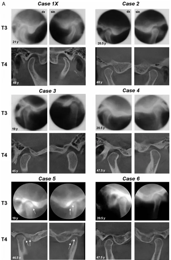
The individual TMJ findings at T3 and T4 derived from conventional lateral tomography and CBCT, clinical analyses, and questionnaires are presented in Table 3. The individual TMJ radiographs (lateral tomography and CBCT) from all patients are shown in Figures 1A through C.
TMJ Osteoarthritis
TMJ osteoarthritis was present in 1 of the 14 patients at T3 and in 3 of the 14 patients at T4. In the patient with osteoarthritis at T3 (Case 5; Figure 1A; Table 3), bone changes were present bilaterally. The right condyle had erosions and an osteophyte anteri-orly, and the left condyle was flattened and also had an anterior osteophyte. At T4 in Case 5, erosion had developed on the posterior part of the articular eminence in the right TMJ, and the previous osteo-phyte remained. However, the erosion on the anterior part of the right condyle had disappeared. At T4 the structural bone changes in the left TMJ were unchanged. In another two patients at T4 (Cases 8X and 14; Figures 1B and 1C, respectively; Table 3) structural bone changes had developed in the TMJs. Cases 8X had developed an erosion and an osteo-phyte on the right condyle as well as subcortical sclerosis and an osteophyte on the left condyle. Case 14 had developed an erosion on the posterior part of the articular eminence in the left TMJ. At T4, for one patient (Case 13; Figure 1C) osteoarthritis in the left TMJ was indeterminate.
TMJ Symptoms
Seven patients did not report any TMJ symptoms at T3 or T4. At T3, TMJ clicking was present in three TMJs (in two patients). At T4, TMJ clicking was present in eight TMJs (in six patients) (Table 3). According to the RDC/TMD, all clicking was due to disc displacement with reduction. At T3 none of the patients had TMJ crepitus, but at T4 crepitus was registered bilaterally in one patient. Both at T3 and T4, no TMJ locking was reported by any of the patients.
At T3 none of the patients reported TMJ pain. At T4, one patient experienced unilateral pain (Table 3) when Table 2. Osseous Diagnoses for the Temporomandibular Joint
from Conventional Lateral Tomography and Cone-Beam Computed Tomography Using Scoring Options A, B, or Ca
A. No osteoarthritis
1. Normal relative size of the condylar head; and
2. No subcortical sclerosis or articular surface flattening; and 3. No deformation due to subcortical cyst, surface erosion,
osteophyte, or generalized sclerosis. B. Indeterminate for osteoarthritis
1. Normal relative size of the condylar head; and
2. Subcortical sclerosis with/without articular surface flattening; or 3. Articular surface flattening with/without subcortical sclerosis;
and
4. No deformation due to subcortical cyst, surface erosion, osteophyte, or generalized sclerosis.
C. Osteoarthritis
1. Deformation due to subcortical cyst, surface erosion, osteo-phyte, or generalized sclerosis.
chewing tough food. No other patients reported difficul-ties in chewing food.
DISCUSSION
The loss of subjects at follow-up represents a potential source of bias.11 At the recall of the present
22 patients, 30–33 years after therapy, the attendance rate was 70% (when excluding the two persons who were deceased), which must be considered accept-able, especially in relation to the long follow-up period. Furthermore, with respect to dentoskeletal character-istics, length of treatment, immediate treatment, and 6-year posttreatment results,12,13
the six not attending subjects did not differ from the 14 attending ones. Thus, the participating subjects must be considered an unbiased group for a reliable long-term follow-up study. A possible study limitation was the lack of a control group, but the results reported were compared with those from epidemiologic studies on TMD.
In 1990, at the time of the T3 evaluations,6 lateral
tomography was the method of choice. The CBCT technique was not available until several years later. However, the two different radiographic modalities are not likely to affect the results significantly as the diagnostic accuracy of TMJ bone changes for CBCT and conventional tomography has been found to be comparable.14
In the clinical and anamnestic evaluations of the TMJs, at T3 the procedure of Carlsson and Helkimo9
was used. As RDC/TMD10
is widely used today, the T4 clinical evaluations were based on RDC/TMD. High reliability has been found for the clinical criteria.15,16
Both examination protocols at T3 and T4 included TMJ palpation and anamnestic information to achieve information regarding TMJ pain, sounds, and locking.
TMDs are common among children and adults. In previous epidemiologic, mainly cross-sectional, studies, the prevalence has been reported to vary between 6% and 71%.17–19 Furthermore, longitudinal studies have
disclosed a significant and unpredictable fluctuation of TMD over time.20,21 In the aforementioned studies of
TMD both the TMJ and the associate musculature were considered. This study, on the other hand, was confined to TMJ disorders exclusively. Thus, a direct compari-son, between the present findings and those from epidemiologic studies is difficult.
The criteria for TMJ osteoarthritis used in this study included surface erosion, osteophyte, subcortical cyst, or sclerosis.8
The results showed that 1 of the 14 patients (7% of TMJs) had osteoarthritis at the 6-year follow-up and 3 of the 14 patients (18% of TMJs) at the 32-year follow-up. These results are in line with the frequencies of osteoarthritis found in a recent CBCT study on subjects without ongoing pain.22
Table 3. TMJ Findings from Radiography, Clinical Examination and Anamnestic Recordings in 14 Class II, Division 1 Subjects (Cases 1–14) Treated with a Herbst Appliance and Followed 6 Years (T3) and 32 Years (T4) after Therapya
Osteoarthritisc TMJ Clicking/Crepitus TMJ Pain
Gender Lateral Tomography at T3 CBCT at T4 Clinical/ Questionnaire at T3 Clinical/ Questionnaire at T4 Questionnaire at T3 / T4 Caseb Male / Female TMJ Right TMJ Left TMJ Right TMJ Left TMJ Right TMJ Left TMJ Right TMJ Left TMJ Right TMJ Left
1X Male Clicking Clicking
2 Male
3 Male
4 Male
5 Male OA OA OA OA Clicking Clicking Clicking Clicking
6 Male
7 Female
8X Male OA OA
9 Male Clicking
10 Male Crepitus Clicking Clicking
11 Female Crepitus Crepitus Pain at T4
12 Male Clicking
13 Male Indeterminate Clicking
14 Male OA
Summary 12 males 1/28 TMJs with OA at T3 5/28 TMJs with OA at T4 3/28 TMJs with click-ing at T3 8/28 TMJs with click-ing at T4 1/28 TMJ with pain at T4 2 females 1 TMJ indeterminate OA at T4 1/28 TMJ with crepitus at T3 2/28 TMJs with crepi-tus at T4
aClinical indicates clinical examination, questionnaire, anamnestic recording; CBCT, cone-beam computed tomography; TMJ indicates temporomandibular joint; OA, osteoarthritis; RDC/TMD, Research Diagnostic Criteria for Temporomandibular Disorders.
bCases 1X and 8X were treated by extractions after active Herbst therapy. cOsteoarthritis diagnosis according to Ahmad et al.8(Table 2).
Figure 1. (A) TMJ radiographs (right and left side) from 14 Class II division 1 malocclusions (Cases 1–6) treated with the Herbst appliance. Conventional lateral tomograms from T3 (6 years after treatment) and CBCT examinations from T4 (32 years after treatment). Note the osteoarthritic changes in Case 5 at T3 and T4 (arrows). (B) TMJ radiographs (right and left side) from 14 Class II division 1 malocclusions (Cases 7–12) treated with the Herbst appliance. Conventional lateral tomograms from T3 (6 years after treatment) and CBCT examinations from T4 (32 years after treatment). In the Cases 9 and 11 (females) no lateral tomograms existed from T3. Note osteoarthritic changes in Case 8X at T4.
Figure 1. Continued. (arrows). (C) TMJ radiographs (right and left side) from 14 Class II division 1 malocclusions (Cases 13 and 14) treated with the Herbst appliance. Conventional lateral tomograms from T3 (6 years after treatment) and CBCT examinations from T4 (32 years after treatment). Note osteoarthritic changes in Case 14 at T4 (arrow). The T3 lateral tomograms are from the publication of Hansen et al.6with kind permission of theEuropean Journal of Orthodontics.
In asymptomatic patients, TMJ osteoarthritis has been reported to be more common in Class II (43%) and Class III (20%) than in Class I (3%) malocclu-sions.23 Furthermore, it has been shown that TMJ
osteoarthritis is more frequent in older than in younger persons, thus being an age-related change.24A similar
age dependency was also noted in our material: osteoarthritis was diagnosed at T3 in two (7%) joints and at T4 in five (18%) joints.
Previous studies have found a poor correlation between TMJ osteoarthritis and TMJ pain.22,25 The
result of this study supports these findings, as none of the patients with TMJ osteoarthritis had TMJ pain. The poor correlation between CBCT findings of osteoar-thritis and TMJ symptoms emphasizes the importance of evaluating the TMJs both radiographically and clinically. In our patients, TMJ pain was registered in only one TMJ with crepitus at T4. None of the patients had any TMJ locking or difficulties in chewing tough food. The results indicate that when TMJ symptoms were present, they were mild.
The clinical diagnosis of disc displacement was based on the RDC/TMD.10 As it has been proven by
magnetic resonance imaging (MRI)26 that a disc
displacement without reduction can exist without any clinical signs or symptoms, clinical diagnostic proce-dures will underestimate the actual prevalence of disc displacements.27
So in the absence of any MRI data, the present RDC/TMD clinical findings will not reveal all disc displacements.
A previous clinical Herbst study has shown that TMJ clicking, which was present before treatment, had
disappeared after therapy and remained absent during the first 12 months after treatment.5Furthermore, two
MRI investigations revealed that the position of the disc became relatively more retrusive by Herbst treatment.28,29
Thus, from the results of the aforemen-tioned three studies it was concluded that Herbst therapy did not result in any adverse changes in TMJ disc position. On the contrary, it was suggested that Herbst treatment may possibly promote repositioning of a displaced disc in patients with anterior disc displacement.28In the present patients the prevalence
of TMJ clicking was 11% (3/28 TMJs) at T3 and 25% (7/28 TMJs) at T4 (Table 3), and was thus of the same magnitude as that found in subjects representing normal populations.22,30,31
Summarizing our results, there was no indication that any of the long-term follow-up findings on TMJ disorders were related to Herbst therapy.
CONCLUSIONS
N The TMJ findings 6 years and 32 years after Herbst treatment corresponded to those in the general population.
N In the very-long-term perspective, the Herbst appli-ance was not thought to be harmful to the TMJ. ACKNOWLEDGMENT
The authors would like to thank Professor Arne Petersson, Department of Oral and Maxillofacial Radiology, University of Malmo¨/Sweden, for the help with the evaluations of the TMJ radiographs and for the valuable advice throughout the study.
REFERENCES
1. Pancherz H, Ruf S.The Herbst Appliance. Research-based Clinical Management. London, UK: Quintessence Publish-ing Co Ltd; 2008.
2. Foucard JM, Pajoni D, Carpentier P, Pharaboz C. MRI study of the temporomandibular joint disk behavior in children with hyperpropulsion appliances.Orthod Fr. 1998;69:79–91. 3. Ruf S, Pancherz H. Long-term TMJ effects of Herbst
treatment: a clinical and MRI study.Am J Orthod Dentofacial Orthop. 1998;114:475–483.
4. Ruf S, Pancherz H. Does bite-Jumping damage the TMJ? A prospective longitudinal clinical and MRI study of Herbst patients.Angle Orthod. 2000;70:183–199.
5. Pancherz H, Anehus-Pancherz M. The effect of continuous bite jumping with the Herbst appliance on the masticatory system: a functional analysis of treated Class II malocclu-sions.Eur J Orthod. 1982;4:37–44.
6. Hansen K, Pancherz H, Petersson A. Long-term effects of the Herbst appliance on the craniomandibular system with special reference to the TMJ.Eur J Orthod. 1990;12:244–253. 7. Pancherz H. The mechanism of Class II correction in Herbst
appliance treatment. A cephalometric investigation. Am J Orthod. 1982;82:104–113.
8. Ahmad M, Hollender L, Andersson Q, et al. Research diagnostic criteria for mandibular disorders (RDC/TMD): development of images analysis criteria and examiner reliability for image analysis. Oral Surg Oral Med Oral Pathol Oral Radiol Endod. 2009;107:844–860.
9. Carlsson GE, Helkimo M. Funktionell underso¨kning av tuggapparaten. In: Host JJ, Nygaard O¨ stby B, Osvald O, eds. Nordisk Klinisk Odontologi. Copenhagen, Denmark: A/S Forlaget for faglitteratur; 1972:8-I-1–8-I-21.
10. Dworkin SF, LeResche I. Research diagnostic criteria for temporomandibular disorders: review, criteria, examinations and specifications, critique.J Craniomandib Disord. 1992;6: 301–355.
11. Zelle BA, Bhandari M, Sanches AI, Probst C, Pape HC. Loss of follow-up in orthodontic trauma: is 80% follow-up still acceptable?J Orthop Trauma. 2013;27:177–181.
12. Pancherz H, Bjerklin K, Lindskog-Stockland B, Hansen K. A 32-year folllow-up study of Herbst therapy. A biometric dental cast analysis.Am J Orthod Dentofacial Orthop. 2014; 145:15–27.
13. Pancherz H, Bjerklin K, Hashem K. Late adult skeletofacial growth after adolescent Herbst therapy. A 32-year longitudinal follow-up study.Am J Orthod Dentofacial Orthop. 2015. In press.
14. Hintze H, Wiese M, Wentzel A. Cone beam CT and conventional tomography for the detection of morphological temporomandiblar joint changes.Dentomaxillofacial Radiol. 2007;36:192–197.
15. John MT, Dworking SF, Mancl LA. Reliability of clinical temporomandibular disorder diagnoses. Pain. 2005;118: 61–69.
16. Schmitter M, Ohlmann B, John MT, Hirsch C, Rammelsberg P. Research diagnostic criteria for temporomandibular disorders: a calibration and reliability study.Cranio. 2005; 23:212–218.
17. Vanderas AP. Prevalence of craniomandibular dysfunction in children and adolescents: a review.Pediatr Dent. 1987;9: 312–316.
18. Dibbets JM, van der Weele LT. Prevalence of structural bony changes in the mandibular condyle.J Craniomandib Disord. 1992;6:254–259.
19. dos Anjos Pontual ML, Freire JSL, Barbosa JMN, Frazao MAG, dos Anjos Pontual A, Fonseca da Silveria MM. Evaluation of bone changes in the temporomandibular joint using cone beam CT.Dentomaxillofac Radiol. 2012;41:24–29. 20. Magnusson T, Egermark I, Carlsson GE. A longitudinal epidemiologic study of signs and symptoms of temporo-mandibular disorders from 15 to 35 years of age.J Orofac Pain. 2000;14:310–319.
21. Va¨nman A, Agerberg G. Recurrent headaches and cranio-mandibular disorders in adolescents: a longitudinal study. J Craniomandib Disord. 1987;1:229–236.
22. Bakke M, Petersson A, Wiese M, Svanholt P, Sonnesson L. Bony deviations revealed by cone beam computed tomog-raphy of the temporomandibular joint in subjects without ongoing pain. J Oral Facial Pain Headache. 2014;28: 331–337.
23. Krisjane Z, Urtane I, Krumina G, Ragovska I. The prevalence of TMJ osteoarthritis in asymptomatic patients with dentofacial deformities: a cone-beam CT study. Int J Oral Maxillofac Surg. 2012;41:690–695.
24. Widmalm SE, Westesson PL, Kim IK, Pereira FJ Jr, Lundh H, Tasaki MM. Temporomandibular joint pathosis related to sex, age, and dentition in autopsy material.Oral Surg Oral Med Oral Pathol. 1994;78:416–425.
25. Palconet G, Ludlow JB, Tyndall DA, Lim PF. Correlating cone beam CT results with temporomandibular joint pain of osteoarthritic origin. Dentomaxillofac Radiol. 2012;41: 126–130.
26. Ribeiro RF, Tallents RH, Katzberg RW, et al. The prevalence of disc displacement in symptomatic and asymptomatic volunteers aged 6 to 25 years.J Orofac Pain. 1997;11:37–47.
27. Wiese M, Wenzel A, Hintze H, et al. Osseous changes and condyle position in TMJ tomograms: impact of RDC/TMD clinical diagnoses on agreement between expected and actual findings.Oral Surg Oral Med Oral Pathol Oral Radiol Endod. 2008;106:e52–e63.
28. Pancherz H, Ruf S, Thomalske-Faubert C. Mandibular articular disc position changes during Herbst treatment: A prospective longitudinal MRI study.Am J Orthod Dentofacial Orthop. 1999;116:207–214.
29. de Arruda Aidar LA, Dominguez GC, Abrahao M, Yamashita HK, Vigorito JV. Effects of Herbst appliance treatment on temporomandibular joint disc position and morphology: a prospective magnetic resonance imaging study.Am J Orthod Dentofacial Orthop. 2009;136:412–424.
30. Dworkin SF, Huggins KH, LeResche L, et al. Epidemiology of signs and symptoms in temporomandibular disorders: clinical signs in cases and controls.J Am Dent Assoc. 1990; 120:273–281.
31. Sale´ H, Bryndahl F, Isberg A. Temporomandibular joints in asymptomatic and symptomatic volunteers: a prospective 15-year follow-up clinical and MR imaging study.Radiology. 2013:267183–194.