DOCT OR AL DISSERT A TION IN ODONT OL OG Y DURER ISKANDER ANI MALMÖ TEMPOR OMANDIBUL AR JOINT IMA GIN G USIN G C ONE-BEAM C OMPUTED T OMOGR APHY
DURER ISKANDERANI
TEMPOROMANDIBULAR
JOINT IMAGING USING
CONE-BEAM COMPUTED
TOMOGRAPHY
T E M P O R O M A N D I B U L A R J O I N T I M A G I N G U S I N G C O N E - B E A M C O M P U T E D T O M O G R A P H Y
Malmö University, Faculty of Odontology
Doctoral Dissertation 2020
© Copyright: Durer Iskanderani, 2020 Front cover illustration: Aisha Iskanderani ISBN 978-91-7877-106-6 (print) ISBN 978-91-7877-107-3 (pdf) DOI 10.24834/isbn.9789178771073 Holmbergs, Malmö 2020
Department of Oral and Maxillofacial Radiology
Faculty of Odontology
DURER ISKANDERANI
TEMPOROMANDIBULAR
JOINT IMAGING USING
CONE-BEAM COMPUTED
TOMOGRAPHY
This publication is also available in electronic format at: www.mau.diva-portal.org
To My Beloved Family, Hussien, Maya & Malak and My Parents, Faisal & Hala
CONTENTS
PREFACE ... 9 ABBREVIATIONS ... 11 THESIS AT A GLANCE ... 13 ABSTRACT ... 14 POPULÄRVETENSKAPLIG SAMMANFATTNING ... 17 INTRODUCTION ... 19 Temporomandibular disorders ... 19Cone-beam computed tomography ... 23
Image interpretation ... 23
Radiation dosimetry ... 25
Dose optimisation ... 26
AIMS ... 30
MATERIALS AND METHODS ... 31
Imaging modalities ... 31
Panoramic examinations ... 31
Cone-beam computed tomography examinations ... 31
Study I and II ... 32
Radiographic examinations ... 32
Participated observers ... 33
Radiographic image assessment ... 33
CBCT educational tools development ... 33
Subjective evaluation of the web-based programme ... 34
Data analysis ... 34
Study III ... 35
Phantoms and Gafchromic film ... 35
Dose measurements ... 35
Study IV ... 36
Participants ... 36
Radiographic examinations ... 36
Radiographic image assessment ... 37
Data analysis ... 38 RESULTS ... 39 Studies I and II ... 39 Study III ... 43 Study IV ... 45 DISCUSSION ... 49
CBCT educational tools (Study I and II) ... 50
Dose measurements (Study III) ... 52
Dose optimisation (Study IV) ... 58
Implementation of the results ... 60
CONCLUSIONS ... 61 FUTURE RESEARCH ... 62 ACKNOWLEDGEMENTS ... 63 REFERENCES ... 65 PAPERS I-IV ... 75
PREFACE
This thesis is based on the following Papers, which are referred to in the text by their Roman numerals. All papers are reprinted with permission from the copyright holders. The papers are appended at the end of the thesis.
I. Assessment of a training programme on detection of temporomandibular joint osseous changes applying pre-defined 2D multiplane cone-beam computed tomography reconstructions. Iskanderani D, Alstergren P, Hellén-Halme K.
J Oral Rehabil. 2018; 45:282-288.
II. Web-based educational programme for temporomandibular joint assessment with cone-beam computed tomography.
Iskanderani D, Alstergren P, Ekberg EC, Shi XQ, Hellén-Halme K. J Oral Rehabil. 2020; 00:1-7.
III. Dose distributions in adult and child head phantoms for panoramic imaging and cone beam computed tomography imaging of the temporomandibular joint.
Iskanderani D, Nilsson M, Alstergren P, Hellén-Halme K. Oral Surg Oral Med Oral Pathol Oral Radiol. 2020; 130:200-208. IV. Evaluation of a low-dose protocol for cone beam computed
tomography of the temporomandibular joint.
Iskanderani D, Nilsson M, Alstergren P, Xie-Qi Shi, Hellén-Halme K. Dentomaxillofac Radiol. 2020; 49:20190495.
The author has performed most of the planning, data collection, data analysis, and writing of the papers in collaboration with the supervisors.
ABBREVIATIONS
3D Three-dimensional
2D Two-dimensional
ALADA As low as diagnostically acceptable
ALARA As low as reasonably achievable
Az Area under the ROC curve
CBCT Cone-beam computed tomography
CT Computed tomography
DC/TMD Diagnostic Criteria for
Temporomandibular Disorder
DJD Degenerative joint disease
FOV Field of view
GDP General dental practitioner
Gy Gray
ICRP International Commission on
Radiological Protection
k Kappa value
kV Kilovolt
LD Luminescent dosimeter
mA Milliampere
mAs Milliampere second
MPR Multiplanar reconstruction or
reformation
MRI Magnetic resonance imaging
µGy Microgray
NCRP National Council on Radiation
Protection and Measurements
OSL Optically stimulated luminescence
PPT PowerPoint
ROC Receiver operating characteristic
s Second
SE Standard error
SEDENTEXCT Safety and Efficacy of a New and
Emerging Dental X-ray modality
TLD Thermoluminescent dosimeter
TMJ Temporomandibular joint
THESIS AT A GLANCE
Study I: Observational study
An educational tool for the
assessment of temporomandibular joint osseous tissues applying pre-defined 2D multiplane cone-beam computed tomography reconstructions was assessed.
Study II: Observational study
A web-based programme for
temporomandibular joint evaluation using cone-beam computed tomo-graphy was developed and evaluated.
Study III: Phantom study
Dose distributions were obtained in adult and child head phantoms for panoramic imaging and cone-beam computed tomographic imaging of the temporomandibular joint.
Study IV: Clinical study
A low-dose protocol for cone-beam computed tomography imaging of the
temporomandibular joint was
ABSTRACT
Cone-beam computed tomography (CBCT) is one of the most revolutionary innovations in dentistry, and was introduced into the dental field two decades ago as a three-dimensional (3D) imaging modality. Since then, it has gained general clinical acceptance, and is widespread among many dental specialties. The CBCT examination is a fast and user-friendly technique that provides multiplanar images with high spatial resolution, providing information that is unattainable with two-dimensional (2D) imaging in many diagnostic tasks. Thus, CBCT is an essential examination tool that can replace or complement other examinations. CBCT imaging plays a valuable role when hard tissue abnormalities are suspected in the temporomandibular joint (TMJ) by providing diagnostic information on cortical and subcortical bone integrity or destruction/production changes.
The growing availability and use of CBCT at dental clinics has led to concerns as to whether the information obtained by CBCT imaging justifies the additional exposure of the patient to radiation and the training required to examine and interpret the images. The radiation protection principles: justification and optimisation, should always be applied as the basis for protection. This thesis presents four studies on TMJ imaging using CBCT. In line with recent trends towards digital online education, two CBCT educational tools were developed for the interpretation of CBCT images of the TMJ and published on the Malmö University web site. The first was an educational tool including 35 CBCT examinations of TMJ, presented as 2D multiplane CBCT images. The second was a web-based programme containing 15 CBCT examinations of TMJ, presented as 3D multiplane CBCT images. Both tools included the image analysis criteria of the Diagnostic Criteria for Temporomandibular disorder (DC/TMD) as an assessment module, and were tested by dental students. It was found that they could be useful educational tools for TMJ assessment using CBCT images.
The aim of the third study was to map and compare the distribution of absorbed doses using radiochromic film dosimeters in panoramic radiography and CBCT examinations of the TMJ using adult and child anthropomorphic head phantoms. Sheets of Gafchromic film (XR-QA2) were placed at five levels, corresponding to the radiographic examination, in the phantoms. The clinical protocols for panoramic and CBCT imaging of the TMJ of three dental X-ray units were used. The mean absorbed doses to a number of radiosensitive tissues within the oral and maxillofacial regions were estimated. The absorbed doses varied considerably among and within the radiosensitive tissues with examination type, X-ray unit, clinical setting, and patient age. The bone surface and salivary glands received the highest absorbed doses in both radiographic examinations. The radiation burden was lower when using two small right and left fields of view (FOVs) than when using a single larger FOV. Accurate measurements of the absorbed dose in small dental radiation fields is challenging due to steep dose gradients. The use of Gafchromic film has shown promising results, allowing dose comparisons between different radiographic imaging.
The last study was carried out to investigate the possibility of dose optimisation in CBCT examinations of the TMJ, in line with the recommendations of the International Commission on Radiological Protection (ICRP) and the National Council on Radiation Protection and Measurements (NCRP). Thirty-four adult patients referred for CBCT imaging of the TMJ underwent two examinations with different scanning protocols, a manufacturer-recommended protocol (default), and a low-dose protocol in which the tube current was reduced to 20% of that in the default protocol. Three image stacks were reconstructed: the default protocol, the low-dose protocol, and the low-dose protocol processed using a noise reduction algorithm. Four radiologists evaluated the visibility of TMJ anatomic structures, image quality and radiographic findings. It was found that the visibility of the TMJ anatomical structures, the overall image quality and the radiographic findings using the low-dose protocol were comparable to those in the default protocol, indicating that diagnostically comparable results could be obtained with a five times lower radiation dose.
Finally, this thesis highlights the diagnostics of TMJ imaging using CBCT in the context of image interpretation, imaging dosimetry and dose optimisation, all with the purpose of improving and optimising radiological TMJ diagnostics.
POPULÄRVETENSKAPLIG
SAMMANFATTNING
Denna avhandling belyser olika aspekter vid avbildning med Cone-beam computed tomography (CBCT) teknik, allt i syfte att förbättra och optimera käkledsdiagnostiken.
CBCT är en tredimensionell avbildningsmetod som introducerades för cirka två decennier sedan inom tandvården. CBCT-undersökningen är en snabb och användarvänlig teknik som i tre olika plan ger röntgenbilder med hög detaljupplösning. Detta kan medföra att information som inte kan avbildas i vanliga intraorala röntgenbilder kan visas och analyseras. Ett exempel är röntgenundersökningar av käklederna. Dessa kan med fördel avbildas med CBCT teknik när man misstänker hårdvävnadsförändring i leden Den stora nackdelen med CBCT tekniken är dock att den ger en betydligt högre stråldos till patienterna. Genom strikta kriterier för när CBCT ska användas och genom noggrann optimering av hur man ska utföra själva undersökningen kan man genomföra undersökningarna med minsta möjliga stråldos men som ändå ger tillräckligt bra bildunderlag för en analys. I Sverige får ansvaret för användning av denna metod enbart innehas av tandläkare med specialistkompetens inom odontologisk radiologi.
Denna avhandling presenterar fyra studier om CBCT och avbildning av käkleden. I linje med den senaste trenden mot digital online-utbildning utvecklades två pedagogiska verktyg för tolkning av CBCT-bilder av käkleder. Den första, ett utbildningsverktyg med 35 CBCT-undersökningar, presenterade som 2D-multiplanara CBCT-bilder. Den andra, ett webbaserat program som innehöll 15 CBCT-undersökningar, presenterade som 3D-multiplanara CBCT-bilder. Båda visade sig vara
Syftet med den tredje studien var att kartlägga och jämföra fördelningen av stråldos med hjälp av strålningskänslig film som placerades i två olika fantom, ett som symboliserade en vuxen patient och ett som motsvarade ett barn. Kliniska protokoll för panorama- och CBCT-avbildning av käkleden för tre olika röntgenapparater användes. Stråldoserna varierade avsevärt mellan och inom de strålkänsliga vävnaderna med undersökningstyp, röntgenenhet, klinisk inställning och patientens ålder. Benytor samt spottkörtlarna i området erhöll de högsta stråldoserna för båda röntgenundersökningarna.
Den sista studien genomfördes för att undersöka möjligheten till dosopimering vid CBCT-undersökningar av käkleder i linje med rekommendationerna från International Commission on Radiological Protection (ICRP) och National Council on Radiation Protection and Measurements (NCRP). Trettiofyra vuxna patienter genomgick två undersökningar med olika undersökningsprotokoll, ett standardprotokoll och ett lågdosprotokoll där rörströmmen reducerades till 20 %. Det visade sig att avbildningen av de anatomiska strukturerna i käkleden, den totala bildkvaliteten och de röntgenfynd som gjordes med lågdosprotokollet var jämförbara med dem för standardprotokollet, vilket indikerar att diagnostiskt jämförbara resultat kunde uppnås med en femtedel av stråldosen.
INTRODUCTION
Temporomandibular disorders
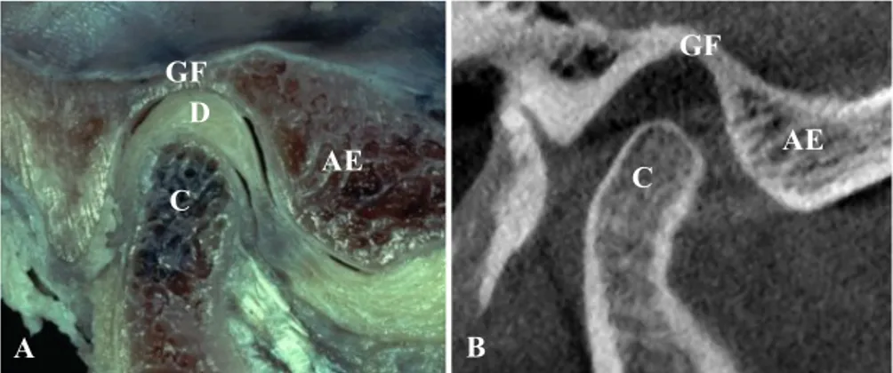
Temporomandibular disorders (TMDs) are a heterogeneous and often multifactorial group of disorders affecting the temporomandibular joint (TMJ), the masticatory muscles, and their associated structures (Figure 1). They represent a major source of orofacial pain of non-dental origin. The TMDs prevalence varied widely due to variation in the studied population, examination methods, and diagnostic criteria, however, they affect up to 12% of the population, with increased prevalence among younger individuals and women (National Institute of Dental and Craniofacial Research). These disorders commonly have characteristic clinical features such as pain, limited jaw function and joint sound, which can influence daily activities, psychosocial functioning and quality of life. The central attribute of all these disorders is pain, particularly when it becomes chronic, and usually leads to treatment seeking. Other symptoms include headache, jaw asymmetry or locking, neck pain, oral parafunctions and muscle tenderness. The TMDs include pain-related and joint-related disorders, with degenerative joint disease (DJD) being among the most common pathology affecting the TMJ (Tanaka et al., 2008; Ohrbach and Dworkin, 2016).
Figure 1. The temporomandibular joint. (A) Photograph of an
anatomical dissection (reprinted with permission from Lars Eriksson and Per-Lennart Westesson), and (B) A radiographic image. AE: Articular Eminence, C: Condyle, D: Disc, GF: Glenoid Fossa.
DJD is a non-inflammatory degenerative disease of the joints characterized by destruction and repair of the articular fibrocartilage and underlying osseous structures. The degenerative changes associated with DJD are believed to result from dysfunction of the remodelling capacity of the articular structures due to reduced adaptive capacity or excessive functional loading that exceeds the adaptive capacity (Tanaka et al., 2008). It is commonly related to trauma, the aging process, systemic illness, or hormonal or mechanical factors. A reduction or loss of adaptive response can lead to loss of the articulating cartilage together with gradual progressive destruction of the osseous tissues. Clinically, this can be associated with mild or symptom-free disease, and radio-graphic examination usually is part of the diagnostic process (Tanaka et al., 2008).
Historically, considerable effort has been devoted to defining a reliable modus for the diagnosis of TMDs (Dworkin and LeResche, 1992). However, many controversies have arisen, together with a lack of consensus among clinicians and researchers, who have reported a lack of scientific evidence supporting aetiology, diagnosis and treatment strategies related to TMDs (Ohrbach and Dworkin, 2016). The introduction of the Diagnostic Criteria for Temporomandibular Disorder (DC/TMD) (Schiffman et al., 2014) formerly RDC/TMD, the Research
AE GF C A C AE GF D B
Diagnostic Criteria for TMD (Dworkin and LeResche, 1992) has facilitated TMD diagnostics. It was developed based on a multicentre, population, and experimental and clinical studies. The DC/TMD offers an evidence-based diagnostic system when identifying TMDs, intended to expand scientific knowledge and standardize patient care. It has been proposed that the DC/TMD be used in general dentistry for screening and examination to identify patients suspected of having a TMD, and in decision-making regarding treatment or referral to a specialist, as it would be a part of a clinical examination.
The DC/TMD has been proven to be a reliable and valid comprehensive diagnostic tool, which has been supplemented with descriptions of the most common disorders (Schiffman et al., 2014) and expanded to cover more uncommon disorders (Peck et al., 2014) affecting the TMJ. It also provides definitions of the history, examination, and imaging procedures that are needed to render a reliable clinical diagnosis using decision trees. It therefore helps in the systematic investigation of the patient’s complaint in order to obtain the best possible diagnostic information, and consequently to provide the best possible treatment. It is widely accepted, and serves as a sound connection between research and the clinical setting worldwide (Schiffman et al., 2014).
Proper disease management entails a comprehensive process of history reviewing and thorough physical investigations. Although imaging with ionizing radiation is important in augmenting the diagnostic process after ruling out an extracapsular source of pain, the indication for TMJ imaging is based on whether the information gained from the imaging will influence the diagnosis and/or treatment plan. (Petersson, 2010). Panoramic imaging is one of the most widely used dental radiographic investigations as an adjunct to clinical investigations. It provides an overview of the dentition, jaw and alveolar bone, and the TMJ and maxillary sinuses. It has the advantages of being readily accessible and easy to conduct, and both the radiation dose and cost are low. Although it enables the detection of gross osseous changes in the TMJ, it cannot be used to evaluate all the surfaces of the TMJ due to superimposition and distortion. Panoramic imaging has been reported to have poor reliability and low sensitivity for the detection of TMJ osseous changes
questioned its usefulness for TMJ diagnostics (Ahmad et al., 2009; Leon, 2004; Mawani et al., 2005; Schmitter et al., 2006). Computed tomography (CT) has been recommend as the modality of choice for evaluating the whole spectrum of osseous-related changes in the TMJ (Ahmad et al., 2009; Boeddinghaus and Whyte, 2013). Comprehensive TMJ diagnostic image analysis criteria were developed within the RDC⁄TMD framework by Ahmad et al. in 2009, which proved to be reliable for diagnosing osseous and non-osseous conditions of TMJ using CT and magnetic resonance imaging (MRI), respectively (Ahmad et al., 2009). It was found to be useful in increasing the observer agreement, thus advantageous in order to standardize TMDs diagnostics (Hellen-Halme et al., 2012; Iskanderani et al, 2018). However, the limited availability of CT and cost decrease the use of this technology for dental applications. Cone-beam computed tomography (CBCT) has been found to be superior to CT in imaging the hard tissues in the maxillofacial region, while the radiation dose to the patient is also considerably lower (Hashimoto et al., 2003; Hashimoto et al., 2007). Comparative studies of the accuracy of TMJ diagnosis have revealed that CBCT is comparable to CT (Honda et al., 2006; Zain-Alabdeen and Alsadhan, 2012). CBCT is thus now considered to be a dose- and cost-effective alternative to CT for TMJ osseous evaluation (Honda et al., 2006).
The growing availability of CBCT units at dental clinics, and the relatively low radiation dose associated with it as a three-dimensional (3D) imaging method, have led to an increase in the number of such investigations. However, concern has arisen as to whether the information obtained with CBCT imaging justifies the additional exposure of the patient to radiation, and the training required for image interpretation (Horner et al., 2009). Therefore, the work presented in this thesis was carried out to investigate different aspects of CBCT imaging of the TMJ, including image interpretation, imaging dosimetry and dose optimisation.
Cone-beam computed tomography
CBCT is an X-ray imaging technique in which a cone-beam of X-rays is rotated around the object, the signal being collected on a two-dimensional (2D) detector. It provides a 3D volumetric dataset composed of a series of 2D projections. These images are then reconstructed and visualized in different planes, such as multiplanar reconstruction or reformation (MPR), including axial, sagittal and coronal images, reconstructed panoramic images, cross-sectional images, as well as 3D volumetric rendering. These reconstructions are used for specific anatomic regions and diagnostic tasks. CBCT was first introduced in the field of dentistry in the late 1990s by Mozzo et al. (Mozzo et al., 1998) and Arai et al. (Arai et al., 1999).
CBCT represents a radical change in dental imaging as it overcomes the limitations associated with plain radiographic techniques. The information obtained from CBCT imaging has led to improvements in patient care by improving the diagnosis and/or treatment planning. Since its introduction, CBCT technology has developed rapidly. This has led to increased interest in, and clinical acceptance of, CBCT among many dental specialties and a wide range of oral and maxillofacial applications, ranging from diagnosis to treatment and follow-up. Compared with CT, the scanning time is shorter and the radiation dose relatively lower, and the images have high spatial resolution allowing fine details to be observed (Miracle and Mukherji, 2009). Images can be acquired using multiple fields of view (FOVs) adapted to the size of the region of interest and the particular diagnostic task, ranging from few centimetres to a full head scan. Several new systems also provide a hybrid solution combining 3D reconstruction CBCT images and 2D acquisition as digital panoramic images and cephalograms.
Image interpretation
manipulate the entire scanned volume in many viewing planes and ways, in both two and three dimensions. Therefore, thorough knowledge of CBCT anatomy, anatomic variations, tissue abnormalities and path-ologies is required for the interpretation of CBCT images. The clinician who requests or performs CBCT imaging is responsible for generating a task-specific images that provide an answer to the clinical question based on examination of the dataset. It is thus very important that clinicians have the skills required for the interpretation of CBCT images. Studies on under- and/or postgraduate dental education have revealed that little is known regarding some basic aspects of CBCT imaging, such as the appearance of normal anatomy and pathology in CBCT images, radiation safety, patient dose, and image interpretation (Lavanya et al., 2016; Buchanan et al., 2017). Further studies and educa-tional materials targeting some of these gaps are thus necessary to increase knowledge and further the educational process.
Various educational tools are available for improving skills in the inter-pretation of CBCT images (Al-Rawi et al., 2007; Hassan et al., 2007; SEDENTEXCT Project). However, they are limited in number and scope. Recent advances in digital and web technologies, and their use in education, have focused attention on alternative methods of education, and digital online learning is now a common and promising method of providing information. Several studies have highlighted the advantages of using computer-assisted learning in dental education, including the facts that it is useful, practical, cheap, or sometimes free, can easily be updated, and is accessible at any time or place, in contrast to traditional study materials (Francis et al., 2000; Clark, 2001; Scarsbrook et al., 2005; Al-Rawi et al., 2007; Mitov et al., 2010). Furthermore, online learning has been found to have similar outcomes to traditional learning, as well as being well-received by students (Howerton et al., 2004; Al-Rawi et al., 2007).
Knowledge gap
A shortage of educational tools and training material for the interpretation of CBCT images of the TMJ motivated the development of tools that could facilitatedental education in the field of radiographic interpretation.
Radiation dosimetry
Radiation dosimetry involves the measurement and assessment of the energy imparted to matter by ionizing radiation. The absorbed dose, i.e., the energy deposited per unit mass, is the fundamental physical dose quantity that is related to biological, chemical, or physical changes in the material or in living tissue. The mean absorbed dose to a specified organ or tissue can be referred to as the organ dose (Dance and Castellano, 2014). Thus, measuring the organ dose allows the associated potential harm to be estimated.
The energy imparted to the patient can be determined by measuring the distribution of the absorbed dose (Carlsson et al., 1999). In dental and maxillofacial radiology, this can be measured using anthropomorphic head phantoms made of human-tissue-equivalent materials to simulate the X-ray attenuation and scattering properties of the tissues, thus simulating the clinical situation. Different dosimetric approaches can be used with phantoms such as luminescent dosimeters (LDs) and films. The extent of the field used in dental X-ray examinations is usually small, and characterized by steep dose gradients, which means that it is necessary for a dosimeter to be able to measure high and low doses over small distances. Thus, obtaining the dose distribution from point dosimetric measurements using LDs is a significant challenge. Luminescent dosimetry is also a time-consuming technique, and requires careful handling and a large number of dosimeters to estimate the dose distribution. Therefore, using radiochromic film is advantageous in such cases.
Radiochromic film
Radiochromic films were first introduced for high-dose measurements in radiotherapy. However, several types of film have been developed for applications in diagnostic radiology where the dose is lower (≤ 50 Gy). Gafchromic™ film is a self-developing radiochromic film that is used to measure the absorbed dose and its distribution. It has a radiosensitive layer containing a crystalline diacetylene monomer that polymerizes and darkens when irradiated; the amount of darkening being related to the absorbed dose (Watanabe et al., 2006). Its composition is nearly
tissue-desired size and shape, and the handling and reading processes are simple. Gafchromic film has evolved through sequential versions, corresponding to different dynamic ranges, i.e., dose sensitivity, for different purposes (Devic, 2011; Das, 2017). The latest versions of Gafchromic film, such as XR-QA and XR-QA2, are characterized by a low dynamic range, which can be used for diagnostic radiology. Gafchromic film has high spatial resolution with low energy dependence, making it suitable as a dosimeter in dental radiology (Butson et al., 2010; Devic, 2011; Das, 2017). However, it requires calibration due to its non-linear sensitometric response (Watanabe et al., 2006). Previous radiation dosimetry studies have reported the feasibility of Gafchromic film, however, few trials have been performed in dental radiology (Rampado et al., 2014; Kadesjö et al., 2015; Hellen-Halme et al., 2013; Al-Okshi et al., 2013).
Knowledge gap
The limited number of reports in the literature regarding the use of Gafchromic film as a radiation dosimeter for dental imaging led to investigations on its use for panoramic and CBCT imaging of the TMJ.
Dose optimisation
X-rays are today widely used in the medical care process with ever-increasing applications. At the same time, efforts are constantly being made by the radiological community to reduce the amount of ionizing radiation used, and to protect both patients and radiation workers. As part of these efforts, the International Commission on Radiological Protection (ICRP) has defined three fundamental principles of radiological protection: justification, limitation and optimisation (ICRP, 2007).
The principle of justification means that no radiographic examination should be carried out unless its advantages are greater than the harmful effects. The principle of dose limitation means that the total radiation dose to individuals from regulated sources in planned exposure situations other than patient medical exposure shall not exceed the limits recommended by the ICRP (ICRP, 2007). Finally, optimisation of protection means that all exposures should be kept as low as reasonably
achievable (the ALARA principle) considering economic and societal factors. The ALADA principle (as low as diagnostically acceptable) was later proposed, to emphasize the importance of dose optimisation taking into account the diagnostic task in medical imaging, i.e., the radiation dose to the patient must be appropriate for the intended medical purpose (Bushberg, NCRP, 2015). Therefore, special attention should be paid to justification and optimisation in any radiographic examination in order to limit the risks associated with radiation. As dose optimisation is a multidisciplinary task, radiologists and radiographers [in collaboration with medical physicists] must be aware of the risks and benefits of radiation associated with CBCT imaging, and must understand and be able to implement the principles of radiation protection. Further edification and training of referring clinicians may also be important. The CBCT radiation dose varies substantially depending on which X-ray unit and imaging protocol is used. It is not always necessary to obtain images with the least possible level of noise, as some diagnostic tasks do not require high-resolution images to answer the clinical question. Furthermore, the patient may be exposed to an unnecessarily high dose of radiation. Thus, the radiation dose should be adjusted so as to provide an image quality that is suitable for the diagnostic task in question (Lofthag-Hansen et al., 2011). In current dental practice, the exposure settings recommended by the manufacturer, and giving the best image quality, are commonly used in examination protocols. This practice usually produces good image quality, but may lead to a higher radiation dose than necessary.
Several scanning parameters can affect the radiation dose, such as the tube current (mA), tube voltage (kV), exposure time (s), voxel size, FOV and rotation arc. Understanding the exposure parameters, and their relation to the radiation dose and radiographic image quality, is of the utmost importance when optimising the exposure. Dose optimisation is usually achieved by reducing the tube current, using partial rotations, reducing the number of projections, and using larger voxel sizes or selecting a more appropriate FOV. Reducing the tube voltage is an uncommon approach (Yeung et al., 2019). The most straightforward and simple optimisation approach is to adapt the FOV to the size and
position of the area of interest, as a larger FOV increases the radiation dose to the patient (Lofthag-Hansen et al., 2011). Another practical approach to dose optimisation is to adjust the tube current to a level that provides the clinician with sufficient information for a given diagnostic task, at the expense of image quality (Sur et al., 2010; Pauwels et al., 2014).
The tube current and exposure time determine the number of X-ray photons emitted, and increasing the tube current thus increases the signal measured by the X-ray detector and decreases image noise. The tube current is linearly proportional to the radiation dose. Thus, reducing the tube current by 50%, while keeping the tube voltage and exposure time constant, reduces the delivered radiation dose by half. However, the image quality will also be reduced, principally due to the increase in quantum noise. The influence of the tube voltage on the radiation dose is complicated as it determines both the quality and quantity of the X-ray photons. Reducing the tube current thus results in less image degradation than reducing the voltage (Pauwels et al., 2014). Several studies have proposed the potential for dose reduction through reducing the tube current (Sur et al., 2010; Neves et al., 2014; Pauwels et al., 2014; Pauwels et al., 2015).
Image noise influences both contrast resolution and spatial resolution and, consequently, the representation of anatomical structures on the image. Despite the presence of noise, the radiologist may still be able to see the detail required in the structures to make a reliable diagnosis. Indeed, this enables the optimisation principle to be applied. It is therefore important that the radiologist is aware of the diagnostic task when assessing the outcome of optimisation. Task-based assessment measures the quality of an examination as the ability of an observer to perform a well-defined task, based on a series of images (Reiser, 2014). Image quality in CBCT examinations has usually been based on subjective analysis, which is in turn based on the visibility of anatomical landmarks. Visual grading is a common and practical method used to assess task-specific anatomical structures and image quality in comparison to a reference image. Deciding whether the visibility of anatomical landmarks in reduced image quality justifies the lower dose, would be answered depending on the diagnostic task.
Knowledge gap
Published data on dose optimisation in relation to different diagnostic tasks are limited, and there is thus a need for CBCT dose optimisation adapted for different imaging indications.
AIMS
The overall aim of the studies presented in this thesis was to investigate the diagnostics of TMJ imaging using CBCT, in the context of image interpretation, imaging dosimetry and dose optimisation, all with the common purpose of improving and optimising radiological TMJ diagnostics.
Specific aims
The specific aims of the studies were:
• to investigate the usefulness of an educational tool for the assessment of TMJ osseous tissues using CBCT images (Study I);
• to develop and evaluate an educational web-based programme for the interpretation of CBCT images of the TMJ (Study II); • to map and compare absorbed doses distribution with
Gafchromic films in panoramic radiography and CBCT
examinations of the TMJ using adult and child phantoms (Study III); and
• to evaluate a low-dose protocol for CBCT examination of the TMJ (Study IV).
MATERIALS AND METHODS
Imaging modalities
All radiographic imaging was performed with X-ray units from J. Morita Corp., Kyoto, Japan.
Panoramic examinations
The exposures were performed with two panoramic combination X-ray units, namely, Veraviewepocs 3D F40 and Veraview X800 (Study III). The imaging protocols and exposure parameters used for panoramic examinations are given in Table 1.
Cone-beam computed tomography examinations
Three CBCT units were used: Veraviewepocs 3D F40 was used in all the studies, while Veraview X800 and 3D Accuitomo 170 were used in Study III. The imaging protocols and exposure parameters used for CBCT examinations are given in Table 1.
Table 1. Imaging protocols and exposure parameters for the
radiographic imaging used in all studies
N/A, not applicable; A, adult; C, child; FOV, field of view
Study I and II
Radiographic examinations
Fifty CBCT examinations of TMJ, which were obtained for the assessment of DJD, were retrieved from the database at the Faculty of Odontology, Malmö University for Studies I and II. None of the
St
udy
Protocol X-ray unit Tube
voltage (kV) Tube current-exposure time product (mAs) Rotation angle (°) FOV (cm) I, II CBCT, TMJ, A Veraviewepocs 3D F40 90 47 180 4x4
III Panoramic, A 70 74 220 N/A
Panoramic, C 64 65 220 N/A Panoramic, A Veraview X800 75 119 220 N/A Panoramic, C 75 104 220 N/A CBCT, TMJ, A Veraviewepocs 3D F40 90 47 180 4x4 CBCT, TMJ, C 90 47 180 4x4 CBCT, TMJ, A Veraview X800 100 107 360 4x4 CBCT, TMJ, A 100 56 180 4x4 CBCT, TMJ, C 100 90 360 4x4 CBCT, TMJ, C 100 47 180 4x4 CBCT, TMJ, A 3D Accuitomo 170 90 105 360 4x4 CBCT, TMJ, A 90 54 180 4x4 CBCT, TMJ, C 90 105 360 4x4 CBCT, TMJ, C 90 54 180 4x4 CBCT, TMJ, A 90 105 360 14x5 CBCT, TMJ, A 90 54 180 14x5 CBCT, TMJ, C 90 105 360 14x5 CBCT, TMJ, C 90 54 180 14x5 IV CBCT, TMJ, A Default Veraviewepocs 3D F40 90 47 180 4x4 CBCT, TMJ, A Low-dose 90 9.4 180 4x4
examinations could be related to any other underlying disease or malignancy, and they covered a wide range of osseous changes in the TMJ. Images of TMJs showing no osseous changes were also included. All personal, clinical and radiograph-related data were anonymized. Study I included 35 of these examinations, and 3 MPR images for each examination were exported. The remaining 15 examinations were used in Study II, and 3 MPR volumes for each examination were exported and converted to JPEG file format to be compatible with web browsers using Synedra image viewer.
Participated observers
Seven International Master’s dental students who were general dental practitioners (GDPs), took part as observers in Study I, and 36 undergraduate dental students in their final year at the Faculty of Odon-tology, Malmö University, Malmö, Sweden, participated in Study II. None of them had any previous training in the interpretation of CBCT images of the TMJ.
Radiographic image assessment
The assessment module used in Studies I and II for the TMJ osseous tissues was structured according to the DC/TMD image analysis criteria (Ahmad et al., 2009). The reference findings were compiled through consensus by three oral and maxillofacial radiologists.
CBCT educational tools development
Two PowerPoint (PPT) files (Study I) and a web-based programme (Study II) were developed as educational tools for the assessment of TMJ osseous tissues in CBCT images. Both tools presented the CBCT examinations of the TMJ together with the assessment module. The first PPT file was used as a training session and included five CBCT examinations of the TMJ. The second PPT file included 30 CBCT examinations, which were assessed by the observers who participated in the training session. The observers assessed the images twice without a
training period in between. (The two PPT files can be accessed at
https://mau.app.box.com/s/rs6t84nxxac8ibe3bnzaq9su9b18dle0.
The web-based programme was designed and implemented with the help of professional web designers (programme development described in detail in Paper II). During image assessment, it enables the observer to scroll through the whole image volume and enlarge the images. The observers were given an introduction to the programme, and then assessed the examinations on their own computers, twice [given an open accessibility to the programme in between the two assessments for practice]. Their responses were saved in real-time on a remote server, and the examination cases were presented in randomized order each time the programme was started. The programme is now freely available at http://www.cbct-tmj.mau.se.
Subjective evaluation of the web-based programme
The participated observers were asked to evaluate their experience of using the web-based programme. The questions concerned accessibility, design, radiographic image quality, the results matrix, the knowledge gained, and the web-based programme as a learning tool. Responses were given on a 5-point Likert scale: 1, extremely satisfied; 2, moderately satisfied: 3, neutral; 4, moderately dissatisfied and 5, extremely dissatisfied.
Data analysis
The observers’ performance with the CBCT educational tools was expressed as the percentage of correct findings. The changes in overall response between the first and second assessments were tested using the pairwise t-test in Study I and the sign test in Study II. The level of significance (p) was set to ≤ 0.05.
Study III
Phantoms and Gafchromic film
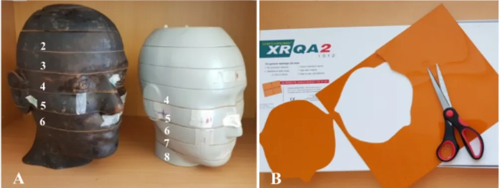
Two cross-sectional anthropomorphic head phantoms for absorbed dose measurements were used in Study III: an adult RANDO phantom and a 10-year-old child CIRS phantom (Figure 2A). The sectional surfaces are flat and smooth. The adult phantom is made of human bone and the child phantom is made of bone-equivalent material; both embedded in soft-tissue-equivalent materials. Organs of interest were traced for each selected phantom slices. Gafchromic XR-QA2 films were used for absorbed dose measurements (Figure 2B). These were calibrated against an ionization chamber before use, as described in detail in Paper III.
Figure 2. (A) The two anthropomorphic head phantoms loaded with
Gafchromic film at five levels, as indicated by the numbers. Left: the adult phantom, for temporomandibular joint examination, and right: the child phantom, for panoramic examination. (B) Gafchromic film cut to the correct size and shape.
Dose measurements
The phantoms were loaded with Gafchromic films at five levels. For panoramic examinations, the films were centred around the occlusal planes, while for the CBCT examinations, they were centred around the TMJ region. The phantoms were carefully placed so that the central beam of the radiation field did not coincide with the film at the third level, i.e. the film at the third level had a slight offset in the Z direction
2 3 4 5 6 7 4 5 6 8 A B
(length axis of the phantom). Multiple exposures were made to obtain a dose that was within the dynamic range of the film. The exposed films were scanned using an Epson Perfection 4990 Photo flatbed scanner and converted into 8-bit greyscale images using ImageJ software. Net pixel values were calculated (mean pixel value in the exposed image minus the mean pixel value for the background). The absorbed doses were estimated by applying the dose calibration equation to the net pixel value data after converting these data into 32-bit floating-point data. The absorbed dose matrix was then divided by the number of exposures, which provided an absorbed dose matrix for 1 exposure, as in the clinical situation. The mean doses absorbed by the radiosensitive structures within the oral and maxillofacial region (brain, eyes, salivary glands, oral mucosa and bone surface) were estimated by overlapping the traced organs on the dose distribution matrices and then calculating the mean absorbed dose inside each organ.
Data analysis
The relative radiation burden and mean absorbed dose to the radiosensitive tissues were compared according to the examination type, X-ray unit, clinical settings and phantom.
Study IV
ParticipantsThirty-four adult patients (mean age 57 years) who had been referred for CBCT examination of the TMJ were recruited for Study IV. The Regional Ethics Review Board in Lund, Sweden, approved this study (Dnr. 2017/434).
Radiographic examinations
The participants underwent two consecutive CBCT examinations: one using the default (manufacturer’s) protocol and the other with a low-dose protocol. The CBCT examinations were reconstructed in sagittal volumes bilaterally (giving 68 volumes). These were retrieved,
anonymized, randomly coded and grouped into three groups using the ImageJ software: a default protocol, a low-dose protocol, and a processed protocol, which is low-dose stack processed with a noise reduction algorithm (giving a total number of 204 stacks) (Rudin et al., 1992) (Figure 3).
Figure 3. Cone-beam computed tomography images for two
temporomandibular joints using the three protocols: (A) default, (B) low-dose, and (C) processed. 1 (A, B, and C) show no signs of degenerative joint disease, while 2 (A, B, and C) show signs of degenerative joint disease.
Radiographic image assessment
Four calibrated oral and maxillofacial radiologists evaluated all the images. They independently assessed the visibility of five anatomical structures in the TMJ region, namely: the outlines of the condyle, the articular eminence, and the articular fossa, and the trabecular patterns of the condyle and the temporal bone, under the same viewing conditions. A three-point scale was used to assess the visibility of each anatomical
2 3 4 5 6 7 4 5 6 8 A B 1 2 A A B C B C
structure: 1, definitely visible; 2, questionably visible and 3, not visible. Furthermore, the observers were asked to give their overall subjective impression of image quality ranked as: 1, diagnostically acceptable; 2, diagnostically questionable and 3, not diagnostically acceptable. To assess the radiographic findings and thereby, the diagnostic performance of the low-dose protocols, the observers were asked to record their level of confidence concerning the presence of DJD as 1, definitely not; 2, probably not; 3, questionable; 4, probably and 5, definitely, according to the DC/TMD (Ahmad et al., 2009). Intraobserver agreement was determined by asking each observer to re-evaluate 40 TMJs after an interval of at least 14 days.
Data analysis
The data from the observed TMJ anatomical structures and overall image quality ratings were pooled and compared pairwise using the sign test. Receiver operating characteristic (ROC) curves were used to analyse the radiographic findings concerning the presence of DJD. The areas under the curves (Az) were calculated. Intraobserver agreement
was estimated using kappa (k) statistics according to Altman (Altman, 1991): k = 0.81–1.00, excellent; k = 0.61–0.80, good; k = 0.41–0.60, moderate; k = 0.20–0.40, fair; k < 0.20, poor.
RESULTS
Studies I and II
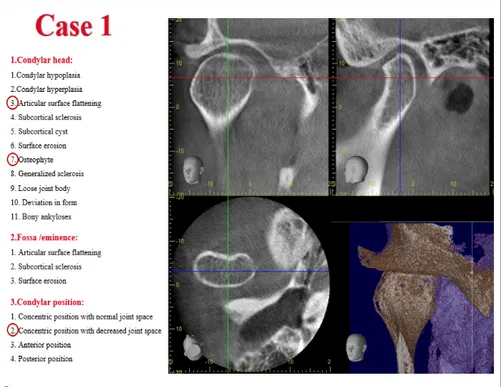
Two educational tools for the assessment of TMJ osseous tissues in CBCT images were developed, and can be accessed from the Malmö University web site. Both were designed based on DC/TMD image analysis criteria. The first tool consisted of 2 PPT files including pre-defined 2D multiplane CBCT reconstructions of the TMJ (Figure 4). The second was a web-based programme including 3D multiplane CBCT reconstructions (Figure 5).
Figure 4. A screenshot of the educational tool that presented in Study I
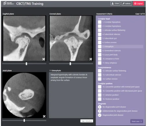
Figure 5. A screenshot of the web-based programme that developed in
Study II for the interpretation of CBCT images of the TMJ.
The observers demonstrated good performance in the first assessment in both tools; the mean correct responses being 86% in Study I and 76% in Study II. The improvements in the second assessment were not statistically significant (p > 0.05), (Figure 6).
Figure 6. Percentage of correct responses in the two educational tools
developed in Studies I and II.
According to the observers’ subjective evaluation (Figure 7), more than 90% were moderately or extremely satisfied with the web-based programme, and thought that it was easy to access and had a user-friendly design, while more than 65% were satisfied with the radiographic image quality and the results matrix (Figure 8). Seventy-six percent of the observers were satisfied with the knowledge gained after using this programme and with its value as a learning tool.
0 20 40 60 80 100
Educational tool in Study I Educational tool in Study II First assessment Second assessment Pe rc en t of c or re ct r es p on se s (% )
Figure 7. Observers’ subjective evaluation of the web-based programme
after the second assessment.
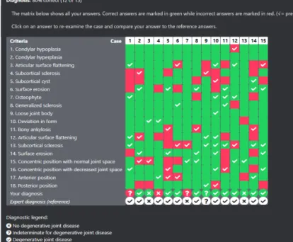
Figure 8. A screenshot of the user’s result matrix that compares user
and reference responses for all examination cases.
53 67 45 26 33 40 45 26 29 38 43 36 2 7 24 19 17 17 2 10 7 5 7 2 0% 20% 40% 60% 80% 100% Extremely satisfied Moderately satisfied Neutral Moderately dissatisfied Extremely dissatisfied Ob se rv er s’ s at is fa ct io n ( % )
Accessibility Design Radiographic
image quality Results matrix
Knowledge
gained learning toolWeb-based
100
Study III
The absorbed doses varied considerably among and within the radiosensitive tissues with all the protocols used, due to variations in the type of examination, the X-ray unit, clinical settings and phantoms. Tables for organ doses in panoramic and CBCT examinations of the TMJ are available in Paper III. Bone surface and salivary glands received the highest absorbed doses in both panoramic and CBCT examinations of the TMJ. The absorbed doses to the other tissues ranged between 70 µGy and 270 µGy in the panoramic examination. In half-rotation CBCT examinations, the absorbed doses varied between 190 µGy and 760 µGy, whereas the corresponding values for full-rotation examinations were 380 µGy and 1530 µGy. Absorbed doses distribution in panoramic examination and two CBCT examinations of TMJ with different FOVs illustrated in Figure 9.
Figure 9. Absorbed doses distribution given as isodose lines in µGy for
the adult head phantom. (A) Panoramic examination, (B) TMJ examination using CBCT with two 4 x 4 cm FOVs, (C) TMJ examination using CBCT with one 14 x 5 cm FOV.
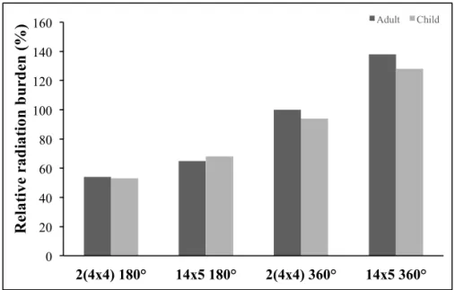
Most of the relative radiation burden to the adult head phantom was slightly higher than that to the child head phantom (Figures 10). Meanwhile, when using two small FOVs, for right and left TMJs examination, the relative radiation burden was lower than when using
μGy 1100 1000 900 700 500 300 200 μGy 4a300 4000 3500 3000 2500 1500 1200 μGy 7300 7000 6500 6000 5500 5000 4000 A B C
Figure 10. The percentage of relative radiation burden to adult and child
head phantoms when comparing different radiographic examinations, rotation angles and X-ray units.
Figure 11. The percentage of relative radiation burden to adult and child
head phantoms when comparing different FOVs in CBCT imaging. 0 20 40 60 80 100 120 140 160 2(4x4) 180° 14x5 180° 2(4x4) 360° 14x5 360° Adult Child Re lat ive r ad iat ion b u rd en ( % ) 0 100 200 300 400 500 600 Adult Child Veraviewepocs 3D F40 PanoramicCBCT 180° CBCT360° CBCT 180° CBCT180° Veraview X800 3D Accuitomo 170 Panoramic CBCT 360° Re la ti ve r ad ia ti on b u rd en ( % )
Large differences were found in the radiation burden between the X-ray units used (Figure 10). The Veraviewepocs 3D F40 unit exhibited the highest radiation burden of the three units for half-rotation CBCT scans, while the Veraview X800 unit delivered the highest radiation burden in both panoramic and full-rotation CBCT examinations. The 3D Accuitomo 170 unit delivered the lowest radiation burden during the CBCT examinations, at both full- and half-rotation scans.
Study IV
Upon establishing the reference standard, two observers evaluated the default protocol using the DC/TMD image analysis criteria (Ahmad et al., 2009). It showed that half of the examinations had radiographic findings indicating the presence of DJD while the rest were without, agreement being obtained among all the examinations except one. None of the observers reported any difference in the visibility of the five anatomical structures or overall image quality between the default protocol and the two low-dose protocols (p ≤ 0.05) (Table 2). Only one observer reported an improvement in the visibility of the structures and image quality between the low-dose protocol and the processed protocol (Observer 3, Table 2).
Table 2. Level of statistical significance (p) for the visibility of five
anatomical structures and image quality of CBCT images produced using the three protocols: default protocol, low-dose protocol, and processed protocol Protocol Ob se rv er 1 Ob se rv er 2 Ob se rv er 3 O bs er ve r 4 Visibility Default vs. Low-dose 0.00 0.00 0.00 0.00 Default vs. Processed 0.00 0.00 0.00 0.00 Low-dose vs. Processed 0.00 0.00 0.79 0.00
No statistically significant difference was observed in the radiographic findings concerning the presence of DJD between the default protocol and the two low-dose protocols (p ≤ 0.05) (Table 3). The mean area under the ROC curves for all observers was 0.93 for the low-dose protocol and 0.94 for the processed protocol. Figures 12 and 13 show the superimposition of the ROC curves for the low-dose and processed protocols, respectively, assessed by all the observers according to the radiographic findings concerning the presence of DJD.
Table 3. Area under the receiver operating characteristic curves (Az)
when four observers assessed the radiographic findings concerning the presence of DJD in CBCT images produced using the default protocol as reference standard, for the low-dose protocol and the processed protocol
SE = standard error
Low-dose protocol Processed protocol
Az SE p-value Az SE p-value
Observer 1 0.92 0.04 0.00 0.95 0.03 0.00
Observer 2 0.95 0.03 0.00 0.94 0.03 0.00
Observer 3 0.92 0.03 0.00 0.93 0.03 0.00
Figure 12. Receiver operating characteristic curves for assessing the
radiographic findings concerning the presence of DJD by four observers when using the low-dose protocol.
Figure 13. Receiver operating characteristic curves for assessing the
radiographic findings concerning the presence of DJD by four observers
Low-dose protocol 1- Specificity Se ns it iv it y 0 0 1 1 Observer 1 Observer 2 Observer 3 Observer 4 Reference line Processed protocol 1- Specificity Se ns it iv it y 0 0 1 1 Observer 1 Observer 2 Observer 3 Observer 4 Reference line
Intraobserver agreement regarding the radiographic findings of the presence of DJD was good to very good for all observers (k values = 0.75–0.85).
DISCUSSION
Radiographic imaging adds a new dimension to the medical diagnostic procedure by allowing clinicians to see structures inside the body in two or three dimensions, without surgical procedures; the greater goal being to treat patients more efficiently and effectively. It may be used for excluding or confirming suspected disease and/or adding information when the clinical diagnosis is uncertain. The popularity of dental CBCT imaging has been growing since its introduction in the late 1990s. However, there is still controversy concerning different aspects such as the radiation dose, the diagnostic efficacy in different diagnostic tasks, the time needed to interpret the image, the cost, knowledge of 3D anatomical structures reconstructed by CBCT images, and image interpretation skills. Nevertheless, it plays an important role in the diagnosis and treatment of many dental and osseous diseases in the jaws.
Radiographic imaging should be used to obtain a definitive diagnosis of osseous-related pathology in the TMJ, although referral for TMJ imaging must be justified based on individual assessment. CBCT examination of the TMJ has acceptable diagnostic accuracy for osseous TMJ abnormalities together with high specificity and fairly high sensitivity, and has been reported to be comparable to CT (Honda et al., 2006; Sirin et al., 2010; Zain-Alabdeen and Alsadhan, 2012). The indication for CBCT imaging of the TMJ is related to the clinician’s questions after obtaining a detailed medical history and thorough clinical investigation. These questions are usually related to failed treatment, persistent symptoms without explanation, or other questions that could be resolved by a CBCT examination, which thus plays a role in diagnosis and/or treatment planning (Petersson, 2010). In the work described in this thesis, different aspects of CBCT imaging of the TMJ were studied, namely image interpretation, imaging dosimetry, and dose
interpretation of CBCT images of the TMJ. The dose distribution was assessed using Gafchromic film in Study III, and a low-dose protocol for CBCT imaging of the TMJ was evaluated in Study IV.
CBCT educational tools (Study I and II)
During the evaluation of the three-dimensional CBCT datasets, a broad and systematic approach is recommended. The observer can reorient, and manipulate the images in many viewing planes. Thus, such images provide extensive and detailed information. The observer needs to assess all the structures in the FOV and gather as much as possible information to answer clinical questions and observe any incidental findings. However, thorough knowledge of 3D anatomy images, anatomic variations, tissue abnormalities, and pathologies is required to interpret CBCT images, and this is not extensively covered during undergraduate dental education.
The need to strengthen the knowledge of TMJ diagnostics, treatment decisions among GDPs as well as continuous education were addressed (Lindfors et al., 2016). Digital online educational tools could provide a means of increasing knowledge and coherence among the users worldwide. In addition, as is the case with the current global situation where the educational process has largely been transferred to the internet, the development of digital online educational tools should be encouraged to support the education. Well-defined criteria, together with calibrated practitioners, will facilitate image interpretation for the evaluation of TMJ pathology (Petersson, 2010). The reliability and validity of the DC/TMD image analysis criteria have been critically appraised providing a solid basis for TMJ evaluation, which is now widely accepted (Ahmad et al., 2009). The application of these criteria could help in providing standardized knowledge and thus facilitate the diagnostic process. Hellen-Halme et al. (Hellen-Halme et al., 2012) have confirmed the value of these criteria, and suggested that their use could improve the consensus or agreement of the observers, thus increase the diagnostic outcome.
The educational tool presented in Study I was intended to provide dental practitioners an overview for the assessment of osseous tissues in the
TMJ through a number of CBCT examinations presented as pre-defined 2D multiplane CBCT reconstructions. The observers were able to identify osseous changes, and their ability to identify TMJs without osseous changes were even better. As a result of these findings, it was decided to expand the project by developing a web-based programme that simulates the clinical situation, allowing the observers to evaluate the whole CBCT volume and diagnose the DJD case.
During development of the web-based program, CBCT DICOM images were converted to least compressed JPEG format. The conversion was performed to manage the size of images to fit within the web pages, with reasonable page loading time, while maintaining the accuracy of the assessment. Upon the comparison between these two image formats, no noticeable differences were found (Figure 14).
Figure 14. Comparison between (A) A DICOM image, and (B) A
converted JPEG image for the TMJ.
On average, the observers’ performance in assessing the CBCT examinations was good; however, their performance was lower in certain criteria. Although efforts were made to include all criteria within the examinations, some of the most uncommon criteria were missing. Assessing the size of the abnormalities was challenging, as has been reported in other studies (Marques et al., 2010; Patel et al., 2014). Also, the lack of grading in the extent of osseous changes in the DC/TMD
image analysis criteria and differentiation between normal morphological variations and small pathological changes (for example, differentiation between thickening of the anterior aspect of the condyle and small osteophyte) could also have affected the performance of the observers, as also stated by Larheim et al. (Larheim et al., 2015). Another challenge lies in distinguishing between a deviation in the shape of the condyle from the normal “rounded” shape without any underlying pathology, such as concavity in the cortical outline, and a deviation associated with pathology. DJD could not be confirmed when only articular surface flattening and/or sclerosis was observed, instead indeterminate for DJD was suggested by Ahmad et al. (Ahmad et al., 2009). Image interpretation is dependent on knowledge, practice and experience, which could explain why no significant difference between results of the two assessments was reported. The educational tools developed in this work are intended for use by general dental practitioners, undergraduate and postgraduate dental students, and specialists for training, and/or improving knowledge on the subject. The web-based programme can be used on a range of devices, including laptops, iPads/tablets, and smartphones. Comparable results have been reported for a web-based programme for training in the anatomical interpretation of maxillofacial CBCT images, and was also very positively received by the observers (Hassan et al., 2007; Al-Rawi et al., 2007). A web-based program for the joint’s soft tissue evaluation, developed by Hellen-Halme et al. (Hellen-Halme et al., 2012), can be reviewed for a wider knowledge over the TMJ assessment.
Dose measurements (Study III)
Although dental radiographic imaging is associated with relatively low radiation doses, the risk arising from ionizing radiation is cumulative, and it is especially important to bear this in mind for children and younger patients. The various organs and tissues in the body differ in their sensitivity to radiation. Undifferentiated and rapidly dividing cells are more radiosensitive which, together with the longer life expectancy of younger patients, stresses the need for extra radiation protection of young people.