Surface Modification of Cellulose Materials
From Wood Pulps to Artificial Blood Vessels
N. Lage Ahrenstedt
Licentiate Thesis
School of Biotechnology Royal Institute of Technology
© N. Lage Ahrenstedt School of Biotechnology Royal Institute of Technology AlbaNova University Centre SE-106 91 STOCKHOLM Sweden Printed at UniversitetsService US AB Box 700 14 SE-100 44 STOCKHOLM Sweden TRITA-BIO-Report 2007-6 ISSN 1654-2312 ISBN 978-91-7178-680-7
ABSTRACT
This thesis describes the improvement of two radically different cellulose materials, paper and artificial blood vessels, constructed from two diverse cellulose sources, wood pulp and Acetobacter xylinum. The improvement of both materials was possible due to the natural affinity of the hemicellulose xyloglucan for cellulose.
Chemical and mechanical pulps were treated with xyloglucan in the wet-end prior to hand sheet formation or by spray application of dry hand sheets, loading a comparable amount of xyloglucan. The tensile strength increases for the wet-end treatment and spray application were 28% and 71% respectively for bleached soft wood, compared to untreated sheets (20.7 Nm/g). The corresponding strength increases for hand sheets made of thermo-mechanical pulp were 6% and 13% respectively compared to untreated sheets (42.4 Nm/g). The tendency for chemical pulp to be superior to mechanical pulp with respect to strength increase was valid even for tear strength and Scott-Bond. These results suggest, in agreement with other studies, that adhesion of xyloglucan to wood fibres is dependent on their degree of surface lignification.
Also, a method was developed to increase the blood compatibility of artificial blood vessels constructed of bacterial cellulose. Xyloglucan was covalently linked to the endothelial cell adhesion motif (Arg-Gly-Asp). To obtain this, new solid-phase coupling chemistry was developed. Xyloglucan oligosaccharides (XGO) were transformed into XGO-succinamic acid via the corresponding XGO-β-NH2 derivative prior to coupling with the N-terminus of the solid-phase synthesised Gly-Arg-Gly-Asp-Ser peptide. The resin-bound glyco-peptide was then cleaved and enzymatically re-incorporated into high molecular weight xyloglucan. The glyco-peptide was further adsorbed onto bacterial cellulose scaffolds, increasing the adhesion and proliferation of endothelial cells and therefore blood compatibility.
SAMMANFATTNING
Den här avhandlingen beskriver en vidareutveckling av två vitt skilda cellulosamaterial, papper och blodkärl, tillverkade från två helt olika cellulosakällor, traditionell pappersmassa och bakterien Acetobacter xylinum. Materialförbättringarna var möjliga tack vare hemicellulosan xyloglukans naturliga bindningsförmåga till cellulosa.
Både kemiskt och mekaniskt framställda pappersmassor behandlades med xyloglukan, dels genom adsorption till den våta massan före arkformning och dels genom att spraya torra ark. Båda metoderna tillförde en jämförbar mängd xyloglukan. Dragstyrkeökningen för ark gjorda av xyloglukanbehandlad pappersmassa och för torra ark spraybehandlade med xyloglukan var 28% och 71% vardera för blekt Kraft-massa av barrträd, jämfört med obehandlade ark. Motsvarande styrkeökning för ark gjorda av termomekanisk massa av barrträd var 6% och 13% vardera. Även för rivstyrka och Scott-Bond var styrkeökningen för ark gjorda av kemiskt framställd massa klart överlägsen den för ark gjorda av mekaniskt framställd massa. Resultaten antyder, och får stöd av tidigare studier, att mängden xyloglukan bunden till cellulosafibrer är relaterad till ligninhalten på dess yta.
En metod utvecklades också för att öka artificiella blodkärls blodkompatibilitet. För det utvecklades en syntesmetod där de avgörande reaktionerna sker i skiktet mellan en fast och en flytande fas. På så sätt kopplades xyloglukan kovalent till en peptidsekvens som känns igen av endotelceller, vilka täcker insidan av naturliga blodkärl. Glykopeptiden adsorberades sedan till artificiella blodkärl gjorda av bakteriell cellulosa, vilket ökade bindningsförmågan och celldelningen av odlade endotelceller, vilket därmed ökar kärlets blodkompatibilitet.
ABBREVATIONS
BHW bleached hard wood BSW bleached soft wood
CV column volume
DCC N,N'-dicyclohexyl carbodiimide
DCM dichloromethane
DIEA N,N-diisopropylethylamine
DIP de-inked pulp
DMAP 4-dimethylaminopyridine
DMF N,N-dimethylformamide
DMSO dimethylsulphoxide DTT 1,4-dithiothreitol
EDC 1-ethyl-3-(3-dimethyl aminopropyl) carbodiimide hydrochloride ESI-MS electro-spray ionisation mass spectrometry
FITC Fluorescein isothiocyanate Isomer I Fmoc fluorenyl methyloxycarbonyl
HBTU 2-(1H-benzotriazole-1-yl)-1,1,3,3-tetramethyluronium hexafluorophosphate
HOBt 1-hydroxybenzotriazole IR infrared
NMP N-methylpyrrolidone
NMR nuclear magnetic resonance SPPS solid-phase peptide synthesis tBME tert-butylmethyl ether
TCEP tris(2-carboxyethyl) phosphine TFA trifluoroacetic acid
TIS tri-isopropyl silane TLC thin layer chromatography
TMP thermo-mechanical pulp
UV ultraviolet
VIS visual
XET xyloglucan endotransglycosylase (EC 2.4.1.204)
XG xyloglucan
LIST OF PUBLICATIONS
I Lage Ahrenstedt, Antti Oksanen, Kristian Salminen, Harry Brumer III
Paper dry strength improvement by xyloglucan addition is critically dependent on the purity of cellulose fibre surfaces: wet-end application, spray coating, and synergism with borate
Submitted.
II Aase Bodin, Lage Ahrenstedt, Helen Fink, Harry Brumer III, Bo Risberg, Paul
Gatenholm
Modification of nano cellulose with a xyloglucan-RGD conjugate enhances adhesion and proliferation of endothelial cells: implications for tissue engineering
TABLE OF CONTENTS
Introduction ...8
Cellulose materials...9
Cellulose fibre structure ...9
Cellulose sources ...11
Traditional cellulose modification ...11
Paper ...12
Paper strength...12
Artificial blood vessels ...14
Blood vessel-blood interactions...15
History ...16
Bacterial cellulose scaffolds...17
Cellulose surface modification ...18
XET method ...18
Xyloglucan...18
Cellulose-Xyloglucan interactions ...19
Xyloglucan oligosaccharides ...21
Xyloglucan endotransglycosylase ...22
Cellulose surface modification by XET...23
Experimental ...24
Materials...24
Methods...24
Synthesis of GRGDS-SGGSS-GRGDS ...24
Attempted solid-phase coupling of GRGDS-SGGSS-GRGDS I ...24
Attempted solid-phase coupling of GRGDS-SGGSS-GRGDS II ...25
Attempted solid-phase coupling of GRGDS-SGGSS-GRGDS III ...26
Synthesis of SGGSS-GGSSG-GRGDS...27
Attempted solid-phase coupling of SGGSS-GGSSG-GRGDS I...27
Attempted coupling of SGGSS-GGSSG-GRGDS II...28
Synthesis of SGGSS-GRGDS...28
Solid-phase coupling of SGGSS-GRGDS ...29
Results and discussion ...30
Paper strength (Publication I) ...30
Adhesion of endothelial cells (Publication II)...34
XG-GRGDS synthesis ...34
Glyco-conjugates with 10 and 15-mer peptides (unpublished results) ...40
Conclusions ...42
Future work...42
Acknowledgements ...44
Introduction
Tamarind (Tamarindus indica) is one of the most important trees in India, since every part of it is useful in one way or another (Gerard 1980; Shankaracharya 1998). One of the products is tamarind kernel powder which finds extensive use as a sizing agent in the textile industry and as a thickener in food products. Tamarind kernel powder contains ∼60% xyloglucan (XG), a seed storage polysaccharide, and also a part of the cellulose fibre composite in the plant cell wall. In recent years, the potential role of XG in the pulp and paper industry has been partly investigated (Christiernin et al. 2003; Lima et al. 2003; Stiernstedt et al. 2006), where a fundamental characteristic of XG is an intrinsic cellulose affinity. This affinity has also been utilised to tailor cellulose surface properties by conjugating XG to a molecule of choice and further adsorb the XG-conjugate to a cellulose surface. Hence, XG acts as an anchor for the desired functionality (Brumer et
al. 2004; Zhou et al. 2005; Zhou et al. 2006b).
The use of strength additives in the paper making process enables raw material and energy savings, hence economical profits. This thesis presents the influence on paper strength for hand sheets treated with XG. The hand sheets were made of both chemical and mechanical pulps. The treatments occurred either in the wet-end prior to sheet formation, or by spray application of dry hand sheets. The modified sheets were tested with focus on their dry tensile strength, but tear strength and Scott-Bond was also measured for a selected range of the sprayed sheets.
Arterial sclerosis is a growing health concern in the industrialised countries and does not seldom lead to death. This brings an increased frequency of vascular surgery and an increased demand of artificial blood vessels. This thesis describes the development of a method to increase the blood compatibility of artificial blood vessels constructed of bacterial cellulose by connecting XG to an adhesive peptide recognised by endothelial cells, and adsorbing the cell adhesion glyco-peptide on the bacterial cellulose scaffold.
Cellulose materials
Cellulose fibre structure
Cellulose is argued to be the most abundant polymer in nature and constitutes the main component of plant fibres giving the plant rigidity. Cellulose is built up by β(1→4)-linked D-glucoseunits. The most stable conformation about the β(1→4) linkage involves alternating 180° flips of every second glucose unit, so that the repeating unit of cellulose is rather a cellobiose molecule than a glucose unit (Figure 2). The β-configuration of the glycosidic bond allows the polymer to adopt a fully extended conformation. Amylose, in contrast, is built up by α(1→4) glycosidic linkages, which are naturally bent, conferring a gradual turn to the polymer chain resulting in a helical conformation.
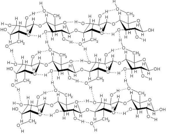
Figure 1: The structure of a cellulose sheet showing the intra- and interchain hydrogen bonds.
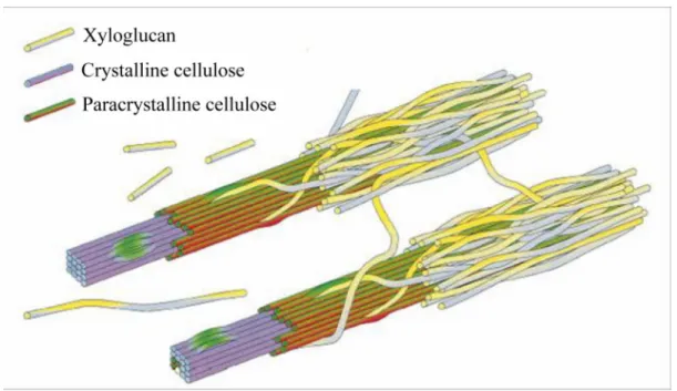
Juxtaposition of several extended cellulose chains permits extensive intra- and interchain hydrogen bonding. The extended cellulose chains form an array with the flat glucose units fixed edge to edge by hydrogen bonds (Figure 1). These sheets are further stacked on top of each other and held together by hydrogen bonds and van der Waals interactions. This network of up to 40 cellulose chains makes up the crystalline core of the cellulose fibril.
Native cellulose (cellulose I) has two crystalline allomorphs, Iα and Iβ (VanderHart et al. 1984). The main difference between the two crystalline phases is the relative position of the chains to each other. This difference brings a staggered conformation to the cellulose chains in the Iβ phase, stabilising the structure like bricks in a wall. Allomorph Iα can irreversibly be transformed into Iβ by annealing (slightly heating), thus Iβ is the most stable phase.
Cellulose in higher plants (woody tissue, cotton etc.) consists mainly of the Iβ phase whereas primitive organisms (bacteria, algae etc.) are enriched in the Iα phase.
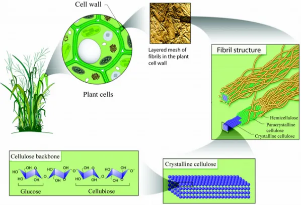
Figure 2: The cell wall hierarchy, from a single cellulose polymer to the cellulose fibre composite (reprinted with permission from the U.S. Department of Energy Genome Programs, http://genomics.energy.gov).
The crystalline cellulose fibril is further covered by para crystalline cellulose and finally surrounded by hemicelluloses. Hemicelluloses incorporate the amorphous cellulose layer and cross-link the individual fibrils, and form covalent associations with lignin, an aromatic polymer involved in binding the cellulose fibres together. Collectively with some proteins and lipids, these fibre composites make up the cell wall (Figure 2).
Cellulose sources
Cellulose is produced by various species in nature, both plants and other organisms. The cell walls of plants, algae and oomycetes (e.g. water moulds) are primarily built up by cellulose. Only one group of animals, the tunicates, can synthesise cellulose, while it is digestible by all grass and leave-eating species. Some acetic acid bacteria, notably
Acetobacter xylinum, are known to synthesise and secrete cellulose into the surrounding
media.
Traditional cellulose modification
Plant fibres have excellent load bearing properties as a result of their well organised structure. Their high strength in combination with light weight makes cellulose fibres an interesting material to reinforce polymer composites. But the difference in surface polarity between hydrophilic cellulose fibres and hydrophobic polymers can result in compatibility difficulties. To overcome this problem, fibre surfaces have been treated with benzyl chloride (Lu et al. 2003) or maleated polymers (Feng et al. 2001). The attachments of these species make the cellulose surface more hydrophobic and consequently more compatible with the polymer matrix. However, to link cellulose with these hydrophobic groups the cellulose surface hydroxyl groups are used as reactive sites. These are the very same hydroxyl groups that are involved in the hydrogen bonding responsible for the delicate fibre integrity. Disruption of fibre integrity is connected with loss of fibre strength and the fibre strength does drop dramatically during the surface modification (Lu et al. 2003). But, even though the strength of the individual fibre decreases, the resulting fibre-polymer composites gain on the fibre modification.
For applications primarily based on the cellulose fibre properties, such as paper and board, the fibre is rather modified by adsorption interactions or by coating or spraying of the paper web.
Paper
Wood is by far the most abundant source of cellulose, and paper the dominating cellulose product. The process of turning wood into paper is complex and involves many stages. Since the production is on such a large scale, even small improvements bring huge savings. In the work described in this thesis the ambition was to increase paper dry strength, which conveys the possibility to decrease the paper grammage (resulting in raw material savings) or the degree of pulp refining (energy savings) without compromising the strength properties of paper. An increased tensile strength enables higher paper machine speed and a greater Z-strength could result in a shorter drying section. These examples are part of the consequences of a stronger paper.
Good paper formation is one way to obtain paper strength, but the upper limit for this is presumably reached. So, to further increase paper strength a range of strength additives have been investigated (discussed in Publication I). For optimising the use of these strength additives, a fundamental understanding of their mechanism is of greatest importance.
Paper strength
When considering paper strength in general there are two fundamental factors that have to be taken into account, the fibre-fibre joint strength and the strength of the individual fibre. A smooth and soft fibre promotes a better fibre-fibre joint formation, since smooth fibres allow greater contact surface at the fibre interface. A stiff and straight fibre is stronger than an accordion like fibre and takes more strain. Davison suggested that the weak link in paper strength is the fibre-fibre joint since the individual fibres withdrawn from a paper were twice as strong as the paper itself (Davison 1972).
During the consolidation and drying process the fibres come close to each other and form an intimate contact, an important process for the fibre-fibre joint formation.
Different intermolecular forces hold the fibres together at the contact area and these are responsible for the fibre-fibre joint strength. Of these forces, a general opinion is that hydrogen bonds are the most important. Hydrogen bond forces are short-ranged (<5 Å) and are probably responsible for holding the fibres together only when the fibres are very close to each other and occupy a correct conformation towards each other. For long-range interactions (~100 Å) van der Waals and ionic forces may be of higher importance, and these do not depend on a special conformation. Moreover, there are also mechanical interactions that hold the fibres together, like inter-diffusion of polymers across the interface and interlocking of two fibre surfaces at the fibre-fibre joint where the fibres are in partial molecular contact. All interactions mentioned above take place at the fibre-fibre joint. Hence, a soft fibre, allowing greater surface contact between the fibres, would provide the paper with more strength.
Addition of hemicelluloses to the pulp in the wet-end is known to increase the general paper strength. The strength increase may originate in an increased retention of fines, known to increase paper strength, or in an increased softening of the fibre surface, creating more surface area.
Dry strength additives commonly contain cationic groups, like cationic starch (Formento et al. 1994; Mendes et al. 2001), which adhere to the negatively charged cellulose surface (Moeller 1966). Cationic additives interacting with cellulose through ion-ion interactions may be disturbed and shielded by anionic substances in the process water. These interactions will decrease the adsorption of the additives to the cellulose fibres, decreasing the efficiency of the additive. It is therefore of interest to find strength additives which interact with cellulose in a non charge-mediated manner. Some uncharged polysaccharides avoid this problem through irreversible adsorption to cellulose by extensive hydrogen bonding and van der Waals interactions, which increase the tensile strength (Rojas et al. 1999; Hannuksela et al. 2002; Lima et al. 2003; Suurnakki et al. 2003; Hannuksela et al. 2004; Ren et al. 2006).
Artificial blood vessels
A result of the lifestyle and eating habit developments in the U.S.A., Europe and other parts of the world is an increased frequency of mortality caused by arterial scleroses. Lack of activity in combination with an unbalanced diet give rise to an unhealthy level of cholesterol (i.e. when the low-density lipoproteins are saturated with cholesterol) which in turn may lead to deposits of plaque on the blood vessel walls, decreasing the lumen calibre and hindering a smooth blood flow. These deposits may finally lead to thromboses and occlusion of the vessel, and if this occurs in the coronary arteries, to a potentially lethal heart attack. Therefore, there is a huge need for developing new approaches and methods to help this category of people, either by introducing healthier habits or replacing the damaged blood vessels with artificial ones.
There are three main routes to artificial blood vessels, including synthetic grafts, biodegradable grafts and tissue engineered blood vessels. The details and differences of these methods are well described in the literature (Parikh et al. 2000; Heyligers et al. 2005; Kakisis et al. 2005). Their common feature is that they mimic the layered structure of the natural blood vessel in one way or another.
The natural vessel is mainly built up by three layers (Figure 3). The layer in contact with blood, the tunica intima is lined by a monolayer of endothelial cells, further discussed in the next section. Smooth muscle cells make up the tunica media and regulate the blood flow by contractions and relaxations. Outermost, the vessel is covered with fibroblasts in a loose and porous network that facilitates incorporation of the surrounding collagen (Parikh et al. 2000).
Blood vessel-blood interactions
In vivo, thromboses are prevented by interactions between the blood components and the
endothelium, the innermost layer in the natural vessel, made up by a monolayer of endothelial cells. The endothelial cells provide a physical barrier to the blood and resist deposition of blood proteins and pro-coagulants, e.g. by supplying a negatively charged barrier. The endothelium is highly sensitive and regulates diminutive changes of the blood composition by secreting a variety of substances (e.g. prostacyclin, nitric oxide and heparin) which controls the blood cell trafficking, growth and vascular remodelling (Lüscher et al. 1997; Parikh et al. 2000).
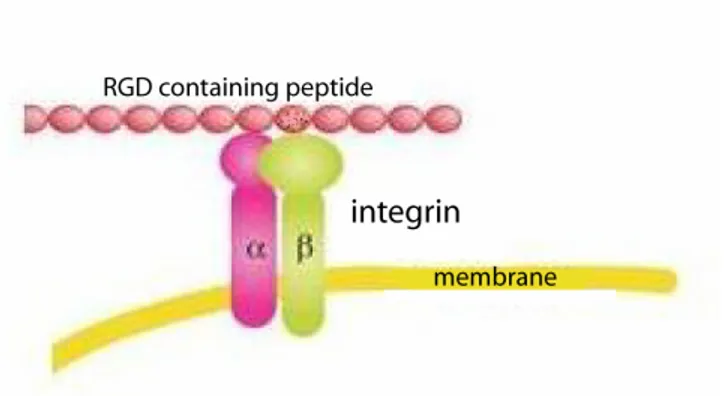
Figure 4: Endothelial cells interfere with different blood proteins, containing the RGD-motif, via the integrin surface receptors. Adapted from Gao et al. (2006).
Many of these interactions between the endothelial cells and the blood components are dependent on the integrin receptors attached to the cell surface. The integrin protein superfamily is built up by two subunits (alpha and beta) of which at least 20 combinations are known. The common feature for proteins recognised by integrin receptors is the RGD (Arg-Gly-Asp) amino acid motif (Joshi et al. 1993), as
schematically illustrated in Figure 4. Even though various protein ligands contain the RGD cell attachment sequence, individual integrins are capable of recognising only a single adhesive protein, or in some cases a limited number. The key to the binding selectivity of the integrins for the different RGD-containing peptides is that the integrins not only recognise the RGD sequence but also the surrounding residues (Ruoslahti et al. 1987; Jois et al. 1996; Addicks et al. 2002).
History
Almost certainly the first attempt to replace arteries with bare synthetic grafts was performed in the early 50’s (Voorhees et al. 1952) and was followed by a number of studies. The results were promising and showed good long-term patency rates, but only for large-calibre grafts. For the narrow grafts (< 6mm inner diameter) early occlusion occurred. Since then, the complications related to long-term graft viability have been investigated resulting in the general opinion that most drawbacks with a bare graft is due to its inability to withstand one or more of the phases of vascular response to injury (e.g. intimal hyperplasia and thrombosis), effects that often lead to a total occlusion of the graft (Parikh et al. 2000; Heyligers et al. 2005).
In vivo, a monolayer of endothelial cells prevents thrombosis. Consequently, the
next step in the development of artificial blood vessels was to introduce the highly blood compatible endothelial cells. This was not possible until the early 70’s, when the techniques to isolate and culture pure endothelial cells were developed. Using the new methods Mansfield et al. (1975) seeded a synthetic polyester surface with endothelial cells. First then, the tools for producing a synthetic graft seeded with endothelial cells were available and it did not take long before the first animal trial was reported (Herring
et al. 1978), where seeded prostheses were implanted in dogs. Some years later the same
research group published the first study of seeded synthetic grafts implanted in humans (Herring et al. 1984). Two long-term investigations of the patency of such grafts implanted in humans are reported. The 6 years follow-up of the first study reported 67% primary patency after 6 years (Leseche et al. 1995) and the other study presented a 9 years post-implantation patency of 65% (Deutsch et al. 1999).
Bacterial cellulose scaffolds
Acetobacter xylinum synthesises cellulose and extrudes it extracellularly into the
surrounding growth medium. The individual cellulose chains aggregate into fibrils, which bundle to form ribbons. These ribbons build up an ultra fine network that is highly hydrated and swollen (∼99% water). The formed network of bacterial cellulose ribbons is much finer than wood cellulose fibres (Publication II, Figure 2). Also, the bacterial cellulose network is pure, in contrast to extracted wood cellulose which contains remainders from the plant composite (e.g. hemicellulose and lignin).
Since the extracellularly secreted cellulose deposits on whatever object present in the growth medium, different cellulose structures can be shaped. When silicon tubing is present in the medium, the secreted cellulose deposits in a lamella structure around the tubing and upon removal of the silicon scaffold, tubular vessel like constructs are obtained, suitable for producing artificial blood vessels (Klemm et al. 2001; Bodin et al. 2005). This moulding technique brings a convenient structure to the cellulose vessel. The lumen, which has been in contact with the silicon tubing, gets a smooth surface while the outside gets a bit more fibrillated.
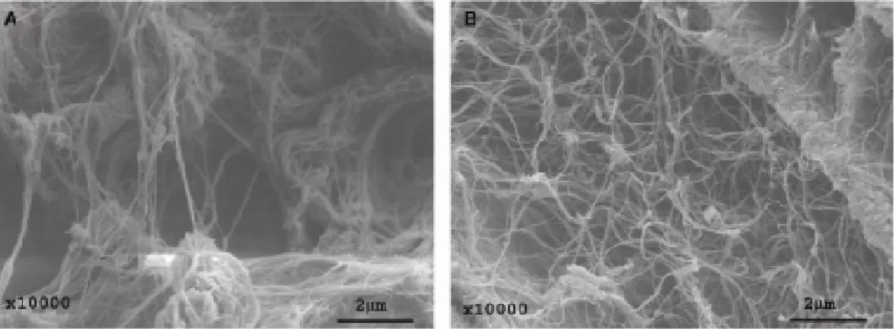
Figure 5: SEM-micrographs of (A) collagen and (B) bacterial cellulose. Reprinted with permission from Bäckdahl et al. (2006).
The fibrillated surface of bacterial cellulose is in the same scale as collagen fibrils, and its surface structure resembles that of collagen as observed by electron microscopy (Figure
5). Collagen is a major compound of the tissue that surrounds blood vessels in vivo.
maintained function. Helenius et al. (2006) performed a biocompatibility study of bacterial cellulose in pigs and concluded that bacterial cellulose is fully biocompatible.
Cellulose surface modification
XET method
The main principle of the method for fibre modification developed in the laboratory of Wood Biotechnology at KTH (Brumer et al. 2004) is to perform chemo-enzymatic synthesis on xyloglucan (XG) instead of the hydroxyl groups on the cellulose surface. To facilitate the procedure, XG is first enzymatically digested to xyloglucan oligosaccharides (XGOs). After several chemical modifications, the modified XGOs are re-incorporated into high molecular weight XG by xyloglucan endotransglycosylase (XET) and finally irreversibly adsorbed to cellulose.
Xyloglucan
Xyloglucan (XG) is the principal hemicellulose in the primary cell walls of dicotyledonous and non-commelinoid monocotyledonous plants. XG embeds and cross-links the cellulose fibrils and ties the primary and secondary cell walls together by inter-boundary bridging. Also, XG is the predominant seed storage polysaccharide in certain species, especially tamarind and nasturtium. Deoiled tamarind (Tamarindus indica) kernel powder was used for the work in this thesis and contains ∼60% XG by weight.
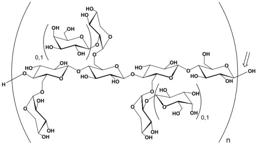
O O HO HO O O OHO OH O O HO OH OH O O HO OH O OH O OH O OH O OH HO HO HO O HO O OH O HO OH OH O OH O OH OH H OH n 0,1 0,1
Figure 6: The structure of tamarind XG, with possible galactose substitution indicated, n ≈700. The arrow indicates the site for endo-glucanase cleavage.
Xyloglucan and cellulose are built up by the same backbone structure, additionally XG is extensively branched. Tamarind XG is built up by four repeating units; XXXG, XLXG, XXLG and XLLG (in the molar ratio 15:7:32:46), where each letter represents a glycopyranosyl unit of the backbone, with or without substituents (Fry et al. 1993). X represents a Xylp α(1→6)-Glcp unit, L represents a Galp β(1→2)Xylp α(1→6)Glcp unit, and G represents an unbranched Glcp residue. When written in sequence, the backbone glyco-pyranosyl units are conjugated with Glcp β(1→4)-Glcp linkages (Figure 6).
Cellulose-Xyloglucan interactions
Xyloglucan (XG) associates tightly and cross-links the cellulose fibrils in the primary cell wall through extensive co-operative hydrogen bonds and van der Waals interactions (Hanus et al. 2006), as illustrated in Figure 7.
Figure 7: XG cross-links cellulose fibrils. Adapted from Rose et al. (1999).
The basis for these interactions is thought to originate in the structural similarity of XG and cellulose, which share the same glucan backbone, since the adhesion of XG is dependent on a sterically accessible backbone and its conformation. The decoration pattern of XG determines the access and conformation of the backbone and hence the attraction for cellulose (Levy et al. 1997; Chambat et al. 2005; Hanus et al. 2006).
The amount of XG bonded to cellulose increases when lowering the molecular weight (Lima et al. 2004) until a certain limit (Vincken et al. 1995). Computer simulations confirm this behaviour and explain it with the adoption of certain favourable cellulose-binding conformations along short sections of the backbone (Levy et al. 1997). Binding of these sections induces a continued flattening of the backbone and appropriate side-chain folding, hence further binding.
Adsorption studies of XG to wood fibres reveal that XG has a specific affinity for the cellulose component and that fibres contaminated with lignin or other remainders from the pulping process exhibit a lower adsorption capacity for XG (Zhou et al. 2006a). The cleanest wood cellulose surface in the trials by Zhou et al. (pine/spruce Kraft, refined) adsorbed ∼40% of the loaded XG (50 mg/g dry pulp). Bacterial cellulose fibrils on the other hand are very pure and adsorb XG almost quantitatively (Chambat et al. 2005), at a loading amount of 50 mg/g dry material.
The ability of XG to adsorb onto wood pulps has been investigated as a non-ionic wet-end additive in papermaking to increase strength and formation properties (Christiernin et al. 2003). The presence of XG in the papermaking process helps the retention of fines and increases the tensile strength (Christiernin et al. 2003; Lima et al. 2003). Addition of XG also results in fewer large flocs during sheet formation (Christiernin et al. 2003), possibly by decreasing the friction between the cellulose fibres (Stiernstedt et al. 2006). These observations make it interesting to further investigate the role of XG in papermaking.
Xyloglucan oligosaccharides
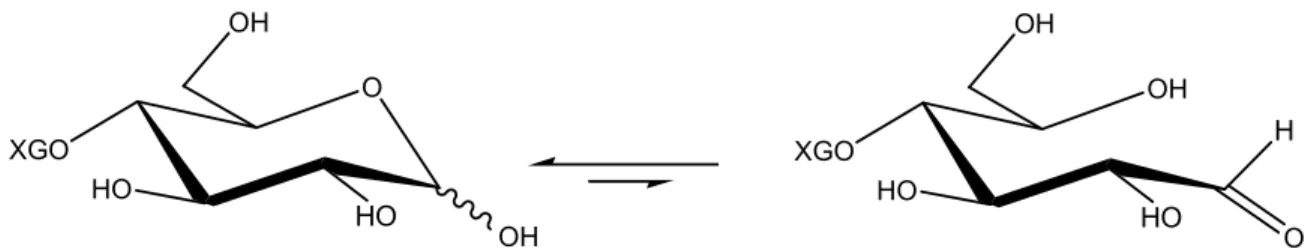
Xyloglucan (XG) is cleavable at the unsubstituted glycopyranosyl units by endo-glucanases (Figure 6). The resulting xyloglucan oligosaccharides (XGOs) have different properties (e.g. solubility and viscosity) than the high molecular weight XG, which makes them suitable for further reactions.
XGO O HO HO OH OH XGO OH HO HO OH O H
Figure 8: The reducing end of XGO in equilibrium between the closed hemiacetal and the open aldehyde conformations.
Nucleophilic coupling reactions with sugars are most often performed at the reducing end. All aldoses without substituents at the anomeric carbon are in equilibrium between the closed and open conformation (Figure 8). This equilibrium is strongly pushed towards the hemiacetal. The existing amount of aldehyde is enough to provide the saccharide with a selective reactive site at the anomeric carbon.
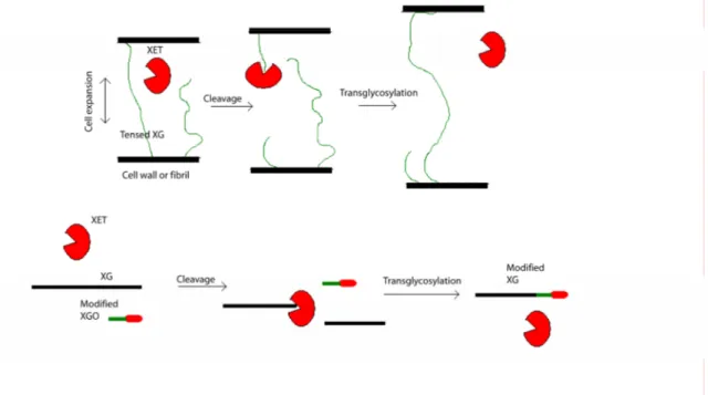
Xyloglucan endotransglycosylase
When the plant cell grows the xyloglucans (XGs), which cross-links the cellulose fibrils and the primary and secondary cell wall, get tensed and have to be loosened to allow the cell to expand, and subsequently reinforced to maintain the integrity of the cell wall. In
vivo, xyloglucan endotransglycosylase (XET) specifically cuts the XG polymer and
attaches it to another loose XG chain (Figure 9, upper part). This transglycosylation ability is mimicked in vitro, using heterogeneously expressed Populus tremula x
tremuloides PttXET16A (Figure 9, lower part).
Figure 9: Transglycosylation of XG by XET (a) in vivo during cell expansion (b) in vitro during cellulose surface modification.
XET is related to the retaining glycosyl hydrolases, which carry out carbohydrate degradation. These enzymes catalyse, in the first reaction step, the cleavage of a glycosidic bond, which leads to the formation of a covalent enzyme-glycosyl intermediate (Vocadlo et al. 2001). In the case of hydrolases this intermediate is decomposed by water with carbohydrate hydrolysis as the net result. In contrast to the hydrolases, XET transfers the enzyme-glycosyl intermediate to a carbohydrate acceptor.
Cellulose surface modification by XET
The route of fibre surface modification starts at xyloglucan (XG), which in the first step is digested to xyloglucan oligosaccharides (XGOs). To prepare the XGOs for further reactions, the Kochetkov amination is performed (Likhosherstov et al. 1986), resulting in 1-deoxy-1-amino xyloglucan oligosaccharides (XGO-β-NH2). The amination step turns the aldehyde of the reducing end of XGO into a primary amine, which is a strong and selective nucleophile, clearly distinguished from the hydroxyl groups. The XGOs are thereby activated and XGO-β-NH2 is the chemical handle to which a whole range of coupling reactions can be implemented.
When the desired function is attached to XGO-β-NH2, the modified XGOs are incorporated into XG by using the strict transglycosylating ability of XET.
The configuration of native and modified XGOs occupying the acceptor binding site of XET16A from Populus tremula x tremuloides have been investigated by X-ray crystallography (Johansson et al. 2004). The X-ray structures revealed good interaction of the non-reducing end of XGO with the enzyme, with the glycosyl unit at the reducing end fully exposed to the solvent. Since the reducing end of the XGOs and its attached modification is fully exposed to the solvent it does not interact with the active site of the enzyme and hence, does not restrict the transglycosylation.
The obtained modified XG is finally adsorbed onto cellulose, anchoring the functional group onto the cellulose surface. The adsorption occurs in water at room temperature with moderate stirring. These reaction conditions are mild compared to organic synthesis using the surface hydroxyl groups and result in a total maintenance of the structure and integrity of the individual fibres and fibre network during the modification (Zhou et al. 2006a; Ahrenstedt et al. submitted). This method has previously been used with success to introduce a number of functional groups to Whatman Nr 1 filter papers (Brumer et al. 2004; Zhou et al. 2006b).
Experimental
This section describes the methods used in unpublished experiments, which still will be discussed in the next section.
Materials
Thiolated XGOs (XGO-SH), synthesised as previously described (Brumer et al. 2004), were kindly provided by Dr. Qi Zhou. Iodoacetic acid was obtained from Sigma-Aldrich (Steinheim, Germany). All other chemicals were obtained as stated in the attached publications.
Methods
Synthesis of GRGDS-SGGSS-GRGDS
The GRGDS-SGGSS-GRGDS peptide was synthesised with solid-phase peptide synthesis (SPPS) as described in Publication II, resulting in resin-bound peptides (0.29 mmol peptide/g resin). The peptide was cleaved from the resin (79 mg) with simultaneous removal of the side-chain protection groups with 1 mL TFA/H2O/TIS (95:2.5:2:5) for 30 minutes at room temperature. The reaction was diluted with water (20 mL), extracted with tBME (3 x 20 mL) and filtered through glass fibres. Freeze-drying the aqueous phase yielded a white solid (27 mg, 87%). ESI-MS: [M +Na +2H]3+, 453.8591 calculated (453.8660 observed); [M +2H]2+, 669.2938 calc. (669.3047obs.).
Attempted solid-phase coupling of GRGDS-SGGSS-GRGDS I
XGO-NH-succinamic acid was manually conjugated to the resin-bound peptide in a reaction vessel equipped with a glass frit filter (pore size P2). XGO-NH-succinamic acid
(460 mg, 0.33 mmol, 2 equiv.), DIEA (120 μL, 0.88 mmol, 6 equiv.), HOBt (160 mg, 1.0 mmol, 7 equiv.) and HBTU (390 mg, 1.0 mmol, 7 equiv.) were dissolved in DMF (8 mL) and added to the resin-bound peptide (500 mg, 0.14 mmol, 1 equiv). The coupling was terminated after 1 hour by extensive washing of the resin with ethanol, NMP, DIEA (5% in DCM), NMP and DCM (∼10 mL each). The resin was subsequently dried under vacuum. The glycopeptide was cleaved from the resin with simultaneous removal of the side-chain protection groups with 3 mL TFA/H2O/TIS (95:2.5:2:5) for 30 minutes at room temperature. The reaction was diluted with water (40 mL), extracted with tBME (3 x 40 mL) and filtered through glass fibres. Freeze-drying the aqueous phase yielded a white solid (139 mg). ESI-MS: GRGDS-SGGSS-GRGDS [M +2H]2+, 669.2938 calculated (669.3101 observed) among other unidentified peaks. The MS data indicates only cleaved and purified peptide, which is consistent with observations by 1H NMR spectroscopy (data not shown).
Attempted solid-phase coupling of GRGDS-SGGSS-GRGDS II
XGO-NH-succinamic acid was manually conjugated to the resin-bound peptide in a reaction vessel equipped with a glass frit filter (pore size P2). XGO-NH-succinamic acid (48 mg, 0.035 mmol, 2 equiv.), DIEA (12 μL, 0.089 mmol, 4 equiv.), HOBt (16 mg, 0.1 mmol, 5 equiv.) and HBTU (40 mg, 0.1 mmol, 5 equiv.) were dissolved in a mixture of DMF (0.14 mL) and DMSO (0.06 mL) and added to the resin-bound peptide (60 mg, 0.02 mmol, 1 equiv). The coupling was terminated after 1 hour by extensive washing of the resin with ethanol, NMP, DIEA (5% in DCM), NMP and DCM (∼10 mL each). The resin was subsequently dried under vacuum. The glycopeptide was cleaved from the resin with simultaneous removal of the side-chain protection groups with 1 mL TFA/H2O/TIS (95:2.5:2:5) for 30 minutes at room temperature. The reaction was diluted with water (20 mL), extracted with tBME (3 x 20 mL) and filtered through glass fibres. Freeze-drying the aqueous phase yielded a white solid (18 mg). ESI-MS: GRGDS-SGGSS-GRGDS [M +2H]2+, 669.2938 calculated (669.3112 observed); [M +2Na]2+, 691.2756 calc. (691.2902 obs.); XXLG-GRGDS-SGGSS-GRGDS and XLXG-GRGDS-SGGSS-GRGDS [M +2H
+Na]3+, 888.9986 calc. (889.3615 obs.); XLLG-GRGDS-SGGSS-GRGDS [M +2H +Na]3+, 943.0186 calc. (943.3995 obs.) among other unidentified peaks. The MS data indicate mostly starting material and unidentified side products.
Attempted solid-phase coupling of GRGDS-SGGSS-GRGDS III
The resin-bound peptide was manually conjugated with iodoacetic acid prior to solid-phase coupling with XGO-SH.
Iodoacetic acid (15 mg, 0.081 mmol, 5 equiv.) was pre-activated with DCC (34 mg, 0.16 mmol, 10 equiv.) in DCM (1 mL) for 30 minutes in an ice bath. The mixture was further transferred to the resin-bound peptide (56 mg, 0.016 mmol, 1 equiv.) and mixed with DMF (1 mL) and DIEA (28 μL, 0.16 mmol, 10 equiv.) in a reaction vessel equipped with a glass frit filter (pore size P2). The coupling was terminated after 1 hour by extensive washing of the resin with DMF and DCM (∼10 mL each). The resin was subsequently dried under vacuum. The iodoacetic-peptide derivative was either further reacted in the next step or cleaved from the resin with simultaneous removal of the side-chain protection groups with 1 mL TFA/H2O/TIS (95:2.5:2:5) for 30 minutes at room temperature. The cleaving mixture was diluted with water (20 mL), extracted with tBME (3 x 20 mL) and filtered through glass fibres. Freeze-drying the aqueous phase yielded a white solid (22 mg, 49%). ESI-MS: [M +Na +2H]3+, 509.8282 calculated (509.8418 observed); [M +2Na +H]3+, 517.1555 calc. (517.1683 obs.); [M +2H]2+, 753.2474 calc. (753.2595 obs.).
The resin-bound iodoacetic-peptide derivative (0.011 mmol, 1 equiv.) was further conjugated with XGO-SH (39 mg, 0.028 mmol, 2.6 equiv.) in the presence of DIEA (2 μL, 0.011 mmol, 1 equiv) and TCEP (2 mg, 0.0056 mmol, 0.5 equiv.) in DMF (1 mL). The coupling was terminated after 1 hour by extensive washing of the resin with DMF and DCM (∼10 mL each). The resin was subsequently dried under vacuum. The glycopeptide derivative was cleaved from the resin with simultaneous removal of the side-chain protection groups with 1 mL TFA/H2O/TIS (95:2.5:2:5) for 30 minutes at room temperature. The reaction was diluted with water (20 mL), extracted with tBME (3 x 20 mL) and filtered through glass fibres. Freeze-drying the aqueous phase yielded a
white solid. ESI-MS: XXLG-SH and XLXG-SH [M +2Na]2+, 686.7133 calculated (686.2014 observed); XLLG-β-NH2 [M +Na +H]2+, 705.7439 calc. (705.7693 obs.); [M +2Na]2+, 716.7348 calc. (716.7429 obs.); GRGDS-SGGSS-GRGDS-NH-CO-CH2-Cl [M +2H]2+, 707.2796 calc. (707.2859 obs.); GRGDS-SGGSS-GRGDS-NH-CO-CH2-I [M +2H]2+, 753.2474 (753.2602 obs.) among other unidentified peaks. The MS data indicates only starting materials.
Synthesis of SGGSS-GGSSG-GRGDS
The SGGSS-GGSSG-GRGDS peptide was synthesised with SPPS as described in Publication II, resulting in resin-bound peptides (0.32 mmol peptide/g resin). The peptide was cleaved from the resin (83 mg) with simultaneous removal of the side-chain protection groups with 1 mL TFA/H2O/TIS (95:2.5:2:5) for 30 minutes at room temperature. The reaction was diluted with water (20 mL), extracted with tBME (3 x 20 mL) and filtered through glass fibres. Freeze-drying the aqueous phase yielded a white solid (21 mg, 65%). ESI-MS: [M +Na +2H]3+, 411.5009 calculated (411.4893 observed); [M +2H]2+, 605.7565 calc. (605.7229 obs.); [M +Na +H]2+, 616.7474 calc. (616.7114 obs.); [M +2Na]2+, 627.7383 calc. (627.7173 obs.).
Attempted solid-phase coupling of SGGSS-GGSSG-GRGDS I
XGO-NH-succinamic acid was manually conjugated to the resin-bound peptide in a reaction vessel equipped with a glass frit filter (pore size P2). XGO-NH-succinamic acid (370 mg, 0.27 mmol, 2 equiv.), DIEA (100 μL, 0.55 mmol, 4 equiv.), HOBt (130 mg, 0.82 mmol, 6 equiv.) and HBTU (310 mg, 0.82 mmol, 6 equiv.) were dissolved in DMF (11 mL) and added to the resin-bound peptide (420 mg, 0.14 mmol, 1 equiv). The coupling was terminated after 1 hour by extensive washing of the resin with ethanol, NMP, DIEA (5% in DCM), NMP and DCM (∼10 mL each). The resin was subsequently dried under vacuum. The glycopeptide was cleaved from the resin with simultaneous removal of the side-chain protection groups with 3 mL TFA/H2O/TIS (95:2.5:2:5) for 30 minutes at room temperature. The reaction was diluted with water (40 mL), extracted
with tBME (3 x 40 mL) and filtered through glass fibres. Freeze-drying the aqueous phase yielded a white solid (139 mg). ESI-MS: SGGSS-GGSSG-GRGDS [M +Na +2H]3+, 411.5009 calculated (411.5153 observed); [M +2H]2+, 605.7565 calc. (605.7662 obs.); [M +Na +H]2+, 616.7474 (616.75 obs.); 627.7383 calc. (627.7372 obs.) among other unidentified peaks. The MS data indicates only unreacted peptide, which is consistent with observations by 1H NMR spectroscopy (data not shown).
Attempted coupling of SGGSS-GGSSG-GRGDS II
XGO-NH-succinamic acid (57 mg, 0.04 mmol, 2 equiv.), DMAP (1mg, 0.01 mmol, 0.5 equiv.) and EDC (8 mg, 0.04 mmol, 2 equiv.) were dissolved in de-ionised water (1.5 mL) and added to the non-resin-bound peptide (25 mg, 0.02 mmol, 1 equiv.). After 23 hours, the volume was reduced and the crude reaction mixture was fractionated with reversed phase chromatography as follows: the resin (Silice C18 60Å, 40-63 μm, obtained from SDS, Peypin, France) was packed in 100% acetonitrile and prepared for the water phase by successively changing the gradient to 100% water with 2 column volumes (CVs) of 100% acetonitrile, 1 CV 75% acetonitrile, 1 CV 50% acetonitrile, 1 CV 25% acetonitrile and 2 CVs 0% acetonitrile. After loading the crude product, diluted to a half CV, a gradient of acetonitrile was applied and fractionated. 1 CV 0%, 1 CV 2%, 1 CV 4% and 1 CV 6%. TLC (70/30 acetonotrile/water) indicated in which fractions the desired product was. The fractions were pooled, concentrated and freeze-dried. ESI-MS: XXXG-succinamic acid [M +2Na]2+, 603.6798 calculated (603.7169 observed); XXLG and XLXG [M +2Na]2+, 635.1898 calc. (635.2196 obs.); XLLG [M +2Na]2+, 716.2148 (716.2535 obs.); XLLG-succinamic acid [M +2Na]2+, 765.7348 calc. (765.7790 obs.) among other unidentified peaks. The MS data indicate only starting material.
Synthesis of SGGSS-GRGDS
The SGGSS-GRGDS peptide was synthesised with SPPS as described in Publication II, resulting in resin-bound peptides (0.38 mmol peptide/g resin). The peptide was cleaved from the resin (101 mg) with simultaneous removal of the side-chain protection groups
with 1 mL TFA/H2O/TIS (95:2.5:2:5) for 30 minutes at room temperature. The reaction was diluted with water (20 mL), extracted with tBME (3 x 20 mL) and filtered through glass fibres. Freeze-drying the aqueous phase yielded a white solid (26 mg, 70%). ESI-MS: [M +2H]2+, 433.1923 calculated (433.2840 observed); [M +H]+, 865.3766 calc. (865.3837 obs.).
Solid-phase coupling of SGGSS-GRGDS
XGO-NH-succinamic acid was manually conjugated to the resin-bound peptide in a reaction vessel equipped with a glass frit filter (pore size P2). XGO-NH-succinamic acid (71 mg, 0.05 mmol, 2 equiv.), DIEA (18 μL, 0.11 mmol, 4 equiv.), HOBt (24 mg, 0.16 mmol, 6 equiv.) and HBTU (60 mg, 0.16 mmol, 6 equiv.) were dissolved in DMF (2 mL) and added to the resin-bound peptide (70 mg, 0.026 mmol, 1 equiv). The coupling was terminated after 1 hour by extensive washing of the resin with ethanol, NMP, DIEA (5% in DCM), NMP and DCM (∼10 mL each). The resin was subsequently dried under vacuum. The glycopeptide was cleaved from the resin with simultaneous removal of the side-chain protection groups with 1 mL TFA/H2O/TIS (95:2.5:2:5) for 30 minutes at room temperature. The reaction was diluted with water (20 mL), extracted with tBME (3 x 20 mL) and filtered through glass fibres. Freeze-drying the aqueous phase yielded a white solid (29 mg, 49%). ESI-MS: XXXG-SGGSS-GRGDS [M +2Na +H]3+, 684.9087 calculated (684.9163 observed), [M +Na +H]2+, 1015.8682 calc. (1015.9092 obs.); XXLG-SGGSS-GRGDS and XLXG-SGGSS-GRGDS [M +2Na +H]3+, 738.9264 calc. (738.9304 obs.), [M +Na +H]2+, 1096.89 calc. (1096.91 obs.); XLLG-SGGSS-GRGDS [M + 2Na +H]3+, 792.9440 calc. (792.9438 obs.), [M +Na +H]2+, 1177.92 calc. (1177.94 obs.); SGGSS-GRGDS [M +H]+, 865.3766 calc. (865.3837 obs.).
Results and discussion
In the process of developing new materials, paper or blood vessels, the material characteristics are very essential for the outcome. In the case of paper, strength, optical properties and printability are fundamental for the final product. For blood vessels, blood- and tissue-compatibility is crucial to maintain their function.
Paper strength (Publication I)
Xyloglucan (XG) has previously been shown to increase paper dry strength when adsorbed to pulp in the wet-end. The tensile strength increases reported in this thesis were comparable or better than those (Christiernin et al. 2003; Lima et al. 2003). In the wet-end treatments with XG, three different pulps were used: pulp made from bleached soft wood (BSW; mixed pine/spruce), de-inked pulp (DIP) and thermo-mechanical pulp (TMP).
A main characteristic to distinguish the different pulps from each other is their degree of de-lignification during the pulping process. Chemically bleached pulps contain ∼5% surface lignin, while unbleached mechanical pulps contain 30- 80% surface lignin. The de-inked pulp used for the work reported in this thesis consists mainly of mechanical pulp and, hence, would be more similar to mechanical than chemical pulp with respect to surface lignin. These surface lignin numbers are based on two studies, which measured the content of surface lignin for a range of pulps using X-ray photoelectron spectroscopy and electron spectroscopy for chemical analysis respectively (Hannuksela et al. 2003; Zhou et al. 2006a). Both studies found a relation between the surface lignin content and the adsorbed amount of hemicelluloses, as follows: the lower the surface lignin content, the higher the adsorption of hemicelluloses (galactoglucomannan and XG).
These observations are in good agreement with the results in this thesis, where XG was loaded (1% w/w) to the chemical and mechanical pulps in the wet-end prior to hand sheet formation. The hand sheets were further conditioned and tested for tensile
strength, which was much greater for the chemical pulp (BSW; 28%) than for the thermo-mechanical (TMP, 6%) and de-inked pulps (DIP, 11%).
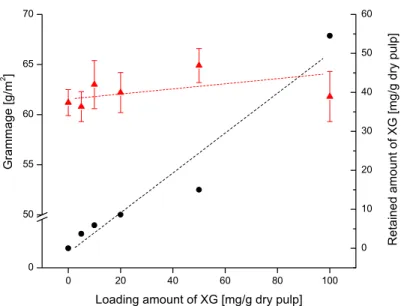
0 20 40 60 80 100 0 50 55 60 65 70 Retai ned amount of X G [mg/ g dr y pulp] G rammage [ g /m 2 ]
Loading amount of XG [mg/g dry pulp]
0 10 20 30 40 50 60
Figure 11: The average grammage of five hand sheets formed of BSW pulp (red triangles) treated with different amounts of XG in the wet-end. The amount of XG adsorbed after 18 hours is represented by black circles.
The retained amount of XG (described in Publication I) corresponded to the increased tensile strength for the different pulps (BSW retained more than DIP which retained more than TMP). The retained amount was also related to the tensile strength increases, as shown by increasing the loading amount (Publication I, Figure 2).
The explanation for the strength increases may depend on an increased softening of the fibre surface, or on the fact that the adsorbed XG results in an increased retained amount of fines. By plotting the grammage and the retained amount against the loading of XG, it was clear that an increased loading amount did not increase the grammage, even though the retained amount of XG increased (Figure 11). Consequently, the strength increase was due to XG and not to an increased amount of fines. The observed variations in grammage may be explained by insufficient skills in the manual hand sheet production. Thus to increase the retained amount of XG, our focus turned to spray application of dry sheets. In addition to the pulps used for wet-end application, sheets made of bleached hard wood pulp (BHW; birch) were also sprayed. Spraying the sheets twice on
each side loaded a similar amount of XG to the sheets as was retained in the wet-end application, except for TMP (∼0.8% vs. ∼0.7% for BSW; ∼0.6% vs. ∼0.4% for DIP and ∼0.9% vs. ∼0% for TMP). The sheets were further conditioned and tested with respect to their tensile strength, tear strength and Z-strength (Scott-Bond).
The strength development for sprayed sheets mirrored that of the wet-end treatment. Sheets made of chemical pulp revealed a huge tensile strength increase (71% for BSW and 27% for BHW), while the effects on sheets made of mechanical pulp were more modest (10% for DIP and 13% TMP) when comparing sheets sprayed twice on each side with unsprayed sheets. Although the general strength increases were higher than for the wet-end treatment (except for DIP-sheets which showed a similar increase for the two treatments), the chemical pulps responded more significantly to the treatment than the mechanical pulps. Consequently, the XG-cellulose contact is of great importance even in the spray application, more important than covering the sheets with a XG film.
Borates are known to form complexes with polysaccharides (Pezron et al. 1988; O'Neill et al. 1996; Martin et al. 2003; Bishop et al. 2004). This is possible due to boron’s ability to switch ligands via hydrolysis (Figure 12). Hence, a mixture of XG and borax was sprayed to the sheets with the aim to introduce covalent bonds between XG and cellulose or between two adsorbed XG molecules. An introduction of covalent bonds was expected to strengthen the network of cellulose fibres and consequently the paper strength in general.
B
OH
OH
HO
RO
B
OR
OR
OR
ROH
H
2O
Figure 12: Borate di-ester formation where R is XG or cellulose.
The combination of XG and borax did boost the effect of XG, again more obviously for the chemical pulps (88% for BSW and 39% for BHW) than for the mechanical pulps (12% for DIP and 20% for TMP), when comparing the tensile strength indices of sheets sprayed twice on each side to unsprayed sheets.
The tear strength (Publication I, Figure 4) was increased for sprayed hand sheets made of BSW and BHW pulp when treated with XG (70% and 56% respectively) or XG +borax (51% and 55% respectively). The increase for DIP sheets sprayed with XG or XG +borax was lower (4% and 8%), as for TMP sheets (12% and 9%). The standard deviations were notably greater for the BSW sheets (∼20-30%) than for the BHW, DIP and TMP sheets (∼10%).
The Z-strength of sprayed sheets was measured as Scott-Bond (Publication I,
Table 2). Resistance to de-lamination increased mostly for sheets made of BSW (254%)
and BHW (260%), while the strength increase for sheets made of DIP (30%) and TMP (35%) was slightly lower, compared to unsprayed sheets. Spraying of XG and borax in combination boosted the strength effect for the pulps as follow: BSW (301%), BHW (316%), DIP (32%) and TMP (45%) compared to the corresponding unsprayed sheets.
The bulk of the sheets increased during the spraying, due to re-wetting, but it did not seem to influence the paper strength. The bulk increased in an equal extent for sheets sprayed with water without affecting the strength, as for sheets sprayed with XG increasing the strength significantly.
A possible drawback with the spray application is the increase of surface roughness due to re-wetting of the sheets. However, the increased roughness can be compensated for with calendering after the spraying or prevented by spraying the wet web in the press section of the paper machine.
This would bring a better paper formation and, consequently, even greater paper strength increases. Since the spraying itself decreases the paper strength as verified by spraying only water to the sheets (Publication I, Figure 3), the strength additive have to compensate for this loss and improve the original strength. If the strength decrease due to rewetting was eliminated, the effect of the strength additives could be utilised to its full power.
Adhesion of endothelial cells (Publication II)
To obtain an artificial blood vessel, which can prevent intimal hyperplasia (abnormal growth of tissue) and thrombosis, the bacterial cellulose scaffold was seeded with endothelial cells. The seeding was enhanced by adsorbing a XG-RGD conjugate to the bacterial cellulose scaffold. The XG-RGD conjugate was obtained by using a method developed in the laboratory of Wood Biotechnology at the Royal Institute of Technology in Stockholm, Sweden (Brumer et al. 2004). The main principle of this method for cellulose surface modification is to perform the chemical synthesis on the polysaccharide XG instead of directly on the hydroxyl groups on the cellulose surface, and then adsorb the modified XG to the cellulose surface.
XG-GRGDS synthesis
Xyloglucan oligosaccharides (XGOs) were obtained by endo-glucanase digestion of XG, essentially as previously described (Greffe et al. 2005) and constitutes the platform for further synthesis. The complete digestion of XG was confirmed by HPLC analysis on a Dionex (Sunnyvale, USA) carbopac PA100 column (Figure 13).
XXXG
XLLG
XLXG XXLG
The obtained advantages of using XGOs instead of XG for the synthetic work are essential. First, the solubility of XGOs in water is about ten times higher than for XG and in contrast to XG, neither heating nor extensive stirring is necessary. In addition, XGOs are soluble in common organic solvents such as DMF, NMP and DMSO which makes possible the use of a wide range of anhydrous chemistry. Further, their molecular size and solubility properties make XGOs suitable for standard analytical methods such as NMR, MS, TLC, IR and UV-Vis absorption spectroscopy for determining structure, purity and yield. O (XGO) HO OH OH XGO
aq. succinic anhydride Sat aq. NH4HCO3
XG Endoglucanase, pH 4.5 1. HOBt/HBTU DIEA, DMF XGO-GRGDS 4 XET/XG XG-GRGDS 5 2 2. TFA, TIS, H2O H N OH O O O (XGO) HO OH OH NH2 1 3 SDGRG-NH2
Prior to amide formation with the N-terminus of the resin-bound peptide, the reducing end of XGO was modified into a carboxylic acid via a glycosylamine intermediate. Glycosylamines are generally accessible by two routes: either via azide formation which requires protection and glycosyl-halide formation followed by halogen replacement and deprotection, or alternatively, the amination can be obtained by one single reaction step using the Kochetkov method. In the first step on the path towards the glycopeptide conjugate the latter strategy was used to form 1-deoxy-1-amino-XGO (1) as follows (Scheme 1). An aqueous solution of XGOs (50 mg/mL) was saturated with ammonium hydrogen carbonate and stirred at 42° C for 28 hours to stereo selectively convert the saccharides into β-glycosylamine (Kallin et al. 1989; Meinjohanns et al. 1998), as confirmed by NMR (Figure 14).
Figure 14: NMR spectrum of 1-deoxy-1-amino-XGO (1; top) compared with a spectrum of a mixture of XGOs (bottom).
The excess of ammonium hydrogen carbonate was removed (>99%) by three consecutive cycles of freeze drying to yield a white powder consisting of a mixture of XGOs and 1-deoxy-1-amino-β-XGOs (1). The extent of conversion was of 75%, based on integration of 1H-NMR signals from the anomeric protons of the starting material and product. Kallin
et al. (1989) investigated the stability of formed glycosylamines and found them rather
stable in the pH range 8-10, which includes the pH used in our experiments. Further, Kallin et al. (1989) pointed out the corresponding carbamate and bis-β-glycosylamine as possible side products to glycosylamine formation. However, XGO-carbamate was not detected by MS nor by IR and there was no trace of the dimer in the MS or NMR spectra. According to Bejugam et al. (2004) the dimer formation was significantly increased at temperatures above 50° C when they carried out the Kochetkov reaction in DMSO, thus the reaction temperature was set to 42° C.
The advantage of introducing an anomeric primary amine is central for the following synthetic work since the amine is a stronger nucleophile than the saccharide hydroxyl groups and can therefore be used selectively for nucleophilic reactions. Consequently, the crude mixture of XGOs and (1) was further mixed with succinic anhydride (2 equiv.) to quantitatively afford XGO-succinamic acid (2), using a similar setup as described by Xia et al. (2001). Reversed phase chromatography separated (2) from unreacted XGOs, which were brought with the crude starting material. The total removal was confirmed by NMR (Figure 15).
Figure 15: NMR spectrum of XGO-NH-succinamic acid (2) after reversed phase chromatography. There are no traces of the XGO 1H signals at δ =4.6 and δ =5.2. The multiple at δ =2.6 shows the two CH2 groups of incorporated succinic anhydride.
The GRGDS peptide (3) was synthesised on a 0.25 mmol scale by conventional solid-phase peptide synthesis using Fmoc protection strategy, essentially according to the protocol described by Engfeldt et. al. (2005). Prior to amide formation with the resin-bound peptide (3), the modified saccharide (2) was activated with HBTU/HOBt in presence of DIEA, to form a tetramethyluronium complex. The obtained glycopeptide (4) was cleaved from the resin with simultaneous removal of the side chain protection groups with TFA, isolated by extraction with tBME and freeze-dried to obtain the product, typically at 50% yield. Under identical conditions, cleavage and de-protection of the unmodified peptide from the resin yielded the peptide, typically at 90% yield. The successful linking of the peptide and XGO was confirmed by NMR (Figure 16).
The modified oligosaccharides (4) were incorporated into high molecular weight XG using the strict transglycosylating ability of XET to obtain the final glyco-conjugate (5). The molecular weight and polydispersity of (5) was determined by GPC (Figure 17).
0 50 100 150 200 250 300 350 0 5 10 15 20 25 30 Time [min] In t
Figure 17: GPC curve for XG-GRGDS, Mw = 28 kDa and PDI = 2.0.
Later, the produced glyco-peptide (5) was adsorbed to the bacterial cellulose scaffolds in aqueous conditions at room temperature. The scaffolds adsorbed ∼90% of the loaded XG-GRGDS (200 mg/g dry material) after 24 hours (Bodin et al. submitted). The RGD-substituted scaffolds were further seeded with endothelial cells and the cell attachment was confirmed by confocal microscopy. The cellulose surface modified with XG-RGD revealed a significantly greater proliferation of endothelial cells than a surface modified with XG only or an unmodified surface (Publication II, Figure 11).
Glyco-conjugates with 10 and 15-mer peptides (unpublished
results)
The HOBt/HBTU aided coupling of XGO-NH-succinamic acid (2) and the resin-bound 5-mer peptide (3) yielded the desired glyco-peptide (4) without much further development of the coupling method. This was definitely not the case for the longer 15-mer peptides, GRGDS-SGGSS-GRGDS (6) and SGGSS-GGSSG-GRGDS (7). Both resin-bound peptides failed the HOBt/HBTU aided coupling with (2), as confirmed by MS and NMR (not shown).
Tickler et al. (2004) proposed that resin-bound peptides may aggregate in such manner that subsequent acylations of the N-terminus is hindered. Solvent mixtures containing DMSO were proposed to inhibit these aggregations. Consequently, the HOBt/HBTU aided coupling of (6) and (7) with (2) was performed in a DMSO/DMF mixture (30% v/v), without success.
An attempt to couple the cleaved SGGSS-GGSSG-GRGDS peptide with (2) in water, aided by EDC was made, without success.
SDGRG-GSSGG-SDGRG-NH2 HO O I DMF/DCM DCC/DIEA SDGRG-GSSGG-SDGRG-NH O I 1. XGO-SH DTT/DIEA DMF 2. TFA/TIS/H2O N H O S O N H GRGDS-SGGSS-GRGDS XGO 6 9 SDGRG-GSSGG-SDGRG-NH O I 8 TFA/TIS/H2O
Scheme 2: Alternative route for glyco-peptide synthesis, in this case XGO-GRGDS-SGGSS-GRGDS.
The common methodology in the previously described couplings is the formation of an amide linkage between the sugar and the peptide species. Another approach was investigated for the conjugation, using a SN2 type reaction in the crucial step (Scheme 2). The resin-bound peptide (6) first conjugated with iodoacetic acid prior to coupling with XGO-SH.
The conjugation of (6) and iodoacetic acid revealed no difficulties and the cleaved iodoacetic-peptide derivative (8) was confirmed by MS. The second step, on the other hand, did not yield the desired glyco-peptide (9). Only the starting material was detected by MS.
Since the HOBt/HBTU aided coupling of (2) with the resin-bound 5-mer peptide worked properly, but not with (6) and (7), a middle sized 10-mer peptide (SGGSS-GRGDS) was synthesised. The 10-mer was further conjugated with (2) as described and the correct outcome was confirmed by MS.
These trials showed that resin-bound 5 and 10-mers can be attached to XGO without difficulties, while this seems much more complex for the 15-mers and has yet not been obtained.
Conclusions
The use of xyloglucan (XG) as a strength agent in the paper making industry seems to have a bright future. Xyloglucan increases paper strength when added in the wet-end, but even more so when applied by spraying. In both application modes, chemical pulp shows a greater response than mechanical pulp to the treatments. Together with earlier studies, this suggests that the fibre surface composition is important for maximal efficiency of XG. Spraying of the hand sheets increased their bulk, but this did not seem to be the reason for the strength increase, since the sheets sprayed with water did not get stronger. Instead, the strength increase is thought to originate from an increased softness of the fibre surface when coated with XG.
The XG-cellulose interactions were also used to attach a biologically significant cell adhesion peptide sequence (RGD) to a cellulose surface. The RGD motif is recognised by integrin receptors on endothelial cells, responsible for an even blood flow in blood vessels. The cellulose used was produced by Acetobacter xylinum and moulded into a vessel-like shape. The presence of the RGD motif enhanced the growth and proliferation of endothelial cells, which is believed to increase the blood compatibility of the produced artificial blood vessel.
Future work
Future investigations will focus upon other paper properties, such as bending stiffness, which is expected to be increased due to an increased bulk and greater strength properties. Another interesting property awaiting investigation is the penetration depth of sprayed XG in the sheets. The penetration depth can be determined by stripping the hand sheet into layers in the z-direction and measuring the intensity on the withdrawn layers of an adsorbed fluorescently labelled xyloglucan derivative (XG-FITC, previously synthesised by Brumer et al.).
The glyco-peptide conjugation method will be developed, by investigating the iodoacetic acid route, so that even larger (>10 amino acids) peptides can be attached to XGO and further adsorbed to the cellulose surface. Also, other derivatives of XG will be produced, e.g. XG-heparin, in the context of artificial blood vessels.
Acknowledgements
This thesis is the product of many people’s work, and I would like to take the opportunity to give them proper attention. Docent Harry Brumer III, my supervisor, is gratefully thanked for guiding and steering my projects in a fruitful direction. I owe gratitude to Professor Tuula Teeri. She is responsible for my being in the Wood Biotechnology laboratory, by accepting me as a diploma worker in the beginning.
The success of the blood vessel project is much due to the rich collaboration with Assistant Professor Amelie Eriksson Karlström and Torun Engfeldt (Molecular Biotechnology, KTH) who provided valuable supervision of the solid-phase peptide synthesis, Professor Paul Gatenholm and Dr. Aase Bodin (Biopolymer technology, Chalmers) and Professor Bo Risberg and Helen Fink (Vascular Engineering Centre, Sahlgrenska University Hospital) .
The results from the hand sheet modifications are much attributed to our finish co-workers in Jyväskylä, Antti Oksanen, Vesa Kunnari and Kristian Salminen, who shared their equipment and knowledge with us.
At home ground, Dr. Qi Zhou, Martin Bauman and Farid Ibatullin are sincerely credited for being such heroes in the lab, helping me in every situation and explaining whatever stupid question one can ask. Also Ela, Fredrika, Jens, Johanna, Lotta R, Maria, Nomchit and Ulla are valued for their help and great support.
Anne Fält at STFI is acknowledged for introducing me to the tensile strength testing and for being a good friend.
References
Addicks, E., R. Mazitschek and A. Giannis (2002). "Synthesis and biological investigation of novel tricyclic benzodiazepinedione-based RGD analogues". Chembiochem 3(11): 1078-1088.
Ahrenstedt, L., A. Oksanen, K. Salminen and H. Brumer (submitted).
Backdahl, H., G. Helenius, A. Bodin, U. Nannmark, B. R. Johansson, B. Risberg and P. Gatenholm (2006). "Mechanical properties of bacterial cellulose and interactions with smooth muscle cells". Biomaterials 27(9): 2141-2149.
Bejugam, M. and S. L. Flitsch (2004). "An efficient synthetic route to glycoamino acid building blocks for glycopeptide synthesis". Organic Letters 6(22): 4001-4004.
Bishop, M., N. Shahid, J. Z. Yang and A. R. Barron (2004). "Determination of the mode and efficacy of the cross-linking of guar by borate using MAS B-11 NMR of borate cross-linked guar in combination with solution B-11 NMR of model systems". Dalton Transactions 17): 2621-2634.
Bodin, A., L. Ahrenstedt, H. Fink, H. Brumer, B. Risberg and P. Gatenholm (submitted).
Bodin, A., H. Backdahl, G. Helenius, L. Gustafsson, B. Risberg and P. Gatenholm (2005). "Bacterial cellulose as scaffold for tissue engineering." Abstracts of Papers of the American Chemical Society 229(U297-U297. Brumer, H., Q. Zhou, M. J. Baumann, K. Carlsson and T. T. Teeri (2004).
"Activation of crystalline cellulose surfaces through the chemoenzymatic modification of xyloglucan". Journal of the American Chemical Society
126(18): 5715-5721.
Chambat, G., M. Karmous, M. Costes, M. Picard and J. P. Joseleau (2005). "Variation of xyloglucan substitution pattern affects the sorption on celluloses with different degrees of crystallinity". Cellulose 12(2): 117-125. Christiernin, M., G. Henriksson, M. E. Lindstrom, H. Brumer, T. T. Teeri, T.
Lindstrom and J. Laine (2003). "The effects of xyloglucan on the properties of paper made from bleached kraft pulp". Nordic Pulp & Paper Research Journal 18(2): 182-187.
Davison, R. W. (1972). "Weak link in paper dry strength". Tappi 55(4): 567-573. Deutsch, M., J. Meinhart, T. Fischlein, P. Preiss and P. Zilla (1999). "Clinical
autologous in vitro endothelialization of infrainguinal ePTFE grafts in 100 patients: A 9-year experience". Surgery 126(5): 847-855.
Engfeldt, T., B. Renberg, H. Brumer, P. A. Nygren and A. E. Karlstrom (2005). "Chemical synthesis of triple-labelled three-helix bundle binding proteins for specific fluorescent detection of unlabelled protein". Chembiochem
6(6): 1043-1050.
Feng, D., D. F. Caulfield and A. R. Sanadi (2001). "Effect of compatibilizer on the structure-property relationships of Kenaf-fiber/polypropylene composites". Polymer Composites 22(4): 506-517.