*Corresponding author: Henrik C Ahn, Department of Cardiothoracic Surgery, Institution of Medical and Health Sciences, Linkoping University, Linkoping, Sweden, Tel: +46707256463; E-mail: henrik.casimir.ahn@liu.se
Received July 03, 2016; Accepted July 25, 2016; Published July 30, 2016 Citation: Ahn HC, Delshad B, Baranowski J (2016) An Implantable Pressure Sensor for Long-term Wireless Monitoring of Cardiac Function- First Study in Man. J Cardiovasc Dis Diagn 4: 252. doi: 10.4172/2329-9517.1000252
Copyright: © 2016 Ahn HC, et al. This is an open-access article distributed under the terms of the Creative Commons Attribution License, which permits unrestricted use, distribution, and reproduction in any medium, provided the original author and source are credited.
An Implantable Pressure Sensor for Long-term Wireless Monitoring of
Cardiac Function- First Study in Man
Henrik Casimir Ahn1*, Baz Delshad1 and Jacek Baranowski2
1Department of Cardiothoracic Surgery, Institution of Medical and Health Sciences, Linkoping University, Linkoping, Sweden 2Department of Physiology, Institution of Medical and Health Sciences, Linkoping University, Linkoping, Sweden
Abstract
Background: Heart failure is a huge health problem. The possibility of long-term monitoring heart function
more accurately in these patients has gained increasing interest. The primary aim of this study was to see if a wireless pressure sensor can be safely implanted to give accurate and reproducible long-term intracardiac pressure recordings. Another aim was to see if there are any adverse effects connected with the implant. A control group was included for comparison of clinical data.
Methods: Forty patients with heart failure, 31 scheduled for open heart surgery and 9 for transcatheter aortic
valve replacement (TAVR) were included to test the safety and feasibility of the Titan™ pressure sensor. The patients were randomized to the implant or control group.
Findings: Initial sensor measurements showed very good correlation with reference pressure values from
a fluid-filled catheter, and there was no need for calibration of the sensor. At the 6-month follow-up 11 patients had been wearing the implant for >1 year with a median time of 560 days. Ten of these had adequate sensor function. Compared to the control group there was no difference in adverse clinical events and the overall number of complications was low.
Conclusions: This first study in man on a new implantable wireless hemodynamic monitor showed favorable
results regarding our primary endpoints; accuracy of recordings over time and safety profile. The technology has great potential for monitoring at home since it is easy to use in the out-patient setting.
Keywords: Heart failure; Diagnostics; Pressure monitoring;
TelemedicinIntroduction
Heart failure is a huge health problem. The disease often follows a similar course; stable in the initial stages but progressive deterioration develops with exacerbations that eventually lead to recurrent hospitalization. To begin with, patients usually respond to standard medical treatment, but the course is unpredictable and in the final stages inotropic support is often required to preserve life.
The possibility of monitoring heart function more accurately in these patients has gained increasing interest. The ability to detect a sudden increase in filling pressure would provide early warning of an imminent exacerbation and enable medical intervention before the development of clinical symptoms [1].
There are a few implantable cardiac hemodynamic monitors on the market, some already in use while others have gone through clinical studies with successful safety records. These devices can be divided into two categories; those monitoring pressure of the left side of the heart, and those on the right.
A catheter-based technology with a device introduced into the pulmonary artery allowing wireless recordings of pulmonary artery pressure and indirectly pulmonary artery wedge pressure is currently approved by the Food and Drug Administration [2,3].
We have used a newly developed device that provides rapid, detailed, continuous real time cardiac pressure measurement, implanted in a series of patients undergoing open chest heart surgery or transcatheter aortic valve replacement. One primary aim was to see if this pressure sensor can be safely implanted to give accurate and reproducible long-term intracardiac pressure recordings similar to the ones obtained with fluid- filled catheters that are traditionally used during open chest surgery and on intensive care units. Another primary aim was to see
if there are any adverse effects connected with the implant. A control group was included for comparison of clinical data.
Material and Methods
Forty patients with heart failure, 31 scheduled for open heart surgery and 9 for transcatheter aortic valve replacement (TAVR) were included to test the safety and feasibility of the Titan™ pressure sensor. This first study in man was approved by the Swedish Medical Products Agency Dnr: 461:2012/518610 and the Regional Ethics Committee Dnr: 2013/50-31. After written consent the patients were randomized, using a closed envelope system, to the implant or control group.
Patient demography is shown in Table 1. The Clinical Report Form (CRF) included follow-up investigation with patient history, echocardiography, blood samples and QoL assessment 1, 3 and 6 months after surgery. Exercise testing was also done after 6 months.
The Titan device
The wireless implantable hemodynamic monitor system Titan™ (ISS Inc., MI, USA) comprises two parts; an implantable, telemetric sensor (no implanted power source/battery is required) and an extracorporeal companion readout electronics and user interface (Figure 1). There is
Surgical procedure
Left ventricular (LV) implantation was done in conjunction with transapical transcatheter aortic valve replacement where the apex of the left ventricle is exposed through a small thoracotomy. The wall thickness of the heart was measured with ultrasound before selecting an appropriately sized implant. After puncture and insertion of a small guidewire we introduced a 14 French dilator and then the implant. The proximal part of the implant was secured with sutures to the epicardium using the four flange holes. The implant should protrude at least 4-5 mm inside the endocardium to enable optimal transmission conditions. A separate pressure line with a needle through the LV was used for reference measurements. Sensor and pressure line curves were recorded simultaneously to confirm agreement between sensor output signal and reference values.
The left atrial (LA) implant was introduced, at the end of open chest surgery, through the incision in the border between the LA and the right upper pulmonary vein used intraoperatively for blood drainage of the left heart. The proximal flange was sutured to the epicardium as described above.
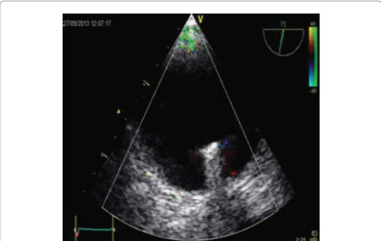
An adequate position free from the wall of the pulmonary vein or atrium was confirmed with ultrasound (Figure 2). A standard LA fluid-filled reference catheter was inserted and simultaneous baseline sensor and reference catheter measurements were made to confirm agreement, as with the LV sensor. After closure of the surgical incision repeated recordings were performed to confirm reproducibility of the pressure recordings. The length of time the reference LA catheter was left in place depended on the patient’s hemodynamic condition.
Statistics
Data were prospectively registered on a clinical report form, and transferred to and saved in an Excel database for future analysis. The study design and sample size were chosen with basic function and safety of the implant as primary endpoints. Linear regression was used to study the relationship between intraoperative sensor and reference catheter pressure values. Non-parametric tests were used to test differences between continuous variables, as the criteria for normality using the Shapiro-Wilks test were not fulfilled. Categorical variables were compared using the chi-square test or Fischer´s exact test when the number of observations was very small (<5). A p-value<0.05 was regarded as significant. Statistica software (StatSoft Inc.) was used for the calculations.
no physical connection between the implant and the rest of the system (extracorporeal components). Using radio frequency (RF) magnetic telemetry, the receiver transmits power to the sensing implant and communicates with it. The RF interface requires very little power. The wireless communication transmits detailed cardiac pressure waveforms and implant information such as implant power enabling advanced dynamic power transmission.
The miniature implant has two main components; polyether ketone housing and a cylindrical pressure-sensing probe placed inside. The probe contains a miniature micro-electromechanical pressure sensor along with custom electronics and a telemetry antenna.
An important feature of the implant is that the pressure sensing element is located at the distal flat side of the cylindrical implant. This small flat side is the only part of the implant that must be placed within the vessel whose pressure is to be monitored. There are 4 implant lengths between 18 and 30 mm and the implant is secured in place with sutures through 4 small holes in a flange at the proximal end of the housing.
Before embarking on this first human study, more than 60 animal studies in 4 different models had been performed at 5 qualified institutes since 2003 with favorable results regarding biocompatibility, thrombogenicity, and function. The Titan sensor has been tested in all heart chambers and in the aortic and pulmonary arteries. The sensor recordings have been compared with measurements from Millar catheters with overlapping pressure forms and very similar pressure values without the need of calibration of the sensor. The longest of all the animal studies prior to 2012 were performed at the Wayne State University between 2006 and 2007 with periods ranging from 8 to 27 weeks [4,5]. No thromboembolic complications were observed in the animal long term studies.
Figure 1: a) An 18 mm implant. The sensor is at the distal end, and the proximal part has a flange with 4 holes for fixation sutures. b) shows an antenna and a tablet pc on a read-out unit. With the permission of ISS Inc.
Figure 2: Echocardiographic image of a sensor implanted in the left atrium. Patient characteristics (n=40) Sensor (n=20) Control (n=20)
Age, mean ± SD years, (range) 71 (58-84) 71 (59-85)
Gender (M/F) 15/5 12/8 NYHA (III/IV) 15/5 17/3 Etiology (AS,AI,MI,CAD) 5/4/7/4 5/5/5/5 AVR/TAVR/MI-repair/CABG 5/4/7/4 6/5/4/5 On warfarin 10 9 On antiplatelets 10 11
Sensor location LA/LV 16/4
---Table 1: Patient characteristics: M = Male, F = Female, NYHA = New York Heart Association, AS = Aortic Stenosis, AI = Aortic Insufficiency, MI = Mitral Insufficiency, CAD = Coronary Artery Disease, AVR = Aortic Valve Replacement, TAVR = Transcatheter Aortic Valve Replacement, CABG = Coronary Artery Bypass Grafting, LA = Left Atrium, LV = Left Ventricle.
hypertrophic LV and underwent TAVR, the sensor was implanted in the LV at the end of the procedure. Signal capture was poor and we changed the position of the implant. After relocation it was still difficult to visualize the implant with TOE, but since signal capture was improved we accepted this position. At the 6-month follow-up the signal had drifted and this was confirmed with heart catheterization. Two LA implants suffered signal drift within the first 3 months, but in one the pressure curve was restored shortly after the 6-month follow-up. There were no signs of hemolysis, with a median haptoglobin value of 1.15 g/L (reference<1.9 g/L).
Follow-up data
Accumulated clinical data from the 1, 3 and 6-month follow-ups are shown in Table 2. Overall there were a few complications in each group with no statistically significant differences between them. The sensor curves allowed us to follow filling pressure on a daily basis and these were helpful in the early detection of arrhythmias which were thus treated at an early stage.
Serious adverse events (SAEs)
An adverse event is defined as any untoward medical occurrence, unintended disease or injury, or any untoward clinical signs in a patient, whether or not related to the medical device/procedure under investigation.
All such events were extracted from the patients’ records and analysed. Those classified as serious, i.e with the criteria in Table 3, were graded according to degree of severity. There were no events in Categories 1 and 3. Pt #2 in the device group suffered from treatable urosepticemia, and Pt # 13 in the control group had temporary ventricular tachycardia (Category 2). No relationship could be found between the SAE Categories 2, 4, 5 recorded and the use of the sensor device.
Adverse device effect (ADE)
No ADE related to the use of the sensor device was observed at the time of implantation or during the postoperative period.
Pat #2 underwent transapical TAVR Sept. 10, 2013. The myocardial wall was very thick and it was difficult to visualize the tip of the implant with intraoperative echocardiography. The implant was relocated twice after which a good signal was obtained. The patient underwent
Results
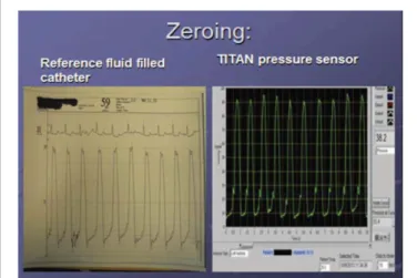
All sensors were successfully implanted within 15 min regardless of LA or LV location. Figure 3 shows concomitant recordings from fluid filled catheter as reference and sensor pressure at the end of TAVR in our first patient. They have very similar waveform and pressure values. There was no need for calibration of the sensor in this patient or in any of the cases with LA or LV implants.
Figure 4 shows an original LA pressure recording from the sensor after open chest cardiac surgery with baseline variation synchronous with the patient´s respiration.
The sensor recordings from the implant in the LA in 16 patients were compared with the LA pressure measurements with a routinely used fluid-filled catheter. Initial intraoperative sensor pressure and reference values from the fluid-filled catheter showed a significant linear correlation without the need for calibration of the sensor output (Figure 5).
Initial transesophageal echocardiography (TOE) confirmed that the sensor was in a satisfactory position in 18/20 cases. Signal capture was accurate in 19/20 patients. In patient #2 who had a very
Figure 3: Intraoperative recording after implant of the pressure sensor in the LV after TAVR. The curves from the reference catheter (left panel) and pressure sensor (right panel) have the same waveform and pressure values.
Figure 4: Pressure curve from a LA implant. The baseline variation correlates with the patient´s breathing. Mean value is calculated by the software. ISS inc. Lab View-software.
heart catheterization Feb 28, 2014 due to unexpected signal values. Dysfunction of the implant was verified where neither the curve form nor the signal values, that were very high, correlated with catheter values. It is possible that the implant was initially covered with tissue or became covered soon after the procedure.
Pt #29 was operated upon Oct 9, 2014 with mitral and tricuspid
valve repair, aortic valve replacement and CryoMaze ablation. On Dec 18, 2014, the signal started to drift giving higher and higher values. By June 1 the curve became more natural again with normal signal values. Pt # 36 underwent surgery Nov 25, 2014 with aortic valve replacement and coronary artery bypass grafting. Initially the sensor functioned normally but Jan 25, 2015 the signal drifted giving high values and shortly afterwards it disappeared. Echocardiography at the 6-month follow-up showed the implant to be in a good position.
No Serious Adverse Device Effect (SADE) was observed and no implant had been explanted by the 6 month follow-up.
Table 4 shows a summary of data from 11 consecutive patients receiving the pressure sensor more than 1 year previously. The LV sensors were implanted at the end of TAVR in 3 patients and the LA sensors at the end of an open chest procedure in 8 patients. There were no adverse events associated with the implant during a total time of 6186 days. The follow-up time for the patients is now 560 (407-786) days (Median + range).
The adequacy of LA pressure values can be tested by a Valsalva maneuver using a manometer. Cardiac filling pressures and airway pressure have been shown to equalize after 5 s of strain [6].
All patients were trained to monitor pressure at home. An 84 year-old woman, was initially motivated for the study but the lack of basic knowledge on how to handle the computer caused frustration. We therefore stopped home monitoring and she was regularly taken into hospital for measurements. All the other patients were able to do their own regular measurements at home.
Discussion
Initial sensor measurements showed very good correlation with reference pressure values from a fluid-filled catheter, and there was no need for calibration of the sensor. At the 6-month follow-up 11 patients had been wearing the implant for >1 year with a median time of 560 days. Ten of these had adequate sensor function. Compared to the control group there was no difference in adverse clinical events and the overall number of complications was low.
Postoperative antiplatelet medication or warfarin was a prerequisite Figure 5: Correlation between initial intraoperative sensor pressure and
reference values from a fluid-filled catheter. There was a good correlation (r=0.96, p<001). The dotted lines indicate 95% confidence interval.
22 20 18 16 14 12 10 8 6 4 2 2 4 6 8 10 12 14 16 18 20 22 Sensor 20 Reference 20
Sensor group Control group between groupsDifference
Stroke 0 0 ---Pericardial bleeding 1 0 ns Endocarditis 0 0 ---Malignant arrhythmia 1 4 ns Systolic function, 1/2/3/4 10/8/1/1 11/4/1/3 ns NYHA I/II/III/IV 10/8/2/0 11/4/1/3 ns NT-proBNP median 620 875 ns Exercise test % reference 75 75 ns
Elevated Filling pressure
on TOE, Yes/No 9/11 8/8, 4NA ns
Death 0 0
---Table 2: Accumulated clinical data at 6 months. There were no significant differences between the two groups. 1/2/3/4 = normal/mildly depressed/moderately depressed/ severely depressed, NYHA = New York Heart Association, NT-proBNP = N-terminal of the Prohormone Brain Natriuretic Peptide, TOE = Transesophageal Echocardiogram.
Serious adverse event (SAE) Sensor group Control group between groupsDifference
1. Death 0 0
---2. Life-threatening illness or injury 1 1 ns 3. Permanent impairment of a bodily
function 0 0
---4. Hospitalization or prolonged
postoperative care 13 16 ns 5. Requires medical intervention to
prevent life-threatening illness or injury, or permanent
impairment to a bodily function
7 11 ns
Table 3: Serious adverse events (SAE) Categories 1-5. No significant difference between groups was found.
Patient # and surgical
procedure Implant site
Time with implant, days Adequate pressure curve Adequate pressure values
1 AVR LA 786 Yes Yes
2 TAVR LV 710 No No (offset)
3 MI repair LA 680 Yes Yes
4 CABG +
LV-patch LA 653 Yes Yes
5 MI+TI repair +
CryoMaze LA 575 Yes Yes
6 TAVR LV 560 Yes Yes
7 TAVR LV 530 Yes Yes
8 MI+TI repair LA 449 Yes Yes
9 MI repair LA 428 Yes Yes
10 MI+TI repair + CABG LA 408 Yes Yes
11 AVR+ CryoMaze LA 407 Yes Yes
∑= 6186 days
Table 4: Patients in chronological order receiving implant in LV (left ventricle) or LA (left atrium) and with sensor time, median 560 days. TAVR = Transcatheter Aortic Valve Replacement, AVR = Aortic Valve Implantation, MI = Mitral, TI = Tricuspid, CABG = Coronary Artery Bypass Grafting.
for sensor implantation. We did not observe thrombotic covering of the sensor or thrombo-embolic events. Even though the sensor only needs minimal protrusion inside the heart to work, we chose to use a relatively long sensor (18 mm) in the LA to avoid the risk of cell growth covering the surface. The length of the LV implant was selected after careful measurement of the LV wall thickness using echocardiography. Introduction of the LV sensor using the Seldinger technique was a straightforward procedure, and insertion of the sensor in the LA via the incision made for LV drainage was easily performed. Daily recordings during the postoperative hospital stay were feasible to do, and the patients were trained either in hospital or soon after discharge to do their own recordings at home. One patient was unable to learn to do her own measurements. Trend curves were used to show pressure over time and enabled us to detect sudden changes in intracardiac pressure and arrhythmias. Although we did not focus on detailed surveillance of heart failure symptoms in this study, data so far obtained show the potential of home monitoring to detect early signs of increasing filling pressure and arrhythmias enabling therapeutic intervention before severe symptoms occur.
Chronicle® by Medtronic and HeartPOD® by St. Jude Medical are large sized, pacemaker type of devices under development and in early clinical phase trials [7,8]. Chronicle® is an IHM that measures right ventricular pressure. The COMPASS-HF study showed some promising results for this monitor but failed to reach the primary goal of reducing heart failure (HF) events compared to a group with optimal medical management [7]. The efficacy of LAP monitoring by the HeartPOD® device in the management of HF was investigated in the HOMEOSTASIS trial [8,9]. LAP values were significantly higher in the weeks prior to a clinical exacerbation of HF, indicating that hemodynamic changes with increased filling pressure regularly precede clinical decompensation. The data suggest that LAP monitoring has the potential to improve hemodynamics, symptoms and outcomes in patients with advanced HF. These promising results have been followed by the LAPTOP-HF study, the purpose of which is to investigate if LAP monitoring combined with a physician-directed self-management system will reduce episodes of HF exacerbation, decompensation and hospitalization. The study is still in progress.
Compared to other system for IHM the Titan™ sensor is small and in this respect is most like the CardioMEMS® device. Until now the Titan™ sensor needs to be implanted at an open chest procedure, whereas the CardioMEMS® involves catheter-based technology [2,3]. The Titan can principally be implanted in any of the heart chambers. The CardioMEMS® is restricted to pulmonary artery placement but the only IMH so far approved by the FDA. We recently performed the first animal implantation of a modified Titan™ device via a catheter with promising results, and this modified device is now ready for long-term studies in animals.
IMH development remains in its early stages and more clinical trials are required before such devices are efficient enough to benefit patient outcome compared to present standard management. Future
studies will show whether IMH has the potential to improve patient care substantially, or if it just becomes an adjunct to standard medical treatment [1].
Limitations
This is the first report on the human application of this IMH device. Our results show that the sensor works well for the wireless measurement of intracardiac pressures, without adverse events. Long-term durability beyond the time range of this study is not known, nor can we say if heart failure patients will benefit from its use.
Conclusion
This first study in man on a new implantable wireless hemodynamic monitor showed favorable results regarding our primary endpoints; accuracy of recordings over time and safety profile. The technology has great potential for monitoring at home since it is easy to use in the out-patient setting.
References
1. Smith SA, Abraham WT (2012) Implantable Cardiovascular Sensors and Computers: interventional heart failure strategies. Curr Cardiol Rep 14: 611-618.
2. Abraham WT, Adamson PB, Bourge RC, Aaron MF, Costanzo MR, et al. (2011) Wireless pulmonary artery haemodynamic monitoring in chronic heart failure: a randomised controlled trial. Lancet 377: 658-666.
3. Loh JP, Barbash IM, Waksman R (2013) Overview of the 2011 Food and Drug Administration Circulatory System Devices Panel of the Medical Devices Advisory Committee Meeting on the CardioMEMS Champion Heart Failure Monitoring System. J Am Coll Cardiol 61: 1571-1576.
4. Najafi N, Ludomirsky A (2004) Initial animal studies of a wireless, batteryless, MEMS implant for cardiovascular applications. Biomed Microdevices 6: 61-65. 5. Hammond RL, Hanna K, Morgan C, Perakis P, Najafi N, et al. (2012) A wireless
and battery less miniature intracardiac pressure sensor: early implantation studies. ASAIO J 58: 83-87.
6. McClean D, Aragon J, Jamali A, Kar S, Ritzema-Carter J, et al. (2006) Noninvasive calibration of cardiac pressure transducers in patients with heart failure: an aid to implantable hemodynamic monitoring and therapeutic guidance. J Card Fail 12: 568-576.
7. Bourge RC, Abraham WT, Adamson PB, Aaron MF, Aranda JM, et al. (2008) Randomized controlled trial of an implantable continuous hemodynamic monitor in patients with advanced heart failure: the COMPASS-HF study. J Am Coll Cardiol 51: 1073-1079.
8. Troughton RW, Ritzema J, Eigler NL, Melton IC, Krum H, et al. (2011) Direct left atrial pressure monitoring in severe heart failure: long-term sensor performance. J Cardiovasc Transl Res 4: 3-13.
9. Ritzema J, Troughton R, Melton I, Crozier I, Doughty R, et al. (2010) Hemodynamically Guided Home Self-Therapy in Severe Heart Failure Patients (HOMEOSTASIS) Study Group. Physician directed patient self-management of left atrial pressure in advanced chronic heart failure. Circulation 121: 1086-1095.
OMICS International: Publication Benefits & Features Unique features:
• Increased global visibility of articles through worldwide distribution and indexing • Showcasing recent research output in a timely and updated manner • Special issues on the current trends of scientific research Special features:
• 700+ Open Access Journals • 50,000+ Editorial team • Rapid review process
• Quality and quick editorial, review and publication processing • Indexing at major indexing services
• Sharing Option: Social Networking Enabled
• Authors, Reviewers and Editors rewarded with online Scientific Credits • Better discount for your subsequent articles
Submit your manuscript at: http://www.omicsonline.org/submission Citation: Ahn HC, Delshad B, Baranowski J (2016) An Implantable Pressure
Sensor for Long-term Wireless Monitoring of Cardiac Function- First Study in Man. J Cardiovasc Dis Diagn 4: 252. doi: 10.4172/2329-9517.1000252